Electrical Impedance Tomography (EIT) to Optimize Ventilatory Management in Critically Ill Patients: A Report of Two Cases


Abstract
:1. Introduction
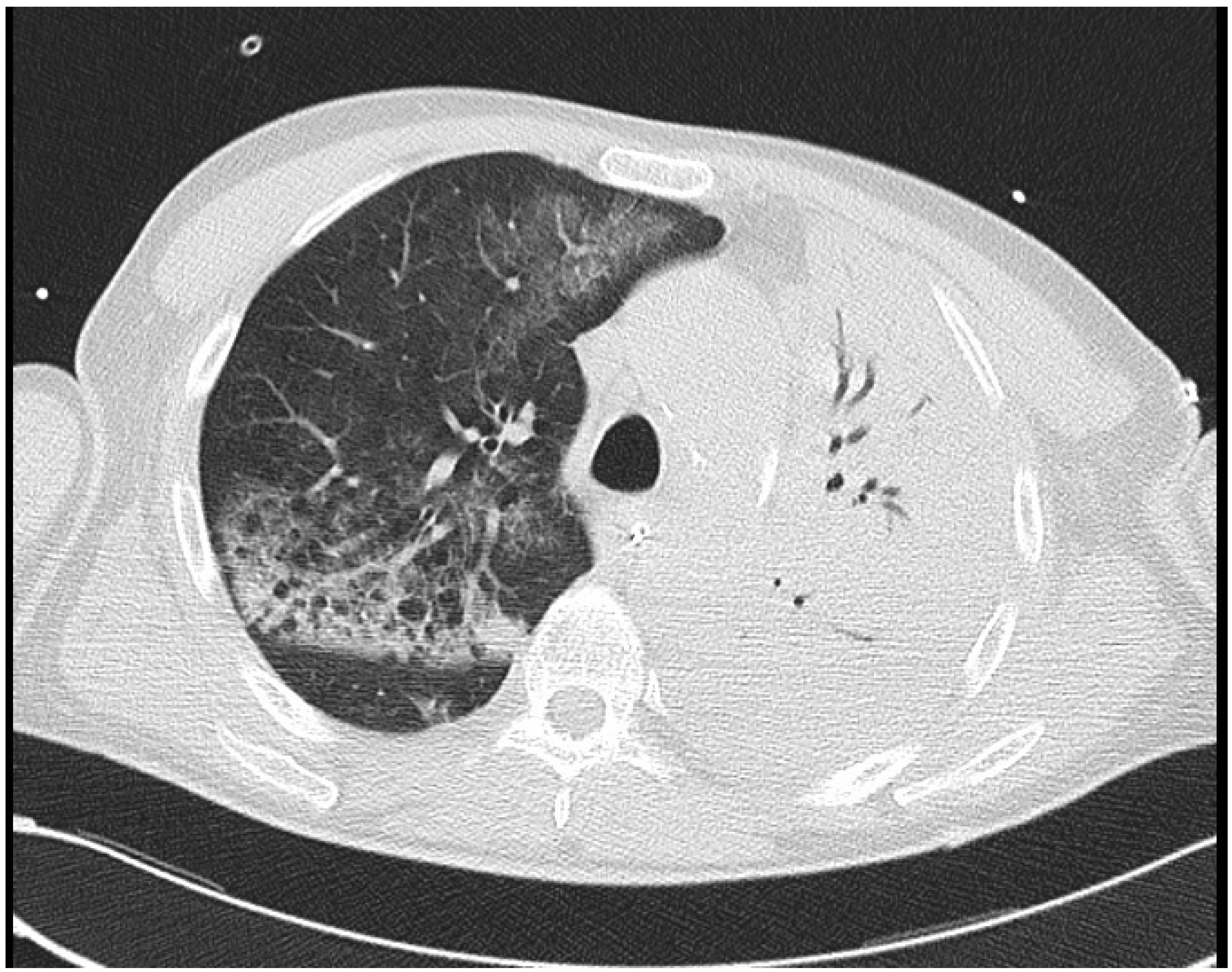
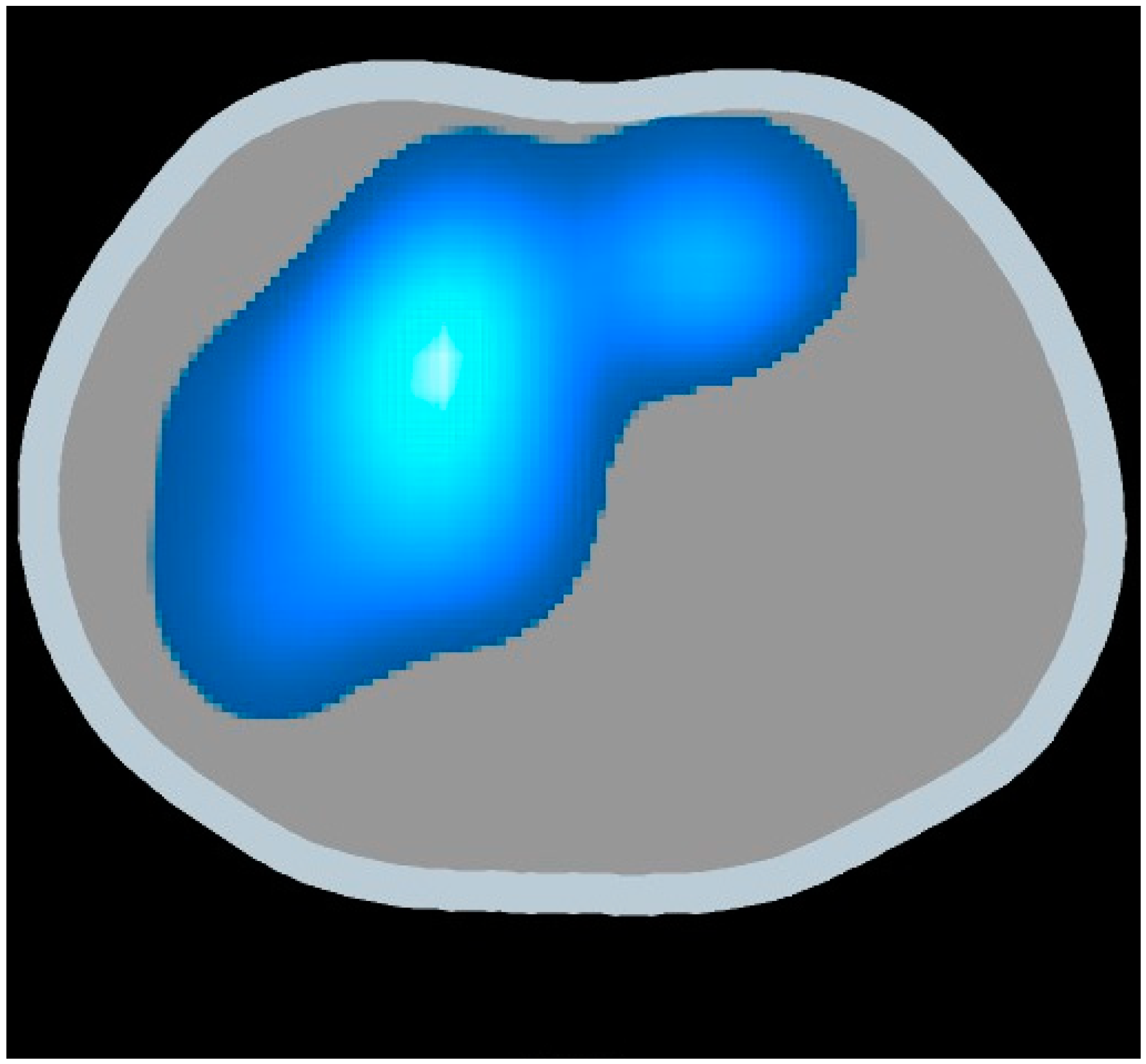
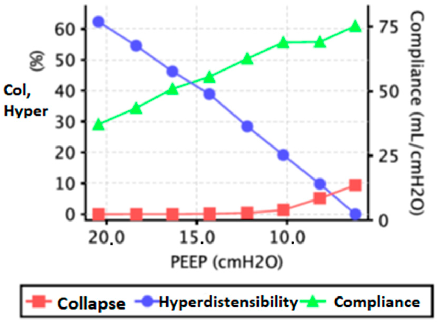
2. Case Report 1
3. Case Report 2
4. Discussion
5. Conclusions
Author Contributions
Funding
Institutional Review Board Statement
Informed Consent Statement
Data Availability Statement
Conflicts of Interest
References
- Maciejewski, D.; Putowski, Z.; Czok, M.; Krzych, Ł. Electrical impedance tomography as a tool for monitoring mechanical ventilation. An introduction to the technique. Adv. Med. Sci. 2021, 66, 388–395. [Google Scholar] [CrossRef]
- Walsh, B.K.; Smallwood, C.D. Electrical Impedance Tomography During Mechanical Ventilation. Respir. Care 2016, 61, 1417–1424. [Google Scholar] [CrossRef] [PubMed]
- Costa, E.L.V.; Amato, M.B.P. Electrical Impedance Tomography in Critically Ill Patients. Clin. Pulm. Med. 2013, 20, 26. [Google Scholar] [CrossRef]
- Zhao, Z.; Zhang, J.-S.; Chen, Y.-T.; Chang, H.-T.; Hsu, Y.-L.; Frerichs, I.; Adler, A. The use of electrical impedance tomography for individualized ventilation strategy in COVID-19: A case report. BMC Pulm. Med. 2021, 21, 38. [Google Scholar] [CrossRef] [PubMed]
- Thomas, P. An Introduction to the Clinical Application and Interpretation of Electrical Impedance Tomography. Respir. Care 2022, 67, 721. [Google Scholar] [CrossRef]
- Bachmann, M.C.; Morais, C.; Bugedo, G.; Bruhn, A.; Morales, A.; Borges, J.B.; Costa, E.; Retamal, J. Electrical impedance tomography in acute respiratory distress syndrome. Crit. Care 2018, 22, 263. [Google Scholar] [CrossRef]
- Prins, S.A.; Weller, D.; Labout, J.A.M.; den Uil, C.A. Electrical Impedance Tomography As a Bedside Diagnostic Tool for Pulmonary Embolism. Crit. Care Explor. 2023, 5, e0843. [Google Scholar] [CrossRef]
- Xu, M.; He, H.; Long, Y. Lung Perfusion Assessment by Bedside Electrical Impedance Tomography in Critically Ill Patients. Front. Physiol 2021, 12, 748724. [Google Scholar] [CrossRef]
- Sella, N.; Pettenuzzo, T.; Zarantonello, F.; Andreatta, G.; De Cassai, A.; Schiavolin, C.; Simoni, C.; Pasin, L.; Boscolo, A.; Navalesi, P. Electrical impedance tomography: A compass for the safe route to optimal PEEP. Respir. Med. 2021, 187, 106555. [Google Scholar] [CrossRef]
- van der Zee, P.; Somhorst, P.; Endeman, H.; Gommers, D. Electrical Impedance Tomography for Positive End-Expiratory Pressure Titration in COVID-19-related Acute Respiratory Distress Syndrome. Am. J. Respir. Crit. Care Med. 2020, 202, 280–284. [Google Scholar] [CrossRef] [PubMed]
- Frerichs, I.; Dargaville, P.A.; Dudykevych, T.; Rimensberger, P.C. Electrical impedance tomography: A method for monitoring regional lung aeration and tidal volume distribution? Intensive Care Med. 2003, 29, 2312–2316. [Google Scholar] [CrossRef] [PubMed]
- Lundin, S.; Stenqvist, O. Electrical impedance tomography: Potentials and pitfalls. Curr. Opin. Crit. Care 2012, 18, 35–41. [Google Scholar] [CrossRef] [PubMed]
- Brown, B.H. Electrical impedance tomography (EIT): A review. J. Med. Eng. Technol. 2003, 27, 97–108. [Google Scholar] [CrossRef]


Disclaimer/Publisher’s Note: The statements, opinions and data contained in all publications are solely those of the individual author(s) and contributor(s) and not of MDPI and/or the editor(s). MDPI and/or the editor(s) disclaim responsibility for any injury to people or property resulting from any ideas, methods, instructions or products referred to in the content. |
© 2023 by the authors. Licensee MDPI, Basel, Switzerland. This article is an open access article distributed under the terms and conditions of the Creative Commons Attribution (CC BY) license (https://creativecommons.org/licenses/by/4.0/).
Share and Cite
Cappellini, I.; Campiglia, L.; Zamidei, L.; Consales, G. Electrical Impedance Tomography (EIT) to Optimize Ventilatory Management in Critically Ill Patients: A Report of Two Cases. Anesth. Res. 2024, 1, 3-7. https://doi.org/10.3390/anesthres1010002
Cappellini I, Campiglia L, Zamidei L, Consales G. Electrical Impedance Tomography (EIT) to Optimize Ventilatory Management in Critically Ill Patients: A Report of Two Cases. Anesthesia Research. 2024; 1(1):3-7. https://doi.org/10.3390/anesthres1010002
Chicago/Turabian StyleCappellini, Iacopo, Laura Campiglia, Lucia Zamidei, and Guglielmo Consales. 2024. "Electrical Impedance Tomography (EIT) to Optimize Ventilatory Management in Critically Ill Patients: A Report of Two Cases" Anesthesia Research 1, no. 1: 3-7. https://doi.org/10.3390/anesthres1010002