Cataract Surgery following Corneal Allogenic Intrastromal Ring Segments and Implantable Collamer Lens


Abstract
:1. Introduction
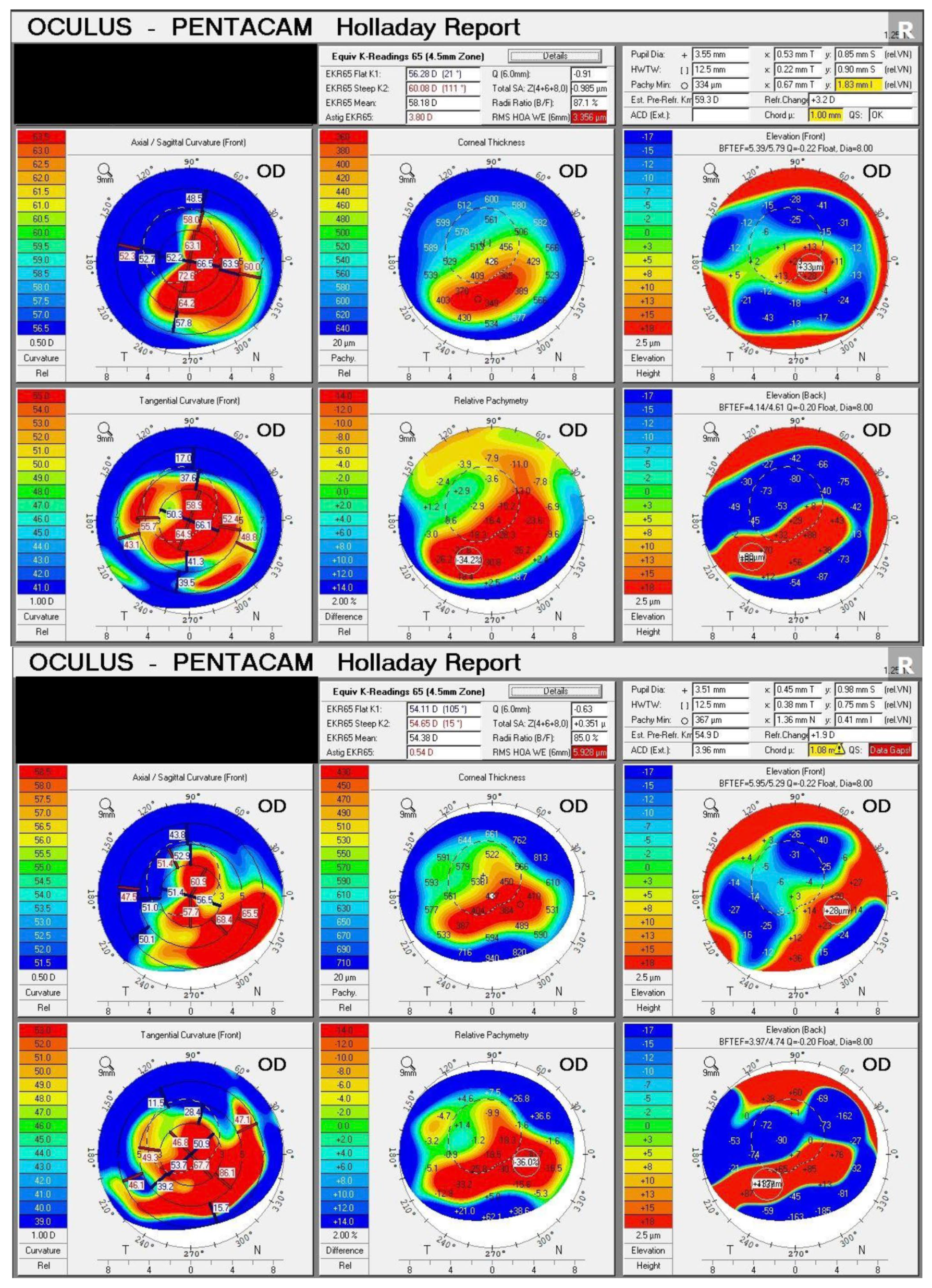
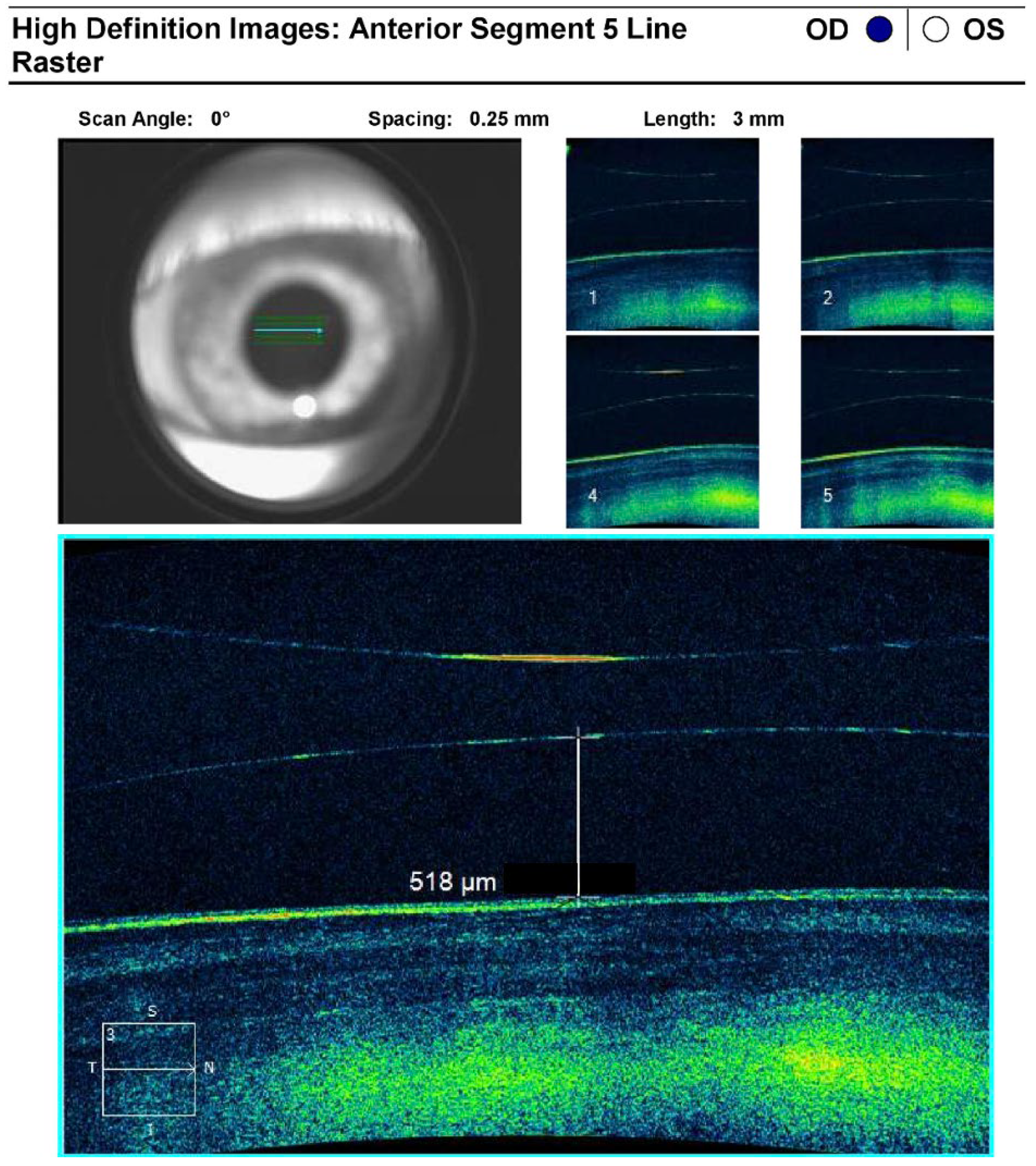
2. Case Report
3. Discussion
4. Conclusions
Supplementary Materials
Author Contributions
Funding
Institutional Review Board Statement
Informed Consent Statement
Data Availability Statement
Conflicts of Interest
References
- Basu, S.; Vazirani, J. Keratoconus: Current perspectives. Clin. Ophthalmol. 2013, 7, 2019–2030. [Google Scholar] [CrossRef] [PubMed] [Green Version]
- Santodomingo-Rubido, J.; Carracedo, G.; Suzaki, A.; Villa-Collar, C.; Vincent, S.J.; Wolffsohn, J.S. Keratoconus: An updated review. Contact Lens Anterior Eye 2022, 45, 101559. [Google Scholar] [CrossRef] [PubMed]
- Jacob, S.; Patel, S.R.; Agarwal, A.; Ramalingam, A.; Saijimol, A.I.; Raj, J.M. Corneal Allogenic Intrastromal Ring Segments (CAIRS) combined with corneal crosslinking for keratoconus. J. Refract. Surg. 2018, 34, 296–303. [Google Scholar] [CrossRef] [PubMed]
- Arej, N.; Chanbour, W.; Zaarour, K.; Amro, M.; El-Rami, H.; Harb, F.; Jarade, E. Management of cataract in keratoconus: Early visual outcomes of different treatment modalities. Int. J. Ophthalmol. 2019, 12, 1654–1658. [Google Scholar] [CrossRef]
- Moshirfar, M.; Walker, B.D.; Birdsong, O.C. Cataract surgery in eyes with keratoconus: A review of the current literature. Curr. Opin. Ophthalmol. 2018, 29, 75–80. [Google Scholar] [CrossRef]
- Ghiasian, L.; Abolfathzadeh, N.; Manafi, N.; Hadavandkhani, A. Intraocular lens power calculation in keratoconus; A review of literature. J. Curr. Ophthalmol. 2019, 31, 127–134. [Google Scholar] [CrossRef]
- Bozorg, S.; Pineda, R. Cataract and keratoconus: Minimizing complications in intraocular lens calculations. Semin. Ophthalmol. 2014, 29, 376–379. [Google Scholar] [CrossRef]
- Chung, J.; Bu, J.J.; Afshari, N.A. Advancements in intraocular lens power calculation formulas. Curr. Opin. Ophthalmol. 2022, 33, 35–40. [Google Scholar] [CrossRef]
- Aiello, F.; Nasser, Q.; Nucci, C.; Angunawela, R.; Gatzioufas, Z.; Maurino, V. Cataract Surgery in Patients with Keratoconus: Pearls and Pitfalls. Open Ophthalmol. J. 2017, 11, 194–200. [Google Scholar] [CrossRef] [Green Version]
- Gonzalez-Salinas, R.M.; Franco, J.J.B.; Reyes-Luis, J.L.; Sánchez-Huerta, V.; de Wit-Carter, G.; Hernández-Quintela, E.M.; Pineda, R.I.M. Cataract surgery in patients with underlying keratoconus: Focused review. J. Cataract Refract. Surg. 2023, 49, 97–102. [Google Scholar] [CrossRef]
- Hashemi, H.; Yekta, A.; Khabazkhoob, M. Effect of keratoconus grades on repeatability of keratometry readings: Comparison of 5 devices. J. Cataract Refract. Surg. 2015, 41, 1065–1072. [Google Scholar] [CrossRef] [PubMed]
- Epitropoulos, A.; Matossian, C.; Berdy, G.J.; Malhotra, R.P.; Potvin, R. Effect of tear osmolarity on repeatability of keratometry for cataract surgery planning. J. Cataract Refract. Surg. 2015, 41, 1672–1677. [Google Scholar] [CrossRef] [PubMed]
- Watson, M.P.; Anand, S.; Bhogal, M.; Gore, D.; Moriyama, A.; Pullum, K.; Hau, S.; Tuft, S.J. Cataract surgery outcome in eyes with keratoconus. Br. J. Ophthalmol. 2014, 98, 361–364. [Google Scholar] [CrossRef] [PubMed]
- Fadlallah, A.; Dirani, A.; El Rami, H.; Cherfane, G.; Jarade, E. Safety and visual outcome of Visian toric ICL implantation after corneal collagen cross-linking in keratoconus. J. Refract. Surg. 2013, 29, 84–89. [Google Scholar] [CrossRef]
- Fairaq, R.; Almutlak, M.; Almazyad, E.; Badawi, A.H.; Ahad, M.A. Outcomes and complications of implantable collamer lens for mild to advance keratoconus. Int. Ophthalmol. 2021, 41, 2609–2618. [Google Scholar] [CrossRef]
- Singh, C.; Joshi, V.P. Cataract surgery in Keratoconus revisited—An update on preoperative and intraoperative considerations and postoperative outcomes. Semin. Ophthalmol. 2022, 38, 57–64. [Google Scholar] [CrossRef]
- Chen, Y.-A.; Hirnschall, N.; Findl, O. Comparison of corneal wetting properties of viscous eye lubricant and balanced salt solution to maintain optical clarity during cataract surgery. J. Cataract Refract. Surg. 2011, 37, 1806–1808. [Google Scholar] [CrossRef]
- Oie, Y.; Kamei, M.; Matsumura, N.; Fujimoto, H.; Soma, T.; Koh, S.; Tsujikawa, M.; Maeda, N.; Nishida, K. Rigid gas-permeable contact lens-assisted cataract surgery in patients with severe keratoconus. J. Cataract Refract. Surg. 2014, 40, 345–348. [Google Scholar] [CrossRef]
- Chan, E.; Mahroo, O.A.R.; Spalton, D.J. Complications of cataract surgery. Clin. Exp. Optom. 2010, 93, 379–389. [Google Scholar] [CrossRef]
- Kamiya, K.; Iijima, K.; Nobuyuki, S.; Mori, Y.; Miyata, K.; Yamaguchi, T.; Shimazaki, J.; Watanabe, S.; Maeda, N. Predictability of Intraocular Lens Power Calculation for Cataract with Keratoconus: A Multicenter Study. Sci. Rep. 2018, 8, 1312. [Google Scholar] [CrossRef] [Green Version]
- Savini, G.; Abbate, R.; Hoffer, K.J.; Mularoni, A.; Imburgia, A.; Avoni, L.; D’eliseo, D.; Schiano-Lomoriello, D. Intraocular lens power calculation in eyes with keratoconus. J. Cataract Refract. Surg. 2019, 45, 576–581. [Google Scholar] [CrossRef] [PubMed]


Disclaimer/Publisher’s Note: The statements, opinions and data contained in all publications are solely those of the individual author(s) and contributor(s) and not of MDPI and/or the editor(s). MDPI and/or the editor(s) disclaim responsibility for any injury to people or property resulting from any ideas, methods, instructions or products referred to in the content. |
© 2023 by the authors. Licensee MDPI, Basel, Switzerland. This article is an open access article distributed under the terms and conditions of the Creative Commons Attribution (CC BY) license (https://creativecommons.org/licenses/by/4.0/).
Share and Cite
Ali, A.A.; Saenz, B.; Nasser, T. Cataract Surgery following Corneal Allogenic Intrastromal Ring Segments and Implantable Collamer Lens. J. Clin. Transl. Ophthalmol. 2023, 1, 72-78. https://doi.org/10.3390/jcto1030009
Ali AA, Saenz B, Nasser T. Cataract Surgery following Corneal Allogenic Intrastromal Ring Segments and Implantable Collamer Lens. Journal of Clinical & Translational Ophthalmology. 2023; 1(3):72-78. https://doi.org/10.3390/jcto1030009
Chicago/Turabian StyleAli, Arsalan Akbar, Bobby Saenz, and Taj Nasser. 2023. "Cataract Surgery following Corneal Allogenic Intrastromal Ring Segments and Implantable Collamer Lens" Journal of Clinical & Translational Ophthalmology 1, no. 3: 72-78. https://doi.org/10.3390/jcto1030009