Article

Perception and Risk Factors Associated with Tuberculosis in the Manyara Region, Tanzania

1
Department of Global Health and Biomedical Science, School of Life Science, Nelson Mandela African Institute of Science and Technology (NM-AIST), P.O. Box 447 Tengeru, Tanzania
2
Kibong’oto Infectious Disease Hospital (KIDH), P.O. Box 12 Sanya Juu Siha, Tanzania
3
Global Health Initiative, Henry Ford Health, Detroit, MI 48202, USA
4
Huck Institutes of the Life Sciences, The Pennsylvania State University, University Park, PA 16802, USA
*
Author to whom correspondence should be addressed.
Zoonotic Dis. 2023, 3(4), 266-278; https://doi.org/10.3390/zoonoticdis3040022
Submission received: 13 July 2023 / Revised: 19 September 2023 / Accepted: 21 September 2023 / Published: 6 October 2023

Abstract

:

Simple Summary

The Manyara region is amongst the tuberculosis hot spot regions in Tanzania; both pulmonary and extra-pulmonary cases have been reported, which raises questions on risk factors of the disease and the Mycobacteria species circulating in the area. A questionnaire was administered to identify risk factors of the disease. Furthermore, pulmonary and extra-pulmonary samples were collected from participants and cultured, and organisms from positive cultures were subjected to molecular speciation using PCR to identify MTBC species. The study found a low awareness of both human TB, animal TB, and zoonotic TB among participants. All specimens, pulmonary or extrapulmonary, that were positive by culture were typed as M. tuberculosis sensu stricto. Effective TB disease educational programs should be implemented to overcome the problem.

Abstract

Tuberculosis (TB) results from infection with members of the Mycobacterium tuberculosis complex (MTBC) and represents a major global public health concern. We here sought to assess the perceptions of human and animal TB and the prevalence of circulating MTBCs lineages and associated risk factors through a cross-sectional survey of 335 individuals presenting with symptoms of pulmonary or extrapulmonary TB in the Manyara region of Tanzania. After the enrollment of participants, a questionnaire survey was conducted, samples were collected for bacterial culture, and real-time multiplex PCR was performed to differentiate amongst primary animal and human MTBC lineages. The results show poor TB awareness: 31.6% of the participants were not aware of human TB; 82.4% were unaware of animal TB and 95.2% lacked awareness of zoonotic TB (zTB) transmission. A total of 18 recovered specimens (5%; 95% CI: 3–8%) were positive by culture, all of which were typed as M. tuberculosis sensu stricto using a lineage-specific PCR assay. While no single risk factor was significantly associated with MTBC culture positivity, the survey revealed considerable self-reported high-risk practices for contracting zTB. Together, the results show that Manyara residents have poor knowledge of diseases caused by MTBCs and high evidence of risky practices for contracting zTB.

1. Introduction

Human tuberculosis (TB) is a leading global cause of death and morbidity [1]. TB is caused by organisms belonging to the Mycobacterium tuberculosis complex species (MTBCs) including the primarily human-associated lineages 1 through 9 of M. tuberculosis sensu stricto, M. africanum, and the distantly related M. canetii as well as animal-associated lineages—M. bovis, M. caprae, M. pinnipedii, M. suricattae, M. mungi, M. orygis, and Dassie bacillus [2]. Mycobacterium tuberculosis has apparently infected one-quarter of the world’s population [3], and in 2021, an estimated 1.6 million people died from TB worldwide [1]. TB manifests mainly as lung infections (pulmonary TB) which account for ~84% of incident TB cases, and as infection of other organs (extra-pulmonary TB), which accounts for ~16% of incident TB cases [3].
Apart from human–human transmission by M. tuberculosis sensu stricto, zoonotic tuberculosis (zTB) is often caused by infection with M. bovis or other animal-associated MTBC lineages as a result of transmission from livestock species, and is more often associated with extrapulmonary (EPTB) and pediatric TB cases [4,5]. Transmission of zTB from animals to humans is thought to occur through the consumption of products such as raw milk, meat, and other infected animal products or potentially through direct contact with infected animals [6]. M. bovis contributes ~1% of human TB cases in high-income countries and 10% in low-and-middle-income countries [7]. Tanzania is among 30 countries identified by WHO as high TB burden countries globally [8]. In 2021, Tanzania National Tuberculosis and Leprosy Programme (NTLP), reported an estimate of 87,415 tuberculosis cases and 14,033 (16.2%) were children under the age of 15 years [9].
A significant increase of 116.6% in extra-pulmonary cases between 1995 and 2009 was reported in Tanzania by WHO (2010) report [10]. During the same period, it was reported that the proportion of extra-pulmonary TB in Arusha (which includes the current Manyara region) was higher (over 30%) despite relatively lower HIV prevalence [11]. However, the fraction of extra-pulmonary cases caused by the zoonotic MTBC lineages including M. bovis remains unknown [12]. Moreover, studies done on livestock in this area have reported herd and individual animals’ bovine TB (BTB) prevalence of 7.9% and 2.7%, respectively [13].
Hence this study aimed to assess perceptions (knowledge, attitudes, and practices) and risk factors associated with human TB, bovine TB, and zoonotic TB, and identify circulating Mycobacterium lineages among TB patients who visited health facilities in Manyara Region, Tanzania.

2. Materials and Methods

The study was carried out in the Manyara region of Tanzania, comprising five districts: Mbulu, Babati, Kiteto, Simanjiro, and Hanang (Figure 1). The Manyara region is located between latitudes 3.381° S and 4.521° S and longitudes 35.01° E and 35. 91°E. Based on the Tanzania National Sample Census of Agriculture 2002/2003, most residents of the Manyara region involve themselves with livestock keeping and farming activities [14]. Manyara is one of the regions reported to have a relatively higher proportion (over 30%) of extra-pulmonary tuberculosis cases [11].

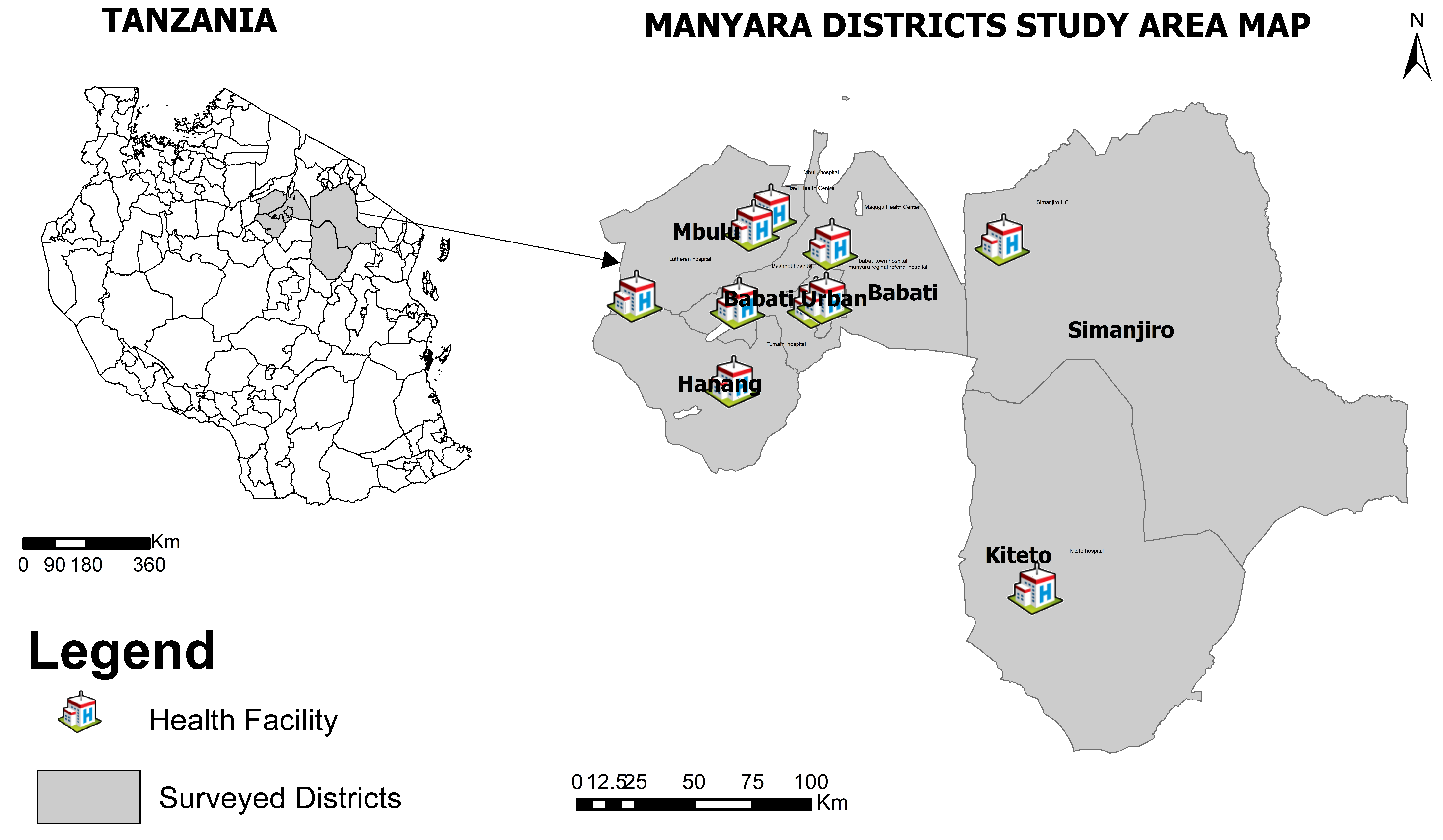
2.1. Study Design and Participant Recruitment

This cross-sectional study recruited patients who were self-represented at hospitals within the Manyara region with pulmonary or extra-pulmonary TB symptoms from October 2020 to December 2021. The inclusion criteria were patients suspected of either pulmonary TB (PTB) or extra-pulmonary TB (EPTB); for example, patients presenting with fever, prolonged cough, malaise, anorexia, or any EPTB symptoms such as pleuritic pain and effusion, tuberculous lymphadenitis of cervical and axillary regions and assented to participate and signed informed consent form. Pregnant, individuals already on TB treatment, and those unable or unwilling to assent or sign informed consent were excluded from the study.

2.2. Sample Size

The sample size was calculated using a previously reported prevalence [15]. An assumed prevalence was considered from the studies which reported a 30% prevalence of EPTB in the Manyara region [11], sensitivities and specificities of 90%, 95% of the level of confidence, and 5% desired precision; a calculated sample size of 335 was determined.

2.3. Specimen Collection

A total of 302 sputa from PTB patients and 33 EPTB specimens (abdominal fluid, lymph node aspirate, or pleural fluid) from EPTB patients were collected by trained medical personnel. Samples were stored at temperatures between 2–8 °C and transported within 24–48 h along with the Case Report Forms (CRFs) to the Kibong’oto Infectious Disease Hospital (KIDH) laboratory for analysis.

2.4. Questionnaire Data Collection

A structured questionnaire was developed based on information from other zoonotic study surveys (Appendix A Table A1). It was aimed at obtaining key data including clinical information, TB history, risk factors, and TB disease knowledge and awareness from patients before the clinical examination.
To assess tuberculosis infection knowledge and awareness (KAP), the questions to measure if the person knows about TB disease were structured with a “yes” or “no” response. Further, to see the level of awareness there was a list of 21 TB symptoms, all were structured, pre-coded, and mainly with the optional “yes”, and “no” responses and were rated on a scale of none (0 questions), poor (below 5 questions), average (above 5 questions) and good (above 10 questions) (Supplementary Materials).
To assess bovine tuberculosis infection, knowledge, and awareness, the question to measure if the person knows about TB disease in animals was structured with a “yes” or “no”, the question was “Before we talked to you about this study, have you heard that cattle can contract tuberculosis?”. Further, to see the level of awareness we had a list of 12 cattle TB symptoms all were structured, pre-coded, and mainly with the expected “yes”, and “no” and their response was rated on a scale of none (0 questions), average (below 5 questions) and good (above 5 questions) (Supplementary Materials).
The questions to measure if the person knows about zTB disease were structured with a “yes” or “no” response, for example, “Do you know that bovine tuberculosis can be transmitted to humans?”. Further, to see the level of awareness we asked the question “Do you know the mode of transmission of tuberculosis between cattle and human beings?” (Supplementary Materials).
The questions to measure common practices towards factors of being infected included four (4) keywords such as unpasteurized milk, untreated meat, aerosol, and direct contact mainly with the expected “yes”, “no” and their response was rated on a scale of none (0 questions), good (at least mention of one mode). Participants were asked about their attitudes regarding the risk of zoonotic TB transmission practices such as consuming raw milk, consuming raw animal products, unsafe attending to animals, sharing the same roof with animals, and having a TB history in their families (Supplementary Materials).

2.5. Laboratory Analysis

The procedure for detecting positive Mycobacterium species in samples included culture to identify positive samples, followed by confirmatory tests for identification and eventually Multiplex real-time PCR (rt-PCR) for speciation as described below.

2.5.1. Detection of Samples That Are Positive for Mycobacteria Culture

Sputa samples were decontaminated using the standard modified Petroff method [16] and mixed with an equal volume of 4% NaOH. The extra-pulmonary samples were not decontaminated, since they are normally sterile [17]. Thereafter both sputa and extrapulmonary samples were centrifuged at 3000× g, the supernatant was discarded, and sediment was re-suspended in 0.8 mL of phosphate buffered saline before inoculation into Mycobacterium Growth Indicator Tube (MGIT). The specimens were cultured on MGIT supplemented with 0.5% pyruvate specifically to support the growth of Mycobacterium bovis as per manufacturer instructions [18]. Incubation was done at 37 °C in the incubator (BACTEC MGIT 960, Becton Dickinson) and positive signal development was regularly monitored for as long as 42 days.

2.5.2. Confirmatory Test for Positive Culture Identification

BACTEC MGIT 960 tubes that yielded a positive culture signal were subjected to confirmation using the Ziehl–Neelsen (ZN)—stain and immunochromatographic test using MPT64 rapid test kit (SD Bio line Kit, Standard Diagnostics Inc., Yongin, Republic of Korea). To rule out contaminations, they were cultured on Blood agar (BA) [19]. Positive cultures with positive ZN staining and MPT64 were confirmed to be MTBC, while those with negative MPT64 were regarded as non-tuberculous mycobacteria (NTM). However, positive cultures that showed growth on BA and were negative in ZN-stain and MPT64 were regarded as contamination.

2.5.3. MTBC Speciation with PCR

This process was undertaken in the containment level 3 laboratory at KCRI. The DNA extraction process was performed using the Cetyl trimethylammonium bromide (CTAB) extraction method with enzymatic digestion and organic solvent extraction to obtain high-quality large fragment DNA [20]. DNA quality was assessed spectroscopically using a Qubit fluorometer. The real-time multiplex PCR was done on the DNA templates to detect MTBC and differentiate between human and animal lineages, including M. bovis, M. caprae, and M. orygis [21].
Two tri-plex multiplex real-time (rt) PCR assays (IS1081, MTCAni, MTCHum) and (Morg, Mcap, Mbov) were developed to screen the MTBC-positive samples. The assays differentiate the human mycobacterial lineages and animal mycobacterial species, and detect specifically Mycobacterium tuberculosis, Mycobacterium bovis, Mycobacterium caprae, and Mycobacterium orygis. A 40× concentration primer-probe mix was prepared first by mixing a forward probe, sterile water, and reverse and forward primer for all six assays, and the mixture was reconstituted to a concentration of 100 micromolar using sterile water. The prime-probe mix reaction is summarized in Appendix A Table A2. From the mix, 0.5 µL was used in the 20 µL PCR reaction mix. The mix was prepared by mixing PrimeTime Gene Expression master mix, 2X ROX dye, Prime time qPCR assays (40X), and nuclease-free water, as shown in Appendix A Table A3.
Thermal cycling reactions were performed using a 7500 Fast Real-Time PCR System (Applied Biosystems, Waltham, MA, USA). The PCR was done at settings of the standard curve, TaqMan reagents, standard properties, and fast 96 well blocks (0.1) at the thermal profile, summarized in Appendix A Table A4. The reporters were FAM for IS1081 and Morg, VIC for MTCAni and Mcap, TAMRA for MTCHum and Mbov, and CY5 for Internal control. The genomic DNA of known M. bovis, M. orygis, M. caprae, and H37Rv were used as positive controls for M. bovis, M. orygis, M. caprae, and M. tuberculosis, respectively. Threshold and baseline were set automatically for analysis, and evaluation of application curves was carried out where the positive control Ct value ranged from 23–27 and negative controls showed no amplification. Appendix A Table A5 summarizes the sequence and melting temperature of primer-probe sets.

2.6. Statistical Analyses
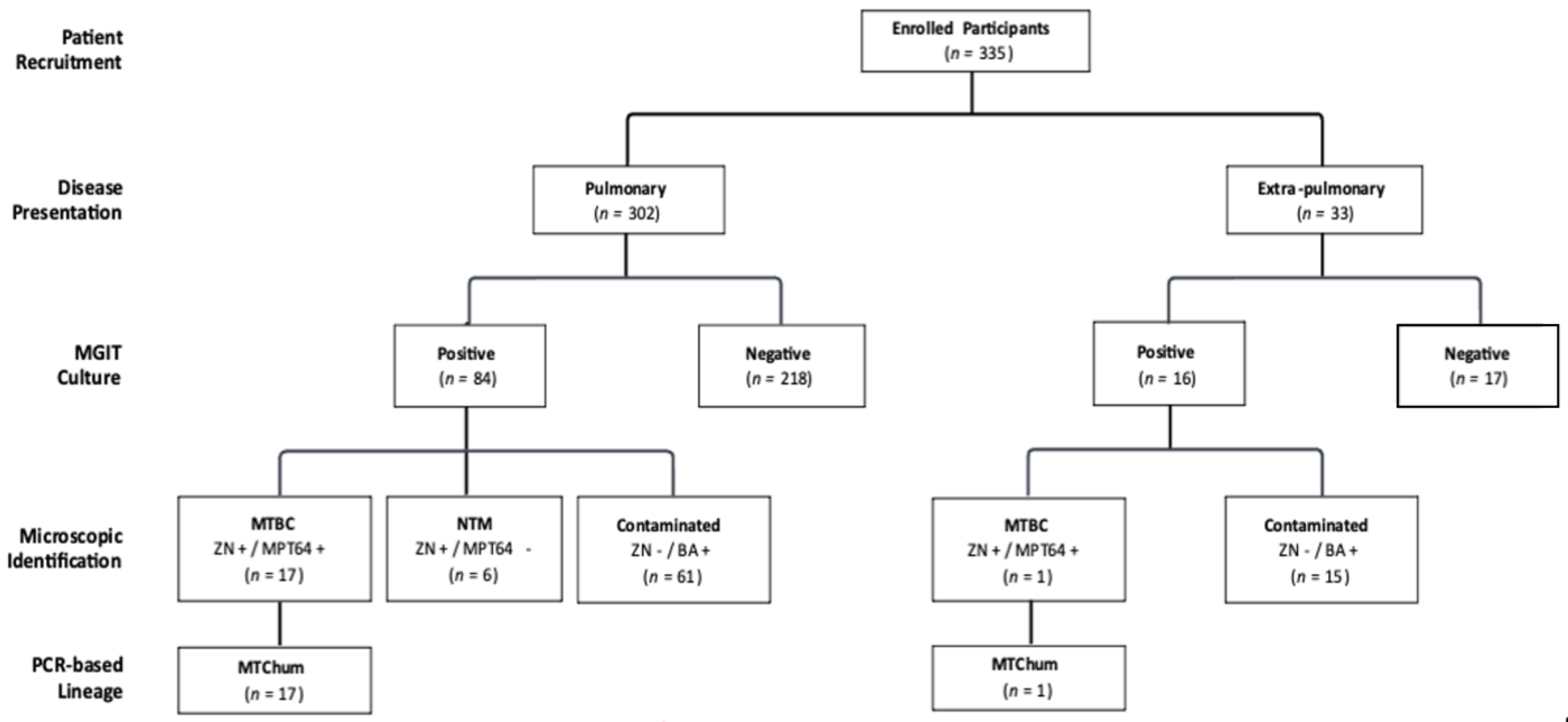
Data analyses were performed using the statistical software RStudio (2022.02.1+461). Descriptive statistics at 95% confidence intervals were estimated to assess baseline demographics, knowledge, awareness, and practices. The chi-squared test (χ2) was used to assess the association between categorical variables at the critical probability of p < 0.05. Logistic regression analysis was applied to determine the association of risk factors with TB-positive cases. All respondents (335 participants) were regarded as one population.

3. Results

3.1. Baseline Characteristics of the Studied Population

A total of 335 self-represented patients were included in the study, and their demographic statistics are represented in Table 1. The majority (60.6%) of participants were practicing livestock-keeping activities as a major source of income and economic well-being.

3.2. Recovery and Identification of Mycobacterium Species from the Target Population

Of 302 pulmonary TB samples, 84 (27.8%) were positive upon MGIT culture, with 17 (5.6%) confirmed as MTBC and 6 (2%) considered to be NTMs (Figure 2). Of the 33 Extra-pulmonary TB samples, 16 were positive on MGIT culture, but only one (3%) was classified as MTBC. Together, the study identified only 18 MTBC, amongst the 335 specimens (5%; 95% CI: 3–8%). In molecular analysis, all 18 MTBCs were identified as M. tuberculosis sensu stricto.

3.3. TB Knowledge and Level of Awareness

On the assessment of knowledge and level of awareness, both human TB and animal TB were reported to be poor in the studied population. Moreover, knowledge of zoonotic TB transmission modes was also quite low.

3.4. Practices That Increase the Risk of Zoonotic TB

The risk factors that could increase zoonotic transmission of TB infection from livestock to humans were analyzed. However, none of the risk factors were found to have a significant association with M. tuberculosis sensu stricto. Nonetheless, a noteworthy observation was that a significant portion of the studied community continued to engage in risky practices, including consuming raw milk (32.4%), drinking soup with raw blood (34.6%), and eating uncooked meat (36.4%).

4. Discussion

Our results showed poor human TB knowledge amongst participants since only 13.1% self-reported a good understanding of the disease symptoms. This may result from the fact that most participants (53.1%) reported attaining only primary education (class 1–7). Previous studies have reported the relationship between poor disease knowledge with low education levels [22,23,24]. Poor knowledge of the disease negatively affects TB management in patients while sustaining the transmission of the disease in the community [25]. Effective TB disease educational programs should be implemented to overcome the problem in Manyara.
It was expected that since this is a pastoral community with a majority engaged in livestock production, they might be familiar with bovine TB. However, to the contrary, the results showed that 82.4% of the participants had never heard of bovine TB and 85.4% had poor knowledge of the disease symptoms (Table 2). These findings are in line with another study conducted in Tanzania, where 64% were unaware of BTB and 78.7% had a poor understanding of the symptoms [26]. The poor knowledge of BTB in pastoral communities may reflect the poor veterinary awareness campaigns on animal diseases since these communities have comparatively better awareness of human TB (34).
Consistent with the general lack of awareness of bTB, zoonotic TB (zTb) awareness was also very low in that 95.2% of participants had never heard of zoonotic TB and 95.8% had a poor understanding of zoonotic transmission (Table 2). The low awareness of the transmission of zoonotic TB in the pastoral society of Manyara, which has been reported by a number of previous studies is surprising but not unusual [11,27,28]. Lack of clear knowledge on zoonotic transmission risks society to poor handling of animals and animal products and highlights a need for campaigns with effective communication addressing human TB including risk of zoonotic transmission from livestock species.
Since the studied population represents individuals presenting with symptoms of TB, the finding that only 5% were culture-confirmed positive is of concern since all individuals were placed on first-line antimycobacterial treatment such as azithromycin, amoxicillin, and cephalosporins while awaiting culture results, providing considerable additional risk to patients from delayed diagnoses of the underlying disease, potential adverse drug outcomes, increased economic costs, and potential contribution to the spread of antimicrobial resistance. This reinforces an urgent need to improve capabilities to culture or molecularly detect TB in suspect patients prior to placing them on treatment.
Of the culture-positive TB participants, 50% (9/18) were livestock keepers. The burden of MTBCs among pastoral societies has been reported in other studies, as well as in Tanzania [29,30]. Although much attention is focused on MTBCs species, there should also be a concern in the non-tuberculous mycobacteria (NTM) group. In this study, 2/6 (33%; 95% CI: 0–71%) of NTM-positive cases were livestock keepers who reported to hospitals with pulmonary complaints. Many other studies have reported an increase in non-tuberculous mycobacteria pulmonary disease (NTM-PD), but the reasons behind the increase are still unclear [31,32]. Therefore, in managing TB in pastoral societies, MTBCs and NTMs must be kept in mind during surveillance.
All the identified M. tuberculosis complex isolates recovered from the patients’ specimens were of human lineage M. tuberculosis sensu stricto. This finding is in line with a study conducted in China, which employed the same methodology but with a larger sample size than the one used in this study, and still, M. bovis was not detected [33]. It is also consistent with the report that modern human tuberculosis (TB) infections, to a large extent, are caused by Mycobacterium tuberculosis sensu stricto and that reported human TB cases due to animal-associated strains are low [34]. However, based on the sample size of the present study, 0/18 (0%; 95% CI: 0–18%), the M. bovis upper bound was still found within the expected zone (10%; 95% CI: 0–18%), therefore the possibility of M. bovis infectious among the human TB cases cannot be excluded. To address this knowledge gap, we recommend future statistically robust epidemiological investigations to assess the contribution of M. bovis or other Mycobacterium lineage infections to human TB in pastoral societies, particularly those that have a high reported prevalence of extra-pulmonary infections.
Furthermore, results from this study show that the studied community practices high-risk behaviors including drinking raw milk (32.4%) or drinking soup mixed with raw blood (34.6%), a recipe for Maasai traditional medicine known as “motori” [35], as well as consumption of uncooked meat (36.4%) (Table 3). Although these risky behaviors were not significantly associated with positive M. tuberculosis sensu stricto cases in this study, this should not mean they do not put individuals at risk. Studies by other researchers have demonstrated that indeed these are significant risk factors for contracting TB [11,36,37,38]. We believe the failure to establish a significant association between the risk factors and positive cases in the present study was due to the fewer numbers of positive cases detected, which calls for further rigorously designed studies to address the problem. Thus, concerted and well-coordinated efforts involving medical and veterinary authorities are likely needed to address traditional pastoral cultures which are risky for contracting tuberculosis and other diseases.
This was a cross-sectional study, and to increase the chance of getting positive patients, only the TB suspects were involved leading to misrepresentation of the population. Further, the items included in the risk factors and disease awareness were based on expert knowledge and literature review. Since the study was not initially designed or powered to carry out or include in-depth qualitative research within the communities, further well-powered and rigorously designed investigations are urgently needed to assess the true risk and risk factors associated with zTB in this population.

5. Conclusions

The study suggests that Manyara residents have poor knowledge and high-risk practices for exposure to TB and for contracting zoonotic TB. Therefore, for better management and effective communication of the disease, there is an urgent unmet need to better quantify the risks and identify the transmission pathways associated with the disease. To this end, well-powered longitudinal case-control studies are needed in agro-pastoralist settings. Also, the study recommends more awareness campaigns on TB to go along with other endeavors aimed at controlling TB in the Manyara region and other agro-pastoralist settings in Tanzania.

Supplementary Materials

The following supporting information can be downloaded at: https://www.mdpi.com/article/10.3390/zoonoticdis3040022/s1.

Author Contributions

P.M. (Prudence Masanga) developed the study protocol, ran the searches, led the sample collection and analysis, and wrote the first draft of the manuscript. J.B., V.K. and S.M. conceptualized the study, sourced the funding, assisted in writing the manuscript, and supervised the research. S.S., I.C. and R.K. provided input on the study protocol and performed formal analysis and validation. S.P., P.M. (Peter Mbelele), P.D. and A.L. performed sample collection and analysis. B.L., L.M. and S.L. developed the methodology. All authors have read and agreed to the published version of the manuscript.

Funding

This project is part of the EDCTP2 program supported by the European Union (grant number TMA2016SF-1463-REMODEL-TZ), Pennsylvania State University, and the African Development Bank (AFDB).

Institutional Review Board Statement

The study was conducted in accordance with the Declaration of Helsinki, and approved by the Northern Tanzania Health Research Ethics Committee (KNCHREC) with registration KNCHREC-00031 on 13 March 2020.

Informed Consent Statement

All participants provided written informed consent after they were fully briefed on study procedures. Children assented and witnessed by their parents/guardian. Written informed consent has been obtained from the patient(s) to publish this paper.

Data Availability Statement

The raw data supporting the conclusions of this article will be made available by the authors without undue reservation to any qualified researcher.

Acknowledgments

The authors acknowledge NM-AIST, KIDH, Manyara Regional, and KCRI authorities for permitting to conduct of this study in their laboratories. In addition, we acknowledge the African Development Bank (AFDB) and the Pennsylvania State University for the provision of reagents, support, and sponsorship in undertaking this research. We also acknowledge patients for voluntarily participating in the study.

Conflicts of Interest

The authors declare no conflict of interest. The funders had no role in the design of the study; in the collection, analyses, or interpretation of data; in the writing of the manuscript; or in the decision to publish the results.

Appendix A

Table A1. Survey questionnaire. The main areas of animal management and practices, habits, and awareness of bovine tuberculosis of respondents to the questionnaires.
Risk Factor GroupGeneral Description
GeneralSex, age, marital status, level of education, occupation, tribe, and residence,
Animal managementHerd type, herd size, husbandry system, animal type and breed, feeding practices, and body condition of animals.
Animal ownership and caretakingReason, sources of cattle, number and type of cattle, other livestock, reasons and rate of exploitation of herds, size of household (involved in animal business).
Housing of animals (especially at nights), Contact with animal (human–animal interactions)Type and degree (duration)of interaction with cattle, contact with other livestock, consumption of unpasteurized milk or milk products, eat raw meat, keeping other animals, abattoirs, cattle markets, vaccination campaign, communal dips.
Contacts of owned cattle with other cattle (animal–animal interactions)Use of same bulls for breeding (group bull), contact with other livestock, transhumance, communal grazing, cattle market, going to or coming from cattle market, vaccination centers, drinking spots, communal dips.
Awareness and recognition of human TBPrevious contact/exposure to TB cases/knowledge, mode of transmission (milk, meat, aerosols), humans affected by bovine TB
Clinical signs in humans and detectionClinical symptoms including fever, cough with expectoration, chills, night sweats, chest pain, abdominal pain, body ache, weight loss, loss of appetite? Detection by acid fast bacilli (AFB) smear and culture of appropriate sputum samples and chest X-ray
Awareness and recognition of TB in animalsPrevious contact/knowledge, Veterinary service, know bovine TB is zoonotic, mode of transmission (milk, meat, aerosol), cattle be affected by human TB
Vaccination programs The request of veterinary servicesVaccines? Routine vaccination? Reasons for veterinary attention, sick animals, average number of veterinary visits per year.
Clinical signs in animals and bovine TB detectionLow productivity, weak, emaciated or diseased? Diagnostic methods? Screening frequency, testing service/agency, awareness and implementation of bovine TB control law, action after test (if positive result), acceptance of routine testing, payment for bovine TB test.
Attached questionnaire form (Supplementary Materials).
Table A2. Primer-Probe Mix.
ParametersConcentrationVolume
Forward Primer100 µM20 µL
Reverse Primer100 µM20l µL
Probe100 µM10 µL
Sterile water 50l µL
Total40×100l µL
Note: The volume of nuclease free water to be added was calculated using the calculator at: https://www.idtdna.com/Calc/resuspension/, accessed on: 3 March 2020.
Table A3. PCR MIX.
Table A3. PCR MIX.
ComponentFinal ConcentrationVolume per Reaction
Prime Time Gene Expression Master10 µL
Mix (2×) with ROX dye0.5 µL
prime Time qPCR Assay 1 (40×)0.5 µL
prime Time qPCR Assay 2 (40×)0.5 µL
prime Time qPCR Assay 3 (40×)0.5 µL
DNA template (with internal control) 1 µL
Nuclease free water 7.5 µL
Total 20 µL
Note: When performing Set 1 multiplex, the PrimeTime qPCR Assays 1, 2 and 3 refer to IS1081, MTCAni and MTCHum. When performing Set 2 multiplex, the PrimeTime qPCR Assays 1, 2 and 3 refer to Morg, Mcap and Mbov.
Table A4. PCR Thermal profile.
StepTempTimeCycles
Polymerase activation95 °C3 min1
Denaturation 95 °C15 s40
Annealing/Extension63 °C1 min
Table A5. Sequence and melting temperatures for Mycobacterium primer probe sets.
AssayPrimerPrimer SequenceTm
IS1081IS1081_FGGCTGCTCTCGACGTTCATC58.2
IS1081_RCGCTGATTGGACCGCTCAT58
IS1081_PCTGAAGCCGACGCCCTGTGC63.8
MTCAniMTCAni_FGGTTTCTCTTCAACGTCTTGCT55.4
MTCAni_RCCGTCCCACGGCTTTGG59.6
MTCAni_PCGGCTGTGCGATCTTCACCGTGAA63.5
MTCHumMTCHum_FCGGTGTTTCTCATGCACGTCTC58.3
MTCHum_RCGTCGCCTTGATCATCGAAAT55.5
MTCHum_PTTACCACGCTGACCCACACCGT63.1
MbovMbov_FAGCCGTAGTCGTGCAGAA56.4
Mbov_RCCCGTAGCGTTACTGAGAAATTG55.7
Mbov_PCAACACTCTTGGAGTGGCCTACAACG61.3
McapRTMcap_FACCGTGCGGATCTTG52.9
Mcap_RCATGGAGATCACCCGT52
Mcap_PTATCGGGTACACAAAGACGA56
MorgMorg_FATTGTCGCGCCGAGACTG58.2
Morg_RGTACCATCTTGGCCGAGCTG58.2
Morg_PCGTCCTCGGCTGACCC58.6

References

  1. World Health Organization. Global Tuberculosis Report 2022. Available online: https://reliefweb.int/report/world/global-tuberculosis-report-2022 (accessed on 15 May 2023).
  2. Huard, R.C.; Fabre, M.; de Haas, P.; Lazzarini, L.C.O.; van Soolingen, D.; Cousins, D.; Ho, J.L. Novel genetic polymorphisms that further delineate the phylogeny of the Mycobacterium tuberculosis complex. J. Bacteriol. 2006, 188, 4271–4287. [Google Scholar] [CrossRef]
  3. World Health Organization. A Situational Analysis of Programmatic Management of TB Preventive Treatment in the WHO South-East Asia Region. Available online: https://iris.who.int/handle/10665/337381 (accessed on 12 May 2023).
  4. De la Rua-Domenech, R. Human Mycobacterium bovis infection in the United Kingdom: Incidence, risks, control measures and review of the zoonotic aspects of bovine tuberculosis. Tuberculosis 2006, 86, 77–109. [Google Scholar] [CrossRef] [PubMed]
  5. Müller, B.; Dürr, S.; Alonso, S.; Hattendorf, J.; Laisse, C.J.; Parsons, S.D.; Van Helden, P.D.; Zinsstag, J. Zoonotic Mycobacterium bovis–induced tuberculosis in humans. Emerg. Infect. Dis. 2013, 19, 899. [Google Scholar] [CrossRef]
  6. Sichewo, P.R.; Vander Kelen, C.; Thys, S.; Michel, A.L. Risk practices for bovine tuberculosis transmission to cattle and livestock farming communities living at wildlife-livestock-human interface in northern KwaZulu Natal, South Africa. PLoS Neglected Trop. Dis. 2020, 14, e0007618. [Google Scholar] [CrossRef]
  7. Etchechoury, I.; Valencia, G.E.; Morcillo, N.; Sequeira, M.; Imperiale, B.; López, M.; Caimi, K.; Zumárraga, M.; Cataldi, A.; Romano, M. Molecular typing of Mycobacterium bovis isolates in Argentina: First description of a Person-to-Person transmission case. Zoonoses Public Health 2010, 57, 375–381. [Google Scholar] [CrossRef] [PubMed]
  8. World Health Organization. Tuberculosis Profile 2017. Available online: https://apps.who.int/iris/handle/10665/259366 (accessed on 18 May 2023).
  9. National Tuberculosis and Leprosy Programme. Final_Joint_External_Program_Review_Report_March-Converted.pdf. Available online: https://ntlp.go.tz/tuberculosis/tb-prevalence-in-tanzania/ (accessed on 28 May 2022).
  10. World Health Organization. Global Tuberculosis Control, Global, Region and Country-Specific for Key Indicators. Available online: http://www.who.int/tb/data (accessed on 25 June 2023).
  11. Mfinanga, S.G.; Mørkve, O.; Kazwala, R.; Cleaveland, S.; Sharp, J.; Shirima, G.; Nilsen, R. The role of livestock keeping in tuberculosis trends in Arusha, Tanzania. Int. J. Tuberc. Lung Dis. 2003, 7, 695–704. [Google Scholar]
  12. Jain, A. Extra pulmonary tuberculosis: A diagnostic dilemma. Indian J. Clin. Biochem. 2011, 26, 269–273. [Google Scholar] [CrossRef] [PubMed]
  13. Mkombozi, F.; Kazwala, R.; Lupindu, A. Prevalence and Risk factors of Bovine Tuberculosis in smallholder dairy cattle in Babati town council of Manyara region, Tanzania. Tanzan. Vet. J. 2014, 29, 30–38. [Google Scholar]
  14. United Republic of Tanzania. National Sample Census of Agriculture 2002/2003. Regional Report: Manyara. Available online: https://www.kilimo.go.tz/uploads/statistics/Manyara_Report.pdf (accessed on 15 December 2022).
  15. Arya, R.; Antonisamy, B.; Kumar, S. Sample size estimation in prevalence studies. Indian J. Pediatr. 2012, 79, 1482–1488. [Google Scholar] [CrossRef] [PubMed]
  16. Tripathi, K.; Tripathi, P.C.; Nema, S.; Shrivastava, A.K.; Dwiwedi, K.; Dhanvijay, A.K. Modified Petroff’s method: An excellent simplified decontamination technique in comparison with Petroff’s method. Int. J. Recent Trends Sci. Technol. 2014, 10, 461–464. [Google Scholar]
  17. Patil, P.S.; Chandi, D.H.; Rangaiahagari, A. Utility of GeneXpert MTB/RIF in the Rapid Diagnosis of Extra Pulmonary Tuberculosis. Indian J. Respir. Care 2023, 12, 46–48. [Google Scholar] [CrossRef]
  18. Siddiqi, S.H.; Rüsch-Gerdes, S. For BACTEC™ MGIT 960™ TB System. In Mycobacteria Growth Indicator Tube (MGIT) Culture and Drug Susceptibility Demonstration Projects; Foundation for innovative new Diagnostic (FinD): Borstel, Germany, 2006. [Google Scholar]
  19. Stewart, L.D.; McCallan, L.; McNair, J.; McGoldrick, A.; Morris, R.; Moyen, J.-L.; Ferré, L.D.J.; Romero, B.; Alonso, E.; Parsons, S.D. Multilaboratory evaluation of a novel lateral flow immunochromatographic assay for confirming isolation of Mycobacterium bovis from veterinary diagnostic specimens. J. Clin. Microbiol. 2017, 55, 3411–3425. [Google Scholar] [CrossRef]
  20. Amaro, A.; Duarte, E.; Amado, A.; Ferronha, H.; Botelho, A. Comparison of three DNA extraction methods for Mycobacterium bovis, Mycobacterium tuberculosis and Mycobacterium avium subsp. avium. Lett. Appl. Microbiol. 2008, 47, 8–11. [Google Scholar] [CrossRef]
  21. Halse, T.A.; Escuyer, V.E.; Musser, K.A. Evaluation of a single-tube multiplex real-time PCR for differentiation of members of the Mycobacterium tuberculosis complex in clinical specimens. J. Clin. Microbiol. 2011, 49, 2562–2567. [Google Scholar] [CrossRef]
  22. Ismail, A.; Josephat, P. Knowledge and perception on tuberculosis transmission in Tanzania: Multinomial logistic regression analysis of secondary data. Tanzan. J. Health Res. 2014, 16, 29–37. [Google Scholar] [CrossRef] [PubMed]
  23. Maja, T.F.; Maposa, D. An investigation of risk factors associated with tuberculosis transmission in South Africa using logistic regression model. Infect. Dis. Rep. 2022, 14, 609–620. [Google Scholar] [CrossRef]
  24. Kazaura, M.; Kamazima, S.R. Knowledge, attitudes and practices on tuberculosis infection prevention and associated factors among rural and urban adults in northeast Tanzania: A cross-sectional study. PLoS Glob. Public Health 2021, 1, e0000104. [Google Scholar] [CrossRef]
  25. Mushtaq, M.U.; Shahid, U.; Abdullah, H.M.; Saeed, A.; Omer, F.; Shad, M.A.; Siddiqui, A.M.; Akram, J. Urban-rural inequities in knowledge, attitudes and practices regarding tuberculosis in two districts of Pakistan’s Punjab province. Int. J. Equity Health 2011, 10, 8. [Google Scholar] [CrossRef]
  26. Katale, B.Z.; Mbugi, E.V.; Karimuribo, E.D.; Keyyu, J.D.; Kendall, S.; Kibiki, G.S.; Godfrey-Faussett, P.; Michel, A.L.; Kazwala, R.R.; Van Helden, P. Prevalence and risk factors for infection of bovine tuberculosis in indigenous cattle in the Serengeti ecosystem, Tanzania. BMC Vet. Res. 2013, 9, 267. [Google Scholar] [CrossRef] [PubMed]
  27. Kilale, A.M.; Ngadaya, E.; Kagaruki, G.B.; Lema, Y.L.; Muhumuza, J.; Ngowi, B.J.; Mfinanga, S.G.; Hinderaker, S.G. Experienced and perceived risks of mycobacterial diseases: A cross sectional study among agropastoral communities in northern Tanzania. PLoS ONE 2015, 10, e0130180. [Google Scholar] [CrossRef]
  28. Shirima, G.; Kazwala, R.; Kambarage, D. Prevalence of bovine tuberculosis in cattle in different farming systems in the eastern zone of Tanzania. Prev. Vet. Med. 2003, 57, 167–172. [Google Scholar] [CrossRef] [PubMed]
  29. Belay, M.; Ameni, G.; Bjune, G.; Couvin, D.; Rastogi, N.; Abebe, F. Strain diversity of Mycobacterium tuberculosis isolates from pulmonary tuberculosis patients in Afar pastoral region of Ethiopia. BioMed Res. Int. 2014, 2014, 238532. [Google Scholar] [CrossRef] [PubMed]
  30. Kazwala, R.; Daborn, C.; Sharp, J.; Kambarage, D.; Jiwa, S.; Mbembati, N. Isolation of Mycobacterium bovis from human cases of cervical adenitis in Tanzania: A cause for concern? Int. J. Tuberc. Lung Dis. 2001, 5, 87–91. [Google Scholar]
  31. Agada, C.; Adesokan, H.; Igwe, D.; Cadmus, S. Mycobacterium africanum and nontuberculous mycobacteria from fresh milk of pastoral cattle and soft cheese in Oyo State--implications for public health. Afr. J. Med. Med. Sci. 2014, 43, 13–20. [Google Scholar] [PubMed]
  32. Katale, B.Z.; Mbugi, E.V.; Botha, L.; Keyyu, J.D.; Kendall, S.; Dockrell, H.M.; Michel, A.L.; Kazwala, R.R.; Rweyemamu, M.M.; Van Helden, P. Species diversity of non-tuberculous mycobacteria isolated from humans, livestock and wildlife in the Serengeti ecosystem, Tanzania. BMC Infect. Dis. 2014, 14, 616. [Google Scholar] [CrossRef] [PubMed]
  33. Jiang, G.; Wang, G.; Chen, S.; Yu, X.; Wang, X.; Zhao, L.; Ma, Y.; Dong, L.; Huang, H. Pulmonary tuberculosis caused by Mycobacterium bovis in China. Sci. Rep. 2015, 5, 8538. [Google Scholar] [CrossRef]
  34. Vågene, Å.J.; Honap, T.P.; Harkins, K.M.; Rosenberg, M.S.; Giffin, K.; Cárdenas-Arroyo, F.; Leguizamón, L.P.; Arnett, J.; Buikstra, J.E.; Herbig, A. Geographically dispersed zoonotic tuberculosis in pre-contact South American human populations. Nat. Commun. 2022, 13, 1195. [Google Scholar] [CrossRef]
  35. Roulette, C.J.; Njau, E.-F.A.; Quinlan, M.B.; Quinlan, R.J.; Call, D.R. Medicinal foods and beverages among Maasai agro-pastoralists in northern Tanzania. J. Ethnopharmacol. 2018, 216, 191–202. [Google Scholar] [CrossRef]
  36. Kazwala, R.; Kambarage, D.; Daborn, C.; Nyange, J.; Jiwa, S.; Sharp, J. Risk factors associated with the occurrence of bovine tuberculosis in cattle in the Southern Highlands of Tanzania. Vet. Res. Commun. 2001, 25, 609–614. [Google Scholar] [CrossRef]
  37. Mfinanga, S.; Morkve, O.; Kazwala, R.; Cleaveland, S.; Sharp, M.; Kunda, J.; Nilsen, R. Mycobacterial adenitis: Role of Mycobacterium bovis, non-tuberculous mycobacteria, HIV infection, and risk factors in Arusha, Tanzania. East Afr. Med. J. 2004, 81, 171–178. [Google Scholar] [CrossRef]
  38. Ameni, G.; Amenu, K.; Tibbo, M. Bovine tuberculosis: Prevalence and risk factor assessment in cattle and cattle owners in Wuchale-Jida district, Central Ethiopia. J. Appl. Res. Vet. Med. 2003, 1, 17–26. [Google Scholar]
Figure 1. Locations of the health facilities which were involved in sample collection. The left panel shows a map of Tanzania with the Manyara region shaded in grey. The right panel shows the districts in the Manyara region and the location of the health facilities involved in the study.
Zoonoticdis 03 00022 g001
Figure 2. Patient Recruitment and Identification of MTBC in Clinics at Study Sites in Tanzania. The flow diagram illustrates the process of patient recruitment and subsequent identification methods for Mycobacterium tuberculosis complex (MTBC) strains circulating amongst patients in Tanzanian clinics. A total of 335 participants were enrolled and categorized into pulmonary (n = 302) and extra-pulmonary (n = 33) presentations. Mycobacterial Growth Indicator Tube (MGIT) culture was performed, resulting in 84 and 16 positive samples from pulmonary and extrapulmonary specimens, respectively. Microscopic identification was conducted using Ziehl–Neelsen (ZN) staining and MPT64 antigen detection, identifying MTBC in 17 samples and nontuberculous mycobacteria (NTM) in 6 samples from pulmonary patients, and 1 MTBC from a patient with extrapulmonary presentation. PCR-based lineage analysis further confirmed MTChum (M. tuberculosis sensu stricto) positivity in all MTBC-positive samples. The flow diagram also includes details of negative results and contaminated samples identified by negative ZN staining and growth in blood agar (BA).
Zoonoticdis 03 00022 g002
Table 1. Demographic characteristics of the studied population.
VariableCategoryn%
Sexfemale16950.5
male16649.6
0–18164.8
19–5419056.7
>5512938.5
Babati8124.2
Hanang7321.8
DistrictsKiteto72.1
Mbulu15546.3
Simanjiro20.6
Other175.1
EducationNo education9227.5
Primary17853.1
Secondary6519.4
Professionallivestock attendants20360.6
Housewives3711.0
Merchant4714.0
peasant226.6
students133.9
office work10.3
driver10.3
unemployed92.7
wildlife workers10.3
craft10.3
Table 2. TB knowledge and level of awareness among studied individuals (n = 335).
Categoryn%
Human TB awarenessYes229 68.4
No106 31.6
Awareness of Human TB symptomsNone106 31.6
Poor82 24.5
Average103 30.8
Good44 13.1
Awareness of Bovine TBYes59 17.6
No27682.4
Awareness of Bovine TB symptomsNone286 85.4
Good49 14.6
Awareness of Zoonotic TBYes16 4.8
No319 95.2
Awareness of Zoonotic TB transmission None32195.8
Good144.2
TB = Tuberculosis.
Table 3. Univariate analysis for factors associated with the transmission of TB.
Exposure Variables Total SamplePositive CasesNegative CasesOR (95%CI)p-Value
Category(n = 335)
n (%)
(n = 18)
n (%)
(n = 317)
n (%)
Raw meatyes122 (36.4)5 (27.8)117 (36.9)0.66 (0.21–1.79)0.436
no213 (63.6)13 (72.2)200 (63.1)
Raw dairy productYes110 (32.8)4 (22.2)106 (33.4)0.57 (0.18–1.77)0.33
No225 (67.2)14 (77.8)211 (66.5)
Soup with bloodyes116 (34.6)6 (33.3)110 (34.7)0.94 (0.32–2.49)0.9
No219 (65.4)12 (66.7)207 (65.3)
Blood mixed with milkyes40 (11.9)2 (11.1)38 (11.9)0.92 (0.14–3.39)0.91
no295 (88.1)16 (88.9)279 (88.0)
Consumed aborted animalYes19 (5.7)2 (11.1)17 (5.4)2.21 (0.33–8.64)0.32
No316 (94.3)16 (88.9)300 (94.6)
Share the roof with cattleYes110 (32.8)6 (33.3)104 (32.8)1.02 (0.35–2.72)0.96
No225 (67.2)12 (66.7)213 (67.2)
Handled aborted productsYes51 (15.2)5 (27.8)46 (14.5)1.81 (0.70–6.33)0.28
No284 (84.8)13 (72.2)217 (85.5)
Family member with TBYes25 (7.5)0 (0)25 (7.9)0 (0–NaN)0.41
No310 (92.5)18 (100)292 (92.1)
TB = Tuberculosis; OR = Odds Ratio; CI = Confidence Interval.
Disclaimer/Publisher’s Note: The statements, opinions and data contained in all publications are solely those of the individual author(s) and contributor(s) and not of MDPI and/or the editor(s). MDPI and/or the editor(s) disclaim responsibility for any injury to people or property resulting from any ideas, methods, instructions or products referred to in the content.

