
Effects of Adper™ Scotchbond™ 1 XT, Clearfil™ SE Bond 2 and Scotchbond™ Universal in Odontoblastic Activity †

 ,
,  , , , ,
  , and
, and 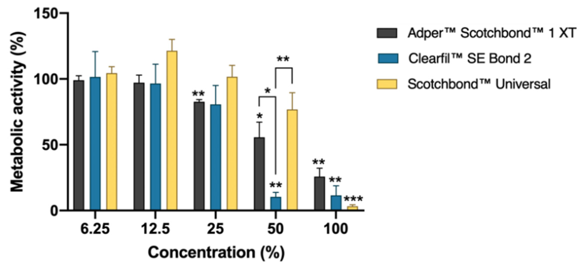
Abstract
:1. Introduction
2. Materials and Methods
2.1. Adhesive Systems Extracts and Cell Cultures
2.2. Metabolic Activity and Protein Content
2.3. Types of Cell Death
2.4. Morphology and Qualitative Cytotoxicity Assessment
2.5. Statistical Analysis
3. Results and Discussion
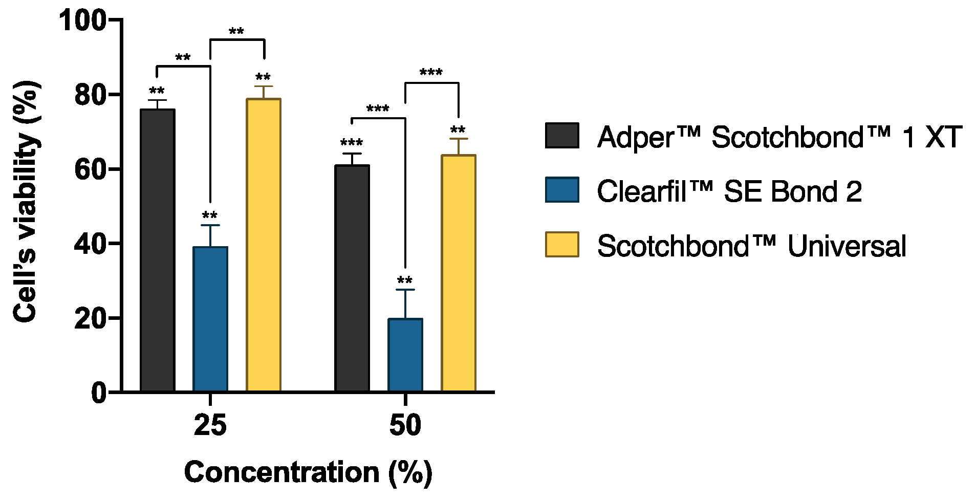
3.1. Metabolic Activity and Protein Content
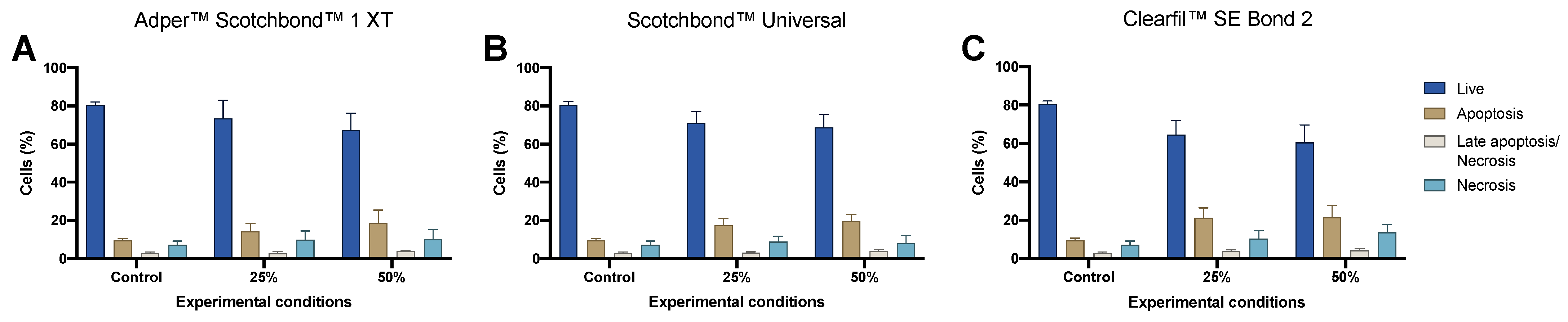
3.2. Types of Cell Death
3.3. Morphology and Qualitative Cytotoxicity Assessment
4. Conclusions
Author Contributions
Funding
Institutional Review Board Statement
Informed Consent Statement
Data Availability Statement
Conflicts of Interest
References
- Van Meerbeek, B.; Yoshihara, K.; Van Landuyt, K.; Yoshida, Y.; Peumans, M. From Buonocore’s Pioneering Acid-Etch Technique to Self-Adhering Restoratives. A Status Perspective of Rapidly Advancing Dental Adhesive Technology. J. Adhes. Dent. 2020, 22, 7–34. [Google Scholar] [PubMed]
- Van Meerbeek, B.; Yoshihara, K.; Yoshida, Y.; Mine, A.; De Munck, J.; Van Landuyt, K.L. State of the art of self-etch adhesives. Dent. Mater. 2011, 27, 17–28. [Google Scholar] [CrossRef] [PubMed]
- Carrilho, E.; Cardoso, M.; Marques Ferreira, M.; Marto, C.M.; Paula, A.; Coelho, A.S. 10-MDP Based Dental Adhesives: Adhesive Interface Characterization and Adhesive Stability-A Systematic Review. Materials 2019, 12, 790. [Google Scholar] [CrossRef] [PubMed] [Green Version]
- Milia, E.; Cumbo, E.; Cardoso, R.J.; Gallina, G. Current dental adhesives systems. A narrative review. Curr. Pharm. Des. 2012, 18, 5542–5552. [Google Scholar] [CrossRef] [PubMed]
- Modena, K.C.; Casas-Apayco, L.C.; Atta, M.T.; Costa, C.A.; Hebling, J.; Sipert, C.R.; Navarro, M.F.; Santos, C.F. Cytotoxicity and biocompatibility of direct and indirect pulp capping materials. J. Appl. Oral. Sci. 2009, 17, 544–554. [Google Scholar] [CrossRef] [PubMed]
- Paula, A.B.; Laranjo, M.; Coelho, A.S.; Abrantes, A.M.; Gonçalves, A.C.; Sarmento-Ribeiro, A.B.; Ferreira, M.M.; Botelho, M.F.; Marto, C.M.; Carrilho, E. Accessing the Cytotoxicity and Cell Response to Biomaterials. JoVE J. Vis. Exp. 2021, 173, e61512. [Google Scholar] [CrossRef] [PubMed]
- ISO 10993-5 Biological Evaluation of Medical Devices–Part 5: Tests for In Vitro Cytotoxicity; International Organization for Standardization: Geneva, Switzerland, 2009.


Publisher’s Note: MDPI stays neutral with regard to jurisdictional claims in published maps and institutional affiliations. |
© 2021 by the authors. Licensee MDPI, Basel, Switzerland. This article is an open access article distributed under the terms and conditions of the Creative Commons Attribution (CC BY) license (https://creativecommons.org/licenses/by/4.0/).
Share and Cite
Cardoso, M.; Coelho, A.; Marto, C.M.; Gonçalves, A.C.; Paula, A.; Botelho, M.F.; Laranjo, M.; Carrilho, E. Effects of Adper™ Scotchbond™ 1 XT, Clearfil™ SE Bond 2 and Scotchbond™ Universal in Odontoblastic Activity. Biol. Life Sci. Forum 2021, 9, 3. https://doi.org/10.3390/ECCM-10866
Cardoso M, Coelho A, Marto CM, Gonçalves AC, Paula A, Botelho MF, Laranjo M, Carrilho E. Effects of Adper™ Scotchbond™ 1 XT, Clearfil™ SE Bond 2 and Scotchbond™ Universal in Odontoblastic Activity. Biology and Life Sciences Forum. 2021; 9(1):3. https://doi.org/10.3390/ECCM-10866
Chicago/Turabian StyleCardoso, Miguel, Ana Coelho, Carlos Miguel Marto, Ana Cristina Gonçalves, Anabela Paula, Maria Filomena Botelho, Mafalda Laranjo, and Eunice Carrilho. 2021. "Effects of Adper™ Scotchbond™ 1 XT, Clearfil™ SE Bond 2 and Scotchbond™ Universal in Odontoblastic Activity" Biology and Life Sciences Forum 9, no. 1: 3. https://doi.org/10.3390/ECCM-10866