1. Introduction
The dromedary’s immune system is one of the most mysterious topics for researchers in the fields of morphology and immunology; moreover, the lack of information about the dromedary immune system led us to conduct the current research, which is based on one of the most important lymphoid organs; the lymph nodes of one-humped camel (camelus dromedarius) from the region of El Oued in southeastern Algeria, in which we aimed to make a full cytological and immunohistochemical study of the axillary, and the mesenteric lymph nodes, in order to highlight the structure and the organization of functional compartments of these organs.
2. Material and Methods
The lymph nodes have undergone meticulous preparation, which starts with the removal of the adipose tissue covering these organs. This process required the use of a scalpel. The sampled organs were then immersed in a 10% formalin solution for 24–48 h for fixation; additional fixation was carried out at room temperature in a 10% formalin solution for 10 to 14 days for the fragments that were meant to be stained using Azur II eosin technique, whereas the identification and the localization of T and B lymphocytes were carried out using immunohistochemistry technique with both anti-CD3 and anti-CD22 monoclonal antibodies to elucidate T and B lymphocytes location.
3. Results and Discussion
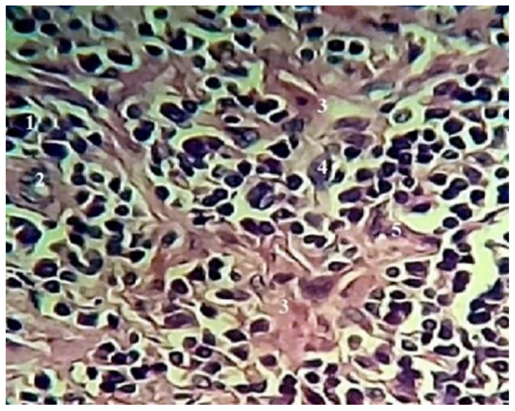
The results of the cytological study of the lymph nodes using Azur II eosin stains revealed that the lymph nodes parenchyma is composed of several cellular populations, in which the lymphocytes were the major population, followed by the macrophages, plasma cells, reticulocytes, and granulocytes such as eosinophils, basophils, and neutrophils (
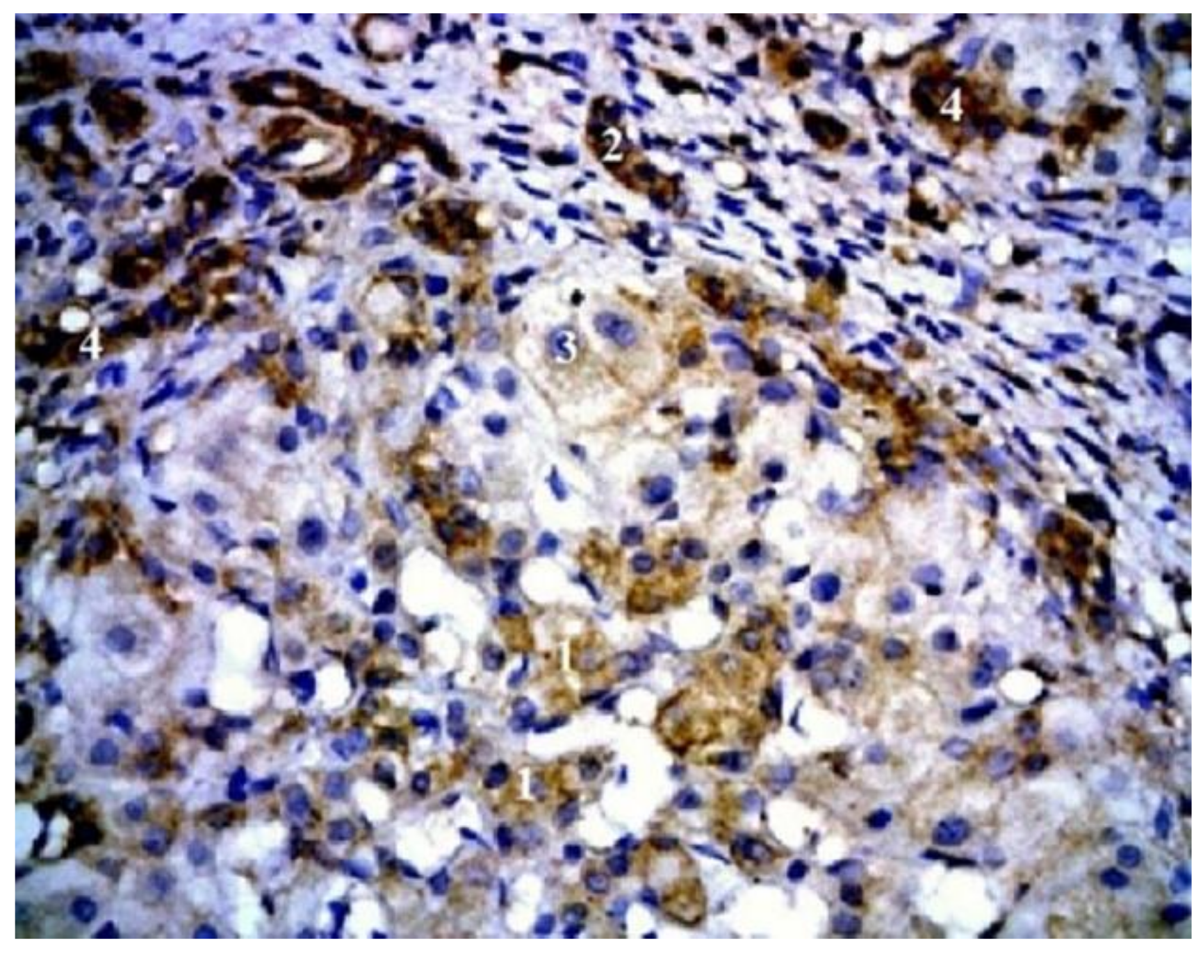
Figure 1). Moreover, it was possible to elucidate that the ratio of lymphocytes in the active follicles and in the paracortical zone was higher than the other functional zones. Furthermore, they presented a diameter of 8–12 µm, with a very dense, rounded nucleus, while the large lymphocytes presented a diameter of 12–15 µm. Whereas macrophages were recognizable by a voluminous, clear, and well-defined cytoplasm. Plasma cells were found to be spreading along the medullary zone, and the dromedary parenchyma was distinguished by a higher rate of reticular cells, plasma cells, and macrophages (
Figure 2). The study of the morphology of lymph nodes of the dromedary in the region of El Oued allowed us to highlight a distinct histological structure formed by several conglomerates consisting of a different number of small nodules scattered within the adipose tissue. Our results indicate that the parenchyma of the lymph nodes in a dromedary has a (compartmentalized) lobular structure that corresponds to the results of the researcher [
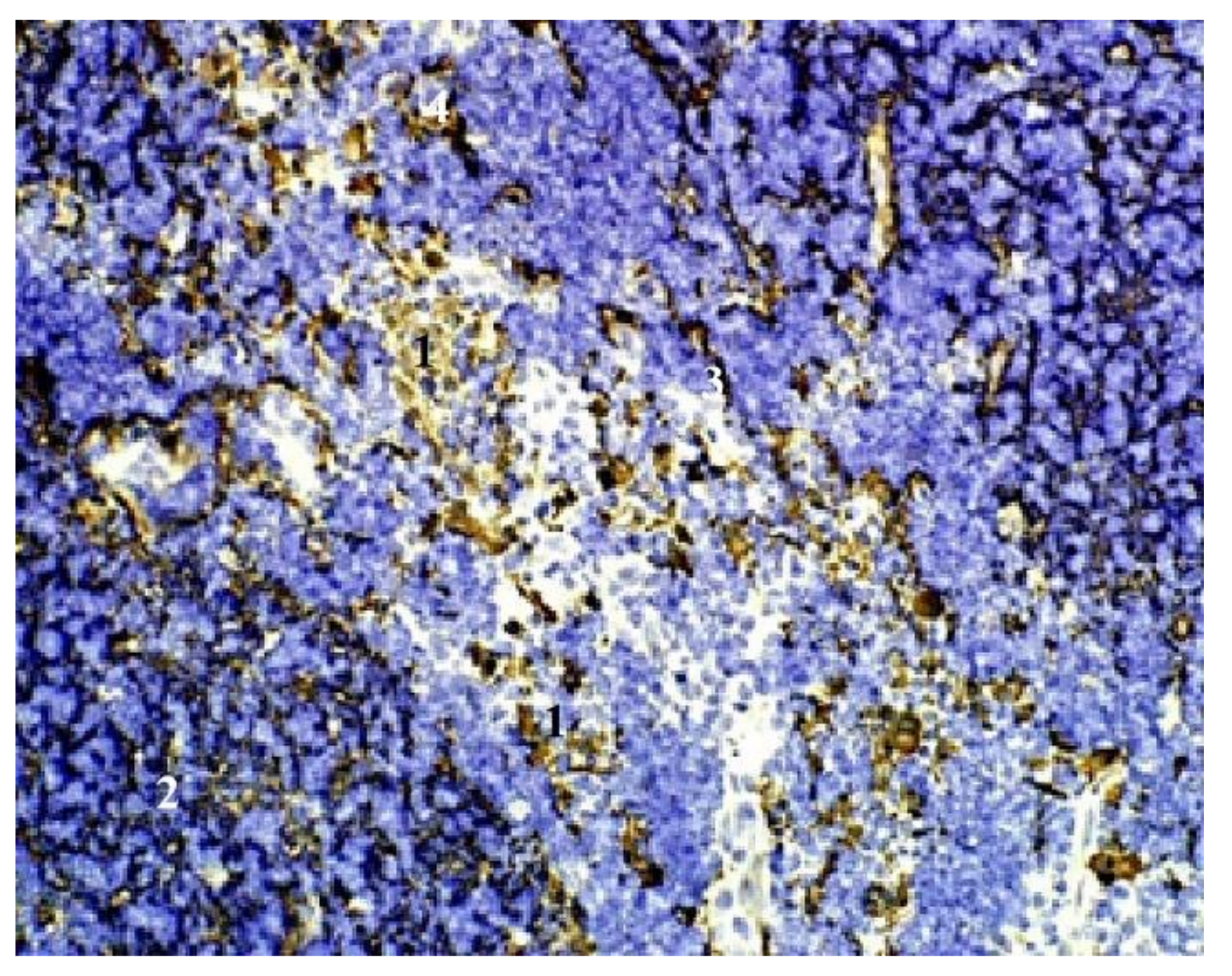
1]. The immunohistochemical results showed that the B lymphocytes were located in the center of the germinal center of the follicles of the lymph nodes, whereas the T lymphocytes were located in the paracortical zone (
Figure 2 and
Figure 3). Nearby identical results were found in the spleen [
2].
4. Conclusions
Cytological and immunohistochemical techniques were used to supplement the microscopic examination of the lymph node of the one-humped camel (camelus dromedarius), using azure II eosine staining technique and immunochemistry using monoclonal antibodies; CD3 and CD22, in order to highlight the location of T and B lymphocytes, and from the obtained results, it is now possible to say that all mammals share the same lymphocyte location, in which the B lymphocytes are located in the germinal center of the primary follicles, while the T lymphocytes are located in the paracortical zone, whereas the B lymphocytes were located in the follicles of both the cortical and the medullary zone and the T lymphocytes were located exclusively in the para-cortical region.
Author Contributions
Conceptualization, T.K., D.E.R. and M.A.F.; methodology, M.A.F.; software, M.A.F.; validation, M.A.F., T.K. and D.E.R.; formal analysis, M.A.F.; investigation, M.A.F.; resources, M.A.F.; data curation, M.A.F.; writing—original draft preparation, M.A.F.; writing—review and editing, M.A.F.; visualization, M.A.F.; supervision, T.K. and D.E.R.; project administration, T.K.; funding acquisition, M.A.F. All authors have read and agreed to the published version of the manuscript.
Funding
This research received no external funding.
Institutional Review Board Statement
The animal study protocol was approved by the Institutional Review Board of the institute of agriculture and veterinary sciences in Taoura, University of Souk Ahras, Algeria.
Informed Consent Statement
Not applicable.
Data Availability Statement
No new data were created or analyzed in this study. Data sharing is not applicable to this article.
Acknowledgments
The authors would thank Yassine Ladjailia (Laboratory Associate in the Institute of Agriculture and Veterinary Sciences, University of Souk Ahras) for the comments that greatly improved the manuscript.
Conflicts of Interest
The authors declare no conflict of interest.
References
- Rahmoun, D.; Lieshchova, M.; Fares, M. Morphological and radiological study of lymph nodes in dromedaries in Algeria. Regul. Mech. Biosyst. 2020, 11, 330–337. [Google Scholar] [CrossRef]
- Amine, F.M.; Tarek, K.; Eddine, R.D. Anatomo-topographic and histo-cytological study of dromedary’s spleen in Algeria. Folia Morphol. 2023, 82, 137–146. [Google Scholar]
| Disclaimer/Publisher’s Note: The statements, opinions and data contained in all publications are solely those of the individual author(s) and contributor(s) and not of MDPI and/or the editor(s). MDPI and/or the editor(s) disclaim responsibility for any injury to people or property resulting from any ideas, methods, instructions or products referred to in the content. |
© 2023 by the authors. Licensee MDPI, Basel, Switzerland. This article is an open access article distributed under the terms and conditions of the Creative Commons Attribution (CC BY) license (https://creativecommons.org/licenses/by/4.0/).


