Article

Lost in Translation: Exploring microRNA Biogenesis and Messenger RNA Fate in Anoxia-Tolerant Turtles

by
Sarah A. Breedon
and
Kenneth B. Storey
*
Department of Biology, Carleton University, Ottawa, ON K1S 5B6, Canada
*
Author to whom correspondence should be addressed.
Oxygen 2022, 2(2), 227-245; https://doi.org/10.3390/oxygen2020017
Submission received: 27 April 2022 / Revised: 23 May 2022 / Accepted: 15 June 2022 / Published: 17 June 2022
(This article belongs to the Special Issue Feature Papers in Oxygen)

Abstract

:
Red-eared slider turtles face natural changes in oxygen availability throughout the year. This includes long-term anoxic brumation where they reduce their metabolic rate by ~90% for months at a time, which they survive without apparent tissue damage. This metabolic rate depression (MRD) is underlaid by various regulatory mechanisms, including messenger RNA (mRNA) silencing via microRNA (miRNA), leading to mRNA decay or translational inhibition in processing bodies (P-bodies) and stress granules. Regulation of miRNA biogenesis was assessed in red-eared slider turtle liver and skeletal muscle via immunoblotting. Hepatic miRNA biogenesis was downregulated in early processing steps, while later steps were upregulated. These contradictory findings indicate either overall decreased miRNA biogenesis, or increased biogenesis if sufficient pre-miRNA stores were produced in early anoxia. Conversely, muscle showed clear upregulation of multiple biogenesis steps indicating increased miRNA production. Additionally, immunoblotting indicated that P-bodies may be favoured by the liver for mRNA storage/decay during reoxygenation with a strong suppression of stress granule proteins in anoxia and reoxygenation. Muscle however showed downregulation of P-bodies during anoxia and reoxygenation, and upregulation of stress granules for mRNA storage during reoxygenation. This study advances our understanding of how these champion anaerobes regulate miRNA biogenesis to alter miRNA expression and mRNA fate during prolonged anoxia.

1. Introduction

Red-eared slider turtles (Trachemys scripta elegans) are capable of surviving extended periods of oxygen deprivation without suffering any apparent tissue damage. These turtles experience both short-term hypoxic conditions during breath-hold diving, as well as long-term anoxic conditions over the winter months while they brumate underwater for weeks to months at a time. During winter brumation, these turtles may be unable to surface for air if bodies of water are ice-locked and the water can soon become hypoxic/anoxic to an extent that turtles are incapable of extracting sufficient oxygen using extrapulmonary methods [1,2]. As such, extended underwater submergence results in the turtles’ blood oxygen levels eventually dropping to near-zero Torr [3,4]. In animals that are not anoxia-tolerant, this lack of oxygen would result in grievous tissue damage, likely leading to organismal death after even a short period of time. However, red-eared slider turtles are able to survive this and quickly return to normal functioning once they have access to ample oxygen again.
The turtle’s ability to survive anoxia is underscored by a set of complex mechanisms that serve to protect the turtle from various cellular insults that can accompany repeated anoxia-reoxygenation cycles. These adaptation mechanisms include the ability to buffer the lactate generated during anaerobic glycolytic fermentation to prevent lethal acidosis, chaperone proteins to protect and refold proteins, and extensive constitutive and inducible antioxidant defenses to mitigate macromolecule damage caused by reactive oxygen species (ROS) [5,6,7,8,9,10,11,12,13]. Besides protective mechanisms such as these, the red-eared slider turtle also has the amazing ability to reduce its metabolic rate by approximately 90% compared to its normoxic rate [2,3,14]. This reduces the cellular demand for adenosine triphosphate (ATP) to levels that can be satisfied by anaerobic metabolic pathways, which are fueled by high organ glycogen reserves, particularly those in the liver [1,15,16]. Metabolic rate depression (MRD) is crucial to the survival of extended bouts of anoxia because if the balance of cellular energetics (i.e., the rate of ATP production versus consumption) is disrupted, the overconsumption of ATP will rapidly lead to glycogen depletion and organismal death. A major characteristic of MRD is the global suppression of nonessential, energetically expensive processes such as protein translation and turnover, and cell cycle progression [17,18,19]. However, despite needing to conserve as much energy as possible via the global suppression of gene expression, there are still a number of important pro-survival pathways that must be maintained or upregulated in order to ensure that the animal can survive prolonged stress exposure [19]. Thus, in addition to global MRD, there must also be a simultaneous reprioritization of energy usage towards the selective expression of certain stress-responsive genes.
MRD and metabolic reorganization are accomplished through the coordinated use of various regulatory mechanisms [19]. In particular, post-transcriptional inhibition of gene expression via microRNA (miRNA) is of particular interest due to their ability to rapidly and reversibly alter gene expression in response to external stimuli [20]. miRNAs are a class of short (~22 nt) non-coding RNA that are highly conserved between species and that function to repress the translation of messenger RNA (mRNA) into protein through miRNA:mRNA binding [20,21]. miRNA regulation has been implicated in MRD of numerous stress-tolerant organisms including wood frogs, milk snails, mouse lemurs, brown bears, and naked mole rats [22,23,24,25]. Notably, previous studies have shown that miRNAs are important regulators of cell proliferation and metabolic processes in red-eared sliders in response to anoxia [17,26,27]. Given their importance to MRD, the regulation of miRNA production is a critical step for analysis to determine how these turtles alter miRNA expression under anoxia.
miRNAs are produced endogenously primarily through the canonical biogenesis pathway (Figure 1). Firstly, nuclear RNA polymerase II transcribes a miRNA-coding gene into the primary miRNA (pri-miRNA) transcript, which is then cleaved by the microprocessor complex [28]. This complex consists of Drosha—a ribonuclease (RNase) III enzyme—and DiGeorge syndrome critical region 8 (DGCR8)—a non-catalytic subunit containing two double-stranded RNA (dsRNA)-binding domains [20,28]. Drosha crops the 5′ and 3′ single-stranded trailing ends of the pri-miRNA, leaving only the ~70 nt stem-loop structure of the precursor miRNA (pre-miRNA) [20,29]. Following cropping, the pre-miRNA is exported through a nuclear pore into the cytoplasm via Exportin-5 (XPO5) in a Ran-GTP-dependent manner [20,29]. Once in the cytoplasm, the terminal loop of the pre-miRNA is cleaved by the RNase III enzyme Dicer, to form a ~22 nt dsRNA duplex of mature miRNA [20,29]. Dicer is supported by its cofactors, TAR RNA-binding protein (TRBP), and protein activator of PKR (PACT), which stabilize Dicer to increase its efficiency [20,29]. Following cleavage, the miRNA duplex is brought to the RNA-induced silencing complex (RISC)-loading complex (RLC), which contains Dicer, TRBP, and Argonaute (AGO) proteins [28,30]. The guide strand of the mature duplex is loaded into AGO to form miRISC, while the remaining strand (the passenger strand or miRNA*) is then degraded [29]. The guide strand is used by RISC to target mRNA that is complementary to the miRNA in order to repress its translation [20,29].
miRNA:mRNA binding suppresses gene expression through mRNA degradation or translational inhibition, depending on the specificity of the miRNA seed sequence [21,29]. Perfect binding complementarity leads to AGO-mediated mRNA cleavage if the AGO protein in RISC contains catalytic activity (AGO2); otherwise, the mRNA is targeted for deadenylation and degradation [29]. If the binding is imperfect, however, the mRNA is silenced and sent to processing bodies (P-bodies) or stress granules [21,29,31]. These messenger ribonucleoprotein (mRNP) granules are non-membranous subcellular compartments that act as sites for mRNA processing [32]. The main function of these foci is the temporary storage of translationally repressed mRNA; however, P-bodies also serve as sites of mRNA degradation (Figure 2) [33,34]. Suppressed mRNAs in both types of mRNP granules can undergo translation once normal environmental/cellular conditions are restored [34].
P-bodies are highly conserved and constitutively expressed, whereas stress granules form in response to environmental stress [33,34,36]. The size, number, and protein composition of these granules depends on the particular stress [33,37,38,39]. Sequestering mRNA in mRNP granules after miRNA binding inhibits their translation and effectively conserves the energy that an organism has available during the stress period [34,40,41]. With their connection to miRNA gene silencing, it is not surprising that RISC proteins have been shown to localize to both P-bodies and stress granules under some stress conditions [31,42,43,44]. GW182—also known as trinucleotide repeat-containing gene 6A protein (TNRCG6A)—is required for miRNA-mediated translational repression of mRNAs [45]. GW182 associates with mRNA-bound AGO proteins to recruit poly(A)-deadenylase complexes and act as a scaffold for P-body formation, with studies showing that it aids in efficient miRNA gene silencing [20,45,46]. Additionally, the 5′–3′ Exoribonuclease 1 (XRN1) protein is crucial to the P-body’s decay function as it is responsible for mRNA degradation [40,47]. The RISC-associated protein, poly(A) binding protein cytoplasmic 1 (PABPC1), improves the efficiency of mRNA deadenylation [20,48,49]. Although its deadenylation function is important for XRN1-mediated mRNA decay, PABPC1 predominantly localizes in stress granules, along with other proteins such as T-cell-restricted intracellular antigen (TIA)-related protein (TIAR), and TIA-1 [33,37,40,50]. PABPC1 promotes stress granule assembly by interacting with mRNP oligomers that form as the aggregation-prone TIAR/TIA-1 proteins join the stalled 48S pre-initiation complex which remains bound to the 5′ UTR of the mRNA [32,51,52]. The mRNA transcripts contained within these granules are stored until conditions are favourable for translation to resume, or they can be transferred to P-bodies to undergo degradation [32,34,40,53].
Altogether, strictly controlled biogenesis and maturation of miRNA are crucial to the normal functioning of a cell, and to an organism’s ability to rapidly alter gene expression in response to environmental factors. Changing the expression of key biogenesis proteins can lead to alterations in miRNA expression, and thus, the expression of target mRNAs. Due to this, it is critical to understand how stress exposure alters the regulation of miRNA synthesis through the differential expression of key proteins associated with miRNA biogenesis. Regulation of miRNA synthesis via the differential expression of key biogenesis proteins has previously been analyzed in the brain of wood frogs (Rana sylvatica) as a response to freezing stress [54], but miRNA biogenesis has not previously been assessed in an anoxia-tolerant species such as the red-eared slider turtle. Hence, the primary aim of this study was to determine how the relative expression of proteins associated with miRNA biogenesis changes in response to anoxia and reoxygenation in turtle liver and skeletal muscle. Additionally, a preliminary analysis of mRNA fate following miRNA targeting was performed by assessing the expression of a select few P-body and stress granule proteins to infer whether these sites of mRNA decay/translational inhibition are altered during anoxia. Comparing the results in liver and muscle presents interesting insights into how different tissues respond to anoxia. The liver must remain metabolically active even during MRD given the vital roles it plays during anoxia, especially the utilization of its large glycogen stores to produce and excrete glucose to support glycolysis in other tissues [55,56,57]. On the other hand, skeletal muscle is largely metabolically suppressed, owing to the fact that turtles remain mostly immobile during anoxia, especially at low temperatures [58,59]. As such, it was expected that there would be increased miRNA biogenesis in skeletal muscle to support global gene silencing, while the liver may show decreased biogenesis to prevent over-suppression of transcripts that are critical to its continued functioning during and after anoxia.

2. Materials and Methods

2.1. Animal Experiments

All turtle experimentation was conducted following the protocol outlined in a previous study [60]. Briefly, adult red-eared slider turtles (Trachemys scripta elegans) weighing 850 ± 131 g with mean plastron lengths of 16.5 ± 0.81 cm were used in this study. Turtles were maintained in large tanks (40 L; two turtles per container) of dechloraminated water in incubators at 5 ± 1 °C for approximately two weeks prior to experimentation. Turtles were fed Wardley Reptile Ten Floating Food Stick (Hartz Mountain Corporation, Secaucus, NJ, USA) ad libitum during this period. Subsequently, turtles were moved into smaller containers in 5 °C incubators and held for two days. Normoxic turtles (n = 4) were sampled from this group.
Turtles in the experimental anoxia group (n = 4) were treated by submergence in sealed containers (two turtles per container) of 40 L of deoxygenated water (previously bubbled for ~6 h with 100% nitrogen gas). Wire mesh was placed approximately 10 cm below the water line to prevent turtles from surfacing during anoxic exposure. Tanks were maintained at 5 ± 1 °C and nitrogen gas bubbling was continued for the first 2 h and was restarted during animal sampling. Turtles were sampled after 20 h of anoxic submergence.
The reoxygenation turtle group (n = 4) was treated for anoxia exposure as described above before being transferred to containers (two turtles per container) of normoxic water in 5 ± 1 °C incubators with full access to oxygen. Turtles were allowed to recover from the anoxic episode for 5 h before being sampled.
All animals were euthanized by decapitation and then tissues were rapidly dissected, and flash frozen in liquid nitrogen. Tissue samples were stored at −80 °C until use. Experiments had the prior approval of the Carleton University Animal Care Committee (protocol #106937) and followed guidelines set by the Canadian Council on Animal Care.

2.2. Total Protein Extraction

Total soluble protein extracts were prepared from turtle whole liver and skeletal muscle of normoxic control, 20 h anoxia, and 5 h reoxygenation conditions as previously described [5]. Samples of frozen tissues (~500 mg) were crushed with a mortar and pestle under liquid nitrogen. Samples were then homogenized 1:2 w/v using a Polytron PT10 homogenizer (Kinematica, Werkstrasse, Switzerland) and ice-cold homogenization buffer (20 mM 4-(2-hydroxyethyl)-1-piperazineethanesulfonic acid [HEPES], 200 mM NaCl, 0.1 mM ethylenediaminetetraacetic acid [EDTA], 10 mM NaF, 1 mM Na3VO4, 10 mM β-glycerophosphate, pH 7.4) with a few crystals of phenylmethylsulfonyl fluoride (PMSF) and 1 μL/mL of protease inhibitor (BioShop Canada Inc., Burlington, ON, Canada; Cat# PIC002) added immediately before homogenization. Homogenates were centrifuged at 11,000× g for 15 min (4 °C) in a Biofuge 15 (Baxter CanLab, Brampton, ON, Canada), and the supernatants containing soluble proteins were collected in a new tube.
Protein concentrations for each sample were quantified via the BioRad protein assay (BioRad Laboratories, Hercules, CA, USA; Cat# 5000002) as per the manufacturer’s instructions. Absorbance was measured at 595 nm using a BioTek (Winooski, VT, USA) PowerWave HT microplate spectrophotometer. Concentrations were adjusted to 10 µg/µL using homogenization buffer, then aliquots were mixed 1:1 v/v with sodium dodecyl sulfate (SDS) buffer (100 mM Tris-HCl, 4% w/v SDS, 20% v/v glycerol, 0.2% w/v bromophenol blue, 10% v/v β-mercaptoethanol, pH 6.8) to give a final sample concentration of 5 µg/µL. Samples were then boiled for 10 min to denature and linearize the proteins, before being stored at −40 °C until use.

2.3. Immunoblotting

Western blotting was used to examine relative levels of various proteins in liver and skeletal muscle from normoxic control, 20 h anoxia, and 5 h reoxygenation of red-eared slider turtles as previously described [10]. Equal amounts (25 µg) of protein homogenates were loaded onto 6–15% discontinuous SDS polyacrylamide gels with a 5% upper stacking gel. Upper stacking gels consisted of 5% acrylamide v/v, 1 M Tris buffer (pH 6.8), 0.1% SDS, 0.1% ammonium persulphate (APS), and 0.1% N,N,N′,N′-tetramethylethylenediamine (TEMED), while the lower resolving gels consisted of 6–15% acrylamide v/v, 1.5 M Tris buffer (pH 8.8), 0.1% SDS, 0.1% APS, and 0.1% TEMED. A 5 µL aliquot of either PiNK Plus pre-stained protein ladder (10.5–175 kDa; FroggaBio, Toronto, ON, Canada; Cat# PM005-0500) or BLUeye pre-stained protein ladder (11–245 kDa; FroggaBio; Cat# PM007-0500) was loaded in one lane, depending on the size of the protein of interest. After sample loading, gels were run in 1× running buffer (25 mM Tris-base [pH 6.8], 190 mM glycine, 0.1% w/v SDS) at 180 V for 1–4 h at 4 °C using a BioRad Mini-PROTEAN 3 System (BioRad Laboratories).
After electrophoresis, resolved gels were transferred to 0.45 µm pore polyvinylidene difluoride (PVDF) membranes (Immobilon-P Transfer Membrane; MilliporeSigma, Darmstadt, Germany; Cat# IPVH85R) in prechilled transfer buffer (25 mM Tris-base [pH 8.8], 192 mM glycine 10% v/v methanol) at 60–160 mA for 1.5–16 h (4 °C). PVDF membranes were then washed in Tris-buffered saline and Tween-20 (TBST) (20 mM Tris-base [pH 7.6], 140 mM NaCl, 0.05% v/v Tween-20, 90% v/v ddH2O) for 5 min before being incubated in non-fat milk (1–10% w/v in TBST) for 30 min on a rocker at room temperature (RT). Milk blocking was performed to limit the non-specific binding of primary and secondary antibodies. Membranes were washed in TBST 3 × 5 min, and then incubated with 5 µL of the primary antibody of interest (1:1000 v/v dilution in TBST) on a rocker overnight at 4 °C. Complete primary antibody supplier information can be found in Supplementary Table S1. Antibody specificity was determined prior to immunoblotting by aligning the antibody immunogen sequence to the red-eared turtle genome using the National Center for Biotechnology Information (NCBI) basic local alignment search tool (BLAST) (https://blast.ncbi.nlm.nih.gov/Blast.cgi). If no epitope was provided on the supplier website, multispecies alignments were used, and only proteins with high sequence conservation were used as targets for this study.
After primary antibody probing, membranes were washed in TBST 3 × 5 min, and then incubated at RT for 30 min with horseradish peroxidase (HRP)-conjugated secondary antibody (1:8000 v/v in TBST; BioShop) with the secondary antibody depending on the primary antibody used. Membranes were washed in TBST 3 × 5 min again prior to being visualized via the ChemiGenius 2 BioImaging System (Syngene, Frederick, MD, USA). After visualization, membranes were stained with Coomassie blue dye (0.25% w/v Coomassie brilliant blue, 7.5% v/v acetic acid, and 50% v/v methanol) for 30 min and then destained with destaining solution (50 mL ddH2O, 50 mL acetic acid, 150 mL methanol) for 10 min to visualize total protein levels, which was used to standardize the protein bands of interest.

2.4. Data Quantification and Statistics

For quantification of relative protein levels, intensities of immunoreactive protein bands of interest were quantified with enhanced chemiluminescence (ECL) using H2O2 and luminol via the ChemiGenius BioImaging System and GeneTools Software (v.4.3.8.0, Syngene) as previously described [10]. Standardization of protein sample loading was performed using a group of several Coomassie blue-stained bands in each lane whose intensity was unchanged between experimental treatments and across the multiple samples. Standardized band intensities were calculated as the ratio of immunoreactive band intensity versus Coomassie blue band intensity. Data were statistically analyzed using one-way analysis of variance (ANOVA) with Dunnett’s post hoc test to compare experimental and control values, and results were considered significantly different when p < 0.05. Statistical analyses were performed using the RBioplot statistical and graphing software package [61]. Immunoblot data are expressed as mean values ± SEM (n = 4), relative to control values.

3. Results

3.1. Liver microRNA Biogenesis Protein Expression

The relative abundance of key proteins involved in the miRNA biogenesis and maturation pathway were measured in the liver of normoxic control, 20 h anoxic, and 5 h reoxygenated red-eared slider turtles using immunoblotting (Figure 3). Levels of the microprocessor complex proteins, Drosha and DGCR8, decreased significantly during anoxia relative to control values, decreasing to 0.29 ± 0.04 and 0.57 ± 0.03 respectively. These decreased levels were sustained into reoxygenation, with Drosha at 0.33 ± 0.06 and DGCR8 at 0.28 ± 0.03, as compared with controls. XPO5 protein levels remained unchanged over anoxia and reoxygenation, whereas Ran-GTP levels decreased to 0.60 ± 0.10 of control values during anoxia, and to 0.39 ± 0.09 during reoxygenation. Relative protein levels of Dicer were unchanged as compared with control values during anoxia and reoxygenation. However, TRBP and PACT protein levels increased significantly to 3.35 ± 0.41 and 1.83 ± 0.19 over controls during anoxia, respectively, before returning to control values during reoxygenation. Relative protein levels of AGO1 and AGO2, as well as their relative phosphorylation levels, p-AGO2Ser387, and p-AGO2Tyr393, remained constant during anoxia and reoxygenation. Levels of AGO3 and AGO4 proteins significantly decreased during anoxia to 0.66 ± 0.05 and 0.17 ± 0.03 of control values, respectively, but whereas AGO3 levels rose again to control values during reoxygenation, AGO4 remained low at 0.25 ± 0.02 relative to the control.

3.2. Muscle microRNA Biogenesis Protein Expression

Immunoblotting analysis of miRNA biogenesis and maturation proteins was also performed for the skeletal muscle of normoxic control, anoxic, and reoxygenated turtles (Figure 4). Levels of the microprocessor proteins, Drosha and DGCR8, were unchanged from control levels during anoxia, but significantly increased to 1.77 ± 0.13 and 1.64 ± 0.07-fold over controls, respectively, during reoxygenation. Relative protein levels of XPO5 did not change during anoxia or reoxygenation. Ran-GTP protein levels increased to 1.28 ± 0.03 during anoxia before dropping to 0.76 ± 0.02 of control values during reoxygenation. During anoxia, Dicer relative protein levels rose significantly to 2.11 ± 0.12 of control and remained elevated at 2.45 ± 0.10 into reoxygenation. Levels of TRBP and PACT did not change during anoxia, but both increased significantly in reoxygenation to 1.37 ± 0.08 and 1.26 ± 0.08 of controls, respectively. Protein levels of AGO1, AGO2, AGO3, and AGO4, as well as relative phosphorylation levels of p-AGO2Ser387 and p-AGO2Tyr393, remained unchanged during anoxia and reoxygenation.

3.3. Liver P-Body and Stress Granule Protein Expression

The relative abundance of select P-body and stress granule proteins in normoxic, anoxic, and reoxygenated turtle liver is shown in Figure 5. The P-body proteins GW182 and XRN1 were both unchanged from control levels during anoxia but increased by 1.69 ± 0.06 and 2.36 ± 0.20-fold, respectively, over controls in the liver from the reoxygenation group. The stress granule proteins PABPC1, TIAR, and TIA-1 all decreased under anoxia to 0.79 ± 0.07, 0.65 ± 0.05, and 0.60 ± 0.09, respectively and remained low after 5 h of reoxygenation at 0.58 ± 0.06, 0.48 ± 0.03, and 0.54 ± 0.02, respectively, as compared to the normoxic controls.

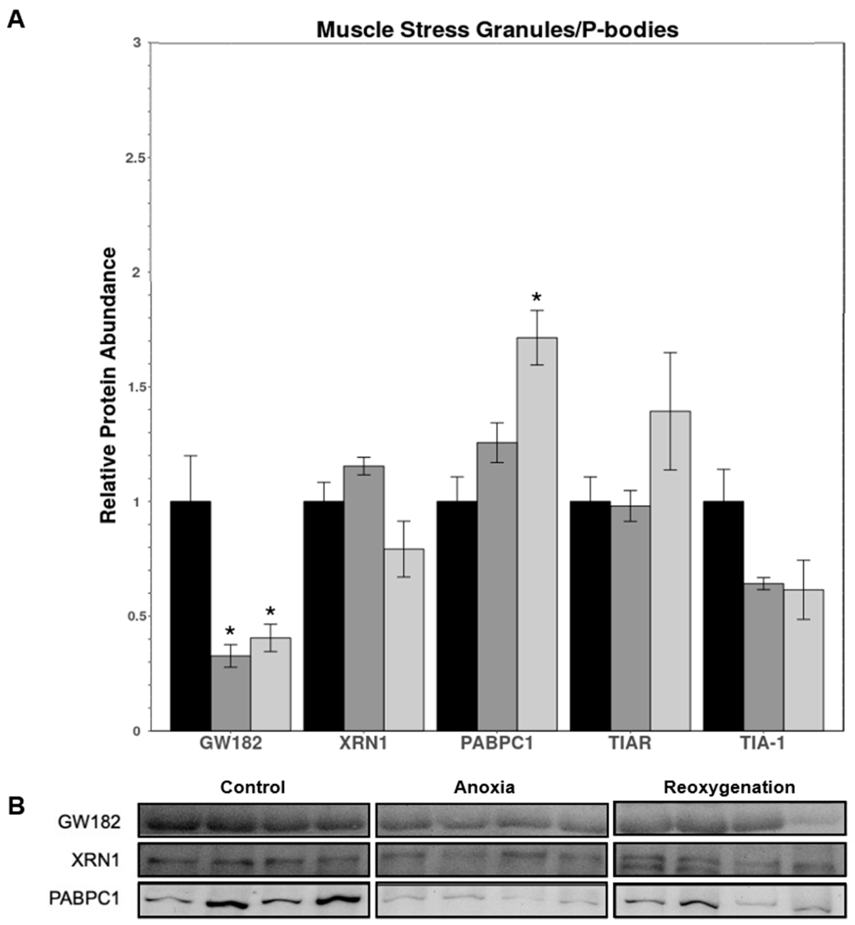
3.4. Muscle P-Body and Stress Granule Protein Expression

Immunoblot analysis of skeletal muscle P-body and stress granule proteins from normoxic, anoxic, and reoxygenated turtles is shown in Figure 6. GW182 levels significantly decreased during anoxia (0.33 ± 0.05) and reoxygenation (0.41 ± 0.06) as compared to control levels, whereas XRN1 levels remained unchanged under both experimental conditions. Levels of PABPC1 were not significantly altered under anoxia but increased by 1.71 ± 0.12-fold over controls during aerobic reoxygenation. TIAR and TIA-1 relative protein levels did not change during either anoxia or reoxygenation.

4. Discussion

Due to their critical role in gene silencing and their ability to rapidly and reversibly alter gene expression, miRNAs are an excellent regulatory mechanism that can be utilized by stress-tolerant organisms as part of their biochemical response to extreme environmental stress. The fine-tuning of gene expression that miRNA enables maximizes the energetic savings associated with MRD, while also allowing for the expression of key pro-survival genes that are critical to an organism’s ability to survive prolonged stress periods without suffering deleterious physiological consequences. This regulation occurs via miRNA:mRNA binding, which silences gene expression by leading to mRNA decay or sequestering for temporary storage. The role of miRNA in anoxia tolerance has been previously studied in red-eared slider turtles, finding tissue-specific changes in expression and an overall increase in the expression of miRNAs associated with metabolic processes and cell cycle suppression [17,26]. However, the regulation of miRNA synthesis has yet to be studied in anoxia-tolerant turtles. Given the critical role that miRNA plays in achieving MRD during environmental stress [17,22,23,24,25,26], it was hypothesized that there would be tissue-specific differential regulation of miRNA biogenesis and mRNA fate in response to anoxia and reoxygenation in the red-eared slider turtle. Indeed, previous studies have shown tissue-specific mRNA expression in response to anoxia in turtles [12,62,63,64,65,66,67], further supporting the notion that miRNA-mediated suppression of mRNA expression would vary based on the function of the tissue in question.

4.1. Regulation of microRNA Biogenesis

The rate of miRNA synthesis can be altered by regulating the activity, function, and abundance of proteins associated with various steps of the biogenesis and maturation process [68]. Regulation of the rate of miRNA biogenesis generally correlates with the cellular abundance of miRNA, thus biogenesis is an important aspect of miRNA-mediated gene regulation to analyze [69]. In the liver, the data presented here showed a contradictory decrease in proteins associated with pri-miRNA processing and nuclear export during anoxia and reoxygenation, but an increase in proteins associated with pre-miRNA processing during anoxia (Figure 3). Reductions in Drosha and DGCR8 contents could lead to a subsequent drop in the cleavage of pri-miRNA into pre-miRNA since these proteins form a complex to remove the trailing ends of the pri-miRNA duplex [20]. Additionally, the decrease in Ran-GTP expression could lead to diminished rates of pre-miRNA export into the cytoplasm for further processing (Figure 3) [20]. Together, these indicate the downregulation of pre-miRNA production and export during anoxia and reoxygenation. Interestingly, this finding contrasted with increases in Dicer cofactors, TRBP and PACT, that stabilize and aid Dicer in its endonucleic excision of pre-miRNA hairpin loops, indicating an increased rate of mature miRNA duplex production (Figure 3) [28,30,70]. This discrepancy could potentially be explained by the fact that miRNA synthesis is a rapid process, and its regulation likely varies between the initial onset of anoxia and prolonged anoxia exposure. Given that the anoxic time point chosen for this study is late (20 h) within the anoxic episode, there could have been a prior buildup of cytoplasmic pre-miRNAs that were initially produced at the onset of anoxia and stored for later processing. This pre-production would ensure that there are sufficient stores of pre-miRNA that could be rapidly and energetically inexpensively made into mature miRNA capable of normal mRNA silencing. Indeed, the selective uridylation of the pre-miRNA 3′ tail has been shown to temporarily disrupt miRNA maturation, which could facilitate this pausing effect [71,72,73]. Certainly, it would be interesting to analyze the biogenesis regulation profile and pre-miRNA uridylation at several time points during anoxic stress to determine if this is indeed the case.
Following processing by Dicer, the now mature miRNA duplex is unwound, and the guide strand is loaded into AGO to form miRISC [29]. Relative protein levels of AGO1 and AGO2 (AGO2 being the only with clear slicer activity) showed no changes in protein expression during anoxia or reoxygenation (Figure 3). This indicates that AGO1/2 miRNA loading, and AGO2-mediated mRNA cleavage may be occurring at the same rate across the anoxia-reoxygenation cycle. Although AGO1 is not as critical as AGO2 to miRNA-mediated mRNA silencing, its constant expression provides additional miRNA loading, allowing for sufficient miRNA:mRNA binding to occur [74]. The maintenance of AGO2 levels is critical to efficient miRNA biogenesis as AGO2 is the main AGO protein in higher-order animals, interacting with approximately 60% of all miRNAs, as opposed to AGO1/3/4, which interact with less than ~30% [74]. This has been demonstrated by studies that have shown that the loss of AGO2 results in reduced miRNA expression [75]. As such, these AGO2 levels indicate either that there is still significant miRNA production occurring during anoxia/reoxygenation, or that the liver maintains sufficient AGO2 levels to avoid a kinetic bottleneck that would slow miRNA production when the turtle enters the reoxygenation phase after anoxia [72]. Additionally, the activity and function of AGO can be modulated by various modifications; for instance, AGO2Ser387 phosphorylation results in reduced mRNA endonuclease cleavage, and increased translational repression and localization in P-bodies, whereas AGO2Tyr393 phosphorylation is associated with decreased binding to Dicer in the RLC and, subsequently, decreased pre-miRNA processing [76,77]. Levels of p-AGO2Ser387 and p-AGO2Tyr393 remained at normoxic levels during anoxia and reoxygenation, further indicating that there was no significant change in AGO2 functionality in the liver (Figure 3). Interestingly, the relative protein expression of AGO3 and AGO4 both decreased under anoxia, as well as during reoxygenation for AGO4 (Figure 3). This could likely be explained by the need to conserve as much ATP as possible during the stress event. Due to the fact that AGO3/4 are not necessary for efficient miRNA loading, the expression of these AGOs could be downregulated under anoxia as an aid to saving energy for the production of other, more critical, proteins [74].
By contrast, significant upregulation was observed in many proteins of the miRNA biogenesis pathway in muscle during anoxia and/or reoxygenation. Increased levels of both Drosha and DGCR8 during reoxygenation could indicate the upregulation of pri-miRNA cleavage and pre-miRNA production when oxygen was again available (Figure 4) [20]. Although the expression of these proteins did not change during anoxia, this could be due to there already being sufficient basal expression to facilitate necessary miRNA synthesis. Next, increased expression of Ran-GTP suggests that nuclear export of pre-miRNAs may be upregulated during anoxia (Figure 4). Interestingly, Ran-GTP levels decreased during reoxygenation, suggesting decreased export (Figure 4); however, this contradictory finding could be explained by a shift to an alternative nuclear export mechanism that is independent of Ran-GTP [28]. Alternatively, this decrease could signal that nuclear export is a key rate-limiting step that cells exploit to fine-tune the rate of miRNA synthesis, in spite of increased expression of multiple other biogenesis proteins. Dicer levels were strongly upregulated in anoxia and reoxygenation, along with TRBP and PACT upregulation during reoxygenation (Figure 4). These observations point towards an increase in Dicer-mediated cleavage of pre-miRNA under anoxia, and especially during reoxygenation with enhanced Dicer stability provided by elevated TRBP and PACT, which would serve to further increase the efficiency of this cleavage (Figure 4) [75,78,79]. None of the AGO proteins, nor of the serine or tyrosine-phosphorylated forms of AGO2s were significantly altered in response to anoxia or reoxygenation (Figure 4). This could be due to the normoxic AGO expression levels being sufficient to achieve the upregulation of miRNA synthesis, as presumed by the increases in other biogenesis proteins.
Taken together, these results indicate that liver miRNA biogenesis is likely either (1) increased if there are sufficient cytoplasmic pre-miRNA stores to supply Dicer-mediated cleavage and subsequent mRNA-targeting, or (2) decreased to conserve vital liver function during anoxia/reoxygenation while maintaining expression of critical pre-miRNA processing proteins to avoid bottlenecks once miRNA synthesis resumes. Analysis of miRNA expression profiles during anoxia and/or reoxygenation to determine how they differ from normoxia could elucidate which of these theories is most accurate. Although the data suggest an overall reduction in pri-miRNA processing and nuclear export in the liver, future studies should be conducted to interrogate the use of various non-canonical biogenesis pathways that could explain the downregulation of these proteins [28]. In particular, the Drosha/DGCR8-independent biogenesis pathway, and pathways that utilize alternative nuclear export mechanisms should be analyzed to see if they are used in place of or in conjunction with the canonical method [28]. miRNA biogenesis appears to be upregulated in skeletal muscle during anoxia and reoxygenation as evidenced by the increased expression of multiple proteins at each significant step in miRNA biogenesis. The increased expression of many proteins throughout the biogenesis process in muscle during anoxia/reoxygenation illustrates that miRNA production is enhanced in this tissue. Upregulation of biogenesis indicates that miRNA-mediated post-transcriptional gene silencing serves an important role in the stress response of these turtles, likely aiding in the global suppression of translation in less metabolically active organs. This suppression would lead to great energetic savings which could then be used to fuel key pro-survival processes in tissues more crucial to the turtle’s survival (e.g., the liver).
Organ-specific differences in miRNA biogenesis may be attributed to the difference in organ function during anoxic episodes. During periods of oxygen deprivation, turtles depend on carbohydrate fermentation to produce ATP. The liver plays a key role in glycogenolysis and synthesis of glucose molecules that is distributed to all other organs in order to maintain their necessary functions. Thus, it follows that liver is likely not as metabolically suppressed as some other organs. This may explain why miRNA biogenesis was significantly more upregulated in muscle as compared to liver, given that when turtles overwinter at the bottom of ponds or lakes, they are essentially comatose, therefore muscle function would not massively affect their survival whereas liver function certainly would. As such, turtles would need to preserve some level of gene expression in the liver, meaning that miRNAs cannot be as widely used for global silencing as they otherwise would in metabolically arrested organs like muscle. Although little work on environmental stress-induced changes in the expression of miRNA biogenesis proteins has been conducted to date, there is evidence that the brain of frozen wood frogs (R. sylvatica) is subject to downregulation of miRNA biogenesis [54]. Similar to the findings in turtle liver, reduced biogenesis in frog brain mainly centered on the regulation of proteins associated with pre-miRNA production and nuclear export, indicating that in metabolically and physiologically critical organs such as the liver and brain, this may be an important regulatory target [54]. Future research on the regulation of biogenesis proteins in other organs and in other stress-tolerant organisms can provide more insight into whether this response is generally conserved, or if there is an organ-, stress-, and animal-specific pattern of biogenesis regulation. Moreover, the expression of a select few miRNA species has been analyzed in anoxic red-eared slider turtles, finding tissue-specific miRNA expression. One study found that both liver and muscle showed upregulation of most of the 11 miRNAs studied, along with the downregulation of more miRNAs in the liver than in muscle [26]. A further study demonstrated an increase in the expression of one miRNA in both liver and muscle, while another study found that two miRNAs were upregulated in liver but not in muscle in response to anoxia [17,27]. Nonetheless, these limited findings do not preclude the idea that a significant number of other miRNA species are upregulated in muscle as indicated by this miRNA biogenesis study, and indeed all of these studies point toward liver miRNA expression also being upregulated in comparison to normoxia. Intriguingly, this seems to support the idea that liver miRNA biogenesis is in fact not significantly decreased during anoxia, but that there are ample stores of cytoplasmic pre-miRNA to continue sufficient mature miRNA expression. Although interesting, these miRNA expression studies are far from comprehensive in terms of how many miRNAs were assessed and future studies analyzing complete miRNAomes would provide a clearer picture of how much miRNA biogenesis is actually occurring.

4.2. Messenger RNA Fate in P-Bodies and Stress Granules

In response to environmental or cellular stress, cytoplasmic mRNP granules form as subcellular foci to sequester mRNA [34]. This can take the form of mRNAs targeted for exonuclease decay in P-bodies, or translationally repressed mRNAs that are temporarily stored in P-bodies or stress granules until favourable conditions are restored [34]. In the liver, GW182 and XRN1 proteins were similarly expressed, with both increasing during reoxygenation (Figure 5). Both of these proteins are associated with P-body formation and mRNA decay, possibly indicating an increase in the amount of mRNA being targeted to P-bodies for degradation [34]. GW182 plays a central role in miRNA-mediated gene silencing through its interactions with AGO proteins, and its knockdown is associated with P-body disassembly [80,81]. Thus, the overexpression of GW182 highlights the potential increased mRNA binding and P-body formation during reoxygenation. Additionally, the exonuclease XRN1 is responsible for mRNA decay, and its elevated abundance points toward increased degradation [40]. Increases in both of these proteins strongly suggest that mRNAs targeted by miRNA may be subject to increased P-body localization and exonuclease decay in the liver of red-eared slider turtles during reoxygenation.
This finding contrasts with the coordinated decrease in the expression of the three proteins associated with stress granule formation assessed (PABPC1, TIAR, and TIA-1) in the liver during anoxia and reoxygenation (Figure 5). PABPC1 complexes with the 3′ poly(A) tails of mRNAs to form mRNP aggregates with TIAR/TIA-1 and leading to the formation of stress granules [32,42]. Due to its key role in joining the target mRNA to stress granule assembly proteins, the reduction in PABPC1 probably indicates that less stress granule formation is occurring. Stress granule induction also depends on the concentration of the key aggregation proteins, TIAR and TIA-1 [42]. Thus, it is likely that the decrease seen in both of these proteins in the liver would signify reduced stress granule formation. Although little work has been conducted on the involvement of P-bodies or stress granules in stress-tolerant organisms, a previous study found no evidence for cytoplasmic stress granule formation in the liver of hibernating thirteen-lined ground squirrels (Ictidomys tridecemlineatus) using indirect fluorescence microscopy and relative protein expression analyses [82]. This corroborates the findings regarding decreased stress granule protein expression presented above. However, microscopy experiments are needed to confirm that this is indeed the case in red-eared slider turtles.
Interestingly, the apparent upregulation of P-bodies and downregulation of stress granules in the liver is opposite to what was observed in skeletal muscle. Protein levels of muscle GW182 were lower during anoxia and reoxygenation than in normoxia (Figure 6), implying that there would be fewer GW182-AGO2 interactions and thus, fewer mRNAs localized to P-bodies. Noteworthy, however, is that XRN1 did not decrease in expression (Figure 6). This suggests that, even though less mRNA is being targeted to XRN1 by virtue of less GW182, that XRN1 was still functioning at basal rates, which may be due to the many roles that XRN1 plays in normal gene expression independent of its actions in sequestering miRNA. Indeed, XRN1 has also been implicated in coordinating transcription, mRNA subcellular localization, and translation initiation [83,84]. These results hint that there may be less use of P-bodies in muscle during anoxia, but additional work should be conducted to elucidate whether a reduction in GW182 expression meaningfully affects mRNA silencing, and if it does in fact represent a transition from the use of P-bodies to the use of stress granules as the primary site of mRNA inhibition during anoxia and reoxygenation. Beyond these P-body proteins, PABPC1 expression was found to increase in reoxygenation, whereas TIAR and TIA-1 remained unchanged in anoxia and reoxygenation (Figure 6). Upregulated PABPC1 expression would lead to increased mRNA interactions, likely inducing stress granule formation when it interacts with TIAR/TIA-1 [32,42]. The lack of change in TIAR and TIA-1 expression likely points towards the basal levels of these proteins being adequate for stress granule formation.
The analysis of mRNA fate seems to indicate that the red-eared slider turtle liver favours P-bodies during reoxygenation over stress granules. This is interesting considering the usual shift towards stress granule formation over the use of P-bodies during periods of cellular stress [40]. Skeletal muscle, however, displayed this transition away from P-body prioritization, as expected. These contrary responses are consistent with the notion that mRNP granule induction is indeed tissue-specific, and posits the need for further, more in-depth research into the matter. Overall, this analysis of mRNA fate in the liver and muscle of red-eared slider turtles provided an interesting starting point for further research into what becomes of the mRNAs targeted by miRNA silencing. Future studies focusing on the expression of additional P-body/stress granule proteins and/or visualizing the number, size, and localization of these cytoplasmic foci would provide additional information about how they are altered during anoxia-reoxygenation cycles and what effect they may have on mRNA fate. These would provide further evidence for altered mRNP granule formation and mRNA sequestration and/or decay in response to anoxic stress in the red-eared slider turtle.

5. Conclusions

This study provides the first analysis of the regulation of miRNA biogenesis and the involvement of P-bodies and stress granules in translational inhibition during anoxia stress and reoxygenation of red-eared slider turtles. Regulated miRNA expression and mRNA suppression are critical to achieving the tissue-specific gene expression that enables turtles to enter MRD and survive prolonged periods of oxygen deprivation. The regulation of miRNA biogenesis in the liver showed that the initial steps of miRNA synthesis were downregulated during anoxia and reoxygenation, whereas the later cytoplasmic processing steps were upregulated in anoxia. Taken together, the results indicate that the liver either has decreased miRNA expression in anoxia/reoxygenation as compared to normoxic conditions, or that there was sufficient pre-miRNA production and nuclear export in early anoxia to be able to sustain the upregulation of select miRNAs as seen in previous studies. Additionally, translationally silenced mRNAs in the liver appear to be sent to P-bodies over stress granules for storage and/or decay, whose proteins were tandemly downregulated. Conversely, muscle showed clear upregulation of various steps of the biogenesis pathway including pri-miRNA processing, pre-miRNA nuclear export, and pre-miRNA cytoplasmic processing. These upregulated steps suggest increased miRNA synthesis occurring in skeletal muscle during anoxia/reoxygenation. Moreover, muscle seemed to favour increased storage of translationally silenced mRNA in stress granules, while downregulating P-body formation. Overall, this study provides new insights into the regulatory mechanisms underlying miRNA synthesis and mRNA storage/degradation in response to anoxia stress and presents interesting areas for future research.

Supplementary Materials

The following supporting information can be downloaded at: https://www.mdpi.com/article/10.3390/oxygen2020017/s1, Table S1: Immunoblot antibody supplier information.

Author Contributions

Conceptualization, S.A.B.; methodology, S.A.B.; validation, S.A.B.; formal analysis, S.A.B.; investigation, S.A.B.; resources, K.B.S.; writing—original draft preparation, S.A.B.; writing—review and editing, S.A.B. and K.B.S.; visualization, S.A.B.; supervision, K.B.S.; project administration, S.A.B.; funding acquisition, K.B.S. All authors have read and agreed to the published version of the manuscript.

Funding

This research was supported by a Discovery Grant (# RGPIN-2020-04733) from the Natural Sciences and Engineering Research Council (NSERC) of Canada and K.B.S. holds the Canada Research Chair in Molecular Physiology.

Institutional Review Board Statement

All animals were cared for in accordance with the guidelines of the Canadian Council on Animal Care and experimental procedures had the prior approval of the Carleton University Animal Care Committee (protocol #106937).

Informed Consent Statement

Not applicable.

Data Availability Statement

The data presented in this study are available from the corresponding author upon reasonable request.

Acknowledgments

We thank J.M. Storey for the editorial review of this manuscript.

Conflicts of Interest

The authors declare no conflict of interest.

References

  1. Jackson, D.C.; Ultsch, G.R. Physiology of hibernation under the ice by turtles and frogs. J. Exp. Zoöl. Part A Ecol. Genet. Physiol. 2010, 313A, 311–327. [Google Scholar] [CrossRef] [PubMed]
  2. Storey, K.B. Anoxia tolerance in turtles: Metabolic regulation and gene expression. Comp. Biochem. Physiol. Part A Mol. Integr. Physiol. 2007, 147, 263–276. [Google Scholar] [CrossRef] [PubMed]
  3. Jackson, D.C. Metabolic depression and oxygen depletion in the diving turtle. J. Appl. Physiol. 1968, 24, 503–509. [Google Scholar] [CrossRef] [PubMed]
  4. Ultsch, G.R. Ecology and physiology of hibernation and overwintering among freshwater fishes, turtles, and snakes. Biol. Rev.—Camb. Philos. Soc. 1989, 64, 435–516. [Google Scholar] [CrossRef]
  5. Breedon, S.A.; Hadj-Moussa, H.; Storey, K.B. Nrf2 activates antioxidant enzymes in the anoxia-tolerant red-eared slider turtle, Trachemys scripta elegans. J. Exp. Zoöl. Part A Ecol. Integr. Physiol. 2021, 335, 426–435. [Google Scholar] [CrossRef] [PubMed]
  6. Bundgaard, A.; James, A.M.; Joyce, W.; Murphy, M.P.; Fago, A. Suppression of reactive oxygen species generation in heart mitochondria from anoxic turtles: The role of complex I S-nitrosation. J. Exp. Biol. 2018, 221, jeb174391. [Google Scholar] [CrossRef] [Green Version]
  7. Davis, E.C.; Jackson, D.C. Lactate uptake by skeletal bone in anoxic turtles, Trachemys scripta. Comp. Biochem. Physiol. Part A Mol. Integr. Physiol. 2007, 146, 299–304. [Google Scholar] [CrossRef]
  8. Kesaraju, S.; Schmidt-Kastner, R.; Prentice, H.M.; Milton, S.L. Modulation of stress proteins and apoptotic regulators in the anoxia tolerant turtle brain. J. Neurochem. 2009, 109, 1413–1426. [Google Scholar] [CrossRef] [Green Version]
  9. Kesaraju, S.; Nayak, G.; Prentice, H.M.; Milton, S.L. Upregulation of Hsp72 mediates anoxia/reoxygenation neuroprotection in the freshwater turtle via modulation of ROS. Brain Res. 2014, 1582, 247–256. [Google Scholar] [CrossRef] [Green Version]
  10. Krivoruchko, A.; Storey, K.B. Regulation of the heat shock response under anoxia in the turtle, Trachemys scripta elegans. J. Comp. Physiol. B 2010, 180, 403–414. [Google Scholar] [CrossRef]
  11. Reiterer, M.; Milton, S.L. Induction of foxo3a protects turtle neurons against oxidative stress. Comp. Biochem. Physiol. Part A Mol. Integr. Physiol. 2020, 243, 110671. [Google Scholar] [CrossRef] [PubMed]
  12. Stecyk, J.A.W.; Couturier, C.S.; Fagernes, C.E.; Ellefsen, S.; Nilsson, G.E. Quantification of heat shock protein mRNA expression in warm and cold anoxic turtles (Trachemys scripta) using an external RNA control for normalization. Comp. Biochem. Physiol. Part D Genom. Proteom. 2021, 7, 59–72. [Google Scholar] [CrossRef] [PubMed]
  13. Willmore, W.G.; Storey, K. Antioxidant systems and anoxia tolerance in a freshwater turtle Trachemys scripta elegans. Mol. Cell. Biochem. 1997, 170, 177–185. [Google Scholar] [CrossRef] [PubMed]
  14. Hochachka, P.W. Metabolic suppression and oxygen availability. Can. J. Zoöl. 1988, 66, 152–158. [Google Scholar] [CrossRef]
  15. Storey, K.B. Adventures in oxygen metabolism. Comp. Biochem. Physiol. Part B Biochem. Mol. Biol. 2004, 139, 359–369. [Google Scholar] [CrossRef] [PubMed]
  16. Farrell, A.P.; Stecyk, J.A.W. The heart as a working model to explore themes and strategies for anoxic survival in ectothermic vertebrates. Comp. Biochem. Physiol. Part A Mol. Integr. Physiol. 2007, 147, 300–312. [Google Scholar] [CrossRef]
  17. Biggar, K.K.; Storey, K.B. Evidence for cell cycle suppression and microRNA regulation of cyclin D1 during anoxia exposure in turtles. Cell Cycle 2012, 11, 1705–1713. [Google Scholar] [CrossRef] [Green Version]
  18. Rider, M.H.; Hussain, N.; Dilworth, S.M.; Storey, K.B. Phosphorylation of translation factors in response to anoxia in turtles, Trachemys scripta elegans: Role of the AMP-activated protein kinase and target of rapamycin signalling pathways. Mol. Cell. Biochem. 2009, 332, 207–213. [Google Scholar] [CrossRef]
  19. Storey, K.B.; Storey, J.M. Metabolic rate depression in animals: Transcriptional and translational controls. Biol. Rev. Camb. Philos. Soc. 2004, 79, 207–233. [Google Scholar] [CrossRef] [PubMed]
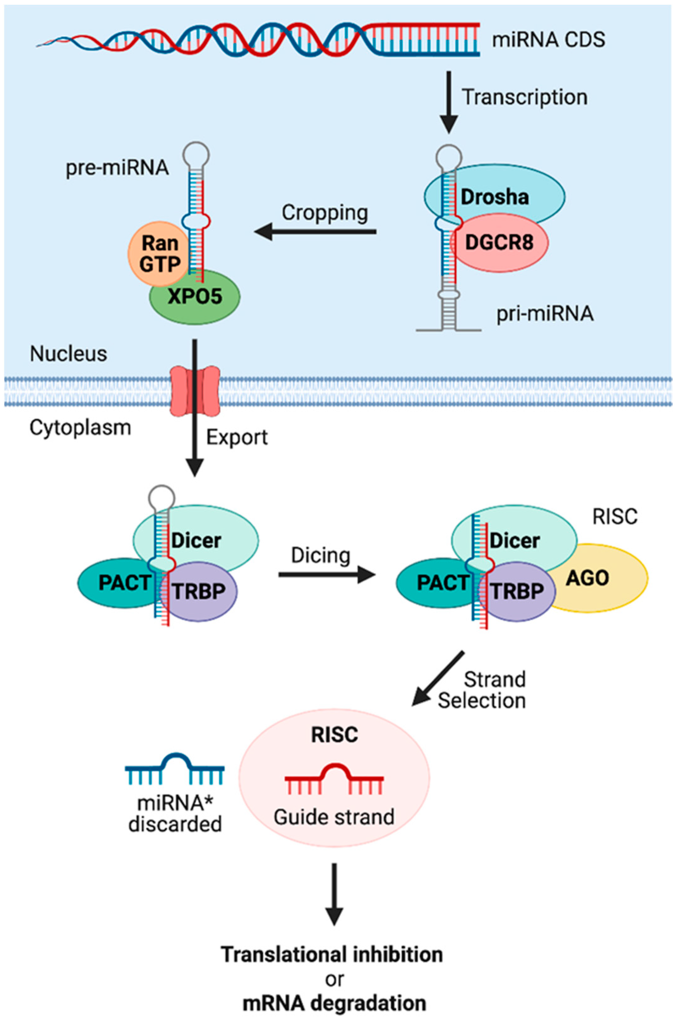
  20. O’Brien, J.; Hayder, H.; Zayed, Y.; Peng, C. Overview of MicroRNA Biogenesis, Mechanisms of Actions, and Circulation. Front. Endocrinol. 2018, 9, 402. [Google Scholar] [CrossRef] [Green Version]
  21. Bartel, D.P. MicroRNAs: Genomics, Biogenesis, Mechanism, and Function. Cell 2004, 116, 281–297. [Google Scholar] [CrossRef] [Green Version]
  22. Hadj-Moussa, H.; Zhang, J.; Pifferi, F.; Perret, M.; Storey, K.B. Profiling torpor-responsive microRNAs in muscles of the hibernating primate Microcebus murinus. Biochim. Biophys. Acta—Gene Regul. Mech 2020, 1863, 194473. [Google Scholar] [CrossRef]
  23. Hadj-Moussa, H.; Pamenter, M.E.; Storey, K.B. Hypoxic naked mole–rat brains use microRNA to coordinate hypometabolic fuels and neuroprotective defenses. J. Cell. Physiol. 2021, 236, 5080–5097. [Google Scholar] [CrossRef] [PubMed]
  24. Hoyeck, M.P.; Hadj-Moussa, H.; Storey, K.B. Estivation-responsive microRNAs in a hypometabolic terrestrial snail. PeerJ 2019, 7, e6515. [Google Scholar] [CrossRef] [PubMed] [Green Version]
  25. Luu, B.E.; Lefai, E.; Giroud, S.; Swenson, J.E.; Chazarin, B.; Gauquelin-Koch, G.; Arnemo, J.M.; Evans, A.L.; Bertile, F.; Storey, K.B. MicroRNAs facilitate skeletal muscle maintenance and metabolic suppression in hibernating brown bears. J. Cell. Physiol. 2020, 235, 3984–3993. [Google Scholar] [CrossRef]
  26. Biggar, K.K.; Storey, K.B. Exploration of low temperature microRNA function in an anoxia tolerant vertebrate ectotherm, the red eared slider turtle (Trachemys scripta elegans). J. Therm. Biol. 2017, 68, 139–146. [Google Scholar] [CrossRef]
  27. Zhang, J.; Biggar, K.K.; Storey, K.B. Regulation of p53 by reversible post-transcriptional and post-translational mechanisms in liver and skeletal muscle of an anoxia tolerant turtle, Trachemys scripta elegans. Gene 2013, 513, 147–155. [Google Scholar] [CrossRef]
  28. Ha, M.; Kim, V.N. Regulation of microRNA biogenesis. Nat. Rev. Mol. Cell Biol. 2014, 15, 509–524. [Google Scholar] [CrossRef]
  29. Finnegan, E.F.; Pasquinelli, A.E. MicroRNA biogenesis: Regulating the regulators. Crit. Rev. Biochem. Mol. Biol. 2013, 48, 51–68. [Google Scholar] [CrossRef] [Green Version]
  30. Koscianska, E.; Starega-Roslan, J.; Krzyzosiak, W.J. The Role of Dicer Protein Partners in the Processing of MicroRNA Precursors. PLoS ONE 2011, 6, e28548. [Google Scholar] [CrossRef] [Green Version]
  31. Leung, A.K.L.; Sharp, P.A. Function and Localization of MicroRNAs in Mammalian Cells. Cold Spring Harb. Symp. Quant. Biol. 2006, 71, 29–38. [Google Scholar] [CrossRef] [PubMed]
  32. Anderson, P.; Kedersha, N. Stress granules: The Tao of RNA triage. Trends Biochem. Sci. 2008, 33, 141–150. [Google Scholar] [CrossRef] [PubMed]
  33. Kedersha, N.; Stoecklin, G.; Ayodele, M.; Yacono, P.; Lykke-Andersen, J.; Fritzler, M.J.; Scheuner, D.; Kaufman, R.J.; Golan, D.E.; Anderson, P. Stress granules and processing bodies are dynamically linked sites of mRNP remodeling. J. Cell Biol. 2005, 169, 871–884. [Google Scholar] [CrossRef] [PubMed] [Green Version]
  34. Standart, N.; Weil, D. P-Bodies: Cytosolic Droplets for Coordinated mRNA Storage. Trends Genet. 2018, 34, 612–626. [Google Scholar] [CrossRef]
  35. Li, Y.R.; King, O.D.; Shorter, J.; Gitler, A.D. Stress granules as crucibles of ALS pathogenesis. J. Cell Biol. 2013, 201, 361–372. [Google Scholar] [CrossRef]
  36. Buchan, J.R. MRNP Granules Assembly, Function, and Connections with Disease. RNA Biol. 2014, 11, 1019–1030. [Google Scholar] [CrossRef] [Green Version]
  37. Buchan, J.R.; Parker, R. Eukaryotic Stress Granules: The Ins and Outs of Translation. Mol. Cell 2009, 36, 932–941. [Google Scholar] [CrossRef] [Green Version]
  38. Parker, R.; Sheth, U. P Bodies and the Control of mRNA Translation and Degradation. Mol. Cell 2007, 25, 635–646. [Google Scholar] [CrossRef]
  39. Parker, R. RNA Degradation in Saccharomyces cerevisae. Genetics 2012, 191, 671–702. [Google Scholar] [CrossRef] [Green Version]
  40. Kedersha, N.; Anderson, P. Mammalian Stress Granules and Processing Bodies. Methods Enzymol. 2007, 431, 61–81. [Google Scholar] [CrossRef]
  41. Spriggs, K.A.; Bushell, M.; Willis, A.E. Translational Regulation of Gene Expression during Conditions of Cell Stress. Mol. Cell 2010, 40, 228–237. [Google Scholar] [CrossRef]
  42. Gilks, N.; Kedersha, N.; Ayodele, M.; Shen, L.; Stoecklin, G.; Dember, L.M.; Anderson, P. Stress Granule Assembly Is Mediated by Prion-like Aggregation of TIA-1. Mol. Biol. Cell 2004, 15, 5383–5398. [Google Scholar] [CrossRef] [Green Version]
  43. Kedersha, N.L.; Gupta, M.; Li, W.; Miller, I.; Anderson, P. RNA-Binding Proteins Tia-1 and Tiar Link the Phosphorylation of Eif-2α to the Assembly of Mammalian Stress Granules. J. Cell Biol. 1999, 147, 1431–1441. [Google Scholar] [CrossRef] [PubMed]
  44. Tourrière, H.; Chebli, K.; Zekri, L.; Courselaud, B.; Blanchard, J.M.; Bertrand, E.; Tazi, J. The RasGAP-associated endoribonuclease G3BP assembles stress granules. J. Cell Biol. 2003, 160, 823–831. [Google Scholar] [CrossRef] [PubMed] [Green Version]
  45. Jakymiw, A.; Lian, S.; Eystathioy, T.; Li, S.; Satoh, M.; Hamel, J.C.; Fritzler, M.J.; Chan, E.K.L. Disruption of GW bodies impairs mammalian RNA interference. Nat. Cell Biol. 2005, 7, 1167–1174. [Google Scholar] [CrossRef] [PubMed]
  46. Eystathioy, T.; Jakymiw, A.; Chan, E.K.L.; Séraphin, B.; Cougot, N.; Fritzler, M.J. The GW182 protein colocalizes with mRNA degradation associated proteins hDcp1 and hLSm4 in cytoplasmic GW bodies: FIGURE 1. RNA 2003, 9, 1171–1173. [Google Scholar] [CrossRef] [Green Version]
  47. Jain, S.; Parker, R. The Discovery and Analysis of P Bodies. Adv. Exp. Med. Biol. 2013, 768, 23–43. [Google Scholar] [CrossRef]
  48. Christie, M.; Boland, A.; Huntzinger, E.; Weichenrieder, O.; Izaurralde, E. Structure of the PAN3 Pseudokinase Reveals the Basis for Interactions with the PAN2 Deadenylase and the GW182 Proteins. Mol. Cell 2013, 51, 360–373. [Google Scholar] [CrossRef] [Green Version]
  49. Jonas, S.; Izaurralde, E. Towards a Molecular Understanding of MicroRNA-Mediated Gene Silencing. Nat. Rev. Genet. 2015, 16, 421–433. [Google Scholar] [CrossRef]
  50. Hoyle, N.; Castelli, L.M.; Campbell, S.G.; Holmes, L.E.; Ashe, M.P. Stress-dependent relocalization of translationally primed mRNPs to cytoplasmic granules that are kinetically and spatially distinct from P-bodies. J. Cell Biol. 2007, 179, 65–74. [Google Scholar] [CrossRef]
  51. Dang, Y.; Kedersha, N.; Low, W.-K.; Romo, D.; Gorospe, M.; Kaufman, R.; Anderson, P.; Liu, J.O. Eukaryotic Initiation Factor 2α-independent Pathway of Stress Granule Induction by the Natural Product Pateamine A. J. Biol. Chem. 2006, 281, 32870–32878. [Google Scholar] [CrossRef] [PubMed] [Green Version]
  52. Mazroui, R.; Sukarieh, R.; Bordeleau, M.-E.; Kaufman, R.J.; Northcote, P.; Tanaka, J.; Gallouzi, I.; Pelletier, J. Inhibition of Ribosome Recruitment Induces Stress Granule Formation Independently of Eukaryotic Initiation Factor 2α Phosphorylation. Mol. Biol. Cell 2006, 17, 4212–4219. [Google Scholar] [CrossRef] [PubMed]
  53. Mollet, S.; Cougot, N.; Wilczynska, A.; Dautry, F.; Kress, M.; Bertrand, E.; Weil, D. Translationally Repressed mRNA Transiently Cycles through Stress Granules during Stress. Mol. Biol. Cell 2008, 19, 4469–4479. [Google Scholar] [CrossRef] [PubMed] [Green Version]
  54. Hadj-Moussa, H.; Storey, K.B. Micromanaging freeze tolerance: The biogenesis and regulation of neuroprotective microRNAs in frozen brains. Cell. Mol. Life Sci. 2018, 75, 3635–3647. [Google Scholar] [CrossRef] [PubMed]
  55. Keiver, K.M.; Hochachka, P.W. Catecholamine stimulation of hepatic glycogenolysis during anoxia in the turtle Chrysemys picta. Am. J. Physiol. 1991, 261, 1341–1345. [Google Scholar] [CrossRef] [PubMed]
  56. Mehrani, H.; Storey, K.B. Enzymatic control of glycogenolysis during anoxic submergence in the freshwater turtle Trachemys scripta. Int. J. Biochem. Cell Biol. 1995, 27, 821–830. [Google Scholar] [CrossRef]
  57. Warren, D.E.; Reese, S.A.; Jackson, D.C. Tissue Glycogen and Extracellular Buffering Limit the Survival of Red-Eared Slider Turtles during Anoxic Submergence at 3 °C. Physiol. Biochem. Zoöl. 2006, 79, 736–744. [Google Scholar] [CrossRef] [Green Version]
  58. Madsen, J.G.; Wang, T.; Madsen, P.T. Hypoxic turtles keep their cool. Temperature 2015, 2, 40–41. [Google Scholar] [CrossRef] [Green Version]
  59. Nilsson, G.E.; Lutz, P.L. Anoxia Tolerant Brains. J. Cereb. Blood Flow Metab. 2004, 24, 475–486. [Google Scholar] [CrossRef] [Green Version]
  60. Wijenayake, S.; Hawkins, L.; Storey, K.B. Dynamic regulation of six histone H3 lysine (K) methyltransferases in response to prolonged anoxia exposure in a freshwater turtle. Gene 2018, 649, 50–57. [Google Scholar] [CrossRef]
  61. Zhang, J.; Storey, K.B. RBioplot: An easy-to-use R pipeline for automated statistical analysis and data visualization in molecular biology and biochemistry. PeerJ 2016, 4, e2436. [Google Scholar] [CrossRef] [PubMed]
  62. Biggar, K.K.; Zhang, J.; Storey, K.B. Navigating oxygen deprivation: Liver transcriptomic responses of the red eared slider turtle to environmental anoxia. PeerJ 2019, 7, e8144. [Google Scholar] [CrossRef] [PubMed]
  63. Couturier, C.S.; Stecyk, J.A.W.; Ellefsen, S.; Sandvik, G.K.; Milton, S.L.; Prentice, H.M.; Nilsson, G.E. The expression of genes involved in excitatory and inhibitory neurotransmission in turtle (Trachemys scripta) brain during anoxic submergence at 21 °C and 5 °C reveals the importance of cold as a preparatory cue for anoxia survival. Comp. Biochem. Physiol. Part D Genom. Proteom. 2019, 30, 55–70. [Google Scholar] [CrossRef] [PubMed]
  64. Douglas, D.N.; Giband, M.; Altosaar, I.; Storey, K.B. Anoxia induces changes in translatable mRNA populations in turtle organs: A possible adaptive strategy for anaerobiosis. J. Comp. Physiol. B 1994, 164, 405–414. [Google Scholar] [CrossRef]
  65. Keenan, S.W.; Hill, C.A.; Kandoth, C.; Buck, L.T.; Warren, D.E. Transcriptomic Responses of the Heart and Brain to Anoxia in the Western Painted Turtle. PLoS ONE 2015, 10, e0131669. [Google Scholar] [CrossRef]
  66. Melleby, A.O.; Sandvik, G.K.; Couturier, C.S.; Nilsson, G.E.; Stecyk, J.A.W. H2S-producing enzymes in anoxia-tolerant vertebrates: Effects of cold acclimation, anoxia exposure and reoxygenation on gene and protein expression. Comp. Biochem. Physiol. Part B Biochem. Mol. Biol. 2020, 243–244, 110430. [Google Scholar] [CrossRef]
  67. Sparks, K.; Couturier, C.S.; Buskirk, J.; Flores, A.; Hoeferle, A.; Hoffman, J.; Stecyk, J.A.W. Gene expression of hypoxia-inducible factor (HIF), HIF regulators, and putative HIF targets in ventricle and telencephalon of Trachemys scripta acclimated to 21 °C or 5 °C and exposed to normoxia, anoxia or reoxygenation. Comp. Biochem. Physiol. Part A Mol. Integr. Physiol. 2022, 267, 111167. [Google Scholar] [CrossRef]
  68. Leung, A.; Sharp, P.A. MicroRNA Functions in Stress Responses. Mol. Cell 2010, 40, 205–215. [Google Scholar] [CrossRef] [Green Version]
  69. Zlotorynski, E. Insights into the kinetics of microRNA biogenesis and turnover. Nat. Rev. Mol. Cell Biol. 2019, 20, 511. [Google Scholar] [CrossRef]
  70. Ma, E.; MacRae, I.J.; Kirsch, J.F.; Doudna, J.A. Autoinhibition of Human Dicer by Its Internal Helicase Domain. J. Mol. Biol. 2008, 380, 237–243. [Google Scholar] [CrossRef] [Green Version]
  71. Bortolamiol-Becet, D.; Hu, F.; Jee, D.; Wen, J.; Okamura, K.; Lin, C.-J.; Ameres, S.L.; Lai, E.C. Selective Suppression of the Splicing-Mediated MicroRNA Pathway by the Terminal Uridyltransferase Tailor. Mol. Cell 2015, 59, 217–228. [Google Scholar] [CrossRef] [PubMed] [Green Version]
  72. Reichholf, B.; Herzog, V.A.; Fasching, N.; Manzenreither, R.A.; Sowemimo, I.; Ameres, S.L. Time-Resolved Small RNA Sequencing Unravels the Molecular Principles of MicroRNA Homeostasis. Mol. Cell 2019, 75, 756–768.e7. [Google Scholar] [CrossRef] [PubMed]
  73. Pinto, M.M.R.; Ignatova, V.; Burkard, T.; Hung, J.-H.; Manzenreither, R.A.; Sowemimo, I.; Herzog, V.A.; Reichholf, B.; Fariña-Lopez, S.; Ameres, S.L. Uridylation of RNA Hairpins by Tailor Confines the Emergence of MicroRNAs in Drosophila. Mol. Cell 2015, 59, 203–216. [Google Scholar] [CrossRef] [PubMed] [Green Version]
  74. Wang, D.; Zhang, Z.; O’Loughlin, E.; Lee, T.; Houel, S.; O’Carroll, D.; Tarakhovsky, A.; Ahn, N.G.; Yi, R. Quantitative functions of Argonaute proteins in mammalian development. Genes Dev. 2012, 26, 693–704. [Google Scholar] [CrossRef] [PubMed] [Green Version]
  75. Winter, J.; Jung, S.; Keller, S.; Gregory, R.I.; Diederichs, S. Many roads to maturity: MicroRNA biogenesis pathways and their regulation. Nat. Cell Biol. 2009, 11, 228–234. [Google Scholar] [CrossRef]
  76. Horman, S.R.; Janas, M.M.; Litterst, C.; Wang, B.; MacRae, I.J.; Sever, M.J.; Morrissey, D.V.; Graves, P.; Luo, B.; Umesalma, S.; et al. Akt-Mediated Phosphorylation of Argonaute 2 Downregulates Cleavage and Upregulates Translational Repression of MicroRNA Targets. Mol. Cell 2013, 50, 356–367. [Google Scholar] [CrossRef] [Green Version]
  77. Shen, J.; Xia, W.; Khotskaya, Y.B.; Huo, L.; Nakanishi, K.; Lim, S.-O.; Du, Y.; Wang, Y.; Chang, W.-C.; Chen, C.-H.; et al. EGFR modulates microRNA maturation in response to hypoxia through phosphorylation of AGO2. Nature 2013, 497, 383–387. [Google Scholar] [CrossRef]
  78. Haase, A.D.; Jaskiewicz, L.; Zhang, H.; Lainé, S.; Sack, R.; Gatignol, A.; Filipowicz, W. TRBP, a regulator of cellular PKR and HIV-1 virus expression, interacts with Dicer and functions in RNA silencing. EMBO Rep. 2005, 6, 961–967. [Google Scholar] [CrossRef]
  79. Lee, Y.; Hur, I.; Park, S.-Y.; Kim, Y.-K.K.; Suh, M.R.; Kim, V.N.; Mi, R.S.; Kim, V.N. The role of PACT in the RNA silencing pathway. EMBO J. 2006, 25, 522–532. [Google Scholar] [CrossRef]
  80. Liu, J.; Rivas, F.V.; Wohlschlegel, J.; Yates, J.R.; Parker, R.; Hannon, G.J. A role for the P-body component GW182 in microRNA function. Nat. Cell Biol. 2005, 7, 1161–1166. [Google Scholar] [CrossRef]
  81. Yao, B.; Li, S.; Chan, E.K.L. Function of GW182 and GW Bodies in siRNA and miRNA Pathways. Adv. Exp. Med. Biol. 2013, 768, 71–96. [Google Scholar] [CrossRef] [PubMed]
  82. Tessier, S.N.; Audas, T.E.; Wu, C.-W.; Lee, S.; Storey, K.B. The involvement of mRNA processing factors TIA-1, TIAR, and PABP-1 during mammalian hibernation. Cell Stress Chaperon. 2014, 19, 813–825. [Google Scholar] [CrossRef] [PubMed] [Green Version]
  83. Begley, V.; Corzo, D.; Jordán-Pla, A.; Cuevas-Bermúdez, A.; De Miguel-Jiménez, L.; Pérez-Aguado, D.; Machuca-Ostos, M.; Navarro, F.; Chávez, M.J.; Pérez-Ortín, J.E.; et al. The mRNA degradation factor Xrn1 regulates transcription elongation in parallel to Ccr4. Nucleic Acids Res. 2019, 47, 9524–9541. [Google Scholar] [CrossRef] [PubMed] [Green Version]
  84. Blasco-Moreno, B.; De Campos-Mata, L.; Böttcher, R.; Garcia-Martinez, J.; Jungfleisch, J.; Nedialkova, D.D.; Chattopadhyay, S.; Gas, M.-E.; Oliva, B.; Pérez-Ortín, J.E.; et al. The exonuclease Xrn1 activates transcription and translation of mRNAs encoding membrane proteins. Nat. Commun. 2019, 10, 1298. [Google Scholar] [CrossRef] [PubMed] [Green Version]
Figure 1. Canonical microRNA biogenesis and maturation pathway. Transcription of a miRNA coding sequence (CDS) results in primary miRNA (pri-miRNA) formation. The pri-miRNA is joined by Drosha and DGCR8 to crop the 5′ and 3′ trailing ends, leaving behind the stem-loop structure of the precursor miRNA (pre-miRNA). Pre-miRNA is exported into the cytoplasm via XPO5 and Ran-GTP where it is bound by Dicer, TRBP, and PACT. Dicer cleaves the hairpin loop leaving only the miRNA duplex. The duplex is unwound, and the guide strand is loaded onto AGO where it can then be used to target mRNA for translational inhibition or degradation. Figure created using BioRender.com.
Oxygen 02 00017 g001
Figure 2. Stress granule and processing body sequestration of mRNA in response to stress. Stress-induced translational stalling can lead to the sequestering of affected mRNA transcripts into P-bodies or stress granules. These granules contain silenced mRNA, RNA-binding proteins, and translation initiation factors, which can be exchanged when they dock. mRNAs contained in P-bodies may undergo degradation in addition to long-term storage. When the stress resolves, mRNAs stored within these granules are released to restart translation. Figure adapted from [35]. Figure created using BioRender.com.
Oxygen 02 00017 g002
Figure 3. Analysis of miRNA biogenesis pathway protein levels in red-eared slider turtle liver under control, anoxia, and reoxygenation conditions. (A) Graph showing relative expression of Drosha, DGCR8, XPO5, Ran-GTP, Dicer, TRBP, PACT, AGO1–4, p-AGO2Ser387, and p-AGO2Tyr393 under normoxic (control), 20 h anoxic, and 5 h aerobic reoxygenation conditions. (B) Representative immunoblots of selected protein targets are shown for the three experimental conditions. Data are mean ± SEM (n = 4 independent trials). Statistical significance for anoxia or reoxygenation values, relative to the standardized control, was determined using one-way analysis of variance (ANOVA) with Dunnett’s post hoc test (*—p < 0.05).
Oxygen 02 00017 g003
Figure 4. Analysis of miRNA biogenesis pathway protein levels in skeletal muscle of red-eared slider turtles under control, anoxia, and reoxygenation conditions. (A) Graph showing relative expression of Drosha, DGCR8, XPO5, Ran-GTP, Dicer, TRBP, PACT, AGO1–4, p-AGO2Ser387, and p-AGO2Tyr393 under normoxic (control), 20 h anoxic, and 5 h reoxygenation conditions where statistically significant changes relative to the standardized control (*—p < 0.05) were determined by ANOVA with Dunnett’s post hoc test. (B) Representative immunoblots of selected protein targets are shown for the three experimental conditions. Other information as in Figure 3.
Oxygen 02 00017 g004
Figure 5. Relative expression analysis of proteins associated with P-body and stress granule in red-eared slider turtle liver. (A) Graph showing relative protein expression of GW182, XRN1, PABPC1, TIAR, and TIA-1 under control, anoxic, and reoxygenation conditions where statistically significant changes relative to the standardized control (*—p < 0.05) were determined by ANOVA with Dunnett’s post hoc test. (B) Representative immunoblots of selected protein targets are shown for the three experimental conditions. Other information as in Figure 3.
Oxygen 02 00017 g005
Figure 6. Relative expression analysis of proteins associated with P-body and stress granule in red-eared slider turtle skeletal muscle. (A) Graph showing relative protein expression of GW182, XRN1, PABPC1, TIAR, and TIA-1 under control, anoxic, and reoxygenation conditions where statistically significant changes relative to the standardized control (*—p < 0.05) were determined by ANOVA with Dunnett’s post hoc test. (B) Representative immunoblots of selected protein targets are shown for the three experimental conditions. Other information as in Figure 3.
Oxygen 02 00017 g006
Publisher’s Note: MDPI stays neutral with regard to jurisdictional claims in published maps and institutional affiliations.

