Article

Effects of Dietary Sodium Propionate on Growth, Digestive Enzyme Activity, and Expression of Immune System Genes in Juveniles of Tropical Gar (Atractosteus tropicus)

by
Jesús G. Arellano-Carrasco
1,
Rafael Martínez-García
2,*,
Alberto Asiain-Hoyos
1,
Juan L. Reta-Mendiola
1,
Pablo Díaz-Rivera
1,
Susana A. Frías-Gómez
3,
Talhia Martínez-Burguete
2,
Gloria Gertrudys Asencio-Alcudia
2,4,
Luis Daniel Jiménez-Martínez
5,
Rocio Guerrero-Zarate
2,
Cesar A. Sepúlveda-Quiroz
2 and
Carlos A. Álvarez-González
2
1
Colegio de Postgraduados—Campus Veracruz, Km. 88.5 Carretera Federal Xalapa-Veracruz, Tepetates, Veracruz C.P. 91690, Veracruz, Mexico
2
Laboratorio de Fisiología en Recursos Acuáticos—División Académica de Ciencias Biológicas, Universidad Juárez Autónoma de Tabasco, Carretera Villahermosa-Cárdenas Km. 0.5, Villahermosa C.P. 86039, Tabasco, Mexico
3
Posgrado en Ciencias del Mar y Limnología (Posgrado en Ciencias del Mar y Limnología), Universidad Nacional Autónoma de Mexico, Av. Universidad 3000, Ciudad Universitaria Coyoacán, Ciudad de México C.P. 04510, Mexico City, Mexico
4
Tecnológico Nacional de México Campus Villahermosa, Km. 3.5 Carretera Villahermosa–Frontera, Cd. Industrial, Villahermosa C.P. 86010, Tabasco, Mexico
5
Laboratorio de Biología Molecular, División Académica Multidisciplinaria de Jalpa de Méndez, Universidad Juárez Autónoma de Tabasco, Carretera Estatal Libre Villahermosa-Comalcalco Km. 27 S/N, Jalpa de Méndez C.P. 86205, Tabasco, Mexico
*
Author to whom correspondence should be addressed.
Aquac. J. 2023, 3(4), 227-237; https://doi.org/10.3390/aquacj3040018
Submission received: 12 September 2023 / Revised: 10 November 2023 / Accepted: 14 November 2023 / Published: 20 November 2023

Abstract

:
We determined the effects of sodium propionate (SP) added to the diets of Atractosteus tropicus juveniles with respect to the growth, survival, digestive enzyme activity, and expression of genes that are associated with the immune system. Five treatments (0, 0.5, 1.0, 1.5, and 2.0%) were evaluated in triplicate on 180 fish (3.65 ± 0.12 g) distributed among 15 (70 L) tanks. The juveniles were fed five times a day with 5% feed in relation to the biomass of the organism. The treatment with 0.5% SP showed a final weight value of 25.7 ± 4.5 g, absolute weight of gain (AWG) of 21.93 ± 4.39 g, and specific growth rate (SGR) of 3.1 ± 0.26. Treatments with 1.5 and 2.0% SP showed the highest survival (91.6%). The control group (0%) showed a greater activity of lipases. There was a tendency that the highest activity of alkaline proteases and chymotrypsin occurred in the 0 and 0.5% treatments. The maximum relative expression of the genes ocln, muc2, and nod2 occurred in the 1.5% treatment. The inclusion of SP in the diet of A. tropicus juveniles could benefit the activity of some digestive enzymes as well as the expression of genes related to the function of the intestinal barrier, therefore benefitting the survival of the organisms.

1. Introduction

Tropical gar (Atractosteus tropicus) is a species with ecological, economic, and cultural importance in several areas of Central America [1]. However, the culture of this species still has some challenges that limit its production. Proper feeding and nutrition play a fundamental role in the health status of these fish and their ability to respond to possible diseases [2].
In Atractosteus tropicus, several feed formulations have been evaluated according to the nutritional requirements of the species, adding additives, probiotics, and prebiotics that could have positive effects on growth and survival; the analysis of these positive effects can be carried out with the support of tools such as digestive enzymatic biochemistry and gene expression [3,4,5].
Short-chain fatty acids (SCFAs) are 1–6 carbon organic fatty acids. SFCAs play important roles as nutrients for the intestine epithelium; as modulators of intracellular pH, cell volume, and other functions associated with ion transport; and as regulators of gene disappearance, differentiation, and expression [6]. The main SCFAs are acetate, propionate, and butyrate [7]. In species such as Acanthopagrus latus, doses of 5 and 10 g/kg of propionate and 10 g/kg of sodium acetate showed an increase in growth, feeding efficiency, and liver antioxidant capacity, and it caused a high activity of pepsin, trypsin, and lipase [8]. In white shrimp (Litopenaeus vannamei), sodium propionate (SP) was evaluated, showing good results for growth and survival at concentrations of 2% [9]; in zebrafish (Danio rerio), good results were found when evaluating parameters of the immune mucosa when incorporating 2% SP [10].
The effects of SP are still unknown for many species; therefore, the objective of this preliminary work was to test different concentrations of SP (0, 0.5, 1.0, 1.5, and 2.0%) in the diets of tropical gar juveniles (A. tropicus) and provide first insights on growth, survival, digestive development, and immune system gene expression as affected by the dietary treatment.

2. Materials and Methods

2.1. Biological Material

The experiment was carried out at the Physiology Laboratory in Aquatic Resources (LAFIRA) at DACBiol-UJAT. Spawning was induced by injecting a female (3 kg, 69 cm) with LHRHa hormone (Sigma-Aldrich, Taufkirchen, Germany) (35 µg kg under the pelvic fin) and six males (1.5 kg, 31 cm no hormone induction). Fish were placed in a 2000 L circular tank conditioned with raffia thread to simulate the natural spawning site. Spawning occurred 18 h after induction; the average dissolved oxygen was 4.9 ± 0.3 mg/L (oxygen meter YSI 85, Yellow Springs, OH, USA), the temperature was 28.5 ± 0.3 °C, and the pH was 7.1 ± 0.1 (HANNA HI 991001, Nusfalau, Romania).
After hatching (72 h post fertilization), larvae were placed in 7 plastic tanks for maintenance in a recirculated system as described in Section 2.2. Once the yolk sac was absorbed and after mouth opening (3 days post hatching), the larvae were fed 5 times per day (8:00, 11:00, 13:00, 15:00, and 18:00 h) with Artemia nauplii, following the feeding scheme proposed by [11], until reaching juvenile size (30 days after hatching (DAH)). For this experiment, 180 juveniles (3.65 ± 0.12 g mean weight and 10.38 ± 0.10 cm mean total length) were used.

2.2. Experimental Design

The design consisted of a completely randomized one-way experiment. Four treatments were evaluated by triplicate with different concentrations of SP (0.5, 1.0, 1.5, and 2.0%) and a control diet (0%). For each experimental unit, 12 organisms were placed in 15 circular tanks of 70 L, connected to a recirculation system with a 1500 L reservoir, and placed in a biological filter, which were all connected to a 1 HP water pump (Jacuzzi, JWPA5D-230A, Delavan, WI, USA). Water quality parameters were monitored daily until the end of the experiment, where we measured data concerning temperature (29.5 ± 0.6 °C) and dissolved oxygen (5.4 ± 0.3 mg/L) using an oxygen meter (YSI 85, Yellow Springs, OH, USA) and pH (7.2 ± 0.1) using a pH meter (HANNA HI 991001, Nusfalau, Romania). The organisms were fed 4 times a day (8:00, 11:00, 14:00 and 18:00 h) with 6% of their biomass following the protocol from De la Cruz-Alvarado et al. [11].

2.3. Experimental Diets

A diet proposed by Frías-Quintana et al. [12] was used as a base diet; different concentrations of SP (Sigma-Aldrich (P1880), Taufkirchen, Germany) were added (Table 1). For the preparation of the experimental feed, the methodology proposed by [13] was followed. Macronutrients, micronutrients, and SP were weighed separately. Macronutrients were mixed for 15 min using an industrial mixer (Bathamex, 178716, Ciuda de México, Mexico); then, the micronutrients and SP were added and mixed for another 15 min. Finally, the liquid ingredients (soy lecithin) and water (400 mL kg) were incorporated and mixed for 15 min. The resulting mixture was processed in a grinder (Torrey, M-22RI, Monterrey, Mexico) to obtain 5 mm pellets, which was subsequently dried in a convection oven (Coriat, HC-35-D, CDMX, México) at 45 °C for 12 h, then crushed with a manual mill, and sieved in order to obtain a 1.5 mm feed size. They were stored at −20 °C for further analysis for 8 h [13]. The proximate composition analysis was determined by following the official methods of the Official Association of Analytical Chemistry from 1990 [14]. Ash was determined by heating the diet sediment at 550 °C for 24 h. For protein determination, the Kjeldahl method was used. Lipids were determined using the 1959 technique from Bligh and Dyer [15], and the nitrogen-free extract calculation was performed by following the 1979 protocol from Brett and Groves [16].

2.4. Growth and Feed Indexes

Every 15 days, the weight and total length of the organisms were measured until the end of the experiment (61 days). Weight was recorded using an electronic balance (Ohaus HH120, precision 120 ± 0.01 g, Shenzhen, China) and length was measured using a digital vernier. Survival was calculated by counting the juveniles at the end of the feeding trial. With the obtained data, the following indexes were calculated:
S u r v i v a l   S %   =   f i n a l   f i s h   n u m b e r i n i t i a l   f i s h   n u m b e r   ×   100
A b s o l u t e   w e i g h t   g a i n   A W G   =   f i n a l   w e i g h t   g     i n i t i a l   w e i g h t   ( g )
S p e c i f i c   g r o w t h   r a t e   S G R   =   ( ln   f i n a l   w e i g h t     ln   i n i t i a l   w e i g h t ) d a y s   o f   f e e d i n g × 100
F e e d   c o n v e r s i o n   r a t e   F C R   =   f e e d   i n t a k e   ( g ) w e i g h t   g a i n   ( g )
C o n d i t i o n   f a c t o r   K   =   f i n a l   w e i g h t   ( g ) t o t a l   f i n a l   l e n g h t ³   ( c m )   ×   100

2.5. Biological Samples and Somatic Indexes

All procedures were performed according to the Official Mexican Norm (NOM-062-ZOO-1999) [17] of Animal Welfare and according to the Declaration of Helsinki. At the end of the feeding trial, 15 fish per treatment were euthanized with a cold temperature shock; fish were placed in a tray and then placed in an ultra-freezer at −80 °C (Lexicon II ultra low freezer, Singapore) for 3 min; then, they were dissected to obtain the visceral package and register the biometric data of the organs.
With the data obtained from the visceral package, somatic indexes were calculated:
H e p a t o s o m a t i c   i n d e x   H S I   =   l i v e r   w e i g h t   g   ×   100 b o d y   w e i g h t   ( g )
V i s c e r o s o m a t i c   i n d e x   V S I   =   v i s c e r a   w e i g h t   g   ×   100 b o d y   w e i g h t   ( g )
R e l a t i v e   i n t e s t i n e   l e n g t h   R I L   =   i n t e s t i n e   l e n g t h   c m   ×   100 b o d y   l e n g t h   ( c m )
For the digestive enzyme analysis, intestines and stomachs from three fish per tank were extracted and stored at −80 °C until further analysis.
For gene expression analysis, intestines and livers from two fish per tank were extracted and stored in RNAlater at −80 °C until processing.

2.6. Digestive Enzyme Activity

The crude enzymatic extract was obtained from the stomachs and intestines, which were macerated and homogenized separately with distilled water at a 1:10 ratio (weight: volume) under cold conditions (4 °C). The mixture was centrifuged at 12,000 rpm for 15 min at 4 °C and the supernatant was aliquoted (1.5 ratio weight: volume) and frozen at −80 °C until analysis. The soluble protein concentration was determined by following the Bradford (1976) [18] protocol using bovine serum albumin (BSA) as the standard protein. Acid proteases (stomach) were determined according to the Anson (1938) [19] methodology, using hemoglobin (0.25%) in L−1 glycine-HCl buffer (100 mM, pH 2) as substrate. The activity of alkaline proteases in intestine was determined using the method described by Walter (1984) [20] using casein (0.25%) as a substrate in 100 mM of Tris-HCL buffer + 10 mM of CaCl2 (pH 9). For both techniques, absorbance was measured at 280 nm (all absorbances were measured using an xMark™ Microplate Absorbance Spectrophotometer (Bio-Rad, Hercules, CA, USA) using a molar extinction coefficient (MOC) of 0.005 mL/µM cm. The methodology described by Erlanger et al. [21] was followed to determine trypsin activity using 2 mM α-Benzoyl-DL-Arginine-p-nitroanilide (BAPNA) as a substrate diluted in 50 mM Tris-HCL + 10 mM CaCl2 buffer (pH 8.2); the absorbance was measured at 410 nm. Chymotrypsin activity was determined according to DelMar et al. [22] using 1.25 mM SAAPNA substrate in 100 mM Tris-HCL + 100 mM CaCl2 buffer (pH 7.8); the absorbance was measured at 410 nm. Amylase activity was determined according to Robyt and Whelan [23] using 2% starch as a substrate in 100 mM citrate-phosphate buffer + 50 mM NaCl, pH 7.5; the reaction product was measured at 600 nm. Lipase activity was determined according to Versaw et al.’s [24] technique using 100 mM β-naphthyl acetate as a substrate in 50 mM Tris-HCL buffer (pH 7.5) with 100 mM of sodium cholate; the reaction was measured at 540 nm. Alkaline and acid phosphatases were determined by following the methodology described by Bergmeyer [25] using 4-nitrophenyl phosphate (2.4%) dissolved in 100 mM L−1 glycine-NaOH (pH 10.1) buffer as a substrate for alkaline phosphatases and 100 mM citric acid (pH 5.5) for acid phosphatases; the absorbance was measured at 410 nm. The enzyme activity was determined using the following equations: units in mL (U/mL) = [Δabs × final reaction volume (mL)]/[ε × time (min) × extract volume (mL)]−1; specific activity (U/mg protein) = U mL/mg of soluble protein.

2.7. RNA Extraction and qPCR

RNA was extracted from intestine and liver samples using Trizol Reagent (Invitrogen, Waltham, MA, USA) in a series of centrifugation–supernatant–sediment steps. RNA concentration and purity were measured with a spectrophotometer (NanoDrop2000, Thermo Fisher Scientific, Madrid, Spain) using an absorbance of 260 and 280 nm. The cDNA synthesis was performed with the Superscript II kit (Invitrogen) using 1 µg of RNA for a final volume of 20 µL. Reactions were performed in a thermocycler (Mastercycler nexus GSX1, Eppendorf AG, Hamburg, Germany) using the following protocol: 5 min at 65 °C, 10 min at 25 °C (alignment), 50 min at 42 °C (cDNA extension), 15 min at 70 °C (RT activation), and finally 20 min at 37 °C.
Genes related to intestinal health, including occludin (ocln), nucleotide-binding oligomerization domain-2 (nod2), and mucin 2 (muc2), were analyzed (Table 2).
The RT-qPCR reaction was performed using a CFX96 Real-Time System (BioRad, Hercules, CA, USA) using 5 µL of SyberGreen, 3 µL of primer mix, and 2 µL of cDNA for a final volume of 10 µL. The protocol followed was 95 °C for 10 min, 40 cycles of 15 s at 95 °C, 60 °C for 30 s, 70 °C for 5 s, and finally a melting curve featuring increments of 0.5 °C from 75 °C to 95 °C. The rpl8 gene was used as the reference gene. Relative gene expression was calculated as the fold change compared with the control and using the 2−ΔΔCt formula (Livak and Schmittgen) [26].
Table 2. Designed primers for qPCR of genes from the intestinal barrier of A. tropicus.
GenPrimer Sequence (5′-3′)Size (bp)Amplification Efficiency (%)ReferenceTemperature (°C)
1Ribosomal proteinF: TGTGCTGCCTGGAAGAGAAG
R: TTTCGGGGTTGTGGGAGATG
9099.82[27] 60
2OccludinF: TGACGAATACCACAGACTGAAG
R: CGATCATAGTCGCTGACCATC
123 [5] 64
3MucinF: GGCCTCCTCAAGAGCACGGTG
R: TCTGCACGCTGGAGCACTCAATG
10090.94[4]70
4Nucleotide-binding oligomerization domain-2F: GTAGTGAACAAGGAGGCGGAC
R: TGAGCTCATCCAGGCCATCG
295 [5] 68
1 Ribosomal protein (rpl8); 2 occludin (ocln); 3 mucin (muc2); 4 nucleotide-binding oligomerization domain-2 (nod2).

2.8. Statistical Analysis

Normality (Kolmogorov–Smirnov) and homoscedasticity (Bartlett) tests were performed for all variables. A one-way ANOVA test was performed to analyze the variables (growth and digestive enzyme activity). A posteriori test (Tukey) was performed when we found significant differences. Gene expression variables were analyzed using the Kruskal–Wallis test and Dunn´s nonparametric test. Statistical analyses were performed in GraphPad Prism software (v.8.0.2) with a significance value of 0.05.

3. Results

3.1. Growth and Survival Rates

The 0.5% SP treatment showed a value of 25.7 ± 4.5 g for final weight. Juveniles of the 1.5% SP treatment achieved a total length of 17.83 ± 2.42 cm; however, there was no significant difference in any treatment (p > 0.05). Treatments with 1.5 and 2.0% SP showed the highest survival rate (91.7 ± 8.33%). Fish fed with 0.5% SP registered 21.93 ± 4.39 for AWG values and 3.1 ± 0.26 (p > 0.05) for SGR values. The 2% SP treatment obtained an FCR of 1.40 ± 0.08 (Table 3). No significant differences were found in HSI, VSI, and RIL (p > 0.05) values (Table 4).

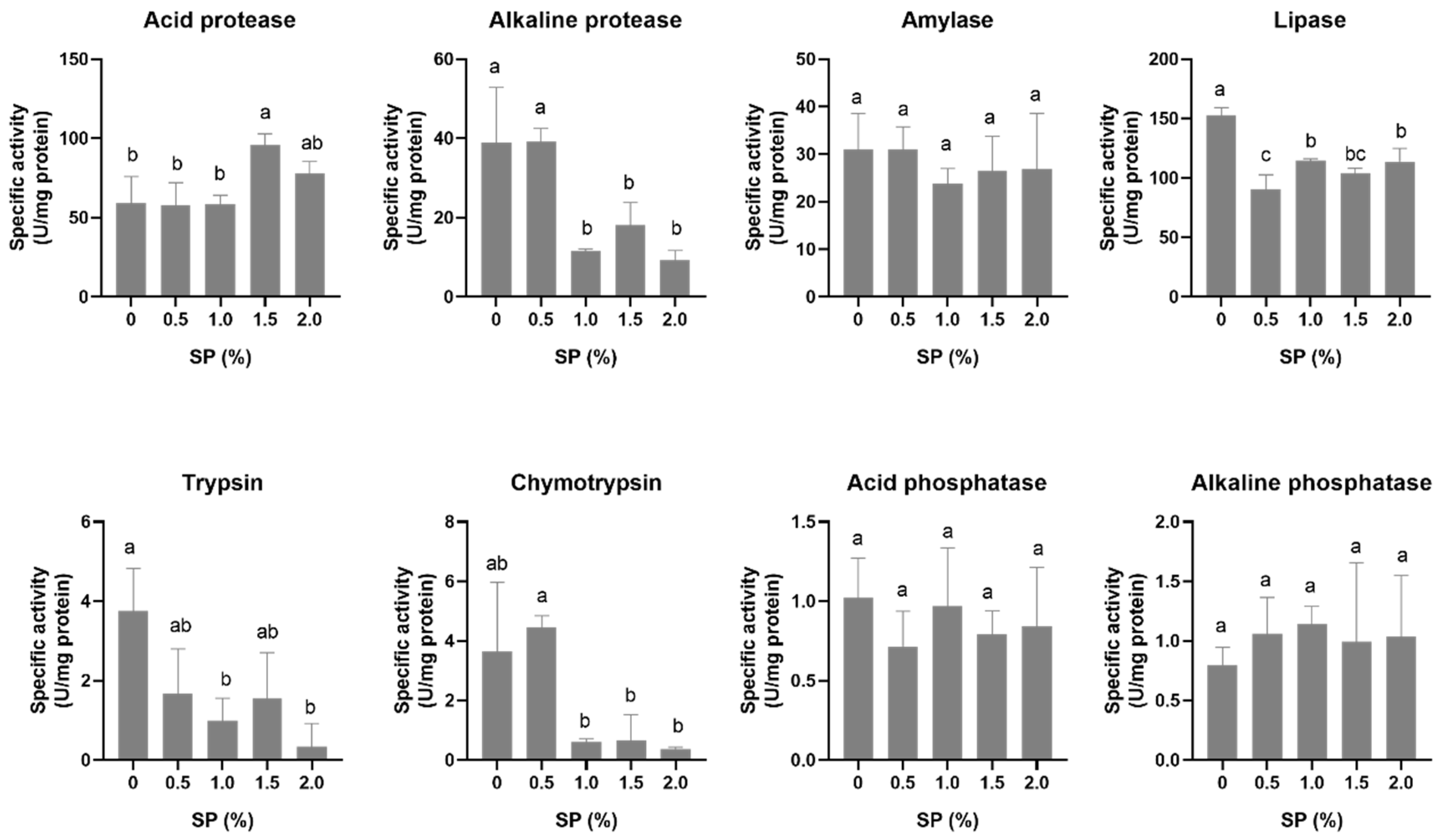
3.2. Digestive Enzyme Activity

For acid proteases, fish fed with 1.5% SP (95.73 ± 7.26 U/mg protein) showed the highest enzyme activity (p < 0.05). Treatment with 0.5% SP showed the highest activity for alkaline proteases (39.16 ± 3.29) and chymotrypsin (0.44 ± 0.039) (p < 0.05). The control group (0%) showed the highest lipase (152.41 ± 6.84) and trypsin (3.13 ± 0.22) activities (Figure 1).

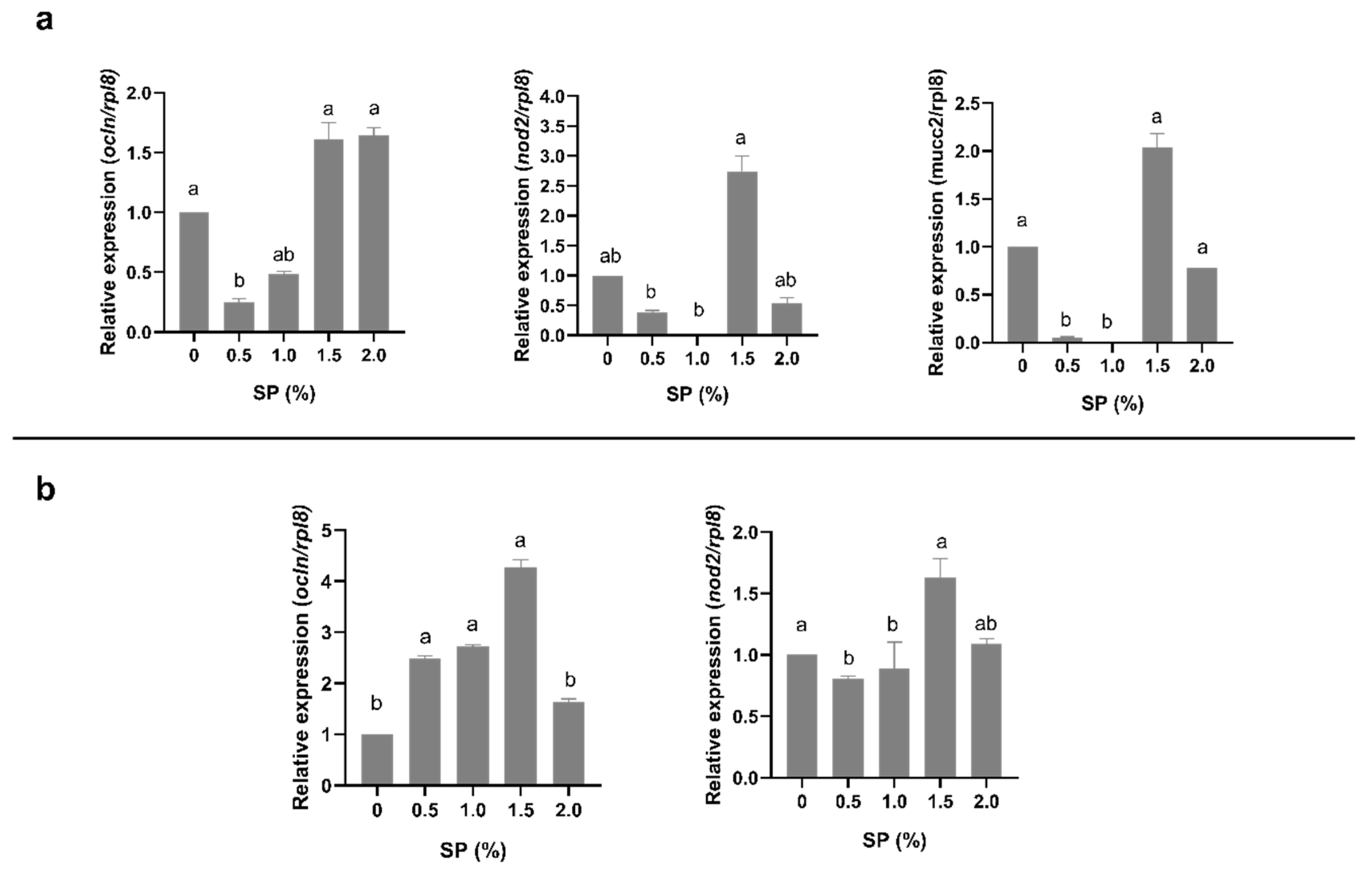
3.3. Gene Expression

According to the qPCR results, in the intestine, fish from the 1.5 and 2% SP treatments showed higher ocln expression when compared with the 0.5% treatment (p < 0.05). The fish from the 1.5% SP treatment showed higher expression of the nod2 and muc2 genes in the intestine when compared with the 0.5 and 1.0% SP treatments (p < 0.05). In liver, the 1.5% SP treatment showed a higher expression of nod2 when compared with the 0.5 and 1.0% SP treatments and a higher expression of ocln gene when compared with the 0 and 2.0% SP treatments (p < 0.05) (Figure 2).

4. Discussion

Innovative trends in aquaculture nutrition include evaluating the effectiveness of several probiotics, prebiotics, and additives that are added to fish diets. Different parameters have been analyzed to characterize the responses to these immunostimulants.
This study evaluated the inclusion of SP at different concentrations. The results did not show significant differences in productive parameters, such as the specific growth rate, feed conversion rate, survival, and somatic indexes. However, Atractosteus tropicus juveniles fed with 0.5% SP showed a trend of higher growth, AWG, and SGR. The highest survival occurred in the 1.5 and 2.0% treatments; this is because SCFAs, in this case SP, could stimulate the activity of one or more intestinal bacteria, which is known to be the action of immune response, performance, and disease resistance [6]. The effects of including SP in diets for fish may vary according to the species, environmental conditions, concentration used, and life stage of the organism. Similar doses were used in other studies. On European seabass (Decentrarchus labrax), final weight, daily weight gain, and survival were higher when including 0.3% SP compared with other treatments [28]. In Caspian white fish (Rutilus frisii kutum), the inclusion of 0.25 and 0.5% SP resulted in a higher final weight and weight gain in addition to promoting immune response parameters [29]. Another study carried out by Hoseinifar [10] with zebrafish (Danio rerio) indicated that inclusion of 1 and 2% SP regulated the expression of genes related to growth hormones (GH and IGF1) and appetite (GHRL). In yellowfin seabream (Acanthopagrus latus), similar results were obtained when including 0.5 and 1% SP to promote growth, immune response parameters, and digestive capacity [8]. In two studies conducted by the same author in white shrimp (Litopenaeus vannamei), it has been shown that including 2% SP increased feed intake; additionally, the concentration of pathogens such as Vibrio spp. in the digestive tract was reduced and final weight was increased when adding 0.5 to 2% SP into the diet [9,30].
A higher HSI in the liver of A. tropicus juveniles may suggest greater glycogen and lipid content, which may be related to the composition of the diets, health, and metabolism of the organism [31,32]. Previous studies with red tilapia (Oreochromis spp.) found no significant differences in HSI and VSI when comparing the effects of different organic acids such as propionate, butyrate, and sodium acetate [33]. However, in A. tropicus, significant differences were found when evaluating this index when adding 0.4% MOS (mannan oligosaccharide) and FOS (fructooligosaccharides) [3,5], suggesting a greater presence of energy reserves in the liver due to the energy intake in the diets. In the present study, no differences were observed in the HSI, VSI, and RIL when including SP. The intestinal length ratio (RIL) suggests a greater area of nutrient absorption; however, it is necessary to perform histological studies to assess the structure of the organs related to these indexes.
The effects of the inclusion of probiotics, prebiotics, and other additives to functional diets for fish has led to the study of various functions of the organism, such as the activity of digestive enzymes [5,8,9,10,34,35].
Our results showed the highest activity in fish fed with 1 and 2% SP; similar results have shown that adding 1% SP and a mix of SP + sodium acetate increased the activity of this enzyme in yellow fin seabream (Acanthopagrus latus) [8]. In contrast, our results showed higher activity of alkaline proteases, trypsin, and chymotrypsin in fish fed with 0 and 0.5% SP. These results may be due to the action of organic acid salts, which reduces pH in the stomach and intestine [36]. Other studies of A. tropicus show an increment in the activity of alkaline proteases, trypsin, and chymotrypsin in fish fed with low concentrations of MOS and FOS (0 and 0.5%) [3,5]. The activity of lipases and amylases were not modified between treatments, similar to the results reported by Nieves-Rodríguez et al. [4] when including β-glucans in diets for juveniles of A. tropicus. The increment in the activity of these enzymes may be due to the specificity that the enzymes have in terms of pH and temperature values [37,38]. In contrast, the modification of the enzymes activities in the group of proteases (acid, alkaline, trypsin, and chymotrypsin proteases) suggest a positive effect of organic acids and/or their salts on protein hydrolysis [39].
Short-chain fatty acids play important roles as nutrients that enhance the intestine epithelium, and they are also regulators of gene disappearance, differentiation, and expression [6]. The effects of SP on the expression of ocln, nod2, and muc2 genes have been rarely reported in fish. However, other immune response parameters have been evaluated in other species when including SP in their diets [10,27,28,40]. Mucus is one of the main components of the intestine, and it is mainly composed of a protein called mucin, which plays an important role in protecting the intestinal barrier, regulating homeostasis, and preventing diseases [41]. Burger-van Paassen [42] suggests that one of the mechanisms that regulates muc2 expression is the use of short-chain fatty acids such as butyrate or propionate. Our study showed a higher expression of this gene in the intestine of A. tropicus juveniles with 1.5% SP. Similar results were reported by Pérez-Jiménez [43], who found a greater expression of muc2 when increasing the concentrations of FOS (7.5%) in larvae of A. tropicus. Other components of the intestinal barrier include the tight junctions (TJs), which provide a barrier within the membrane by regulating paracellular permeability and protein diffusion, thus maintaining cell surface polarity and enhancing the transportation of proteins and macromolecules through cells [44]. Our study showed greater expression of the ocln gene in the intestine at concentrations of 1.5 and 2% SP and in the liver at a concentration of 1.5% SP, suggesting that the addition of short-chain fatty acids also regulates the expression of genes related to tight junctions. Liu [45] reported similar results of greater expression of ocln when adding sodium butyrate in diets for Scophthalmus maximus L. Sun [46] also evaluated the inclusion of 0.5 and 1.0% SP in Scophthalmus maximus L. combined with soybean meal and found similar results in the expression of the ocln gene. Sepúlveda-Quiroz [5] found similar results when evaluating the same genes in juveniles of A. tropicus when adding 1 and 1.5% FOS. Finally, the present study showed a higher expression of the nod2 gene, which has been identified as an important sensor in the first line of defense in invasion by microorganisms in mammals and teleost fish [47]. The results of this study showed a higher expression of nod2 in the intestine and liver of fish fed with 1.5% SP. Highest expression of this gene was observed in the liver, which agrees with J. Li’s study [48], wherein a Vibrio infection was induced (V. anguillarum) in miiuy croaker (Miichthys miiuy), suggesting that the expression of this gene plays an important role in the innate immune response and exerts important immune functions in liver tissues. In contrast, a study involving A. tropicus juveniles did not show significant differences in the expression of ocln and nod2 when incorporating β-glucans into their diets [4]. In our study, the increase in immune-related genes at a higher concentration of SP corresponded with the increasing survival rate, which also increased at a higher concentration of SP.

5. Conclusions

The inclusion of 0.5 to 2% SP in diets for A. tropicus did not influence growth parameters and somatic indexes. However, the inclusion of 1.5% SP showed the highest survival rate and highest enzyme activity for acid proteases. Also, adding 1.5% SP into the diet for A. tropicus may have positive effects on the immune response by increasing the expression of muc2, ocln, and nod2 in the intestine and liver. Nevertheless, for future studies, it is recommended to increase the fish sample in order to find clearer significant differences in relation to genomic expression.

Author Contributions

J.G.A.-C., C.A.Á.-G. and R.M.-G. designed and planned the bioassay; J.G.A.-C., T.M.-B. and S.A.F.-G. conducted the experiment and sample collection; J.G.A.-C., C.A.S.-Q., S.A.F.-G. and G.G.A.-A. carried out the biochemical analyses; J.G.A.-C., L.D.J.-M. and T.M.-B. conducted the molecular analyses; J.G.A.-C., S.A.F.-G., C.A.Á.-G., R.G.-Z. and R.M.-G. performed the statistical analysis; J.G.A.-C., A.A.-H., J.L.R.-M., P.D.-R. and R.M.-G. wrote the paper. All authors have read and agreed to the published version of the manuscript.

Funding

This research was supported by the Consejo Nacional de Ciencia y Tecnología through the grant “Estudio de la fisiología digestiva en larvas y juveniles de pejelagarto (Atractosteus tropicus) con base en técnicas histológicas, bioquímicas y moleculares” CB-2016-1-282765 and by the Colegio de Postgraduados Campus Veracruz through the budget for full-time students 32033001.

Institutional Review Board Statement

This study was conducted in accordance with the Declaration of Helsinki, and the protocol was approved by Secretaría de Agricultura, Ganadería, Desarrollo Rural, Pesca y Alimentación (NOM-062-ZOO-, 1999, 2001).

Informed Consent Statement

Not applicable.

Data Availability Statement

Data set for genes expression could be find at 10.17632/dr9sj5w6hk.1.

Acknowledgments

The authors would like to thank Consejo Nacional de Ciencia y Tecnología (CONACYT) for the financial support of this project through the grant “Estudio de la fisiología digestiva en larvas y juveniles de pejelagarto (Atractosteus tropicus) con base en técnicas histológicas, bioquímicas y moleculares” CB-2016-1-282765 and for the Master degree fellowship (CVU 1081761); to Colegio de Postgraduados, Campus Veracruz, for economic support throughout the Master degree program Agroecosistemas Tropicales; and, finally, to the Laboratorio de Fisiología en Recursos Acuáticos from the Universidad Juárez Autónoma de Tabasco for the technical support during this research project.

Conflicts of Interest

The authors declare no conflict of interest. The funders had no role in the design of the study; in the collection, analyses, or interpretation of data; in the writing of the manuscript; or in the decision to publish the results.

References

  1. Márquez-Couturier, G.; Navarrete-Vázquez, C.; Sánchez-Contreras, W.; Álvarez-González, C.A. Acuicultura Tropical Sustentable, 2nd ed.; Universidad Juárez Autónoma de Tabasco: Villahermosa, Mexico, 2015. [Google Scholar]
  2. Márquez-Couturier, G.; Vázquez-Navarrete, C. Estado de arte de la biología y cultivo de pejelagarto (Atractosteus tropicus). Agroproductividad 2015, 8, 44–51. [Google Scholar]
  3. Nájera-Arzola, I.C.; Álvarez-González, C.A.; Frías-Quintana, C.A.; Peña, E.; Martínez-García, R.; Camarillo-Coop, S.; Méndez-Marín, O.; Gisbert, E. Evaluation of Mannan oligosaccharides (MOS) in balanced diets for tropical gar juveniles (Atractosteus tropicus). Hidrobiologica 2018, 28, 239–246. [Google Scholar] [CrossRef]
  4. Nieves-Rodríguez, K.N.; Álvarez-González, C.A.; Peña-Marín, E.S.; Vega-Villasante, F.; Martínez-García, R.; Camarillo-Coop, S.; Tovar-Ramírez, D.; Guzmán-Villanueva, L.T.; Andree, K.B.; Gisbert, E. Effect of β-Glucans in Diets on Growth, Survival, Digestive Enzyme Activity, and Immune System and Intestinal Barrier Gene Expression for Tropical Gar (Atractosteus tropicus) Juveniles. Fishes 2018, 3, 27. [Google Scholar] [CrossRef]
  5. Sepúlveda-Quiroz, C.A.; Peña-Marín, E.S.; Pérez-Morales, A.; Martínez-García, R.; Alvarez-Villagomez, C.S.; Maytorena-Verdugo, C.I.; Camarillo-Coop, S.; Vissio, P.G.; Sirkin, D.P.; Tovar-Ramírez, D.; et al. Fructooligosaccharide supplementation in diets for tropical gar (Atractosteus tropicus) juvenile: Effects on morphophysiology and intestinal barrier function. Aquac. Res. 2020, 52, 37–50. [Google Scholar] [CrossRef]
  6. Cook, S.; Sellin, J.H. Review article: Short chain fatty acids in health and disease. Aliment. Pharmacol. Ther. 1998, 12, 499–507. [Google Scholar] [CrossRef]
  7. Hijova, E.; Chmelarova, A. Short chain fatty acids and colonic health. Bratisl. Lek. Listy 2007, 108, 354–358. [Google Scholar]
  8. Sotoudeh, E.; Sangari, M.; Bagheri, D.; Morammazi, S.; Mozanzadeh, M.T. Dietary organic acid salts mitigate plant protein induced inflammatory response and improve humoral immunity, antioxidative status and digestive enzyme activities in yellowfin seabream, Acanthopagrus latus. Aquac. Nutr. 2020, 26, 1669–1680. [Google Scholar] [CrossRef]
  9. da Silva, B.C.; Vieira, F.D.N.; Mouriño, J.L.P.; Bolivar, N.; Seiffert, W.Q. Butyrate and propionate improve the growth performance of Litopenaeus vannamei. Aquac. Res. 2014, 47, 612–623. [Google Scholar] [CrossRef]
  10. Hoseinifar, S.H.; Safari, R.; Dadar, M. Dietary sodium propionate affects mucosal immune parameters, growth and appetite related genes expression: Insights from zebrafish model. Gen. Comp. Endocrinol. 2017, 243, 78–83. [Google Scholar] [CrossRef]
  11. De la Cruz-Alvarado, F.J.; Álvarez-González, C.A.; Llera-Herrera, R.; Monroig, Ó.; Kabeya, N.; Rodríguez-Morales, S.; Concha-Frias, B.; Guerrero-Zárate, R.; Jiménez-Martínez, L.D.; Peña-Marín, E.S. Expression of long-chain polyunsaturated fatty acids biosynthesis genes during the early life-cycle stages of the tropical gar Atractosteus tropicus. Comp. Biochem. Physiol. Part B Biochem. Mol. Biol. 2021, 256, 110628. [Google Scholar] [CrossRef]
  12. Frías-Quintana, C.A.; Álvarez-González, C.A.; Tovar-Ramírez, D.; Martínez-García, R.; Camarillo-Coop, S.; Peña, E.; Galaviz, M.A. Use of Potato Starch in Diets of Tropical Gar (Atractosteus tropicus, Gill 1863) Larvae. Fishes 2017, 2, 3. [Google Scholar] [CrossRef]
  13. Alvarez-González, C.; Civera-Cerecedo, R.; Ortiz-Galindo, J.; Dumas, S.; Moreno-Legorreta, M.; Alamo, T.G.-D. Effect of dietary protein level on growth and body composition of juvenile spotted sand bass, Paralabrax maculatofasciatus, fed practical diets. Aquaculture 2001, 194, 151–159. [Google Scholar] [CrossRef]
  14. AOAC. Official Methods of Analysis of the Association of Official Analytical Chemist, 15th ed.; Association of Official Analytical Chemists: Washington, DC, USA, 1990. [Google Scholar]
  15. Bligh, E.G.; Dyer, W.J. A Rapid Method of Total Lipid Extraction and Purification. Available online: https://cdnsciencepub.com/doi/abs/10.1139/o59-099 (accessed on 21 September 2022).
  16. Brett, J.R.; Groves, T.D.D. “Physiological energetics” in Bioenergetics and growth. Fish. Physiol. 1979, 8, 1–786. [Google Scholar] [CrossRef]
  17. NOM-062-ZOO-1999; Especificaciones Técnicas para la Producción, Cuidado y uso de los Animales de Laboratorio. SAGARPA: Mexico City, Mexico, 2001; pp. 1–58.
  18. Bradford, M.M. A rapid and sensitive method for the quantitation of microgram quantities of protein utilizing the principle of protein-dye binding. Anal. Biochem. 1976, 72, 248–254. [Google Scholar] [CrossRef] [PubMed]
  19. Anson, M.L. The estimation of pepsin, trypsin, papain, and cathepsin with hemoglobin. J. Gen. Physiol. 1938, 22, 79–89. [Google Scholar] [CrossRef]
  20. Walter, H.E. Proteinases: Methods with hemoglobin, casein and azocoll as substrates. In Methods of Enzymatic Analysis; Bergmeyer, H.J., Ed.; Verlag Chemie: Weinheim, Germany, 1984; pp. 270–277. [Google Scholar]
  21. Erlanger, B.F.; Kokowsky, N.; Cohen, W. The preparation and properties of two new chromogenic substrates of trypsin. Arch. Biochem. Biophys. 1961, 95, 271–278. [Google Scholar] [CrossRef]
  22. DelMar, E.; Largman, C.; Brodrick, J.; Geokas, M. A sensitive new substrate for chymotrypsin. Anal. Biochem. 1979, 99, 316–320. [Google Scholar] [CrossRef]
  23. Robyt, J.F.; Whelan, W. Amylases. In Starch and Its Derivates; Radley, J.A., Ed.; Chapman and Hall: London, UK, 1968. [Google Scholar]
  24. Versaw, W.K.; Cuppett, S.L.; Winters, D.D.; Williams, L.E. An Improved Colorimetric Assay for Bacterial Lipase in Nonfat Dry Milk. J. Food Sci. 1989, 54, 1557–1558. [Google Scholar] [CrossRef]
  25. Bergmeyer, H.U. Methods of Enzymatic Analysis; Elsevier: Amsterdam, The Netherlands, 1974. [Google Scholar]
  26. Livak, K.J.; Schmittgen, T.D. Analysis of relative gene expression data using real-time quantitative PCR and the 2−ΔΔCT Method. Methods 2001, 25, 402–408. [Google Scholar] [CrossRef]
  27. Aranda-Morales, S.A.; Peña-Marin, E.S.; Jiménez-Martínez, L.D.; Martínez-Burguete, T.; Martínez-Bautista, G.; Alavrez-Villagómez, C.S.; Rosa García, S.R.; Camarillo-Coop, S.; Martínez-García, R.; Guzmán-Villanueva, L.T.; et al. Expression of ion transport proteins and routine metabolism in juveniles of tropical gar (Atractosteus tropicus) exposed to ammonia. Comp. Biochem. Physiol. Part C 2021, 250, 109166. [Google Scholar] [CrossRef]
  28. Wassef, E.A.; Saleh, N.E.; Abdel-Meguid, N.E.; Barakat, K.M.; Abdel-Mohsen, H.H.; El-Bermawy, N.M. Sodium propionate as a dietary acidifier for European seabass (Dicentrarchus labrax) fry: Immune competence, gut microbiome, and intestinal histology benefits. Aquac. Int. 2019, 28, 95–111. [Google Scholar] [CrossRef]
  29. Hoseinifar, S.H.; Zoheiri, F.; Caipang, C.M. Dietary sodium propionate improved performance, mucosal and humoral immune responses in Caspian white fish (Rutilus frisii kutum) fry. Fish Shellfish. Immunol. 2016, 55, 523–528. [Google Scholar] [CrossRef] [PubMed]
  30. da Silva, B.C.; Vieira, F.D.N.; Mouriño, J.L.P.; Ferreira, G.S.; Seiffert, W.Q. Salts of organic acids selection by multiple characteristics for marine shrimp nutrition. Aquaculture 2013, 384–387, 104–110. [Google Scholar] [CrossRef]
  31. Gatlin, D.M., III. Nutrition and Fish Health. In Fish Nutrition, 3rd ed.; Halver, J., Hardy, R., Eds.; Academic Press: Cambridge, MA, USA, 2002; pp. 672–699. [Google Scholar]
  32. Polakof, S.; Panserat, S.; Soengas, J.L.; Moon, T.W. Glucose metabolism in fish: A review. J. Comp. Physiol. B 2012, 182, 1015–1045. [Google Scholar] [CrossRef]
  33. Ebrahimi, M.; Daeman, N.H.; Chong, C.M.; Karami, A.; Kumar, V.; Hoseinifar, S.H.; Romano, N. Comparing the effects of different dietary organic acids on the growth, intestinal short-chain fatty acids, and liver histopathology of red hybrid tilapia (Oreochromis sp.) and potential use of these as preservatives. Fish Physiol. Biochem. 2017, 43, 1195–1207. [Google Scholar] [CrossRef]
  34. Maytorena-Verdugo, C.I.; Peña-Marín, E.S.; Alvarez-Villagómez, C.S.; Pérez-Jiménez, G.M.; Sepúlveda-Quiroz, C.A.; Alvarez-González, C.A. Inclusion of Mannan-Oligosaccharides in Diets for Tropical Gar Atractosteus tropicus Larvae: Effects on Growth, Digestive Enzymes, and Expression of Intestinal Barrier Genes. Fishes 2022, 7, 127. [Google Scholar] [CrossRef]
  35. Hoseinifar, S.H.; Ahmadi, A.; Khalili, M.; Raeisi, M.; Van Doan, H.; Caipang, C.M. The study of antioxidant enzymes and immune-related genes expression in common carp (Cyprinus carpio) fingerlings fed different prebiotics. Aquac. Res. 2017, 48, 5447–5454. [Google Scholar] [CrossRef]
  36. Lim, P.; Lückstädt, C.; Webster, C.; Kesius, C.D. Organic Acids and Their Salts. In Dietary Nutrients, Additives, and Fish Health; Lee, C.D., Lim, C.-S., Gatlin, C., III, Webster, D.M., Eds.; John Wiley & Sons, Inc.: Hoboken, NJ, USA, 2015; pp. 305–315. [Google Scholar]
  37. Guerrero-Zárate, R.; Alvarez-González, C.A.; Olvera-Novoa, M.A.; Perales-García, N.; Frías-Quintana, C.A.; Martínez-García, R.; Contreras-Sánchez, W.M. Partial characterization of digestive proteases in tropical gar Atractosteus tropicus juveniles. Fish Physiol. Biochem. 2013, 40, 1021–1029. [Google Scholar] [CrossRef]
  38. Xiong, D.M.; Xie, C.X.; Zhang, H.J.; Liu, H.P. Digestive enzymes along digestive tract of a carnivorous fish Glyptosternum maculatum (Sisoridae, Siluriformes). J. Anim. Physiol. Anim. Nutr. 2011, 95, 56–64. [Google Scholar] [CrossRef]
  39. Dietary Nutrients, Additives, and Fish Health; Lee, C.-S.; Lim, C.; Gatlin, D.M.; Webster, C.D. (Eds.) John Wiley & Sons, Inc.: Hoboken, NJ, USA, 2015; p. 355. [Google Scholar]
  40. Safari, R.; Hoseinifar, S.H.; Kavandi, M. Modulation of antioxidant defense and immune response in zebra fish (Danio rerio) using dietary sodium propionate. Fish Physiol. Biochem. 2016, 42, 1733–1739. [Google Scholar] [CrossRef]
  41. Liu, Y.; Yu, X.; Zhao, J.; Zhang, H.; Zhai, Q.; Chen, W. The role of MUC2 mucin in intestinal homeostasis and the impact of dietary components on MUC2 expression. Int. J. Biol. Macromol. 2020, 164, 884–891. [Google Scholar] [CrossRef] [PubMed]
  42. Burger-van Paassen, N.; Vincent, A.; Puiman, P.J.; Van Der Sluis, M.; Bouma, J.; Boehm, G.; van Goudoever, J.B.; Van Seuningen, I.; Renes, I.B. The regulation of intestinal mucin MUC2 expression by short-chain fatty acids: Implications for epithelial protection. Biochem. J. 2009, 420, 211–219. [Google Scholar] [CrossRef] [PubMed]
  43. Pérez-Jiménez, G.M.; Peña-Marín, E.S.; Maytorena-Verdugo, C.I.; Sepúlveda-Quiroz, C.A.; Jiménez-Martínez, L.D.; De la Rosa-García, S.; Asencio-Alcudia, G.G.; Martínez, R.; Tovar-Ramírez, D.; Galaviz, M.A.; et al. Incorporation of Fructooligosaccharides in Diets Influence Growth Performance, Digestive Enzyme Activity, and Expression of Intestinal Barrier Function Genes in Tropical Gar (Atractosteus tropicus) Larvae. Fishes 2022, 7, 137. [Google Scholar] [CrossRef]
  44. Treyer, A.; Müsch, A. Hepatocyte polarity. Compr. Physiol. 2013, 3, 243–287. [Google Scholar] [CrossRef]
  45. Liu, Y.; Chen, Z.; Dai, J.; Yang, P.; Xu, W.; Ai, Q.; Zhang, W.; Zhang, Y.; Zhang, Y.; Mai, K. Sodium butyrate supplementation in high-soybean meal diets for turbot (Scophthalmus maximus L.): Effects on inflammatory status, mucosal barriers and microbiota in the intestine. Fish Shellfish. Immunol. 2019, 88, 65–75. [Google Scholar] [CrossRef]
  46. Sun, H.; Zhang, J.; Wang, W.; Shao, R.; Liang, S.; Xu, W.; Li, M.; Ai, Q.; Mai, K.; Wan, M. The Effects of Sodium Propionate Supplementation in the Diet with High Soybean Meal on Growth Performance, Intestinal Health, and Immune Resistance to Bacterial Infection in Turbot (Scophthalmus maximus L.). Aquac. Nutr. 2022, 2022, 8952755. [Google Scholar] [CrossRef]
  47. Li, Y.; Jin, L.; Xia, P.; Sui, W.; Huang, A.; Bu, G.; Meng, F.; Kong, F.; Cao, X.; Han, X.; et al. Identification and functional analysis of NOD2 and its two splicing variants associated with a novel pattern of signal regulation in teleost fishes. Dev. Comp. Immunol. 2021, 120, 104049. [Google Scholar] [CrossRef]
  48. Li, J.; Gao, Y.; Xu, T. Comparative genomic and evolution of vertebrate NOD1 and NOD2 genes and their immune response in miiuy croaker. Fish Shellfish. Immunol. 2015, 46, 387–397. [Google Scholar] [CrossRef]
Figure 1. Enzyme activities (U mg protein−1) of A. tropicus fed with different percentages of SP (0, 0.5, 1.0, 1.5, 2.0%). Values are presented as the mean ± SD. Significant differences between the diets are indicated by different letters (p < 0.05).
Aquacj 03 00018 g001
Figure 2. Relative gene expression of occludin (ocln), mucin2 (muc2), and nucleotide-binding and oligomerization domain-2 (nod2) in the intestines (a) and livers (b) of A. tropicus juveniles fed different SP concentrations using rpl8 as the reference gene. Values are presented as the mean ± SE. Significant differences between the diets are indicated by different letters (p < 0.05).
Aquacj 03 00018 g002
Table 1. Proximal composition of experimental feed with different concentrations of SP.
Sodium Propionate (%)
Ingredients (g/kg)00.51.01.52.0
Pork meal 1250250250250250
Poultry meal 1218.4218.4218.4218.4218.4
Soybean meal 2161.1161.1161.1161.1161.1
Corn starch 3150150150150150
Fish meal 1100100100100100
Wheat meal 255.555.555.540.535.5
Sodium propionate 405101520
Soybean lecithin 53030303030
Grenetin 62020202020
Vitamins and minerals premix 71010101010
Vitamin C 855555
Proximate composition (g/100 g dry matter)
Protein44.3244.8143.9944.8344.74
Lipids15.3215.7415.4315.5315.31
Ash12.7712.7413.2313.7113.53
NFE 928.4928.4328.4328.4828.46
1 Proteínas Marinas y Agropecuarias, S. A. de C. V., Guadalajara, Jalisco. 2 GALMEX Comercializadora de Insumos Agrícolas, Villahermosa, Tabasco, México. 3 MSA Industrializadora de Maíz, Guadalajara, Jalisco, México. 4 Mi granero Ingredientes Selectos. San Pedro Cholula, Puebla, México. 5 Pronat Ultra, Mérida, Yucatán, México. 6 D’gari Productos alimenticios y dietéticos Relámpago, Tlalpan, Edomex, México. 7 Pedregal (para trucha Silver Cup), Toluca, Edo. de Mex. México. 8 ROVIMIX® C-EC (Roche) active agent of 35%. 9 NFE: Nitrogen-free extract.
Table 3. Survival and growth parameters and somatic indexes of A. tropicus fed with different SP concentrations (mean ± standard deviation, SD) for 61 days.
Treatments (SP%)
Growth Parameters 00.511.52
Final weight (g)24.19 ± 1.225.79 ± 4.5 22.17 ± 2.5 23.32 ± 1.921.38 ± 1.2
Final length (cm)17.60 ± 0.117.74 ± 0.617.03 ± 0.517.60 ± 0.516.79 ± 0.3
Survival (%)80.5 ± 12.7 a86.1 ± 4.8 b86.11 ± 9.6 b91.6 ± 8.3 c91.6 ± 8.3 c
AWG (g/fish)20.53 ± 1.2621.93 ± 4.3918.52 ± 2.8119.81 ± 217.69 ± 1.02
SGR (%/day)3.1 ± 0.143.1± 0.263.0 ± 0.343.05 ± 0.172.9 ± 0.14
Condition factor (k)0.44 ± 0.020.46 ± 0.030.45 ± 0.010.41 ± 0.040.45 ± 0.01
FCR1.61 ± 0.091.92 ± 0.381.52 ± 0.231.46 ± 0.141.40 ± 0.08
Note: AWG: absolute weight gain; SGR: specific growth rate; FCR: feed conversion rate. Significant differences are indicated by different letters (p < 0.05).
Table 4. Somatic indexes of A. tropicus fed with different SP concentrations (mean ± standard deviation, SD) for 61 days.
Treatments (SP%)
Somatic Indexes00.511.52
HSI3.26 ± 1.223.04 ± 0.672.80 ± 0.473.47 ± 0.52.95 ± 0.61
VSI9.45 ± 3.658.73 ± 1.098.01 ± 1.0110.53 ± 1.778.10 ± 1.23
RIL35.86 ± 4.9935.18 ± 3.6738.68 ± 4.9140.26 ± 4.2030.92 ± 6.01
Note: HSI: hepatosomatic index; VSI: viscerosomatic index; RIL: relation of intestine length (somatic indexes did not show significant differences).
Disclaimer/Publisher’s Note: The statements, opinions and data contained in all publications are solely those of the individual author(s) and contributor(s) and not of MDPI and/or the editor(s). MDPI and/or the editor(s) disclaim responsibility for any injury to people or property resulting from any ideas, methods, instructions or products referred to in the content.

