Article

Effect of Ischemic Preconditioning (IPC) on Recovery of Exercise Performance Following a Bout of Exercise to Volitional Exhaustion

1
School of Health & Sports Sciences, Liverpool Hope University, Liverpool L16 9JD, UK
2
Liverpool Centre for Cardiovascular Sciences, Liverpool L69 7TX, UK
*
Author to whom correspondence should be addressed.
Physiologia 2023, 3(3), 394-405; https://doi.org/10.3390/physiologia3030027
Submission received: 3 May 2023 / Revised: 23 June 2023 / Accepted: 23 June 2023 / Published: 29 June 2023
(This article belongs to the Special Issue Feature Papers in Human Physiology–2nd Edition)

Abstract

:
The purpose of the present study was to investigate the effect of ischemic preconditioning (IPC) on the recovery of exercise performance following maximal, incremental exercise. A total of 13 healthy males volunteered to participate, undertaking three experimental trials involving a constant work-rate bout of severe intensity exercise undertaken to the limit of tolerance that was preceded by a 40-min recovery period consequent to a maximal, incremental exercise test. During the recovery period, participants underwent IPC at 220 mmHg, sham IPC (SHAM; 20 mmHg), and passive rest (CON). Exercise tolerance time was higher following IPC as compared to SHAM and CON {199 ± 36 (CON) vs. 203 ± 35 (SHAM) vs. 219 ± 34 (IPC), p = 0.03}. This effect was accompanied by a tendency toward an augmented increase in blood lactate from rest to exercise in IPC compared to SHAM and CON (p = 0.08). There was no effect of IPC on oxygen uptake kinetics or muscle oxygenation as indicated via near-infrared spectroscopy. IPC may therefore have the capacity to augment recovery from prior maximal exercise, but this does not appear to be due to enhancements to oxygen uptake kinetics or muscle oxygenation.

1. Introduction

Ischemic preconditioning (IPC) has long been established as a method for reducing infarct size in patients suffering from acute myocardial infarction, but has increasingly been explored as a method for enhancing exercise performance across a variety of sporting modalities [1]. There is significant variation in the outcomes from different studies as to whether there is a consistent effect of IPC on exercise performance and the possible scale of effect. Recent findings have shown conflicting results with data showing improvements in maximum strength [2], strength endurance [3], sprint/repeat sprint performance [4], and anaerobic capacity [5] whilst others have found no effect of IPC on trial performance in swimmers [6], or runners [7]. Whilst improvements in performance have been observed in a growing number of exercise modalities and energy systems, a greater body of evidence has supported the use of IPC for improving aerobic exercise performance [8].
The physiological mechanisms that underpin the possible improvements in exercise performance seen as a result of IPC, have not been fully elucidated [9]. A number of mechanisms have however been postulated for any positive effects of IPC, including augmented oxygen uptake kinetics [10], which are an important determinant of aerobic exercise performance [11,12]. It is proposed that increases in blood flow through an up-regulation of endothelial function following IPC allows for an improved (reduced) time constant () of the fundamental phase of oxygen uptake kinetics, in addition to an attenuation of the slow component [1]. Indeed, Pang et al., [13], Lee and Thompson [14] and Hopper et al., [15], showed that IPC elevated levels of adenosine and ATP sensitive potassium (KATP) channels which facilitates an increase in vasodilation, improved oxygen delivery, and increased substrate availability, leading to improved metabolic dynamics in matching the demands of the muscle [13]. Consequently, any improvements in oxygen uptake kinetics due to IPC would be expected to bring about a reduction in the rate of fatigue development [16]. However, to date, findings from recent studies demonstrate contrasting results on the effect of IPC on oxygen uptake kinetics [1,17,18].
Recovery of function plays a significant role in sports performance where exercise is intermittent in nature, with more rapid recovery between bouts of high intensity exercise being advantageous to the athlete [19]. The persistence of the metabolic by-products of anaerobic metabolism following high intensity exercise appears to limit subsequent performance [20], presumably via a diminution of the gradient of efflux between muscle and blood. IPC has been shown to bring about enhanced convective flow [21] and local perfusion [22] which promotes a more rapid decline of the aforementioned fatigue-related metabolites during the recovery period, thus enhancing subsequent exercise performance [23].
The aim of the present study was therefore to determine the effect of IPC on the immediate recovery of exercise performance capacity following prior exercise to volitional exhaustion. In order to elucidate the mechanisms underpinning any effect of IPC on performance, oxygen uptake kinetics and muscle oxygenation (via near infrared spectroscopy) were observed. It was hypothesised that recovery would be improved by IPC as compared to control, reflected in an improved time to the limit of tolerance during severe intensity, constant work-rate exercise. We further hypothesized that this improvement would be underpinned by faster oxygen uptake kinetics (i.e. reduced) and enhanced muscle oxygenation.

2. Methods

2.1. Participants

Thirteen healthy, recreationally active males (values presented as mean ± SD, age 25 ± 3 yrs; height 178 ± 9 cm; weight 79 ± 6.4 kg) volunteered to participate in this study which was carried out in accordance with the Declaration of Helsinki and approved by the Liverpool Hope University ethics committee. Inclusion criteria were that participants were required to be over 18 years of age, free from cardiometabolic and respiratory disease, and free from musculoskeletal injury that may impede exercise. All participants provided written, informed consent prior to participation and were informed of their right to withdraw at any time.

2.2. Experimental Design

A randomised single-blinded crossover design with three conditions (Control, IPC and Sham) was employed. Pre-participation body mass and height were measured using standard electronic scales and a stadiometer, respectively (Seca, 761, Hamburg, Germany; Avery, 220, Birmingham, UK). All participants refrained from alcohol, caffeine and any additional training supplements 24 h before each experimental visit and were instructed to consume the same dietary intake prior to all three experimental trails. Participants reported to the laboratory on 4 occasions, which consisted of a familiarisation visit, followed by 3 visits consisting of each condition in a randomised order. Each experimental visit consisted of an incremental exercise test undertaken to the limit of tolerance, followed by the experimental intervention, and a subsequent constant work-rate test to the limit of tolerance that was utilised to evaluate recovery. There was at least 48 h between each condition to reduce the acute crossover effects of IPC or sham conditions on the participants [24,25].

2.3. Incremental Exercise

All exercise tests were conducted on an electronically controlled cycle ergometer (Corival, lode, Excalibur sport, Groningen, The Netherlands). The incremental exercise test began with one minute at rest, followed by 3 min at 30 W (baseline). After baseline, power output increased by one watt every 2 s until volitional termination of exercise (with the overall ramp rate at 30 W·min−1). Participants were instructed to maintain a cadence of 80 rpm (±5 rpm) and exercise to the limit of their tolerance. Exercise (in)tolerance was measured to the nearest second and determined as the point when cadence dropped by 10 rpm without the participant being able to return to the required cadence. Participants were strongly verbally encouraged throughout the test.

2.4. Experimental Intervention

Immediately following the incremental test participants conducted one of the three testing conditions: IPC, Sham (SHAM) or control (CON), all in the supine position. The IPC and SHAM protocols consisted of four cycles of bilateral occlusion of the upper thigh with 5 min of cuff inflation to 220 mmHg (IPC) or 20 mmHg (SHAM) (AG101 cuff inflator air source, E20 rapid cuff inflator, Hokanson, Bellevue, WA, USA), respectively, followed by 5 min of reperfusion, where pressure cuffs were completely deflated. Pressure cuffs were placed around the upper thigh at 10 cm above the greater trochanter. A 7-min break between interventions and exercise bouts were maintained for all conditions with participants blinded to cuff pressure. The CON condition consisted of 40 min of supine rest.

2.5. Constant Work-Rate Exercise

Immediately upon termination of the experimental intervention, participants remounted the cycle ergometer and undertook a constant-load bout of exercise to the limit of tolerance at 85%∆ (i.e. equivalent to the power output that elicits 85% of the difference between the gas exchange threshold (GET) and VO2max, as derived from the first experimental incremental exercise test undertaken (regardless of condition). The constant work rate test began with 1-minute rest, prior to a 3 min baseline at 30 W, before an immediate adjustment to the criterion power output. Criteria for the reaching of the limit of tolerance match that for incremental exercise.

2.6. Measurements

Throughout all exercise tests, breath-by-breath measurement of ventilation and pulmonary gas exchange variables (Blue cherry, Geratherm, Ergostik GmBH, Bad Kissingen, Germany). and heart-rate (via short-range telemetry; Polar Electro, S610, Kempele, Finland) was undertaken continuously. Blood lactate was also determined via finger-prick sample taken 1-min prior to, and ~3-min following exercise. During constant work-rate exercise, muscle oxygenation status was also examined via near infrared spectroscopy (NIRS; ISS, OxiplexTS Fluorescence foundation, ISS Champaign, IL USA). The limit of tolerance during constant-load exercise was also recorded to the nearest second. In both exercise tests, exercise was terminated when the participant was unable to maintain a cadence of >70 rpm.

2.7. Pulmonary Gas Exchange and Oxygen Uptake Kinetics

Participants wore an oro-nasal mask tightly secured to the head with adjustable straps. Gas concentration of each breath was continually sampled during the test at 125 Hz with electrochemical cell (O2) and infrared spectroscopy (CO2) concentration signals analysed via a capillary line connected to a variable orifice ventilatory flow sensor attached to the mask at the level of the mouth. To ensure quality and accuracy of the results, gas analysers were calibrated with ambient air and gases containing known precise mixtures and concentrations (16% oxygen, 5% carbon-dioxide and Nitrogen) prior to every exercise test. The flow sensor was calibrated with repeated strokes at different intensities with a 3-litre spirometer calibration syringe. Heart rate was recorded breath-by-breath coincident with the pulmonary gas exchange data. Maximal oxygen uptake (VO2max) was defined as the highest 30-s average value recorded during the incremental exercise test. The GET was estimated via visual procedures as previously described [26].

2.8. Near-Infrared Spectroscopy (NIRS)

The NIRS sensor (OxiplexTS; ISS, Champaign, IL, USA) was placed longitudinally on the right vastus lateralis muscle midway between the greater trochanter and lateral condyle of the tibia; the area was cleaned and shaved to mark the area [27]. The probe was strapped to the leg with a black elastic strap to prevent contamination of ambient light and prevent movement of the probe. The light sensor and NIRS system were calibrated prior to each test with a calibration block of known absorption and scattering coefficients. This was then checked using a second block (check block) with known but different absorption and scattering coefficients. The calibration was adhered to as instructions from the manufacturers. The light source detector separation was at distances of 2.25–3.75 cm and each wavelength was with an assumed water concentration in the cell of 70%, with data sampled at 2 Hz. This NIRS device provides absolute measures of underlying tissue deoxygenated ([HHb + Mb]), oxygenated ([HbO2 + MbO2]), and total ([THb + Mb]) hemoglobin + myoglobin concentration.

2.9. Oxygen Uptake Kinetics

Prior to the interpretation of the data, errant breaths, coughs, swallow and sighs were removed from the data, as this is not reflective of the underlying kinetics [28]. This was through the omission of values that were greater than 4-SD from the local mean. Participants completed one trial in each condition, therefore the confidence by which the onset of the slow component of oxygen uptake kinetics could be identified would be low. Accordingly, a single-exponential model without time delay, with the fitting window commencing at 0 s (i.e. notionally equivalent to the mean response time [MRT]), was used to characterise oxygen uptake kinetics during constant-load exercise [29]:
VO2(t) = VO2(b) + AVO2 × [1 − e(t/τ)]
where VO2(b) is the mean VO2 during the final 30 s of baseline cycling (30 W) prior to exercise onset, AVO2 is the amplitude of the VO2 response and τ is the time constant of the overall response of oxygen uptake to exercise (i.e. the mean response time).

2.10. NIRS Kinetic Analysis

Muscle [HHb + Mb] kinetics during the first 2-minutes of constant work-rate exercise were also modelled via a single exponential function. On transition to exercise, there is typically a period (typically < 20 s) whereby [HHb + Mb] is unchanged, presumably due to a balance in the demands for oxygen at the exercising muscle and the availability of oxygen provided by the muscle pump. Thereafter, the increase in [HHb + Mb] reflects fundamental deoxygenation of the muscle tissue which has been shown to conform reasonably well to an exponential function (REF). The onset of the increase in [HHb + Mb] at the onset of exercise was identified visually as the point at which there is a sustained increase in [Hb + Mb], following an initial steady state reflective of baseline values. Data prior to this point were removed from the modelling process. Accordingly, [HHb + Mb] kinetics were modelled via the following equation:
[HHb](t) = [HHb](base) + AHHb × [1 − e− (t − TD)/τHHb]
where HHb(base) is the average [HHb + Mb] during the final 30 s of baseline cycling at 30 W, TDHHb is the delay relative to the onset of exercise reflecting the onset of the sustained increase in [HHb + Mb], AHHb is amplitude and HHb is the time constant of the response.

2.11. Blood Lactate Sampling

Fingertip blood, capillary samples (20 μL) were taken with a lancet (Accu-check, Roche Diagnostic, Softclix Pro, Lewes, England), then placed into pre-filled reaction tubes (EKF diagnostic 1 mL, hemolyzing solution, The Netherlands). Samples were analysed immediately post testing [La] through an automated analyser (EFK Diagnostic, Biosen C-Line, Barleben, The Netherlands). The automated analyser was calibrated automatically every 60 min using calibration tubes of known solution concentration (12 mM).

2.12. Statistical Analysis

Muscle [HbO2 + Mb], [THb + Mb] and blood lactate data were analysed via 2-way analysis of variance (ANOVA) with repeated measures on both factors (condition × time). All other data were analysed via a 1-Way analysis of variance with repeated measures (condition) (IBM, SPSS Statistical software version, 22.0). Incremental and constant work-rate exercise data were analysed separately. Data were tested for normality via Shapiro-Wilk test. Violations of sphericity were corrected by Greenhouse-Geisser where its Epsilon value was <0.75, and via Hyun-Feldt where the Greenhouse-Geisser Epsilon was >0.75. The location of specific significant effects were identified via planned Difference Contrasts, ordered such that comparisons were: SHAM vs CON; IPC vs SHAM & CON. Significance was accepted at p < 0.05 and data are presented as mean ± standard deviation (SD). Estimates of effect size were calculated using partial eta squared (ŋ2) values where partial ηp2 ≥ 0.01, 0.09 and 0.25 were classified as small, medium and large effect sizes respectively (MRC, 2009).

3. Results

3.1. Incremental Exercise

Time to the limit of tolerance during ramp incremental exercise was no different between trials {550 ± 87 (CON) vs. 544 ± 70 (SHAM) vs. 533 ± 73 (IPC) s; ηp2, = 0.15, F = 1.6, p = 0.24}, accordingly peak ramp power {202 ± 28 (CON) vs. 201 ± 23 (SHAM) vs. 189 ± 36 (IPC) W; ηp2, = 0.15,F = 1.6, p = 0.24} and maximal oxygen uptake {3.24 ± 0.41 (CON) vs. 3.33 ± 0.45 (SHAM) vs. 3.27 ± 0.48 (IPC) L·min−1; 41.3 ± 3.1 (CON) vs. 42.6 ± 5.8 (SHAM) vs. 41.8 ± 6.1 (IPC) mL·kg−1.min−1; ηp2, = 0.04,F = 0.42, p = 0.67} were also no different between trials. Blood lactate increased from baseline exercise {2.09 ± 0.68 (CON) vs. 2.10 ± 0.94 (SHAM) vs. 1.9 ± 1.1 (IPC) mM} to post-exercise {12.5 ± 2.3 (CON) vs. 12.5 ± 3.1 (SHAM) vs. 11.2 ± 2.5 (IPC) mM}, (main effect time, ηp2 = 0.96, F = 258, p < 0.001). There was no difference between trials (main effect condition, ηp2 = 0.18, F = 2.6, p = 0.10) or any significant time × condition interaction (ηp2 = 0.18, F = 2.6, p = 0.16).

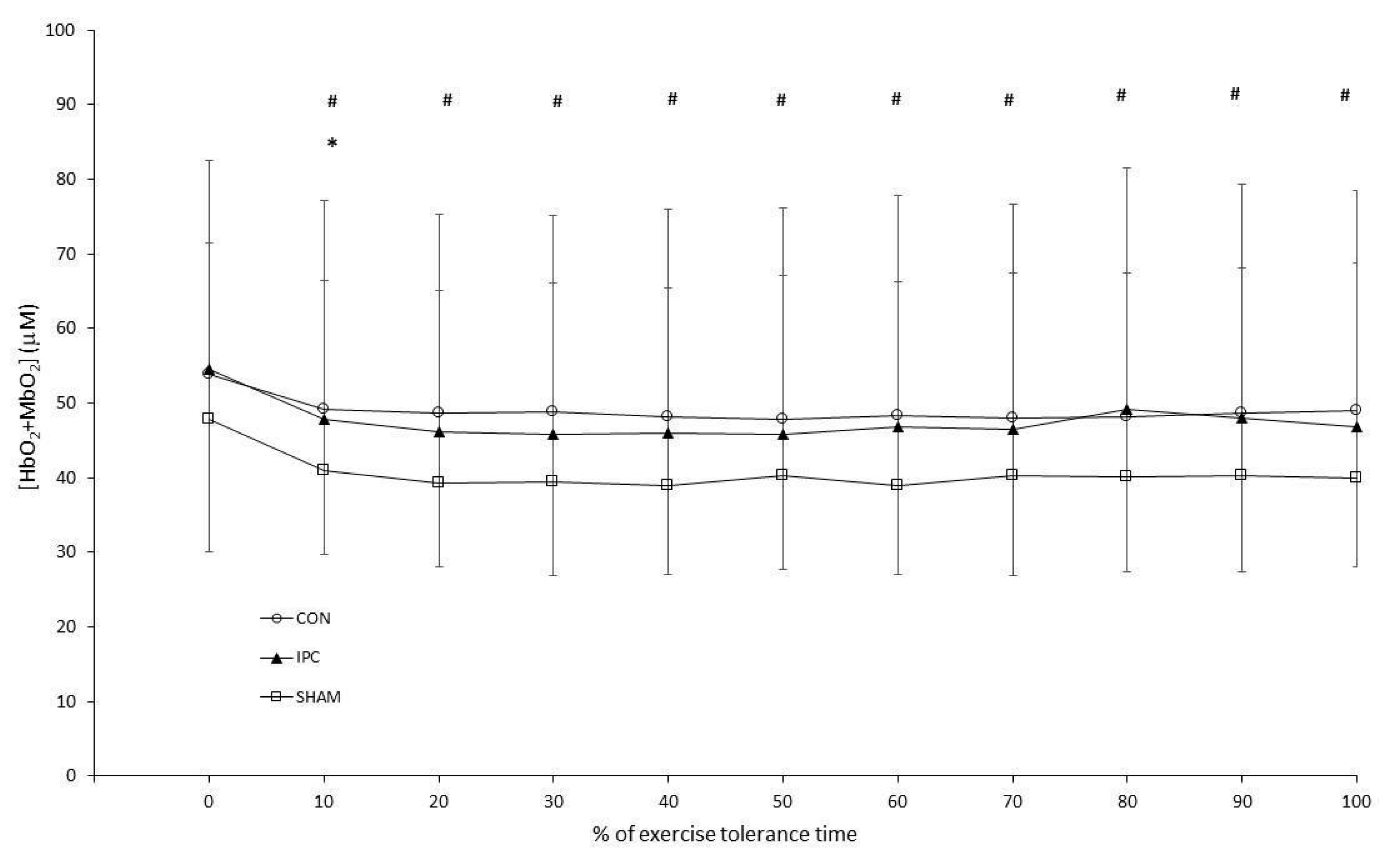
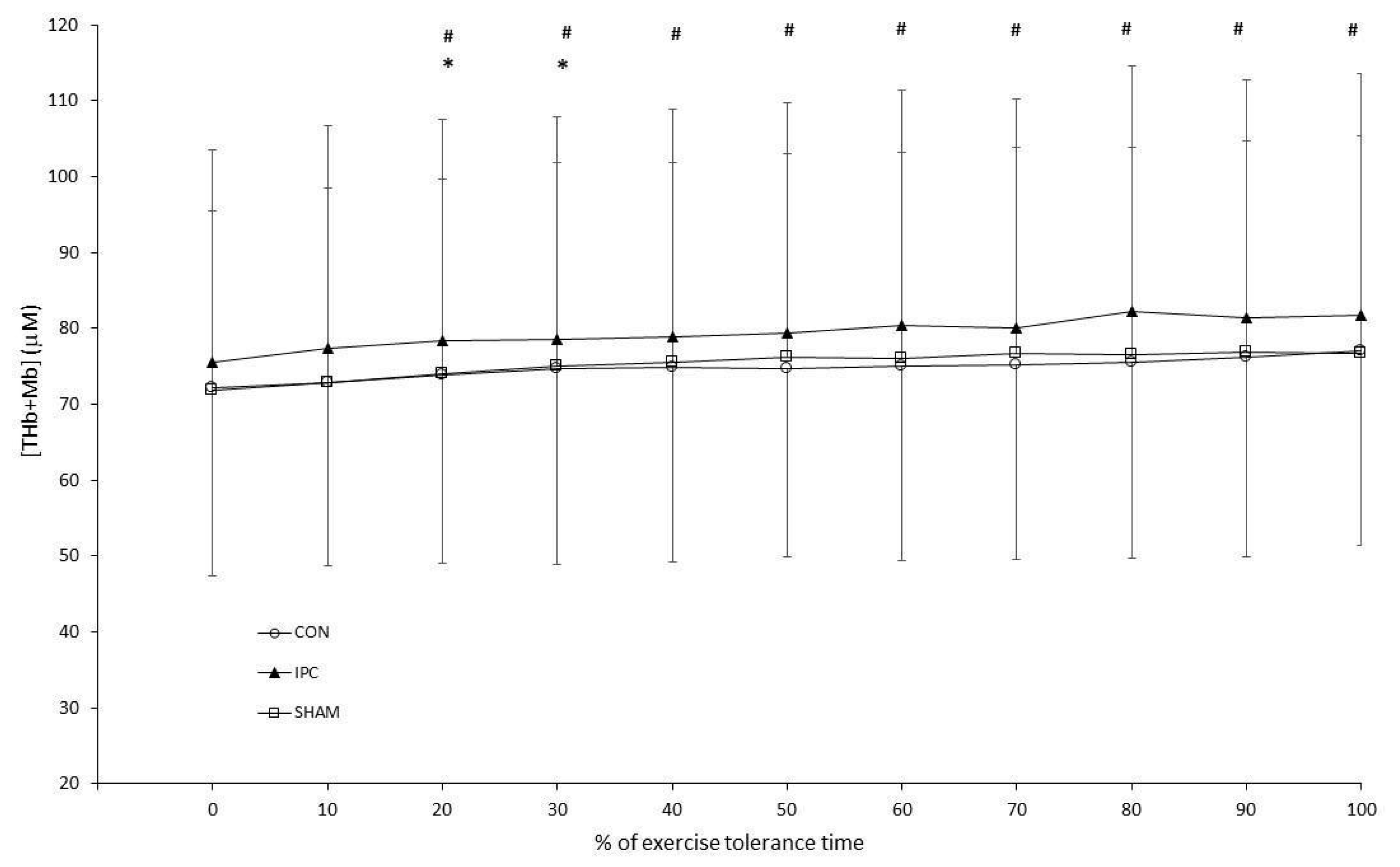
3.2. Constant Work-Rate Exercise

Power during constant load exercise was 278 ± 28 W. There was a significant effect of condition on time to the limit of tolerance (ηp2, = 0.33, F = 4.4, p = 0.03). Difference contrasts revealed no difference between CON and SHAM, whereas the limit of tolerance was longer in IPC versus CON and SHAM {199 ± 36 (CON) vs. 203 ± 35 (SHAM) vs. 219 ± 34 (IPC) s}. The time constant of VO2 and muscle [HHb + Mb] kinetics was not different between conditions {VO2: 55.4 ± 8.8 (CON) vs. 68 ± 21 (SHAM) vs. 59.3 ± 8.6 (IPC) s}, (ηp2 = 0.27, F = 1.8, p = 0.23); {HHb + Mb: 11.4 ± 1.7 (CON) vs. 14.0 ± 6.9 (SHAM) vs. 10.6 ± 3.6 (IPC) s}, (ηp2 = 0.27, F = 2.2, p = 0.18); see Table 1 and Table 2. During exercise, [HbO2 + MbO2] increased throughout exercise (ηp2 = 0.60, F = 12, p < 0.001), however there was no difference in [HbO2 + MbO2] during exercise between conditions (ηp2, = 0.12, F = 1.2, p = 0.37); see Figure 1. There was no condition × time interaction (ηp2 = 0.11, F = 0.94, p = 0.43). [THb + Mb] increased throughout exercise (ηp2 = 0.61, F = 13, p < 0.001, however there was no difference in [THb + Mb] during exercise between conditions (ηp2 = 0.02, F = 0.2, p = 0.82). There was no condition × time interaction (ηp2 = 0.14, F = 1.3, p = 0.31); see Figure 2.
Blood lactate increased from baseline values {4.7 ± 1.6 (CON) vs. 3.7 ± 1.4 (SHAM) vs. 3.32 ± 0.90 (IPC) mM} to post-exercise {12.7 ± 1.7 (CON) vs. 11.6 ± 2.4 (SHAM) vs. 12.8 ± 2.4 (IPC) mM}, (main effect time, ηp2 = 0.97, F = 380, p < 0.001). There was evidence of a condition × time interaction (ηp2 = 0.19, F = 2.7, p = 0.08); difference contrast analysis revealed that this was due to a greater increase in blood lactate between rest and post-exercise in IPC as compared to CON and SHAM.

4. Discussion

The primary aim of the present study was to test whether the use of a bout of ischemic preconditioning following incremental exercise to the limit of tolerance would improve subsequent constant workload exercise performance. The main finding was that IPC resulted in an increased exercise tolerance during constant load exercise when compared to the control and sham conditions. This was not associated with any speeding of pulmonary oxygen uptake kinetics, however there was some indication that IPC altered the metabolic environment since in IPC there was a significantly greater increase in blood lactate during constant work-rate exercise as compared to the other conditions.
The findings from the present study support the findings of Cocking et al [30] who found a significant improvement in 1-h cycle time trial performance following IPC, as compared to a sham treatment. Moreover, Sabino-Carvalho et al [31] also found an increase in exercise time to the limit of tolerance during high intensity, constant speed treadmill running to the limit of tolerance, following an initial incremental exercise test to the limit of tolerance. The present study also shows some agreement with the work of Crisafulli et al [17] and Kido et al [18], whereby an effect of IPC on exercise performance was demonstrated despite maximal oxygen uptake and oxygen uptake kinetics being unaffected by IPC. In addition, Bellini et al [32] also found that upper-body endurance was similarly improved through IPC without alterations to VO2 kinetics.
The exercise intensity selected in the present study was intended to place participants in the severe domain of exercise, that is above critical power but below the threshold beyond which the maximal oxygen uptake could not be attained [33]. The similarity of maximal oxygen uptake during incremental and constant work-rate exercise, and the exercise tolerance times (ranging ~2.5–5 min) indicate that participants were successfully located within the severe domain. Performance in this domain can be well explained by the two-parameter model of exercise performance [33], thus being determined by the proximity of the required power output to the underlying critical power (CP; measured in Watts, W) and the ability to complete work above critical power (W’; measured in kJ).
Critical power is determined, at least in part, by the independent factors of the time constant of oxygen uptake kinetics and oxygen availability during the transition from rest to exercise [11]. In the present study, we demonstrated no effect of IPC on the time constant of oxygen uptake kinetics. Moreover, the lack of effect of IPC on muscle [HHb + Mb] kinetics and [HbO2 + MbO2] during exercise indicate no alterations to muscle oxygenation. By design, the criterion bouts of constant work-rate exercise were preceded (~45 mins) by maximal incremental (ramp) exercise. Such a prior bout of exercise may have had a “priming” effect on oxygen uptake kinetics, demonstrable within this time-frame [34] that could have ablated any effect of IPC. However, during upright cycling exercise, it is not thought that priming exercise confers an important effect on the time constant of oxygen uptake kinetics. Taken together therefore, it appears unlikely that the enhancement to exercise tolerance in the present study was due to an acute improvement in CP conferred by IPC. Indeed, previous studies have also demonstrated no effect of IPC on the time constant of pulmonary oxygen uptake kinetics [35,36] and muscle oxygenation [37].
Although it cannot be ruled out that an increase to CP occurred as a consequence of IPC, the lack of any measurable change to oxygen uptake kinetics or oxygen availability (as determined by [Hbo2 + Mb] is unsupportive of such an outcome. Hence, in accordance with the assumptions of the 2-parameter model of severe intensity exercise performance, if an improvement to performance cannot be explained by CP, then an increase to W’ is suggested, with W’ representing the volume of work available above CP. In the present context, whereby constant work-rate exercise followed a bout of maximal incremental exercise undertaken ~45 min prior, this could alternatively reflect a more complete recovery of W’ following IPC, which may be otherwise incomplete during control conditions. Without knowledge of the underlying W’, it is not possible to know whether there was a fundamental increase to W’ per se, or whether W’ recovery was enhanced by IPC. Nevertheless, the significantly higher increase in blood lactate during constant work-rate exercise in IPC is indicative of a greater involvement of anaerobic metabolism in this condition, which is related to an increased ability to do work above CP (i.e. increased W’). At the termination of maximal exercise in the severe exercise intensity domain, the metabolic milieu of the muscle is similar, regardless of the proximity to CP [38]. This indicates that the limit of tolerance during such exercise is, at least in part, caused by the absolute accumulation of fatigue-related metabolites in the muscle tissue. Hence, the blood lactate data outlined above is indicative of an enhanced ability to clear such metabolites from the circulation, and thus the muscle tissue, during the recovery period between incremental and constant work-rate exercise. In turn, due to a favourable gradient of metabolite export into the circulation from the onset of exercise, this permits a greater reliance on anaerobic metabolism during the criterion bout of exercise, resulting in an increased volume of work undertaken above critical power before a critical accumulation of metabolites occurs. Indeed, the opposite was demonstrated during cycle exercise following prior severe intensity arm exercise whereby W’ was reduced coincident to a prior accumulation of circulating metabolites, as compared to control conditions [39]. Accordingly, the beneficial effect of IPC on exercise performance can be considered as a consequence of an improvement in the ability to recover W’ following prior maximal incremental exercise. Indeed, prior demonstrations of a retardation of the trajectory of the slow component of oxygen uptake [35] following IPC are consistent with the notion of an enhanced ability to undertake work above critical power [40].
The notion that IPC resulted in enhanced blood flow during the recovery period following incremental exercise might be expected to be supported by the finding of either an increase in [HbO2 + MbO2] or a reduction in [HHb + Mb] during the period prior to and/or during constant work rate exercise. The former of these metrics indicates muscle oxygenation, with the latter the regional balance between muscle O2 availability and utilisation. However, this was not the case as there were no differences between conditions in [THb + TMb], [HbO2 + MbO2] or [HHb + Mb] kinetics. However, measurable enhancements to perfusion as a consequence of IPC may have been apparent only during, or in close proximity to the IPC protocol. Accordingly, the brief period of measurements prior to constant work rate exercise may have missed the window during which enhanced blood flow and oxygenation occurred.
A likely candidate mechanism for enhancing blood flow following IPC is an increase in endothelial nitric oxide synthase phosphorylation, and consequently, nitric oxide production [41]. Shear stress on the endothelium has been shown to increase production of NO by endothelium mediated NO synthase [42]. NO plays a role in endothelial function through increased dilation. It is of note therefore that Bailey et al., [43] showed IPC to maintain flow-mediated dilation (FMD) at pre-exercise values following severe intensity exercise performance, as compared to the sham trial where FMD was reduced. An Increased NO availability is believed to improve oxygen extraction during exercise, along with NO attenuating the mitochondria’s oxygen consumption through maintenance of oxygen reserves and the extraction within the cell [44]. Therefore, it is possible to conclude that increased NO synthesis may potentially be an underlying molecular enhancement of IPC.
Further mechanisms linked to the possible performance effects of IPC relate to ATP sparing and lactate flux, with Pang et al., [45] finding IPC increased ATP sparing in pig muscle. This was as a result of improved excitation-contraction coupling of the muscle (mitochondrial flux) and prevention of a loss in power [45,46,47]. Also, it has been suggested there is an up-regulation of the mitochondrial permeability transition pore, which may enhance both local and systemic lactate influx and oxidative metabolism during exercise [48]. Andreas et al., [49] found that during reperfusion phosphocreatine concentrations were substantially higher after IPC than in the control group 4 h after the ischemic period.
The improvement in recovery and in turn performance through enhanced time to exhaustion following IPC may also be as a result of molecular and muscular functional changes [48]. IPC has been shown by Hopper et al., [15], Lawson & Downey, [47] and Pang et al., [13] to enhance levels of Adenosine and intramuscular ATP sensitive potassium (KATP) channels. This can increase vasodilation and thus improve oxygen delivery and substrate availability thereby improving muscle dynamics. Further cellular functions postulated by IPC are shown to effect cyclic adenosine monophosphate (cAMP) [50]. cAMP is involved in the functions in the regulation of substrate metabolism and improving reperfusion, thereby improving performance through increased substrate delivery [50].
Furthermore, metabolic accumulation following occlusion may explain the ergogenic effects seen in exercise. Studies have shown that a build-up of reactive oxygen species, adenosine, bradykinins and opioids initiates a biochemical cascade, which all result in possible improved performance [51]. This may partly help in the understanding of IPC’s effects on aerobic performance as these lead to metabolic accumulation and muscle fatigue.
Changes in muscle contractility rates and force generating capabilities have also been suggested as another possible mechanistic pathway for any ergogenic effect of IPC. Previous studies have suggested enhanced contraction and relaxation rates [52] with Cruz et al [53] observing increased quadriceps activity during cycling following IPC. Whilst we didn’t measure muscle activation signals in the present study, it may explain the difference in performance when IPC was utilised, with the increased muscle activation compensating for the reduction in muscle activity following prior exercise [54].
As with any research in this area, there are some inherent limitations that are important to highlight. A persistent issue with the experimental application of IPC is the inability to fully blind the subjects to which condition they were undergoing due to the distinguishable sensations felt by limb occlusions at 220 mmHg or 20 mmHg, thereby limiting the ability to control for the placebo effect. The psychophysiological impact of the awareness of the different cuff pressures is problematic to mitigate against whilst also being difficult to elucidate through the measures used within this study. Interindividual differences in limb size and composition also impacts the possible scale of occlusion/blood flow limitation in the targeted muscles, impacting the scale of the physiological stress and response across participants.

5. Conclusions

Following a maximal bout of incremental exercise, IPC resulted in an improvement in exercise tolerance during a subsequent bout of constant work rate exercise, as compared to the control conditions. This effect was not accompanied by any speeding of oxygen uptake kinetics or improvement in muscle deoxygenation during exercise, thus indicating that an improvement to critical power was not responsible for the ergogenic effect of IPC. However, the enhanced ability to increase blood lactate during the criterion exercise bout following IPC indicates an improved recovery of the metabolic milieu; taken together with the suggestion that CP was not enhanced by IPC, this indicates an improved recovery of W’ compared to the control conditions. Moreover, these data suggest that IPC exerted its beneficial effect on the recovery of exercise performance via enhanced perfusion of the muscle tissue bed, possibly via enhanced production of NO. Other possible mechanisms have been postulated to contribute to the enhanced exercise performance following IPC, however the determination of such mechanisms is outside the scope of the present study.

Author Contributions

P.J.A. was involved with study design, data collection and analysis, and manuscript writing. S.M. was involved with study design, data analysis and manuscript writing. All authors have read and agreed to the published version of the manuscript.

Funding

This research received no external funding.

Institutional Review Board Statement

Volunteered to participate in this study which was carried out in accordance with the Declaration of Helsinki and approved by the Liverpool Hope University ethics committee.

Informed Consent Statement

All participants provided written, informed consent prior to participation and were informed of their right to withdraw at any time.

Data Availability Statement

Data are not currently publicly available.

Acknowledgments

The authors would like to thank James Webb for his significant contribution to data collection and to Marc Wells for supporting the research process.

Conflicts of Interest

The authors have no known conflict of interest.

References

  1. De Groot, P.C.E.; Thijssen, D.H.J.; Sanchez, M.; Ellenkamp, R.; Hopman, M.T.E. Ischemic Preconditioning Improves Maximal Performance in Humans. Eur. J. Appl. Physiol. 2010, 108, 141–146. [Google Scholar] [CrossRef] [PubMed] [Green Version]
  2. Telles, L.G.; Billaut, F.; de Souza Ribeiro, A.; Junqueira, C.G.; Leitão, L.; Barreto, A.C.; Panza, P.; Vianna, J.M.; da Silva Novaes, J. Ischemic Preconditioning with High and Low Pressure Enhances Maximum Strength and Modulates Heart Rate Variability. IJERPH 2022, 19, 7655. [Google Scholar] [CrossRef]
  3. Da Silva Novaes, J.; da Silva Telles, L.G.; Monteiro, E.R.; da Silva Araujo, G.; Vingren, J.L.; Silva Panza, P.; Reis, V.M.; Laterza, M.C.; Vianna, J.M. Ischemic Preconditioning Improves Resistance Training Session Performance. J. Strength Cond. Res. 2021, 35, 2993–2998. [Google Scholar] [CrossRef]
  4. Ferreira, T.N.; Sabino-Carvalho, J.L.C.; Lopes, T.R.; Ribeiro, I.C.; Succi, J.E.; Da Silva, A.C.; Silva, B.M. Ischemic Preconditioning and Repeated Sprint Swimming: A Placebo and Nocebo Study. Med. Sci. Sport. Exerc. 2016, 48, 1967–1975. [Google Scholar] [CrossRef]
  5. Chen, Y.; Yang, J.; Muradov, O.; Li, X.; Lee, J.K.W.; Qiu, J. Effect of Ischemic Preconditioning on Maximum Accumulated Oxygen Deficit in 400-Meter Runners. Eur. J. Sport Sci. 2022, 23, 789–796. [Google Scholar] [CrossRef] [PubMed]
  6. Williams, N.; Russell, M.; Cook, C.J.; Kilduff, L.P. Effect of Ischemic Preconditioning on Maximal Swimming Performance. J. Strength Cond. Res. 2021, 35, 221–226. [Google Scholar] [CrossRef] [PubMed] [Green Version]
  7. Montoye, A.H.; Mitchinson, C.J.; Townsend, O.R.; Nemmers, C.H.; Serkaian, C.N.; Rider, B.C. Ischemic Preconditioning Does Not Improve Time Trial Performance in Recreational Runners. Int. J. Exerc. Sci. 2020, 13, 1402–1417. [Google Scholar]
  8. Lalonde, F.; Curnier, D.Y. Can Anaerobic Performance Be Improved by Remote Ischemic Preconditioning? J. Strength Cond. Res. 2015, 29, 80–85. [Google Scholar] [CrossRef] [PubMed]
  9. Da Mota, G.R.; Marocolo, M. The Effects of Ischemic Preconditioning on Human Exercise Performance: A Counterpoint. Sports Med. 2016, 46, 1575–1576. [Google Scholar] [CrossRef]
  10. Hausenloy, D.J.; Yellon, D.M. Remote Ischaemic Preconditioning: Underlying Mechanisms and Clinical Application. Cardiovasc. Res. 2008, 79, 377–386. [Google Scholar] [CrossRef]
  11. Goulding, R.P.; Marwood, S. Interaction of Factors Determining Critical Power. Sports Med. 2023, 53, 595–613. [Google Scholar] [CrossRef]
  12. Jones, A.M.; Burnley, M. Oxygen Uptake Kinetics: An Underappreciated Determinant of Exercise Performance. Int. J. Sport. Physiol. Perform. 2009, 4, 524–532. [Google Scholar] [CrossRef] [Green Version]
  13. Pang, C.Y.; Neligan, P.; Xu, H.; He, W.; Zhong, A.; Hopper, R.; Forrest, C.R. Role of ATP-Sensitive K+ Channels in Ischemic Preconditioning of Skeletal Muscle against Infarction. Am. J. Physiol. Heart Circ. Physiol. 1997, 273, H44–H51. [Google Scholar] [CrossRef]
  14. Lee, H.T.; Thompson, C.I. Preconditioning with Ischemia or Adenosine Protects Skeletal Muscle from Ischemic Tissue Reperfusion Injury. J. Surg. Res. 1996, 63, 6. [Google Scholar] [CrossRef] [PubMed]
  15. Hopper, R.A.; Forrest, C.R.; Xu, H.; Zhong, A.; He, W.; Rutka, J.; Neligan, P.; Pang, C.Y. Role and Mechanism of PKC in Ischemic Preconditioning of Pig Skeletal Muscle against Infarction. Am. J. Physiol.-Regul. Integr. Comp. Physiol. 2000, 279, R666–R676. [Google Scholar] [CrossRef]
  16. Goulding, R.P.; Rossiter, H.B.; Marwood, S.; Ferguson, C. Bioenergetic Mechanisms Linking VO2 Kinetics and Exercise Tolerance. Exerc. Sport Sci. Rev. 2021, 49, 274–283. [Google Scholar] [CrossRef]
  17. Crisafulli, A.; Tangianu, F.; Tocco, F.; Concu, A.; Mameli, O.; Mulliri, G.; Caria, M.A. Ischemic Preconditioning of the Muscle Improves Maximal Exercise Performance but Not Maximal Oxygen Uptake in Humans. J. Appl. Physiol. 2011, 111, 530–536. [Google Scholar] [CrossRef] [Green Version]
  18. Kido, K.; Suga, T.; Tanaka, D.; Honjo, T.; Homma, T.; Fujita, S.; Hamaoka, T.; Isaka, T. Ischemic Preconditioning Accelerates Muscle Deoxygenation Dynamics and Enhances Exercise Endurance during the Work-to-Work Test. Physiol. Rep. 2015, 3, e12395. [Google Scholar] [CrossRef] [PubMed] [Green Version]
  19. De Ruiter, C.J.; Jones, D.A.; Sargeant, A.J.; De Haan, A. The Measurement of Force/Velocity Relationships of Fresh and Fatigued Human Adductor Pollicis Muscle. Eur. J. Appl. Physiol. 1999, 80, 386–393. [Google Scholar] [CrossRef] [PubMed]
  20. Karlsson, J.; Bonde-Petersen, F.; Henriksson, J.; Knuttgen, H.G. Effects of Previous Exercise with Arms or Legs on Metabolism and Performance in Exhaustive Exercise. J. Appl. Physiol. 1975, 38, 763–767. [Google Scholar] [CrossRef]
  21. Enko, K.; Nakamura, K.; Yunoki, K.; Miyoshi, T.; Akagi, S.; Yoshida, M.; Toh, N.; Sangawa, M.; Nishii, N.; Nagase, S.; et al. Intermittent Arm Ischemia Induces Vasodilatation of the Contralateral Upper Limb. J. Physiol. Sci. 2011, 61, 507–513. [Google Scholar] [CrossRef]
  22. Paradis-Deschênes, P.; Joanisse, D.R.; Billaut, F. Ischemic Preconditioning Increases Muscle Perfusion, Oxygen Uptake, and Force in Strength-Trained Athletes. Appl. Physiol. Nutr. Metab. 2016, 41, 938–944. [Google Scholar] [CrossRef] [PubMed] [Green Version]
  23. Paradis-Deschênes, P.; Lapointe, J.; Joanisse, D.R.; Billaut, F. Similar Recovery of Maximal Cycling Performance after Ischemic Preconditioning, Neuromuscular Electrical Stimulation or Active Recovery in Endurance Athletes. J. Sports Sci. Med. 2020, 19, 761–771. [Google Scholar] [PubMed]
  24. Bailey, T.G.; Birk, G.K.; Cable, N.T.; Atkinson, G.; Green, D.J.; Jones, H.; Thijssen, D.H.J. Remote Ischemic Preconditioning Prevents Reduction in Brachial Artery Flow-Mediated Dilation after Strenuous Exercise. Am. J. Physiol. Heart Circ. Physiol. 2012, 303, H533–H538. [Google Scholar] [CrossRef]
  25. Loukogeorgakis, S.P.; Panagiotidou, A.T.; Broadhead, M.W.; Donald, A.; Deanfield, J.E.; MacAllister, R.J. Remote Ischemic Preconditioning Provides Early and Late Protection Against Endothelial Ischemia-Reperfusion Injury in Humans. J. Am. Coll. Cardiol. 2005, 46, 450–456. [Google Scholar] [CrossRef] [PubMed] [Green Version]
  26. Beaver, W.L.; Wasserman, K.; Whipp, B.J. A New Method for Detecting Anaerobic Threshold by Gas Exchange. J. Appl. Physiol. 1986, 60, 2020–2027. [Google Scholar] [CrossRef] [PubMed]
  27. Marwood, S.; Roche, D.; Rowland, T.; Garrard, M.; Unnithan, V.B. Faster Pulmonary Oxygen Uptake Kinetics in Trained versus Untrained Male Adolescents. Med. Sci. Sport. Exerc. 2010, 42, 127–134. [Google Scholar] [CrossRef] [PubMed] [Green Version]
  28. Jones, A.M.; Poole, D.C. Oxygen Uptake Dynamics: From Muscle to Mouth—An Introduction to the Symposium. Med. Sci. Sport. Exerc. 2005, 37, 1542–1550. [Google Scholar] [CrossRef] [Green Version]
  29. Bailey, S.J.; Vanhatalo, A.; DiMenna, F.J.; Wilkerson, D.P.; Jones, A.M. Fast-Start Strategy Improves VO2 Kinetics and High-Intensity Exercise Performance. Med. Sci. Sports Exerc. 2011, 43, 457–467. [Google Scholar] [CrossRef]
  30. Cocking, S.; Landman, T.; Benson, M.; Lord, R.; Jones, H.; Gaze, D.; Thijssen, D.H.J.; George, K. The Impact of Remote Ischemic Preconditioning on Cardiac Biomarker and Functional Response to Endurance Exercise. Scand. J. Med. Sci. Sports 2017, 27, 1061–1069. [Google Scholar] [CrossRef] [Green Version]
  31. Sabino-Carvalho, J.L.; Lopes, T.R.; Obeid-Freitas, T.; Ferreira, T.N.; Succi, J.E.; Silva, A.C.; Silva, B.M. Effect of Ischemic Preconditioning on Endurance Performance Does Not Surpass Placebo. Med. Sci. Sport. Exerc. 2017, 49, 124–132. [Google Scholar] [CrossRef]
  32. Bellini, D.; Chapman, C.; Peden, D.; Hoekstra, S.P.; Ferguson, R.A.; Leicht, C.A. Ischaemic Preconditioning Improves Upper-Body Endurance Performance without Altering VO2 Kinetics. Eur. J. Sport Sci. 2022, 1–9. [Google Scholar] [CrossRef]
  33. Jones, A.M.; Burnley, M.; Black, M.I.; Poole, D.C.; Vanhatalo, A. The Maximal Metabolic Steady State: Redefining the “Gold Standard”. Physiol. Rep. 2019, 7, e14098. [Google Scholar] [CrossRef] [PubMed] [Green Version]
  34. Burnley, M.; Doust, J.H.; Jones, A.M. Time Required for the Restoration of Normal Heavy Exercise VO2 Kinetics Following Prior Heavy Exercise. J. Appl. Physiol. 2006, 101, 1320–1327. [Google Scholar] [CrossRef] [PubMed] [Green Version]
  35. Kilding, A.E.; Sequeira, G.M.; Wood, M.R. Effects of Ischemic Preconditioning on Economy, VO2 Kinetics and Cycling Performance in Endurance Athletes. Eur. J. Appl. Physiol. 2018, 119, 961–967. [Google Scholar] [CrossRef]
  36. Wiggins, C.C.; Constantini, K.; Paris, H.L.; Mickleborough, T.D.; Chapman, R.F. Ischemic Preconditioning, O2 Kinetics, and Performance in Normoxia and Hypoxia. Med. Sci. Sports Exerc. 2019, 51, 900–911. [Google Scholar] [CrossRef] [PubMed]
  37. Peden, D.L.; Mitchell, E.A.; Bailey, S.J.; Ferguson, R.A. Ischaemic Preconditioning Blunts Exercise-induced Mitochondrial Dysfunction, Speeds Oxygen Uptake Kinetics but Does Not Alter Severe-Intensity Exercise Capacity. Exp. Physiol. 2022, 107, 1241–1254. [Google Scholar] [CrossRef]
  38. Black, M.I.; Jones, A.M.; Blackwell, J.R.; Bailey, S.J.; Wylie, L.J.; McDonagh, S.T.J.; Thompson, C.; Kelly, J.; Sumners, P.; Mileva, K.N.; et al. Muscle Metabolic and Neuromuscular Determinants of Fatigue during Cycling in Different Exercise Intensity Domains. J. Appl. Physiol. 2017, 122, 446–459. [Google Scholar] [CrossRef] [Green Version]
  39. Johnson, M.A.; Mills, D.E.; Brown, P.I.; Sharpe, G.R. Prior Upper Body Exercise Reduces Cycling Work Capacity but Not Critical Power. Med. Sci. Sports Exerc. 2014, 46, 802–808. [Google Scholar] [CrossRef] [Green Version]
  40. Burnley, M.; Davison, G.; Baker, J.R. Effects of Priming Exercise on VO2 Kinetics and the Power-Duration Relationship. Med. Sci. Sports Exerc. 2011, 43, 2171–2179. [Google Scholar] [CrossRef]
  41. Li, X.-D.; Cheng, Y.-T.; Yang, Y.-J.; Meng, X.-M.; Zhao, J.-L.; Zhang, H.-T.; Wu, Y.-J.; You, S.-J.; Wu, Y.-L. PKA-Mediated ENOS Phosphorylation in the Protection of Ischemic Preconditioning against No-Reflow. Microvasc. Res. 2012, 84, 44–54. [Google Scholar] [CrossRef] [PubMed]
  42. Tinken, T.M.; Thijssen, D.H.J.; Hopkins, N.; Dawson, E.A.; Cable, N.T.; Green, D.J. Shear Stress Mediates Endothelial Adaptations to Exercise Training in Humans. Hypertension 2010, 55, 312–318. [Google Scholar] [CrossRef] [PubMed] [Green Version]
  43. Bailey, T.G.; Jones, H.; Gregson, W.; Atkinson, G.; Cable, N.T.; Thijssen, D.H.J. Effect of Ischemic Preconditioning on Lactate Accumulation and Running Performance. Med. Sci. Sport. Exerc. 2012, 44, 2084–2089. [Google Scholar] [CrossRef] [Green Version]
  44. Kimura, M.; Ueda, K.; Goto, C.; Jitsuiki, D.; Nishioka, K.; Umemura, T.; Noma, K.; Yoshizumi, M.; Chayama, K.; Higashi, Y. Repetition of Ischemic Preconditioning Augments Endothelium-Dependent Vasodilation in Humans: Role of Endothelium-Derived Nitric Oxide and Endothelial Progenitor Cells. ATVB 2007, 27, 1403–1410. [Google Scholar] [CrossRef]
  45. Pang, C.Y.; Yang, R.Z.; Zhong, A.; Xu, N.; Boyd, B.; Forrest, C.R. Acute Ischaemic Preconditioning Protects against Skeletal Muscle Infarction in the Pig. Cardiovasc. Res. 1995, 29, 782–788. [Google Scholar] [CrossRef]
  46. Jean-St-Michel, E.; Manlhiot, C.; Li, J.; Tropak, M.; Michelsen, M.M.; Schmidt, M.R.; McCrindle, B.W.; Wells, G.D.; Redington, A.N. Remote Preconditioning Improves Maximal Performance in Highly Trained Athletes. Med. Sci. Sports Exerc. 2011, 43, 1280–1286. [Google Scholar] [CrossRef] [PubMed] [Green Version]
  47. Lawson, C.S.; Downey, J.M. Preconditioning: State of the Art Myocardial Protection. Cardiovasc. Res. 1993, 27, 542–550. [Google Scholar] [CrossRef]
  48. Hashimoto, T.; Brooks, G.A. Mitochondrial Lactate Oxidation Complex and an Adaptive Role for Lactate Production. Med. Sci. Sport. Exerc. 2008, 40, 486–494. [Google Scholar] [CrossRef]
  49. Andreas, M.; Schmid, A.I.; Keilani, M.; Doberer, D.; Bartko, J.; Crevenna, R.; Moser, E.; Wolzt, M. Effect of Ischemic Preconditioning in Skeletal Muscle Measured by Functional Magnetic Resonance Imaging and Spectroscopy: A Randomized Crossover Trial. J. Cardiovasc. Magn. Reson. 2011, 13, 32. [Google Scholar] [CrossRef] [Green Version]
  50. Lochner, A.; Genade, S.; Tromp, E.; Podzuweit, T.; Moolman, J.A. Ischemic Preconditioning and the β-Adrenergic Signal Transduction Pathway. Circulation 1999, 100, 958–966. [Google Scholar] [CrossRef] [Green Version]
  51. Salvador, A.F.; De Aguiar, R.A.; Lisbôa, F.D.; Pereira, K.L.; Cruz, R.S.D.O.; Caputo, F. Ischemic Preconditioning and Exercise Performance: A Systematic Review and Meta-Analysis. Int. J. Sport. Physiol. Perform. 2016, 11, 4–14. [Google Scholar] [CrossRef] [PubMed]
  52. Barbosa, T.C.; Machado, A.C.; Braz, I.D.; Fernandes, I.A.; Vianna, L.C.; Nobrega, A.C.L.; Silva, B.M. Remote Ischemic Preconditioning Delays Fatigue Development during Handgrip Exercise: RIPC Improves Handgrip Performance. Scand. J. Med. Sci. Sports 2015, 25, 356–364. [Google Scholar] [CrossRef] [PubMed]
  53. De Oliveira Cruz, R.S.; de Aguiar, R.A.; Turnes, T.; Salvador, A.F.; Caputo, F. Effects of Ischemic Preconditioning on Short-Duration Cycling Performance. Appl. Physiol. Nutr. Metab. 2016, 41, 825–831. [Google Scholar] [CrossRef] [PubMed]
  54. Gentil, P.; Oliveira, E.; Bottaro, M. Effects of Exercise Order on Upper-Body Muscle Activation and Exercise Performance. J. Strength Cond. Res. 2007, 21, 1082–1086. [Google Scholar]
Figure 1. [HbO2 + MbO2] at each 10% of exercise tolerance time during exercise to the limit of exercise tolerance in each condition. CON: open circles; IPC: closed triangles; SHAM: open squares. * p < 0.05 versus previous time-point; # p < 0.05 versus onset of exercise.
Physiologia 03 00027 g001
Figure 2. [THb + Mb] at each 10% of exercise tolerance time during exercise to the limit of exercise tolerance in each condition. CON: open circles; IPC: closed triangles; SHAM: open squares. * p < 0.05 versus previous time-point; # p < 0.05 versus onset of exercise.
Physiologia 03 00027 g002
Table 1. Parameters of VO2 kinetics during constant load exercise.
CONSHAMIPC
Baseline (L·min−1)0.99 ± 0.191.02 ± 0.180.91 ± 0.28
Amplitude (L·min−1)2.51 ± 0.162.66 ± 0.142.68 ± 0.23
τVO2 (s)55.4 ± 8.868 ± 2159.3 ± 8.6
Absolute amplitude (L·min−1)3.52 ± 0.303.68 ± 0.293.58 ± 0.21
Table 2. Parameters of [HHb + Mb] kinetics during constant load exercise.
CONSHAMIPC
Baseline (μM)16.5 ± 6.521.3 ± 9.322 ± 12
TDHHb+Mb (s)9.8 ± 3.38.6 ± 3.810.8 ± 3.9
Amplitude (μM)10.7 ± 4.012 ± 1013.7 ± 8.3
τHHb+Mb (s)11.4 ± 1.714.0 ± 6.910.6 ± 3.6
Absolute amplitude (μM)27.2 ± 9.833 ± 1935 ± 20
Disclaimer/Publisher’s Note: The statements, opinions and data contained in all publications are solely those of the individual author(s) and contributor(s) and not of MDPI and/or the editor(s). MDPI and/or the editor(s) disclaim responsibility for any injury to people or property resulting from any ideas, methods, instructions or products referred to in the content.

