Article

Microbial Load of Hand Sanitizer Dispensers—A University Hospital Study

by
Christos Stefanis
1,
Elpida Giorgi
1,*,
Elisavet Stavropoulou
1,2,
Chrysoula (Chrysa) Voidarou
3,
Maria Skoufou
1,
Aikaterini Nelli
3,
Athina Tzora
3,
Christina Tsigalou
1 and
Eugenia Bezirtzoglou
1,*
1
Laboratory of Hygiene and Environmental Protection, Department of Medicine, Democritus University of Thrace, 68100 Alexandroupolis, Greece
2
Infectious Diseases Service, Lausanne University Hospital, University of Lausanne (CHUV), 1011 Lausanne, Switzerland
3
Laboratory of Animal Health, Food Hygiene and Quality, Department of Agriculture, University of Ioannina, 47132 Arta, Greece
*
Authors to whom correspondence should be addressed.
Hygiene 2023, 3(4), 450-464; https://doi.org/10.3390/hygiene3040034
Submission received: 12 September 2023 / Revised: 29 October 2023 / Accepted: 2 November 2023 / Published: 13 November 2023
(This article belongs to the Section Hygiene in Healthcare Facilities)

Abstract

:
Hospital-acquired infections are a significant concern in healthcare settings, leading to patient safety risks, increased morbidity and mortality, and financial burdens. Hand hygiene is crucial in preventing the spread of bacteria in hospitals and communities. Manual hand sanitizer dispensers can harbor presumptive pathogenic bacteria and act as fomites for bacterial transmission. This study aimed to assess the microbial contamination of manual hand sanitizer dispensers in a hospital setting and to study their antibiotic resistance profiles. Samples were collected using sterile cotton swabs and then inoculated into brain heart infusion broth. Subsequent subcultures were performed on both blood and MacConkey agar. The isolates were then identified using the Bruker MALDI Biotyper (Bruker Daltonik, Bremen, Germany) to the species level. Sampling was conducted in various wards and in the hospital and the University areas on dispenser levers and nozzle areas. The results showed that all samples yielded one or more bacterial species. Bacterial isolates identified belonged to species commonly found on the skin microflora and some Gram-negative enteric bacilli. Higher colonization was observed on the dispenser lever. Among Gram+ microorganisms, most bacterial species were shown to be sensitive to β-lactams, with the exception of Staphylococcus spp., resistant to AMP (Ampicillin) and Penicillin. However, no Methicillin resistant isolates were detected. Gram microorganisms such as Pseudomonas luteola were shown to be sensitive to all tested antibiotics, while Pantoea agglomerans was shown to be resistant to AMC (amoxicillin–clavulanic acid). Rifampicin tested only against Bacilli showed resistance. Based on the findings, it is recommended to implement systematic cleaning and proper maintenance of manual dispenser areas or to use automated dispensers to reduce hand contact and minimize microbial contamination. Monitoring the presence of microorganisms in hand sanitizing gels and dispensers is an essential infection control strategy.

1. Introduction

The percentage of infections related to medical practices in the hospital and between patients and medical staff varies between 4 and 9%, adding an extra burden on the health system by escalated morbidity, mortality and hospitalization costs [1,2,3]. Due to the importance of hand disinfection among healthcare workers and patients, in the treatment of hospital-acquired infections (HAIs) and in the development of multi-resistant pathogens, specific and detailed instructions and guidelines for hand hygiene have been issued by international organizations since the 2000s [4,5]. One of the most effective ways to reduce hospital-acquired infections and communicable diseases within the hospital environment is to disinfect hands with alcohol-based hand sanitizers [6,7].
The COVID-19 pandemic crisis has changed many things regarding hygiene and the public perceptions of hand hygiene and simple and practical methods of hand disinfection. Following the recommendations of international organizations and individual state initiatives, one of the measures to avoid the spread of COVID-19 was, among others, the use of hand sanitizers, mainly alcohol-based. Their use spread not only in hospital areas but also in restaurants, supermarkets and people’s daily lives [8]. Their ease of use has made alcohol-based hand sanitizers very popular, driving their global production to tremendous growth. Typical specifications for commercial hand sanitizers include the alcohol type (n-propyl alcohol, isopropyl alcohol, ethanol or a combination of them) and a minimum of 60% alcohol to achieve their disinfecting action properly [9,10].
In addition, hand hygiene is also an important part of hospital hygiene planning that prevents the further transmission of diseases and antimicrobial microorganisms [11]. The use of hand sanitizers placed on wards, at the entrance of clinics and around the patient’s bed was also used before the COVID pandemic. An important link in the chain of hygiene within the hospital premises and the hospital environment is the workers in them. The frequency of using alcohol-based hand rubs among workers is of the utmost importance, and various ways of measuring this hand hygiene practice have been proposed, such as the time of their placement on the hospital premises and the measurement of their consumption [12,13].
The importance of hand hygiene among hospital workers has also been recognized by the World Health Organization, resulting in the development of a methodological framework for hand disinfection at various stages of hospital patient care, as well as training and follow-up plans of practices [13]. This framework presupposes the disinfection of the hands of hospital workers as an imperative practice, fully integrated into the daily workflow, with awareness of the risk of the spread of multi-susceptible microorganisms in the hospital environment [11]. The practical application of this program was called “My five moments for hand hygiene” and includes hand disinfection and hygiene regarding patient treatment, such as before patient contact or before implementing an aseptic task, after body fluid exposure and after contact with the patient and finally after contact with patient surroundings [11,12,13]. Also, compliance with the hand hygiene of the hospital staff does not only concern the bed and the area around the patients in their bed, but the additional practices when moving them to other areas of the hospital for diagnostic tests, as well as their transport to the hospital with any vehicle, where hand hygiene also plays an important role in preventing infections [14].
Hand sanitizers in a hospital must be easily accessible and constantly available to reduce hospital-acquired infections. Spatial location within a hospital is a critical issue for staff utilization. Factors such as the ease of access, the height of the installation on the wall and visibility in patient rooms, at the entrances of special clinics, such as post-surgery rooms, and in waiting areas are among other factors that must be taken into account for the placement and access to hand sanitizers in a hospital environment [12,15]. The ‘accessibility’ factor of hand sanitizers has been highlighted as one of the least important, with the World Health Organization specifying that hand sanitizers should be available in-patient rooms without specific numerical criteria for their density [5]. Countries such as Germany and Sweden have promoted specific guidelines for the number of hand sanitizers per patient bed to at least 0.5 per patient bed for Germany and 2.4 for Sweden, taking into account the hospital type and bed count [16,17].
Hand sanitizers should be available in all hospital wards and areas, and their use by all patients, visitors and hospital staff should be encouraged. The latter should be educated on the correct use of hand sanitizers, which should happen during nursing practice. Also, the nursing and medical staff should be reminded to act as behavioral models regarding hand hygiene because the spread of multi-resistant bacteria and the increase in nosocomial infections partly depend on their compliance [18]. Hand disinfection and its importance in reducing nosocomial infections have resulted in its being proposed as a standard and horizontal practice to minimize the spread of resistant microorganisms, compared to other classic methods, such as using soap and water, which require time and extra costs [16,17]. Combined with an additional network of actions such as staff training and automated wall hand sanitizers to monitor and assess staff compliance, staff and patient [19,20] hand disinfection can be an essential tool against nosocomial infections and protect the health of patients and workers [20].
Although there have been studies concerning the antimicrobial action of hand sanitizers on various microorganisms and from different alcoholic components and combinations, it is impossible to simulate all the realistic hygienic conditions in a hospital [8,21]. Also, tests of the virucidal action of hand sanitizers have shown that the activity of alcoholic antiseptics against specific viruses requires a particular time to achieve their action, e.g., 50 and 70% ethanol reduced the infectivity of feline calicivirus by ≥2.2 log units after exposure for five minutes. Also, increased concentrations of alcohol result in its rapid evaporation from the container and perhaps in reduced effectiveness against some viruses and pathogenic microorganisms. Therefore, the action of hand sanitizers depends on the frequency of their use and the composition and alcohol content, so that the containers and hand sanitizers cannot harbor pathogenic bacteria or viruses and contribute to their further transmission in the hospital environment [22,23,24,25].
This research aims to investigate the presence of bacterial species on hand sanitizers and to determine the antimicrobial resistance traits that can disseminate within the hospital environment while underscoring the critical significance of these findings for public health. Thus, samples will be taken from dispenser levers and nozzles with cotton swabs in various clinics of a tertiary university hospital in Thrace, Northern Greece, which provides health facilities to a region of about 350,000 inhabitants, and in departments of the medical school, in order to identify the microbial flora hosted by hand sanitizers in various hospital and university areas. The sanitizer dispensers in the hospital will be situated in areas that hospital personnel, patients and visitors have access to. Finally, the possibility of a correlation between specific pathogenic bacteria in the hospital and medical school areas will be evaluated.

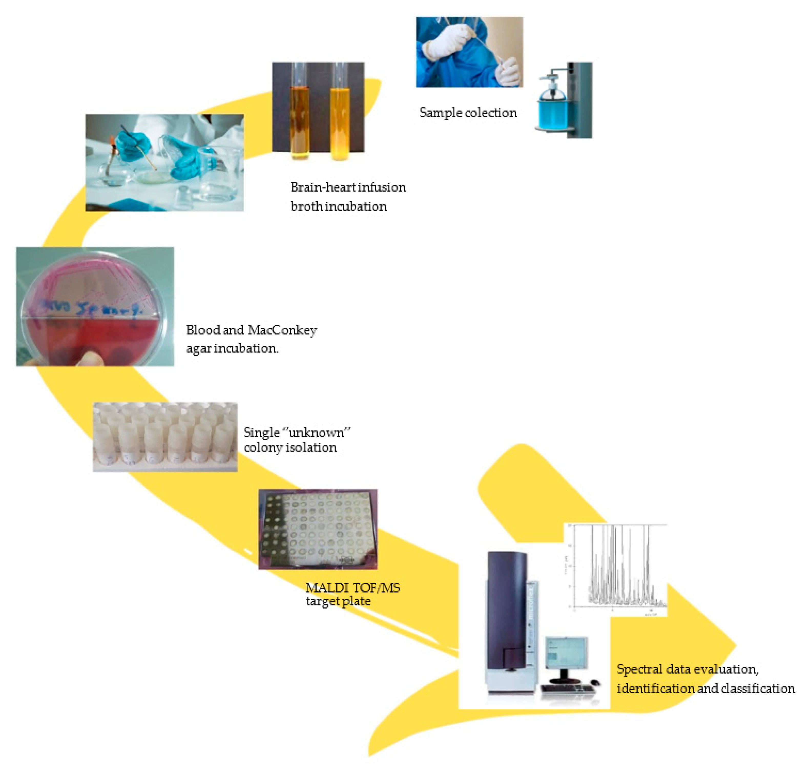
2. Materials and Methods
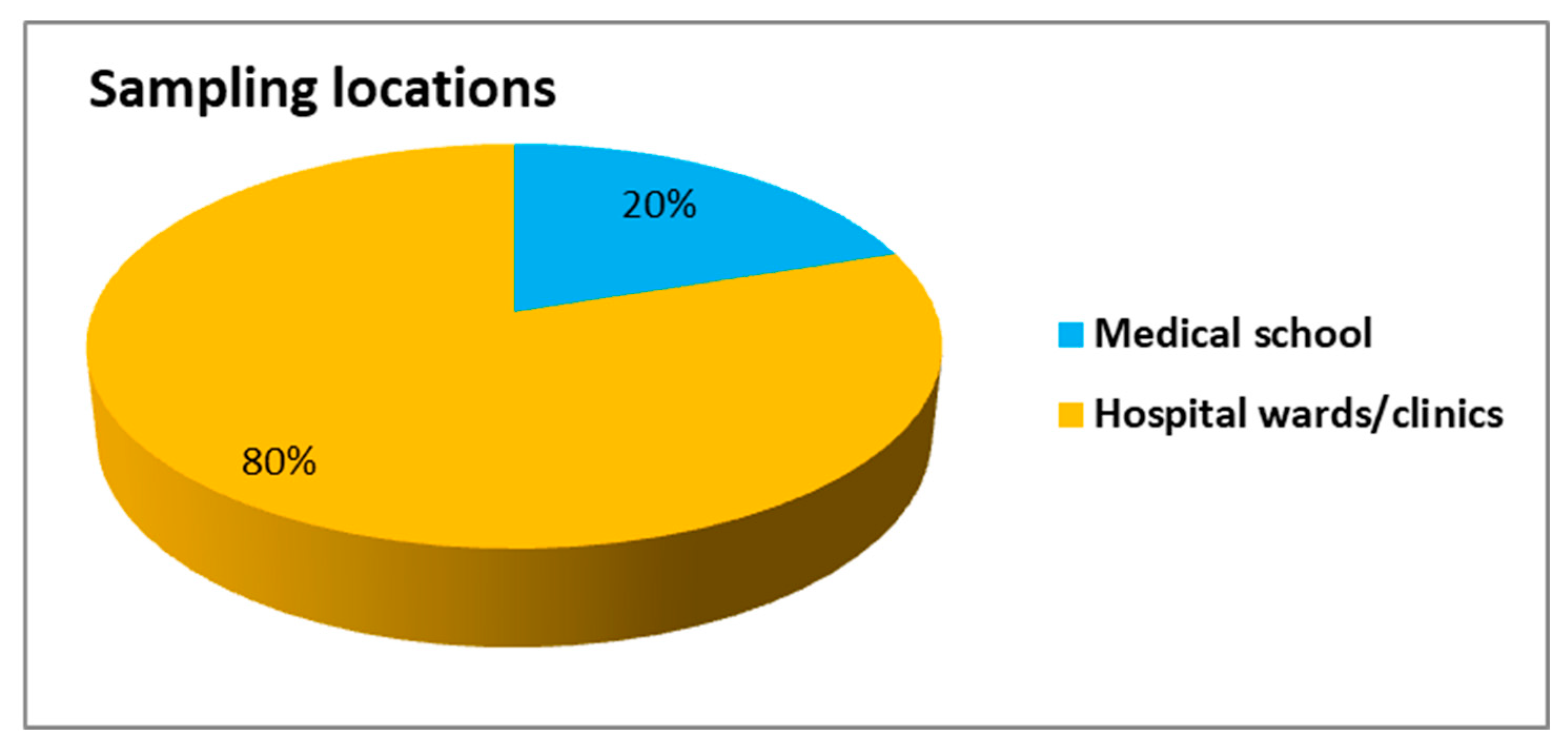
Sampling Procedure

Surface samples were collected from dispensers located in various areas of the hospital and the campus that were available to the public, including hospital wards and clinics, cafeterias, classrooms, etc. Samples were collected from sanitizer dispenser levers and nozzles with cotton swabs and transferred to the laboratory for further investigation. Sample size was assessed according to the accessibility of the hand sanitizer dispensers. Every sample station consisted of up to 3 dispensers. A total of 50 samples were collected from an equivalent number of dispensers in June 2022. After scrubbing the surface of the sanitizer’s dispenser 10 times, swabs were inoculated into brain heart infusion broth and incubated in 37 °C for 24 h. Twenty-four hours later, subcultures were streaked on both Blood and MacConkey agar following common procedures (Figure 1).
Due to COVID-19 regulation policies, access to most clinics was restricted to patients and healthcare staff. All available dispensers from the University Hospital, as well as from the Department of Medicine in the University were tested. Consequently, only 50 sampling stations were available to enter this research study and the results were evaluated accordingly.
Various antibiotics representing different classes commonly employed in clinical practice were chosen. This selection included antimicrobial agents for Gram-negative bacteria, for Gram-positive bacteria and for Bacillus spp.
Pure cultures were identified using Microflex LT MALDI-TOF MS (Matrix-assisted laser desorption/ionization time-of-flight mass spectrometry/Bruker Daltonics, Bremen, Germany). Five single colonies were taken directly from the nutrient agar plate and the sample preparation was performed according to the instructions of Bruker Daltonics and analyzed by running FlexControl 3.4 software. Each sample was spotted in triplicate. For each sample, the MS signals were acquired in a positive linear manner, according to the manufacture’s instructions. The obtained experimental MALDI-TOF mass spectrum profiles were then compared to the reference spectra and the matching between them was expressed by a Biotyper Log (score). The Log (score) considered the number of matching peaks, the total number of peaks, the peak weight representing species specificity and a correlation factor related to the matching peak intensity. All samples were measured on a mass spectrometer (Bruker Daltonics, Germany) using the method “MBT_AutoX” and the peaks were evaluated with the processing method “MBT_Process” using FlexControl (version 3.4, Bruker Daltonics, Germany). A calibration took place by using the Bacterial Test Standard (BTS) from Bruker. All spectra were compared with reference spectra of the BDAL database (version 8.0). Scores lower than 1.700 were interpreted as having no identification, while scores up to 2.000 can be considered species-level identification.
The assessment of antimicrobial resistance profiles in the isolated bacteria was conducted using the agar disk diffusion method, following the criteria established by the National Committee for Clinical Laboratory Standards (NCCLS) [26].

3. Results

A total of 27 isolates of bacterial species were identified in 22 out of 50 sampling stations. As shown in Figure 2, the distribution of samples from hospital and university was 80% and 20%, respectively. The most dominant bacterial species identified belong to the genus of Staphylococcus, comprising 55.5% of the total according to MALDI-TOF analysis. It is worth mentioning that the count of isolated species, and subsequently the number of identified ones, was notably lower than anticipated considering our sample size. Since the sampling from the dispensers occurred at random time intervals throughout the day, this outcome can likely be attributed to the thorough cleaning that might have been performed on the dispensers by the cleaning staff in various areas of the sampling process. Moreover, due to the COVID-19 pandemic, the extensive use of hand sanitizers in everyday routine might contribute to the low percentage of devices’ contamination.
In the Neurosurgery ward, only Staphylococcus cohnii was identified, with a scoring value of 2.32, interpreted as a high-confidence identification. In all maternity ward samples, the identified bacterial isolates included Staphylococcus warneri with a score value of 2.04, Staphylococcus epidermidis with a score value of 2.19, Bacillus megaterium with a score value of 2.20, S. epidermidis with a score value of 2.07 and Staphylococcus hominis with a score value of 2.36. By the main hospital entrance, five species were identified, in particular Solibacillussilvestris with a score value of 1.73 (low confidence identification), while Staphylococcus caprae, S. epidermidis, Micrococcus luteus and Pseudomonas luteola showed high score values: 2.05, 2.13, 2.44 and 2.33, respectively. By the chemotherapy ward, Staphylococcus aureus and Pantoea agglomerans were identified with score values of 2.31 and 2.32, respectively.
Furthermore, from all outpatient clinic samples a total of six species were identified, and they were S. caprae with a score value of 2.21, Staphylococcus lugdunensis with a score value of 2.22, S. epidermidis with a score value of 2.14, Mixta calida with a score value of 1.80, Staphylococcus haemolyticus with a score value of 2.03 and Staphylococcus saprophyticus with a score value of 2.10. In the waiting room of the CAT scan department of the hospital, Lysinibacillus xylanilyticus was identified with a score value of 2.02. In postoperative care, only one of the two bacterial species were identified, namely Bacillus cereus with a score value of 2.03. In the neurological clinic, no bacteria were identified.
In the university area, three different sample stations yielded four identified bacterial isolates. On the dispenser by the cafeteria, B. megaterium was identified with a score value of 2.21; by the histology department, Cytobacillus oceanisediminis with a score value of 2.00; and by the hygiene department, Paenibacillus amyloliquefaciens and Staphylococcus xylosus with score values of 2.00 and 1.82, respectively. Table 1, below, summarizes all findings in three columns that include the sample location, the bacterial species identification and the score value calculated through MALDI-TOF.
In total, 27 isolates were processed for identification by MALDI-TOF. Twenty-three of them were identified to the species level, with a score of up to 2.00, three of them were identified in a low-confidence level, and one was not identified.
All Gram+ bacteria which were isolated from the dispensers and had been identified as previously mentioned were tested against the following thirteen (13) antimicrobial agents: Chloramphenicol (CHL—30 mcg), Ciprofloxacin (CIP—5 mcg), Gentamycin (GEN—10 mcg), Erythromycin (ERY—15 mcg), Trimethoprim/sulfamethoxazole (SXT—1.25 + 23.75 mcg), Tetracycline (TET—30 mcg), Amoxicillin–Clavulanic (AMC—20/10 mcg), Cefoxitin (CFX—30 mcg), Oxacillin (CXI—1 mcg), Ampicillin (AMP—10 mcg), Penicillin G (PIP—10 mcg), Vancomycin (VAN—30 mcg) and Rifampicin (RIF—5 mcg) (Table 2).
Two isolates, one belonging to S. epidermidis species and one belonging to S. caprae species, have shown multi-drug resistance (resistant to more than three classes of antibiotics). The Bacillus cereus isolate was also found to be multi-resistant. Most of the isolates of the bacterial species tested were found to be susceptible to β-lactams, apart from S. caprae, resistant to AMP, and S. aureus and S. epidermidis, resistant to Penicillin. Oxacillin and Cefoxitin were tested only for Staphylococcus spp. isolates to determine if these strains were Methicillin resistant, but none of them were. Rifampicin was tested only against Bacilli, and all were found to be resistant. Pseudomonas luteola was found to be susceptible to all tested antibiotics, while Pantoea agglomerans was found to be resistant to AMC (Amoxicillin–Clavulanic acid) (Table 3). Twenty-three (23) of the isolates showed phenotypical resistance. None of the identified isolates belonging to the Staphylococci group, including both CNS and S. aureus, showed any resistance to Methicillin. In total, 33% of our CoNS isolates displayed multi-drug resistance.

4. Discussion

The challenge of antimicrobial resistance (AMR) in bacterial pathogens continues to be a significant concern in the field of infection control. To comprehensively address this issue, the One Health concept is actively being pursued. This approach acknowledges the equal importance of humans, domestic and wild animals and the environment, recognizing their interconnectedness. It also extends its scope to include commensal and environmental bacteria in order to evaluate the factors that contribute to the development of AMR in pathogens [27].
The coexistence of various microbial communities within shared ecological niches facilitates the broad distribution of genetically encoded resistance characteristics. Furthermore, documented evidence supports the transfer of antimicrobial traits, underscoring the extensive impact of this phenomenon across diverse boundaries and its potential to jeopardize antibiotic-based treatments in both human and veterinary medicine [28].
At first glance, it is noticeable that only B. megaterium was detected in both the hospital, by the maternity ward, and in the university, on dispensers by the cafeteria. In addition, the genus of Staphylococcus seemed to be the dominant one. Score values over 2.00, equaling high-confidence identification, were recorded in 19 out of 27 identified bacteria.
Among the identified microorganisms were species that are considered to be integral part of the skin microbiome. S. lugdunensis, S. aureus, M. luteus, S. caprae, S. epidermidis, S. xylosus, S. warneri, S haemolyticus and S. hominis are regular members of the diverse skin community [29,30,31,32,33,34,35]. However, there have been multiple cases reported where these bacterial species become major nosocomial pathogens leading to severe HAIs. S. aureus might be one of the most popular bacteria reported to be multi-drug-resistant and can remain on various surfaces for up to 5 years, over a wide range of temperatures, and infect patients mainly through contaminated surfaces [36,37,38,39,40,41]. In a study effort to describe the epidemiology of nosocomial outbreaks by Gastmeier et al., S. aureus have been found to contribute to the extent of 14.8% on 1022 nosocomial outbreaks included in the research [42]. S. aureus was detected on the dispenser located by the waiting room of the chemotherapy ward.
S. caprae might naturally colonize the human skin, the nails and nasal mucosa but occasionally act as an opportunistic pathogen infecting immunocompromised patients, leading to HAIs and community-acquired infections [35,36,37,38,39,40,41,42,43]. S. caprae was detected on samples taken from dispensers located by the main hospital entrance and the corridor of outpatient clinics.
M. luteus is a natural bacterial component of our human skin and besides a few reported infectious cases in immunosuppressed patient, M. luteus is not classified as an opportunistic or nosocomial pathogen [33,34]. M. luteus was detected on samples taken from dispensers of the main hospital entrance.
S. epidermidis might be a permanent resident of the human epithelial microflora, but it is also a major pathogen causing HAIs and severe infections among immunocompromised people. This bacteria has the capacity to form biofilms in surfaces that they reside in, and subsequently to colonize patients, especially through medical devices [36,37]. S. epidermidis was identified on dispensers located by the main hospital entrance, the maternity ward and the corridor of outpatient clinics.
S. haemolyticus is classified as a serious nosocomial infection agent. In 2012, an outbreak of S. haemolyticus at the Verona University Hospital Intensive Care Unit was reported. A rather interesting study has revealed both S. haemolyticus and S. hominis species that have acquired resistant properties to all clinically important antibiotics, including those of ß-lactams [38,41]. S. haemolyticus and S. hominis were identified on dispensers located by the outpatient clinics and the maternity ward, respectively.
In summary, Coagulase-negative staphylococci (CoNS) infections, especially in hospital settings, are a significant concern due to their ability to rapidly acquire resistance to multiple antibiotics, form biofilms on medical devices and pose challenges for effective treatment. Preventative measures and surveillance are crucial in managing and controlling these infections in healthcare facilities [44,45]. CoNS are prevalent opportunistic pathogens, while also being widespread commensal microorganisms in both humans and animals. CoNS strains associated with infections in healthcare settings are often distinguished by their notable antimicrobial resistance (AMR) profiles, encompassing resistance to Methicillin and multiple other drugs [27,46] CoNS are commonly found on the skin and mucous membranes, sharing the same ecological niche in the anterior nares of humans with S. aureus and various other bacteria [27,44,46]. This close proximity may facilitate the transfer of resistance genes among these bacterial species [47,48,49,50]. Certainly, CoNS have been recognized as reservoirs and origins of resistance traits that are transmitted within the Staphylococcaceae family [27,51,52,53].
Furthermore, two species from the genus Bacillus were identified: B. megaterium by the cafeteria of the University and the maternity ward at the hospital and B. cereus by the postoperative care ward. B. megaterium can be found ubiquitously across diverse environments, including but not limited to soil, seawater, rivers, seafood and salt lakes. However, it is not considered a cause for nosocomial outbreaks, while up to this date mainly individual case studies were reported regarding its pathogenicity to humans [54,55,56].
B. cereus is ubiquitous and it can be detected in all surfaces, from soil, foods, human skin and salty water to air filtration/ventilation equipment and medical equipment. This bacterium might be associated with food poisoning, but it is also classified as a significant factor in HAIs among immunocompromised hospitalized patients [57,58]. In a cross-sectional study about bacterial cross contamination in ICUs at a French University Hospital by E. Kuczewski et al. [58], 6.3% of the bacteria isolated out of 137 samples belonged to B. cereus and other Bacillus spp. All samples were received from surfaces close to or distant to the patients [59]. The same study has also unveiled that 8.1% of the species identified pertained to P. agglomerans [59].
P. agglomerans environmental reservoirs include plants, water, soil, humans and animals. This bacterial species has been reported to cause HAIs of opportunistic character, mainly in hospitalized immunosuppressed hosts either from contaminated medical devices or fluids [59,60]. P. agglomerans was detected on dispensers by the chemotherapy ward of the hospital.
M. calida, a microorganism widely distributed in diverse environments including plants, humans and food products, was identified on dispensers by the outpatient clinics. So far, no HAI caused by M. calida was reported [61].
The opportunistic pathogen P. luteola was found and identified by the main hospital entrance dispensers. It is isolated mainly from moist environments such as soil and waters. P. luteola has been detected in contaminated solutions such as distilled water, disinfectants and intravenous solutions [62,63]. Severe cases attributed to P. luteola infections in the hospital environment have been reported and thus it is considered to be a rare but significant cause of nosocomial or community-acquired infections [64,65].
By the CAT scan clinic, L. xylanilyticus was identified, but reported a low-confidence score value (<2.00). This bacterium has been isolated initially from plants and no HAI has been associated with it [66,67].
S. silvestris, primarily isolated from soil and plants, was identified by the hospital’s main entrance and scored a low-confidence value (<1.73), and no cases of HAIs have been reported so far. Whilst it is not considered part of the skin microflora, it has been isolated from human hands [68].
Cytobacillus oceanisediminis, detected on dispensers by the university’s department of histology, and Paenibacillus glucanolyticus, by the university’s department of hygiene, scored low-confidence identification values. They are mainly isolated from marine sediments and plants and no reference to HAIs has been reported so far [69,70].
Simon D. Eiref et al. studied the microbial load of 17 hand sanitizer dispensers by 12-bed surgical ICUs of a hospital. They detected one or more bacterial species on every dispenser and among others identified S. saureus, Micrococcus spp. and Bacillus spp. [71].
A limitation of this study might be that the dispensers were placed in areas which visitors, patients and hospital staff all have access to. It would be interesting if samples were taken from hand sanitizers that either only staff or only patients had access to, and to eventually compare these two findings. Also, neither the origin nor the number of dispenser users were known. Another limitation is that dispensers by the campus area are accessible to students and personnel that work both in the university and the hospital, but we do not have a record about this area. Finally, it should also be emphasized that an enrichment culture method in broth was used in the present study, which does not allow the microbial load to be determined. For this reason, we should also consider the possibility that each identified bacterial isolate may represent a single bacterial cell, so that the sampled surface may not act as a potential source of transmission. MALDI-TOF mass spectrometry analysis, on the other hand, delivers sound and valid results. It would be of great interest to define the time necessary for the alcoholic gel to eliminate all bacteria isolates identified to zero. Limitations would be addressed with the continuation of the study.
Hand hygiene is universally acknowledged as a fundamental component in infection control strategies within healthcare settings [6,7]. Physicians recognize its paramount role in preventing HAIs and safeguarding patient safety [11].
Education, along with systematic monitoring and comprehensive training protocols for all hospital personnel, is essential in promoting effective hand hygiene. These protocols must follow rigorous standards, best practices, guidelines and recommendations established by both national healthcare agencies and international health organizations such as the World Health Organization (WHO) [4,5,6].
The medical community is dedicated to continually improving hand hygiene practices [6,7] and ensuring the safety of hand sanitizers and dispensers [8]. Physicians actively contribute to a collective endeavor so as to enhance patient safety and reduce infection risks [6].
Manual hand sanitizer dispensers have a notable effect on hand hygiene, depending on their use and maintenance and ranging from bolstering to diminishing. Understanding their capacity for bacterial transmission is critical for practical applications. Several important factors come into play, including hand contamination and cross-contamination risk [6,7], both of which can lead to bacterial transmission. Dispensers create conditions conducive to these concerns due to the warmth of and the possibility of bacterial growth in residual products [8]. Hence, it is crucial to establish systematic maintenance and cleaning procedures firmly rooted in rigorous hygiene protocols, as well as the training of hospital staff in such practices [4,5].
Finally, it would be remiss not to mention that it is a common belief that even if the dispenser is contaminated by the hands of the user to an unknown level with skin flora, this does not pose a significant risk because the hands of the next user will be treated with the hand sanitizer after contact with the dispenser. At this point, we should digress by pointing out that factors such as heavy workload and fatigue for healthcare stuff and the lack of proper hand hygiene training for hospitals visitors, among others factors, may lead to improper or incomplete hand disinfection, which in turn may lead to the transmission of pathogenic bacteria, a possibility that features the surfaces of the dispensers as major risk determinants.
To summarize, manual hand sanitizer dispensers serve as indispensable tools for promoting hand hygiene [6,7,8], yet they can unintentionally aid in bacterial transmission when not managed properly. To mitigate this risk, it is essential to underscore the importance of proper hygiene practices, regular dispenser maintenance and the investigation of alternatives that do not require direct contact.

5. Conclusions

Hospital-acquired infections (HAIs) introduce substantial risks and complexities within healthcare environments that might result in compromised patient safety and increased morbidity and mortality rates among individuals under medical care. Moreover, HAIs contribute to elevated healthcare expenses and burden hospital staff due to extended patient stays, which is not to mention that HAIs involve antibiotic-resistant bacteria, intensifying the level of concern.
In aggregate, the prevalence of HAIs can markedly influence a hospital’s standing within the healthcare community. Therefore, it is essential to initiate research endeavors that delve into conceivable origins of infections within healthcare institutions and evaluate antibiotic resistance. These examinations are critical in comprehending and efficiently managing these elements with infection control strategies, ultimately diminishing HAIs, enhancing patient safety, and elevating healthcare results.
Based on the MALDI-TOF results, species of the genus Staphylococcus were the most dominant. Other identified bacteria include Bacillus cereus, Bacillus megaterium, Pantoea agglomerans, Pseudomonas luteola, Cytobacillus oceanisediminis, Micrococcus luteus, Mixta calida, Lysinibacillus spp. and Paenibacillus glucanolyticus. Two organisms in the hospital area could not be identified. Among the list of bacteria identified that also scored high-confidence identification scores, eight bacteria, or 29.6% of the total identified, are associated with severe nosocomial infections or outbreaks. Systematic cleaning and the correct use of manual dispenser areas are recommended to minimize the microbial load. Alternatively, automated dispensers are recommended to avoid hand touching and potential bacterial spreading. Undoubtedly, hand hygiene is an essential element for infection control strategy, and thus sanitizing gels and dispenser equipment should be scrupulously controlled for the presence of microorganisms in order to eliminate cross-contamination risks and nosocomial outbreaks. To mitigate the risk of CoNS acting as an AMR reservoir, healthcare institutions must prioritize infection control measures, including strict adherence to antibiotic stewardship programs, proper hand hygiene and the judicious use of antibiotics. Additionally, research into the molecular mechanisms of antibiotic resistance in CoNS and strategies to prevent their spread is crucial in addressing this concern and preventing the further proliferation of antibiotic-resistant bacteria.
One of the concerning aspects of CoNS is their potential to act as a reservoir for antimicrobial resistance (AMR) genes.

Author Contributions

Conceptualization, E.B. and C.T.; methodology, E.B., C.T., A.T., C.V. and A.N.; software, C.S. and E.G.; validation, C.S., E.G., E.S. and M.S.; formal analysis, C.S. and E.G.; investigation, C.S. and E.G.; resources, C.S. and E.G.; data curation, C.S. and E.G.; writing—original draft preparation, C.S., E.G., E.S. and M.S.; writing—review and editing, E.B. and C.T.; visualization, E.B., C.T. and A.T.; supervision, E.B. All authors have read and agreed to the published version of the manuscript.

Funding

This research received no external funding.

Institutional Review Board Statement

Not applicable.

Informed Consent Statement

Not applicable.

Data Availability Statement

Data is contained within the article.

Acknowledgments

This work was supported by the “Master in Food, Nutrition and Microbiome” program of the Medical School Democritus University of Thrace, Alexandroupolis, Greece.

Conflicts of Interest

The authors declare no conflict of interest.

References

  1. Pittet, D.; Allegranzi, B.; Sax, H.; Bertinato, L.; Concia, E.; Cookson, B.; Fabry, J.; Richet, H.; Philip, P.; Spencer, R.C.; et al. Considerations for a WHO European Strategy on Health-Care-Associated Infection, Surveillance, and Control. Lancet Infect. Dis. 2005, 5, 242–250. [Google Scholar] [CrossRef]
  2. Beyersmann, J.; Kneib, T.; Schumacher, M.; Gastmeier, P. Nosocomial Infection, Length of Stay, and Time-Dependent Bias. Infect. Control Hosp. Epidemiol. 2009, 30, 273–276. [Google Scholar] [CrossRef]
  3. Mielke, M. Prevention and Control of Nosocomial Infections and Resistance to Antibiotics in Europe–Primum Non-Nocere: Elements of Successful Prevention and Control of Healthcare-Associated Infections. Int. J. Med. Microbiol. 2010, 300, 346–350. [Google Scholar] [CrossRef] [PubMed]
  4. Boyce, J.M.; Pittet, D.; Healthcare Infection Control Practices Advisory Committee. HICPAC/SHEA/APIC/IDSA Hand Hygiene Task Force Guideline for Hand Hygiene in Health-Care Settings. Recommendations of the Healthcare Infection Control Practices Advisory Committee and the HICPAC/SHEA/APIC/IDSA Hand Hygiene Task Force. Society for Healthcare Epidemiology of America/Association for Prof. MMWR. Recomm. Rep. Morb. Mortal. Wkly. Rep. Recomm. Rep. 2002, 51, 1–45. [Google Scholar]
  5. World Health Organization (WHO). WHO Guidelines on Hand Hygiene in Health Care. Available online: http://whqlibdoc.who.int/publications/2009/9789241597906 (accessed on 21 July 2023).
  6. Picheansathian, W. A Systematic Review on the Effectiveness of Alcohol-Based Solutions for Hand Hygiene. Int. J. Nurs. Pract. 2004, 10, 3–9. [Google Scholar] [CrossRef] [PubMed]
  7. Allegranzi, B.; Sax, H.; Pittet, D. Hand Hygiene and Healthcare System Change within Multi-Modal Promotion: A Narrative Review. J. Hosp. Infect. 2013, 83, S3–S10. [Google Scholar] [CrossRef] [PubMed]
  8. Yeung, Y.W.S.; Ma, Y.; Liu, S.Y.; Pun, W.H.; Chua, S.L. Prevalence of Alcohol-Tolerant and Antibiotic-Resistant Bacterial Pathogens on Public Hand Sanitizer Dispensers. J. Hosp. Infect. 2022, 127, 26–33. [Google Scholar] [CrossRef] [PubMed]
  9. Jairoun, A.A.; Al-Hemyari, S.S.; Shahwan, M. The Pandemic of COVID-19 and Its Implications for the Purity and Authenticity of Alcohol-Based Hand Sanitizers: The Health Risks Associated with Falsified Sanitizers and Recommendations for Regulatory and Public Health Bodies. Res. Soc. Adm. Pharm. 2021, 17, 2050–2051. [Google Scholar] [CrossRef]
  10. Hansen, S.; Schwab, F.; Gastmeier, P.; Pittet, D.; Zingg, W.; Sax, H.; Gastmeier, P.; Hansen, S.; Grundmann, H.; van Benthem, B.; et al. Provision and Consumption of Alcohol-Based Hand Rubs in European Hospitals. Clin. Microbiol. Infect. 2015, 21, 1047–1051. [Google Scholar] [CrossRef]
  11. Toney-Butler, T.J.; Gasner, A.C.N. Hand Hygiene. Available online: https://www.ncbi.nlm.nih.gov/books/NBK470254/ (accessed on 21 July 2023).
  12. Sax, H.; Allegranzi, B.; Uçkay, I.; Larson, E.; Boyce, J.; Pittet, D. ‘My Five Moments for Hand Hygiene’: A User-Centred Design Approach to Understand, Train, Monitor and Report Hand Hygiene. J. Hosp. Infect. 2007, 67, 9–21. [Google Scholar] [CrossRef]
  13. Suzuki, Y.; Morino, M.; Morita, I.; Yamamoto, S. The Effect of a 5-Year Hand Hygiene Initiative Based on the WHO Multimodal Hand Hygiene Improvement Strategy: An Interrupted Time-Series Study. Antimicrob. Resist. Infect. Control 2020, 9, 75. [Google Scholar] [CrossRef]
  14. Kendall, A.; Landers, T.; Kirk, J.; Young, E. Point-of-Care Hand Hygiene: Preventing Infection behind the Curtain. Am. J. Infect. Control 2012, 40, S3–S10. [Google Scholar] [CrossRef]
  15. Cure, L.; Van Enk, R.; Tiong, E. A Systematic Approach for the Location of Hand Sanitizer Dispensers in Hospitals. Health Care Manag. Sci. 2013, 17, 245–258. [Google Scholar] [CrossRef] [PubMed]
  16. Scheithauer, S.; Lemmen, S.W. How Can Compliance with Hand Hygiene Be Improved in Specialized Areas of a University Hospital? J. Hosp. Infect. 2013, 83, S17–S22. [Google Scholar] [CrossRef]
  17. Cosgrove, S.E. The Relationship between Antimicrobial Resistance and Patient Outcomes: Mortality, Length of Hospital Stay, and Health Care Costs. Clin. Infect. Dis. 2006, 42, S82–S89. [Google Scholar] [CrossRef] [PubMed]
  18. Kuster, S.; Roth, J.A.; Frei, R.; Meier, C.A.; Dangel, M.; Widmer, A.F. Handrub Dispensers per Acute Care Hospital Bed: A Study to Develop a New Minimum Standard. Antimicrob. Resist. Infect. Control 2021, 10, 93. [Google Scholar] [CrossRef] [PubMed]
  19. Graves, N.; Weinhold, D.; Tong, E.; Birrell, F.; Doidge, S.; Ramritu, P.; Halton, K.; Lairson, D.; Whitby, M. Effect of Healthcare-Acquired Infection on Length of Hospital Stay and Cost. Infect. Control Hosp. Epidemiol. 2007, 28, 280–292. [Google Scholar] [CrossRef] [PubMed]
  20. Lemmen, S.W.; Lewalter, K. Antibiotic Stewardship and Horizontal Infection Control Are More Effective than Screening, Isolation and Eradication. Infection 2018, 46, 581–590. [Google Scholar] [CrossRef]
  21. Stilo, A.; Troiano, G.; Melcarne, L.; Gioffrè, M.E.; Nante, N.; Messina, G.; Laganà, P. Hand Washing in Operating Room: A Procedural Comparison. Epidemiol. Biostat. Public Health 2022, 13, 11734. [Google Scholar] [CrossRef]
  22. López-Gigosos, R.M.; Mariscal-López, E.; Gutierrez-Bedmar, M.; García-Rodriguez, A.; Mariscal, A. Evaluation of Antimicrobial Persistent Activity of Alcohol-Based Hand Antiseptics against Bacterial Contamination. Eur. J. Clin. Microbiol. Infect. Dis. 2017, 36, 1197–1203. [Google Scholar] [CrossRef]
  23. Godoy, P.; Castilla, J.; Delgado-Rodríguez, M.; Martín, V.; Soldevila, N.; Alonso, J.; Astray, J.; Baricot, M.; Cantón, R.; Castro, A.; et al. Effectiveness of Hand Hygiene and Provision of Information in Preventing Influenza Cases Requiring Hospitalization. Prev. Med. 2012, 54, 434–439. [Google Scholar] [CrossRef]
  24. Park, G.W.; Barclay, L.; Macinga, D.; Charbonneau, D.; Pettigrew, C.A.; Vinje, J. Comparative Efficacy of Seven Hand Sanitizers against Murine Norovirus, Feline Calicivirus, and GII.4 Norovirus. J. Food Prot. 2010, 73, 2232–2238. [Google Scholar] [CrossRef] [PubMed]
  25. Bolon, M.K. Hand Hygiene. Infect. Dis. Clin. N. Am. 2016, 30, 591–607. [Google Scholar] [CrossRef]
  26. 2023 Breakpoint Implementation Toolkit. Available online: https://clsi.org/meetings/ast/breakpoints-in-use-toolkit/ (accessed on 6 October 2023).
  27. Dalton, K.R.; Rock, C.; Carroll, K.C.; Davis, M.F. One Health in hospitals: How understanding the dynamics of people, animals, and the hospital built-environment can be used to better inform interventions for antimicrobial-resistant gram-positive infections. Antimicrob. Resist. Infect. Control 2020, 9, 78. [Google Scholar] [CrossRef]
  28. Palma, E.; Tilocca, B.; Roncada, P. Antimicrobial resistance in veterinary medicine: An overview. Int. J. Mol. Sci. 2020, 21, 1914. [Google Scholar] [CrossRef]
  29. Nelli, A.; Voidarou, C.; Venardou, B.; Fotou, K.; Tsinas, A.; Bonos, E.; Fthenakis, G.C.; Skoufos, I.; Tzora, A. Antimicrobial and Methicillin Resistance Pattern of Potential Mastitis-Inducing Staphylococcus aureus and Coagulase-Negative Staphylococci Isolates from the Mammary Secretion of Dairy Goats. Biology 2022, 11, 1591. [Google Scholar] [CrossRef]
  30. Tzora, A.; Skoufos, S.; Bonos, E.; Fotou, K.; Karamoutsios, A.; Nelli, A.; Giannenas, I.; Tsinas, A.; Skoufos, I. Identification by MALDI-TOF MS and Antibiotic Resistance of Riemerella Anatipestifer, Isolated from a Clinical Case in Commercial Broiler Chickens. Vet. Sci. 2021, 8, 29. [Google Scholar] [CrossRef]
  31. Severn, M.M.; Williams, M.R.; Shahbandi, A.; Bunch, Z.L.; Lyon, L.M.; Nguyen, A.; Zaramela, L.S.; Todd, D.A.; Zengler, K.; Cech, N.B.; et al. The Ubiquitous Human Skin Commensal Staphylococcus hominis Protects against Opportunistic Pathogens. MBio 2022, 13, e00930-22. [Google Scholar] [CrossRef]
  32. Lunjani, N.; Hlela, C.; O’Mahony, L. Microbiome and Skin Biology. Curr. Opin. Allergy Clin. Immunol. 2019, 19, 328–333. [Google Scholar] [CrossRef]
  33. Bieber, L.; Kahlmeter, G. Staphylococcus lugdunensis in Several Niches of the Normal Skin Flora. Clin. Microbiol. Infect. 2010, 16, 385–388. [Google Scholar] [CrossRef]
  34. Kocur, M.; Kloos, W.E.; Schleifer, K.-H. The Genus Micrococcus. In The Prokaryotes; Springer: New York, NY, USA, 2006; pp. 961–971. [Google Scholar]
  35. Seng, P.; Barbe, M.; Pinelli, P.O.O.; Gouriet, F.; Drancourt, M.; Minebois, A.; Cellier, N.; Lechiche, C.; Asencio, G.; Lavigne, J.P.P.; et al. Staphylococcus caprae Bone and Joint Infections: A Re-Emerging Infection? Clin. Microbiol. Infect. 2014, 20, O1052–O1058. [Google Scholar] [CrossRef] [PubMed]
  36. Micropia Staphylococcus epidermidis Lives in Sweaty Places. Available online: https://www.micropia.nl/en/discover/microbiology/staphylococcus-epidermidis/ (accessed on 21 July 2023).
  37. Schleifer, K.H.; Kloos, W.E. Isolation and Characterization of Staphylococci from Human Skin I. Amended Descriptions of Staphylococcus epidermidis and Staphylococcus saprophyticus and Descriptions of Three New Species: Staphylococcus cohnii, Staphylococcus haemolyticus, and Staphylococcus xylosus. Int. J. Syst. Bacteriol. 1975, 25, 50–61. [Google Scholar] [CrossRef]
  38. Iwase, T.; Seki, K.; Shinji, H.; Mizunoe, Y.; Masuda, S. Development of a Real-Time PCR Assay for the Detection and Identification of Staphylococcus capitis, Staphylococcus haemolyticus and Staphylococcus warneri. J. Med. Microbiol. 2007, 56, 1346–1349. [Google Scholar] [CrossRef] [PubMed]
  39. Suleyman, G.; Alangaden, G.; Bardossy, A.C. The Role of Environmental Contamination in the Transmission of Nosocomial Pathogens and Healthcare-Associated Infections. Curr. Infect. Dis. Rep. 2018, 20, 12. [Google Scholar] [CrossRef]
  40. Sharma, N.K.; Garg, R.; Baliga, S.; Bhat, K.G. Nosocomial Infections and Drug Susceptibility Patterns in Methicillin Sensitive and Methicillin Resistant Staphylococcus aureus. J. Clin. Diagn. Res. 2013, 7, 2178–2180. [Google Scholar] [CrossRef]
  41. Podkowik, M.; Park, J.Y.; Seo, K.S.; Bystroń, J.; Bania, J. Enterotoxigenic Potential of Coagulase-Negative Staphylococci. Int. J. Food Microbiol. 2013, 163, 34–40. [Google Scholar] [CrossRef]
  42. Gastmeier, P.; Stamm-Balderjahn, S.; Hansen, S.; Nitzschke-Tiemann, F.; Zuschneid, I.; Groneberg, K.; Rüden, H. How Outbreaks Can Contribute to Prevention of Nosocomial Infection: Analysis of 1,022 Outbreaks. Infect. Control Hosp. Epidemiol. 2005, 26, 357–361. [Google Scholar] [CrossRef]
  43. Rodríguez Fernández, L.; Martín Guerra, J.M.; Dueñas Gutiérrez, C.J. Role of Staphylococcus caprae in Nosocomial Infection. Enfermedades Infecc. Microbiol. Clin. 2020, 38, 455–456. [Google Scholar] [CrossRef]
  44. Becker, K.; Heilmann, C.; Peters, G. Coagulase-negative staphylococci. Clin. Microbiol. Rev. 2014, 27, 870–926. [Google Scholar] [CrossRef]
  45. Widerström, M. Commentary: Significance of Staphylococcus epidermidis in health care-associated infections, from contaminant to clinically relevant pathogen: This is a wake-up call! J. Clin. Microbiol. 2016, 54, 1679–1681. [Google Scholar] [CrossRef]
  46. Marincola, G.; Liong, O.; Schoen, C.; Abouelfetouh, A.; Hamdy, A.; Wencker, F.D.R.; Marciniak, T.; Becker, K.; Koeck, R.; Ziebuhr, W. Antimicrobial resistance profiles of coagulase-negative staphylococci in community-based healthy individuals in Germany. Front. Public Health 2021, 9, 684456. [Google Scholar] [CrossRef] [PubMed]
  47. Heilmann, C.; Ziebuhr, W.; Becker, K. Are coagulase-negative staphylococci virulent? Clin. Microbiol. Infect. 2019, 25, 1071–1080. [Google Scholar] [CrossRef] [PubMed]
  48. Otto, M. Coagulase-negative staphylococci as reservoirs of genes facilitating MRSA infection: Staphylococcal commensal species such as Staphylococcus epidermidis are being recognized as important sources of genes promoting MRSA colonization and virulence. Bioessays 2013, 35, 4–11. [Google Scholar] [CrossRef]
  49. John, J.; George, S.; Nori, S.R.C.; Nelson-Sathi, S. Phylogenomic analysis reveals the evolutionary route of resistant genes in Staphylococcus aureus. Genome Biol. Evol. 2019, 11, 2917–2926. [Google Scholar] [CrossRef] [PubMed]
  50. Zell, C.; Resch, M.; Rosenstein, R.; Albrecht, T.; Hertel, C.; Götz, F. Characterization of Toxin Production of Coagulase-Negative Staphylococci Isolated from Food and Starter Cultures. Int. J. Food Microbiol. 2008, 127, 246–251. [Google Scholar] [CrossRef]
  51. Otto, M. Staphylococcus epidermidis—The “accidental” Pathogen. Nat. Rev. Microbiol. 2009, 7, 555–567. [Google Scholar] [CrossRef] [PubMed]
  52. Szemraj, M.; Grazul, M.; Balcerczak, E.; Szewczyk, E.M. Staphylococcal Species Less Frequently Isolated from Human Clinical Specimens—Are They a Threat for Hospital Patients? BMC Infect. Dis. 2020, 20, 128. [Google Scholar] [CrossRef]
  53. Mazzariol, A.; Lo Cascio, G.; Kocsis, E.; Maccacaro, L.; Fontana, R.; Cornaglia, G. Outbreak of Linezolid-Resistant Staphylococcus haemolyticus in an Italian Intensive Care Unit. Eur. J. Clin. Microbiol. Infect. Dis. 2012, 31, 523–527. [Google Scholar] [CrossRef] [PubMed]
  54. Aksoy, H.M.; Tuncer, C.; Saruhan, I.; Erper, I.; Öztürk, M.; Akca, İ. Isolation and Characterization of Bacillus megaterium Isolates from Dead Pentatomids and Their Insecticidal Activity to Palomena Prasina Nymphs. Akad. Ziraat Derg. 2018, 7, 21–25. [Google Scholar] [CrossRef]
  55. Bocchi, M.B.; Cianni, L.; Perna, A.; Vitiello, R.; Greco, T.; Maccauro, G.; Perisano, C. A Rare Case of Bacillus megaterium Soft Tissues Infection. Acta Biomed. 2020, 91, e2020013. [Google Scholar] [CrossRef]
  56. Kuczewski, E.; Henaff, L.; Regard, A.; Argaud, L.; Lukaszewicz, A.-C.; Rimmelé, T.; Cassier, P.; Fredenucci, I.; Loeffert-Frémiot, S.; Khanafer, N.; et al. Bacterial Cross-Transmission between Inanimate Surfaces and Patients in Intensive Care Units under Real-World Conditions: A Repeated Cross-Sectional Study. Int. J. Environ. Res. Public. Health 2022, 19, 9401. [Google Scholar] [CrossRef] [PubMed]
  57. Glasset, B.; Herbin, S.; Granier, S.A.; Cavalié, L.; Lafeuille, E.; Guérin, C.; Ruimy, R.; Casagrande-Magne, F.; Levast, M.; Chautemps, N.; et al. Bacillus cereus, a Serious Cause of Nosocomial Infections: Epidemiologic and Genetic Survey. PLoS ONE 2018, 13, e0194346. [Google Scholar] [CrossRef] [PubMed]
  58. Bottone, E.J. Bacillus cereus, a Volatile Human Pathogen. Clin. Microbiol. Rev. 2010, 23, 382–398. [Google Scholar] [CrossRef]
  59. Delétoile, A.; Decré, D.; Courant, S.; Passet, V.; Audo, J.; Grimont, P.; Arlet, G.; Brisse, S. Phylogeny and Identification of Pantoea Species and Typing of Pantoea agglomerans Strains by Multilocus Gene Sequencing. J. Clin. Microbiol. 2009, 47, 300–310. [Google Scholar] [CrossRef] [PubMed]
  60. Dutkiewicz, J.; Mackiewicz, B.; Kinga Lemieszek, M.; Golec, M.; Milanowski, J. Pantoea agglomerans: A Mysterious Bacterium of Evil and Good. Part III. Deleterious Effects: Infections of Humans, Animals and Plants. Ann. Agric. Environ. Med. 2016, 23, 197–205. [Google Scholar] [CrossRef]
  61. Palmer, M.; Steenkamp, E.T.; Coetzee, M.P.A.; Avontuur, J.R.; Chan, W.-Y.; van Zyl, E.; Blom, J.; Venter, S.N. Mixta Gen. Nov., a New Genus in the Erwiniaceae. Int. J. Syst. Evol. Microbiol. 2018, 68, 1396–1407. [Google Scholar] [CrossRef]
  62. Casalta, J.-P.; Fournier, P.-E.; Habib, G.; Riberi, A.; Raoult, D. Prosthetic Valve Endocarditis Caused by Pseudomonas luteola. BMC Infect. Dis. 2005, 5, 82. [Google Scholar] [CrossRef]
  63. Bayhan, G.I.; Senel, S.; Tanir, G.; Ozkan, S. Bacteremia Caused by Pseudomonas luteola in Pediatric Patients. Jpn. J. Infect. Dis. 2015, 68, 50–54. [Google Scholar] [CrossRef]
  64. Alhalimi, A.A.; AlShammari, L.T.; Al-Qurayn, A.K.; Al Rashed, A.S. Infective Endocarditis Caused by Pseudomonas luteola in a Pediatric Patient. A Case Report and Review of Literature. Am. J. Case Rep. 2022, 23, 935743. [Google Scholar] [CrossRef]
  65. Yousefi, F.; Shoja, S.; Honarvar, N. Empyema Caused by Pseudomonas luteola: A Case Report. Jundishapur J. Microbiol. 2014, 7, 10923. [Google Scholar] [CrossRef]
  66. Lee, C.S.; Jung, Y.-T.; Park, S.; Oh, T.-K.; Yoon, J.-H. Lysinibacillus xylanilyticus Sp. Nov., a Xylan-Degrading Bacterium Isolated from Forest Humus. Int. J. Syst. Evol. Microbiol. 2010, 60, 281–286. [Google Scholar] [CrossRef] [PubMed]
  67. Nam, Y.-D.; Seo, M.-J.; Lim, S.-I.; Lee, S.-Y. Genome Sequence of Lysinibacillus boronitolerans F1182, Isolated from a Traditional Korean Fermented Soybean Product. J. Bacteriol. 2012, 194, 5988. [Google Scholar] [CrossRef] [PubMed]
  68. Kumar, K.V.; Pal, A.; Bai, P.; Kour, A.; Sheeba, E.; Rajarajan, P.; Kausar, A.; Chatterjee, M.; Prasad, G.; Balayan, S.; et al. Co-Aggregation of Bacterial Flora Isolated from the Human Skin Surface. Microb. Pathog. 2019, 135, 103630. [Google Scholar] [CrossRef]
  69. Yang, Y.; Zhong, W.; Liu, Z.; Xue, X.; Gao, Q.; Wang, D.; Zhang, Y.; Zhang, J. Isolation and Identification of a Cytobacillus oceanisediminis Strain with Ochratoxin A Detoxification Ability. Food Control 2023, 151, 109797. [Google Scholar] [CrossRef]
  70. Mathews, S.L.; Pawlak, J.J.; Grunden, A.M. Isolation of Paenibacillus glucanolyticus from Pulp Mill Sources with Potential to Deconstruct Pulping Waste. Bioresour. Technol. 2014, 164, 100–105. [Google Scholar] [CrossRef]
  71. Eiref, S.D.; Leitman, I.M.; Riley, W. Hand Sanitizer Dispensers and Associated Hospital-Acquired Infections: Friend or Fomite? Surg. Infect. 2012, 13, 137–140. [Google Scholar] [CrossRef] [PubMed]
Figure 1. Study design: general workflow of bacteria collection, growth, isolation, identification and classification with MALDI-TOF-MS.
Hygiene 03 00034 g001
Figure 2. Distribution of sampling locations.
Hygiene 03 00034 g002
Table 1. MALDI-TOF MS identification of the isolates from different sampling locations.
Sample OriginBacterial Species Best Match
(MALDI-TOF)
Score Value 1
Neurosurgery
ward (corridor/entrance)
Staphylococcus cohnii2.32
Maternity ward Ι
(corridor/entrance)
Staphylococcus warneri2.04
Maternity ward ΙΙ
(corridor/entrance)
Staphylococcus epidermidis,
Bacillus megaterium,
Staphylococcus epidermidis
2.19
2.20
2.07
Maternity ward ΙΙΙ
(corridor/entrance)
Staphylococcus hominis2.36
Maternity ward ΙV
(corridor/entrance)
Staphylococcus hominis2.37
Main hospital entrance ISolibacillus silvestris1.73
Main hospital entrance IIStaphylococcus caprae2.05
Main hospital entrance IIIStaphylococcus epidermidis,
Micrococcus luteus
2.13
2.44
Main hospital entrance IVPseudomonas luteola2.33
Chemotherapy ward
(waiting room)
Staphylococcus aureus,
Pantoea agglomerans
2.31
2.32
Outpatient clinics
(entrance)
Staphylococcus caprae2.21
Outpatient clinics
(surgeries)
Staphylococcus lugdunensis
Staphylococcus epidermidis
2.22
2.14
Outpatient clinics
(waiting room)
Mixta calida1.80
Outpatient clinics
(renal clinic waiting room)
Staphylococcus haemolyticus2.03
Outpatient clinics
(gynecology clinic
waiting room)
Staphylococcus
saprophyticus
2.10
CAT scan
(waiting room)
Lysinibacillus xylanilyticus2.02
Postoperative care
(corridor/entrance)
Bacillus cereus2.03
Postoperative care
(info desk)
No organism identification possible1.33
Neurological clinic
(waiting room)
No organism identification possible1.36
Cafeteria (University)Bacillus megaterium2.21
Dept. of Histology
(University)
Cytobacillus oceanisediminis2.00
Dept. of Hygiene
(University)
Paenibacillus amyloliquefaciens,
Staphylococcus xylosus
2.00
1.82
1 Score value interpretation: 0.00–1.69: no organism identification possible; 1.70–1.99: low-confidence identification; 2.00–3.00: high-confidence identification.
Table 2. Antimicrobial resistance pattern of 13 antimicrobial agents against Gram(+) isolates from dispensers.
Antimicrobial Agents
Class of AntibioticsAmphenicolsFluoroquinolonesAminoglycosidesMacrolideSulfonamidesTetracyclinesβ-LactamsGlycopeptideRifampicin
IsolatesCHL
(30 mcg)
CIP
(5 mcg)
GEN
(10 mcg)
ERY
(15 mcg)
TRS
(1.25 + 23.75 mcg)
TET
(30 mcg)
AMC
(20/10 mcg)
CXI
(30 mcg)
OXA
(1 mcg)
AMP
(10 mcg)
PIP
(10 mcg)
VAN
(30 mcg)
RIF
(5 mcg)
1Staphylococcus cohniiSSSSSSSSSSSS-
2Staphylococcus warneriSSSSSSSSSSSS-
3Staphylococcus epidermidisRISRSRSSSSRS-
4Bacillus megaterium-SSSSSS--SSSS
5Staphylococcus epidermidisSSSSSSSSSSRS-
6Staphylococcus hominisSSSRSSSSSSRS-
7Staphylococcus hominisSSSRSSSSSSRS-
8Staphylococcus capraeRSSRSSSSSRRS-
9Staphylococcus epidermidisSSSRSRSSSIRS-
10Micrococcus luteusSSSSSSS--SSS-
11Staphylococcus aureus--SRSSSSSIR--
12Staphylococcus capraeSSSRSSSSSSRS-
13Staphylococcus lugdunensisSSSRSSSSSSSS-
14Staphylococcus epidermidisSSRSSSSSSSSS-
15Staphylococcus haemolyticusSSSISSSSSSSS-
16Staphylococcus saprophyticusSSSRSISSSSSS-
17Lysinibacillus xylanilyticusSSSSSSS--SSSR
18Bacillus
cereus
SSSSISR--RRSR
19Bacillus megateriumSSSSSSS--SSSR
20Cytobacillus oceanisediminisSSSSSSS--SSSR
21Paenibacillus amyloliquefaciensSSSSSSS--SSSR
Interpretation: S: sensitivity, R: resistance, I: intermediate, ”-”: did not perform, CHL: chloramphenicol, CIP: ciprofloxacin, GEN: gentamicin, ERY: erythromycin, TRS: trimethoprim/sulfamethoxazole, TET: tetracycline, AMC: amoxicillin–clavulanic, CXI: cefoxitin, OXA: oxacillin, AMP: ampicillin, PIP: penicillin G, VAN: vancomycin, RIF: rifampicin.
Table 3. Antimicrobial resistance pattern of 13 antimicrobial agents against Gram (-) isolates from dispensers.
Antimicrobial Agents
Class of AntibioticsAmphenicolsPolymyxinFluroquinolonesAminoglycosidesSulfonamidesTetracyclinesβ-LactamsCarbapenem
IsolatesCHL
(30 mcg)
COL
(10 mcg)
CIP
(5 mcg)
NAL
(30 mcg)
GEN
(10 mcg)
TOB
(10 mcg)
SXT
(1.25 + 23.75 mcg)
DOX
(80 mcg)
TET
(30 mcg)
AMC
(20/10 mcg)
AMP
(10 mcg)
IMI
(10 mcg)
MER
(10 mcg)
22Pseudomonas luteolaSSSSSSS--IISS
23Pantoea
agglomerans
SSSSSSS--RSSS
Interpretation: S: sensitivity, R: resistance, I: intermediate, ”-”: did not perform, CHL: chloramphenicol, COL: colistin sulfate, CIP: ciprofloxacin, NAL: nalidixic acid, GEN: gentamicin, TOB: tobramycin, SXT: trimethoprim/sulfamethoxazole, DOX: doxycycline, TET: tetracycline, AMC: amoxicillin–clavulanic, AMP: ampicillin, IMI: imipenem, MER: meropenem.
Disclaimer/Publisher’s Note: The statements, opinions and data contained in all publications are solely those of the individual author(s) and contributor(s) and not of MDPI and/or the editor(s). MDPI and/or the editor(s) disclaim responsibility for any injury to people or property resulting from any ideas, methods, instructions or products referred to in the content.

