Article

Treatment with Gaseous Ozone Significantly Reduced the Number of Bacteria in Extended-Spectrum-β-Lactamase (ESBL)-Producing Escherichia coli Biofilm

1
Department of Microbiology and Parasitology, Faculty of Medicine, University of Rijeka, Braće Branchetta 20, 51000 Rijeka, Croatia
2
Faculty of Health Studies, University of Mostar, Zrinskog Frankopana 34, 88000 Mostar, Bosnia and Herzegovina
*
Author to whom correspondence should be addressed.
Hygiene 2023, 3(2), 125-135; https://doi.org/10.3390/hygiene3020011
Submission received: 13 March 2023 / Revised: 29 March 2023 / Accepted: 7 April 2023 / Published: 17 April 2023

Abstract

:
Ozone is a triatomic allotropic modification of oxygen with very high oxidation potential and strong antimicrobial properties, and can be used as a disinfecting agent. The aim of this work was to investigate the effectiveness of gaseous ozone in reducing the number of bacteria and the total biomass of E. coli biofilm using different methods of quantification and detection. Biofilm of all tested clinical isolates and standard strain was grown on ceramic tiles with dimensions of 1.0 × 1.0 cm over 24 h. These plates were then treated with gaseous ozone for 1 h. After washing, CFU/cm2 was determined, ATP bioluminescence was measured with a luminometer, and the total biomass reduction was measured after crystal-violet staining. Gaseous ozone proved to be very effective in destroying the created bacterial biofilm on ceramic tiles. Treatment caused a reduction in total bacteria number of up to 2.00 log10CFU/cm2, followed by a reduction in total biomass of up to 61.40%. Inhibition rates varied from 35.80% to 99.41%, depending on the method of quantification used. All methods used in this study are effective in determining the anti-biofilm activity of gaseous ozone, but more research is needed.

1. Introduction

Escherichia coli is a member of the bacterial family Enterobacteriaceae and the most widespread commensal inhabitant in humans. It is one of the most important pathogens living in symbiosis with hosts, rarely causing disease in a healthy organism. However, some serotypes are of the most common human and animal pathogens responsible for various diseases [1,2,3,4,5]. E. coli is a gram-negative facultative anaerobe, the most common cause of human urinary tract infections and urosepsis [6,7]. Urinary tract infections (UTI) account for 10–30% of infections in social settings and 25–60% of total hospital infections, representing a serious public health problem [8,9,10]. Most strains of E. coli are harmless, but there are serotypes of E. coli that produce Shiga toxin (STEC), which are highly pathogenic for humans [11,12]. STEC infections cause many diseases, from asymptomatic carriage to hemorrhagic colitis and hemolytic uremic syndrome (HUS). Hospitalization is required in some cases. O157:H7 was the first STEC-associated serotype discovered and is the most common serotype of STEC found in North America and parts of Europe [13]. In treating infections caused by E. coli, a wide spectrum of antibiotics are most commonly used, due to sensitivity to many antimicrobial drugs [4,14]. However, due to the increasingly frequent resistance of strains to antimicrobial drugs and the excessive and frequent use of antibiotics, E. coli can develop resistance. In E. coli, it is crucial to acquire resistance to beta-lactam antibiotics, because the wide spectrum of action and low toxicity are most often used in treating infections [4,5]. The worldwide spread of β-lactamase-producing bacteria (extended-spectrum β-lactamase, ESBL) represents a growing global threat, from bacteria resistant to many antibiotics and, simultaneously, a great concern for developing therapies against multidrug-resistant bacteria [15]. ESBLs are a group of enzymes capable of conferring resistance to penicillin, cephalosporins and aztreonams and rendering them ineffective [16,17]. Beta-lactam antibiotics are imposed as adequate therapy for bacterial infections and have one common feature, the beta-lactam ring. This consists of three carbon atoms and one nitrogen atom, where the nitrogen atom is attached to the β-carbon atom relative to the carbonyl. The class of beta-lactam antibiotics includes penicillin (e.g., amoxicillin, piperacillin), cephalosporins (e.g., cefepime, ceftriaxone), carbapenems (e.g., ertapenem), monobactams (e.g., aztreonam) and beta-lactamase inhibitors [18]. Beta-lactams stop bacterial growth by inhibiting penicillin-binding proteins (PBPs) that are indispensable for cross-linking during cell wall biosynthesis [19]. Because the sequencing of many bacterial genomes has been achieved, the number of PBPs of each bacterium is well determined [20]. Infections caused by E. coli aggregate are difficult to eradicate due to their biofilm formation [21,22]. Biofilm is a highly structured community of microorganisms in their mucosa, attached to the surface. It can be very problematic due to its resistance to biocides and antibiotics and increased tolerance to desiccation [23,24,25]. The formation of bacterial biofilms can occur in hospital wastewater, solid surfaces that are highly sensitive to touch, drug residues, and medical instruments, thus causing potential hospital-acquired infections (HAIs) [26,27]. Biofilm formation is initiated by attachment to the substrate, followed by maturation of the biofilm and formation of the extracellular matrix, which ends with the dispersion of bacteria. Microbial populations adhere to a certain substrate and are incorporated into the matrix, forming a polymer substance that protects them from adverse conditions. Adherence of cells’ bacteria to the surface occurs most often through an extracellular polymer substance (EPS) secreted by the cell. The binding of bacteria and the formation of biofilms is conditioned by various factors, including combinations of chemical, biological and weather conditions. Besides health environments, bacterial biofilms harm the food industry, drinking water distribution systems, marine industries, etc. [28]. EPS accounts for approximately 90% of the biofilm structure and is defensive against environmental conditions, disinfectants, and oxidative stress caused by external factors [24,25]. EPSs are carbohydrate polymers synthesized and released outside the bacterial cell wall [29]. Gaseous ozone has proven to be an effective disinfectant for destroying biofilms. Ozone is a triatomic allotropic modification of oxygen that can be generated using high energy on molecular oxygen [23]. According to the available research on its effects on bacterial biofilms, gaseous ozone has a very high oxidation potential, is cheap to produce, does not leave any toxic residues because it decomposes into oxygen, and has powerful antimicrobial properties [23,30,31,32,33]. In clinical isolates of E. coli, treatment with gaseous ozone can reduce their numbers under laboratory conditions. Because of its instability, ozone must be generated on-site for commercial use. Corona discharge and UV are two common methods for production from air or oxygen. Water vapor in air/oxygen can produce other active compounds, such as hydrogen peroxide (H2O2), nitrous oxide (N2O), and nitric acid (HNO3). Some of the ozone-related effects may be attributed to these compounds. Ideally, ozone should be produced from dry air or oxygen and then humidified before use [34]. However, the effectiveness of disinfection depends on many factors, including ozone concentration, exposure time, pH value, and the presence of other substances [33]. Since gaseous ozone is a potent antimicrobial agent, there are indications that it inhibits the growth of multi-resistant E. coli. However, the effect depends on the concentration and on how ozone is applied. We assume that gaseous ozone affects the synthesis of EPS and thus reduces the possibility of bacteria in the biofilm spreading and multiplying. There is a lack of scientific data on the antimicrobial effect and mechanism of action of gaseous ozone on ESBL E. coli biofilm. Therefore, the aim of this work was to investigate the effectiveness of gaseous ozone in reducing the number of bacteria and the total biomass of E. coli biofilm using different methods of quantification and detection.

2. Materials and Methods

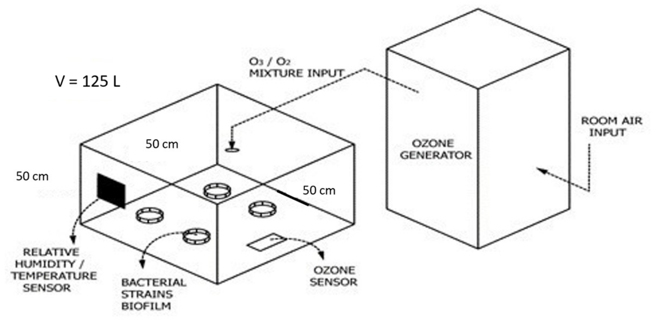
2.1. Test Chamber Characteristics

An in vitro experiment was conducted in a sealed experimental chamber with a removable lid for easier access to the plates and featuring a hole for the ozone input tube. The chamber had a volume of 125 L. The schema of in vitro ozonation is shown in Figure 1. The portable Mozon GPF 8008 ozone generator was used in this study, with the capacity to produce 5 g of ozone/air (O3/O2) mixture. The concentration of ozone in the chamber was continuously monitored using a portable ozone detector model Keernuo GT901, China. A portable station Auriol 4-LD5531, Germany, was used to monitor room temperature and humidity.

2.2. Bacterial Strains

The bacterial strain used for this study was a standard strain of E. coli ATCC 25922 obtained from the culture collection of the Department of Microbiology and Parasitology, University of Rijeka, Croatia. Extended-spectrum-β-lactamase (ESBL)-producing E. coli strains: ESBL E. coli strain 9, ESBL E. coli strain 10, ESBL E. coli strain 11, and ESBL E. coli strain 12, obtained by courtesy of General Hospital, Dr. Ivo Pedišić, Sisak, Croatia, were also used. All ESBL E. coli clinical strains were isolated from urine. Strains were held at −80 °C in 10% glycerol broth. Before being used, the bacteria were plated on Mueller-Hinton agar and cultivated at 37 °C for 24 h.

2.3. Antibiotic Susceptibility Testing Results for All Used Strains

All strains used in this investigation were extended-spectrum-β-lactamase (ESBL)-producing E. coli strains. Results of antibiotic susceptibility testing are provided by the Department of Microbiology at General Hospital Dr. Ivo Pedišić, Sisak, Croatia, and are shown in Table 1.
Considering all the antibiotics listed above, all (100%) tested strains were resistant to amoxicillin and amoxicillin combined with clavulanic acid, whether relating to a systemic infection or a non-complicated urinary tract infection. Sensitivity to tazobactam combined with piperacillin, imipenem, meropenem, ertapenem and fosfomycin showed in all (100%) tested strains. Considering only ESBL clinical isolates, specifically ESBL E. coli strain 9, ESBL E. coli strain 10, ESBL E. coli strain 11 and ESBL E. coli strain 12, all (100%) showed resistance to numerous antibiotics. All (100%) are resistant to cephalexin, cefuroxime, ceftriaxone, ceftazidime, cefpodoxime, cefixime, cefepime, norfloxacin, ciprofloxacin and sulfamethoxazole combined with trimethoprim.
When looking at antibiotic susceptibility testing results for each strain individually, the most sensitive strain appears to be the standard strain, E. coli ATCC 25922. From 21 tests for antibiotic sensitivity, this strain showed sensitivity to 18 antibiotics (85.71%). ESBL E. coli strain 9 showed sensitivity to 8 antibiotics (38.09%), ESBL E. coli strain 10, and ESBL E. coli strain 11 to 7 antibiotics (33.33%). There was no data given for amoxicillin combined with clavulanic acid in noncomplicated urinary infections for ESBL E. coli strain 12. Therefore, this antibiotic was not taken into calculation in antibiotic susceptibility testing. ESBL E. coli strain 12 showed sensitivity to 6 antibiotics among 20 taken into consideration (30.00%), making this strain the most resistant.

2.4. Preparation of Ceramic Tiles and Biofilm Formation

Ceramic tiles with 1 cm × 1 cm dimensions were used to form biofilm. The method for biofilm formation was previously described and modified by Piletić et al. and Kovač et al. [23,35,36,37]. Briefly, the biofilm was formed on the tiles’ top surface, which was thoroughly brushed and sterilized in an autoclave at 121 °C for 15 min. Ceramic tiles were placed in glass Petri dishes and covered with liquified agar (2%, v/v), leaving the top surface of the tiles free and not touched by the agar. The bacterial suspensions of 105 CFU/mL for all tested strains in Mueller Hinton broth were poured over the top surface of the ceramic tiles and then incubated in an orbital shaker at 40 rpm at 35 ± 2 °C for 24 h. After incubation, the ceramic tiles were washed with a sterile saline solution and dried in a laboratory safety cabinet for 1 min. Then, the tiles were removed from the agar, washed out with a sterile saline solution, placed in new Petri dishes, and put in the test chamber.

2.5. Total Bacteria Number Determination

To determine the number of cultivable bacteria, the ceramic tiles with bacterial biofilm were put in sterile Falcon tubes, which were filled with 10 mL of sterile saline solution and then sonicated in an ultrasound bath (Bandelin-BactoSonic, Berlin, Germany) at a frequency of 40 kHz for 1 min to enhance the release of the adhered cells from the tiles. Using a vortex, samples were homogenized one more time to further enhance the detachment of the remaining cells from the biofilm. Afterwards, ten-fold serial dilutions were made and later inoculated onto Mueller Hinton agar and incubated for 24 h at 35 ± 2 °C. Inhibition percentages were calculated using the formula given:
%   inhibition = 1 N treatment N control × 100
where Ntreatment is the mean value of all CFU/cm2 or RLU or CV values after treatment, and Ncontrol is the mean value of CFU/cm2 or RLU or CV values without treatment. The experiment was performed three times in triplicate.

2.6. Determining RLU Values by ATP Bioluminescence

The surface of the ceramic tiles with formed biofilm was wiped with a 3MTM Clean-TraceTM Surface ATP Test Swab UXL100 (3M, Saint Paul, MN, USA) swab pre-immersed for 1 min in luciferin/luciferase reagent. After one minute, the swab was put in a chamber of the 3MTM Clean-TraceTM luminometer (3M, Saint Paul, MN, USA). The amount of light produced was read from the luminometer, expressed as RLU and measured after two, three, four and five minutes. Non–treated ceramic tiles with the biofilm formed served as the control group. Inhibition percentages were calculated using the previously given formula. The experiment was performed three times in triplicate.

2.7. Determining Biomass Reduction by Crystal-Violet Staining

The controls and treated ceramic tiles were rinsed with sterile saline solution and then fixated for 30 min at 80 °C in a dry heat sterilizer (ST-01/02, Instrumentaria, Zagreb, Croatia). After fixation, the tiles were stained with 0.1% crystal violet for 20 min. This step was followed by 15 min of intense mixing and rinsing with 95% ethanol. Afterwards, the optical density (OD) was measured on a spectrophotometer (Eppendorf, Biophotometer, model #6131, Hamburg, Germany) at a wavelength of 600 nm. Inhibition percentages were calculated using the previously given formula. The experiment was performed three times in triplicate.

2.8. Statistical Analysis and Graphing

To determine if there was any statistically significant difference between control and treated ceramic tiles, a Wilcoxon signed-rank test was performed (p < 0.05). To determine if there was any statistically significant difference among tested strains between treated values or untreated values, the Kruskal–Wallis multiple comparison test (p < 0.05) was performed. Statistical analysis was carried out using the TIBCO Statistica 14.0.0. software (StatSoft Inc., Tulsa, OK, USA). Graphing was performed using GraphPad Prism version 9.5.1 (GraphPad Software, San Diego, CA, USA).

3. Results

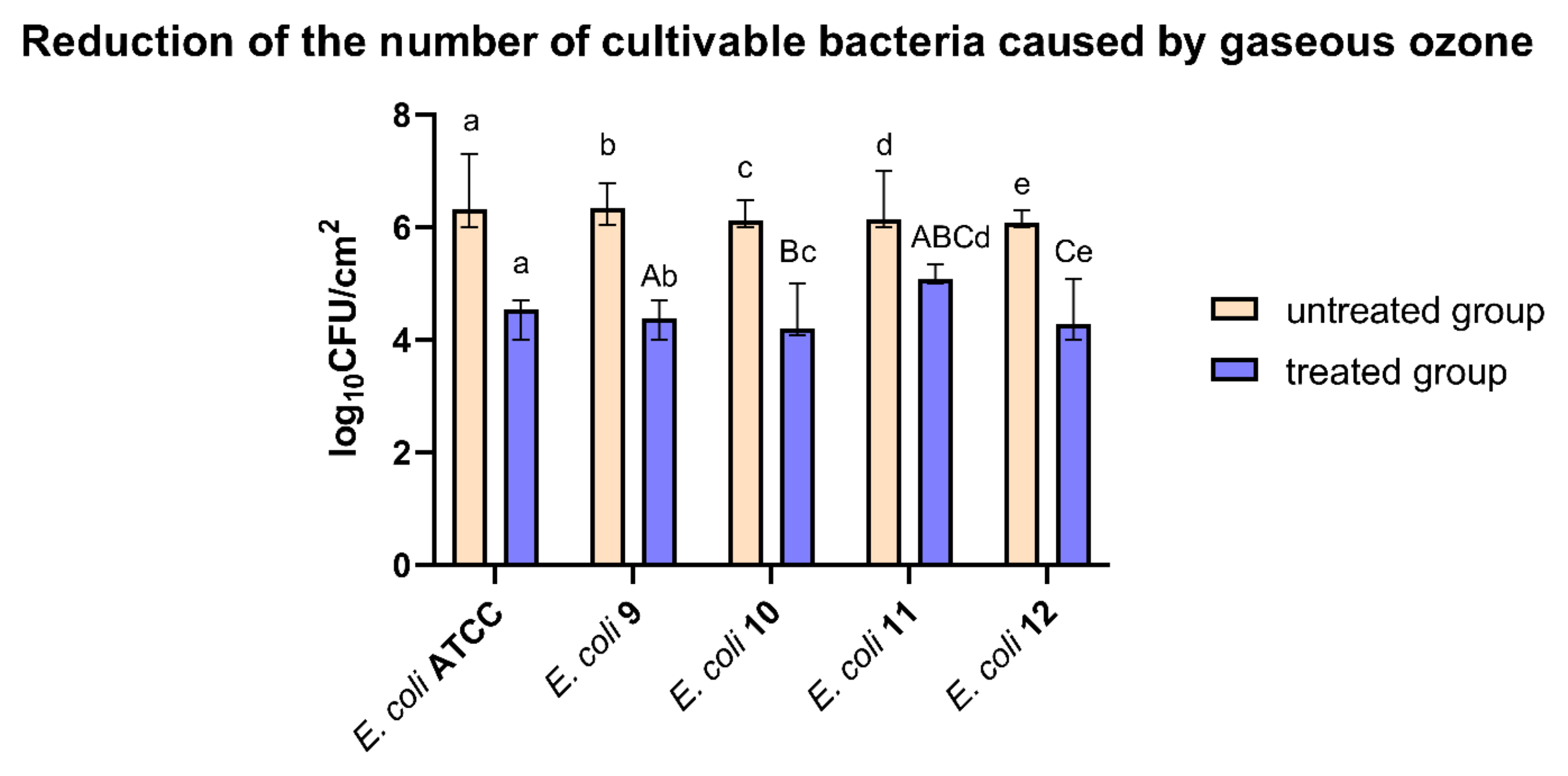
3.1. Reduction of the Number of Cultivable Bacteria Caused by Ozone Treatment

All tested strains formed biofilm on ceramic tile surfaces, and the number of total bacteria varied from 6.14 log10CFU/cm2 to 6.47 log10CFU/cm2 for the untreated (control) group and from 4.35 log10CFU/cm2 to 5.14 log10CFU/cm2 for the treated group. A disinfection treatment with gaseous ozone significantly reduced the number of cultivable bacteria in the biofilm. The numbers of cultivable bacteria are shown in Figure 2.
Gaseous ozone treatment reduces the number of bacteria from approximately 1.25 log10CFU/cm2 to 2.00 log10CFU/cm2, depending on the tested strain, E. coli ATCC 25922 being the most sensitive strain. ESBL E. coli strain 11 (E. coli 11) appears to be the most resistant strain.

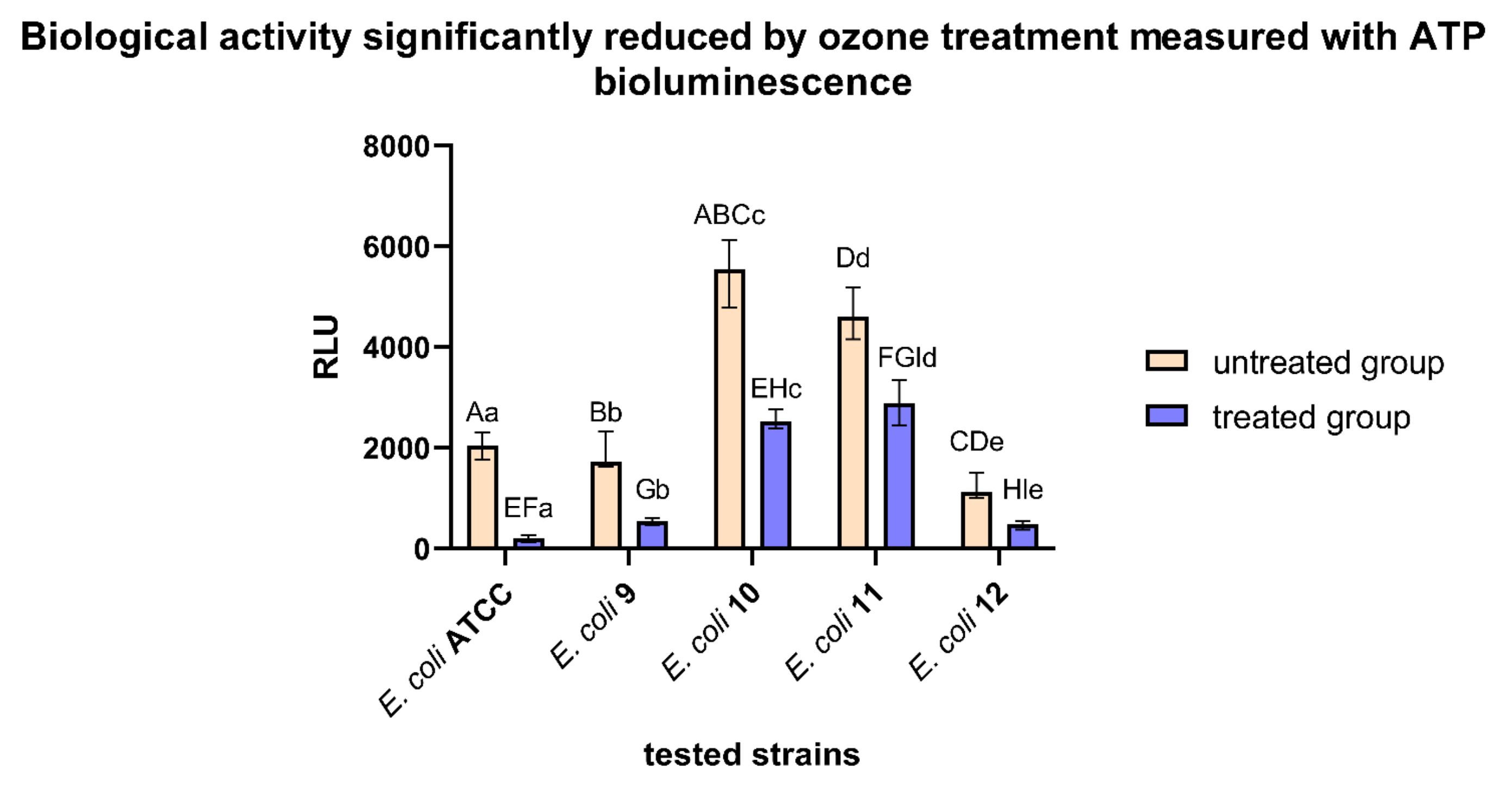
3.2. Biological Activity Significantly Reduced by 1 h Ozone Treatment Measured with ATP Bioluminescence

Biological activity measured using the ATP bioluminescence method showed a statistically significant reduction in biomass after gaseous ozone treatment compared to the control group. A significant difference between different bacteria was observed (p < 0.05). RLU values obtained by the ATP bioluminescence method are shown in Figure 3.
Considering the ATP bioluminescence method, the RLU values on nontreated ceramic tiles varied from approximately 1200 RLU to 5500 RLU, depending on the bacterial strain. After gaseous ozone treatment, values varied from approximately 190 RLU to 2900 RLU, depending on the bacterial strain. Again, the most resistant strain was ESBL E. coli strain 11 (E. coli 11), while the most sensitive was E. coli ATCC 25922.

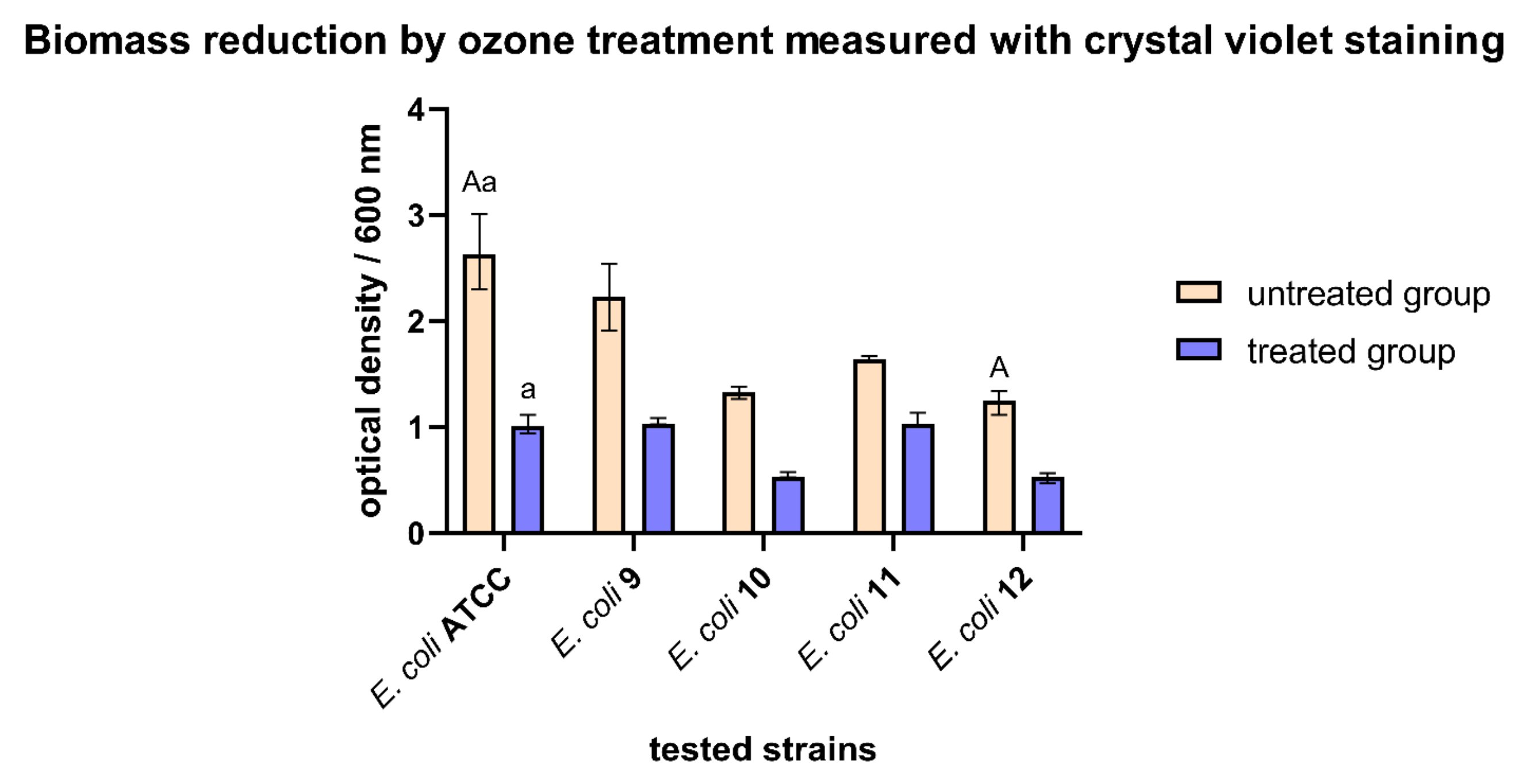
3.3. Biomass Reduction by Ozone Treatment Measured with Crystal Violet Staining

The total biomass reduction was observed for all tested bacteria after gaseous ozone treatment. The reduction was not statistically significant compared to the control group for all tested bacteria except for the E. coli ATCC 25922. Absorbances at 600 nm before and after treatment are shown in Figure 4.
There was no statistically significant difference between values after gaseous ozone treatment. There was a statistically significant difference between untreated values of E. coli ATCC 25922 and ESBL E. coli strain 11. The only case with a statistically significant difference between the untreated and treated values is in E. coli ATCC 25922 biofilm (p < 0.05). Once again, the most sensitive strain was E. coli ATCC 25922, and the most resistant strain was ESBL E. coli strain 11.

3.4. Biofilm Inhibition Rates Varied Using Different Detection Methods

The inhibition percentages were calculated separately after gaseous ozone treatment compared to the control group for each method used. Inhibition rates varied from 35.80% to 99.41%, depending on the method used (Table 2).

4. Discussion

Gaseous ozone has potent antimicrobial activity and strong oxidation properties [23,30,31,32,33,35,36,37]. Although some studies show benefits of gaseous ozone treatment on ceramic tiles for biofilm destruction [23,35,36,37], such as statistically significant reduction of the total bacteria number, reduction of biological activity of bacteria present and reduction of total biomass, scientific data on ESBL E. coli biofilm destruction with gaseous ozone treatment are scarce; therefore, the aim of this research was to investigate the antimicrobial effect of gaseous ozone on ESBL E. coli biofilm.
Three different methods of biofilm quantification were used. Gaseous ozone is efficient as a disinfecting agent against formed biofilm, but not enough for complete biofilm eradication [23,37,38,39,40]. Besides counting total viable bacteria (CFU/cm2), the authors also decided to investigate additional methods for bacterial biomass determination, specifically ATP bioluminescence and crystal violet staining.
Treatment with gaseous ozone showed efficiency in the anti-biofilm effect on the formed biofilm but with certain variations and limitations, considering the quantification methods used. The best inhibition percentages after ozone treatment were shown by E. coli ATCC 25922, from which it can be concluded that the standard strain is the most sensitive strain. On the other side, ESBL E. coli strain 11 (E. coli 11) was the most resistant strain in all methods used. Determining the total number of viable bacteria showed the highest inhibition rates, followed by ATP bioluminescence and crystal violet staining.
The distinct characteristics of each approach and its measuring focus can be used to account for this variance in inhibition rates between methods. With the ability to count live cells, dead cells, EPS, and VBNC cells, ATP bioluminescence and crystal violet can be supplementary biomass estimation methods [41,42,43,44,45]. As a result, inhibition rates determined with these approaches are lower than the total number of viable bacteria. The same results were previously described for the gaseous ozone anti-biofilm effect on K. pneumoniae biofilm and the benzalkonium chloride anti-biofilm effect on the most common food-borne pathogens [23,37]. It is worth emphasizing that not every method used for biofilm detection is equally precise.
The great advantage of CFU is that only viable bacteria are counted, as the CFU excludes dead bacteria and debris. On the other hand, a drawback of this method is that occasionally groups of bacteria are counted as single colonies, which can lead to inaccurate and misleading findings [46]. Because of the ATP reaction with luciferin and luciferase, ATP bioluminescence is a quick, simple, and extremely sensitive method for determining how much light is produced by bacteria in a biofilm. In addition to measuring ATP produced by bacteria, surface product residues are also measured [42,47]. All examined strains’ biofilms on ceramic tiles are successfully detected using ATP bioluminescence, and the value of the biofilms is significantly reduced after treatment with a cleaning agent. On the other side, the absence of established standards or reference values is a drawback of this approach. The likelihood of a false positive result is another drawback. Suppose a surface of no interest is cleaned during swabbing. In that case, certain microorganisms from that surface may react with the luciferin reagent and produce a result irrelevant to the research [48]. The detection of total biofilm biomass using crystal violet staining is inaccurate. One of the most commonly used techniques for measuring biofilm biomass is crystal violet staining. However, this technique has some drawbacks, including unspecific binding to negatively charged molecules and poor reproducibility due to uneven dye extraction or differential removal of biofilm biomass during the steps [49]. Because of this, several researchers advise switching to a fluorescence staining [49].
The aim of this work was to investigate the effectiveness of the gaseous ozone in reducing the number of bacteria and the total biomass on ESBL E. coli strains biofilm using different methods of quantification and detection. A significant reduction was found in all tested methods, except for crystal violet staining, where inhibition percentages are not sufficient and unsatisfactory. Various biofilm detection methods can be used to investigate how efficient gaseous ozone is, but in conclusion, gaseous ozone did not cause complete biofilm eradication.
Although ozone gas significantly reduced bacteria in ESBL E. coli biofilm, complete removal of biofilm was not determined. The authors suggest further investigation with longer exposure time (i.e., two hours), higher relative humidity and temperature. Some of the investigations that can emerge as follow-up from this study are investigations of combined disinfection antibiofilm action using one or more biocidal active substances of natural origin as a potential environmentally friendly disinfection procedure.

5. Conclusions

Gaseous ozone in concentration of 25 ppm during 1 h exposure time significantly reduced the number of bacteria in ESBL E. coli biofilm, no matter which detection method was used, although failing to fully eradicate biofilm from the ceramic surface. In order for complete biofilm removal from the surface, the authors would like to emphasize the importance of thorough mechanical cleaning prior to disinfection with gaseous ozone. This study has shown that gaseous ozone has potential for practical application in industry and further investigation with different biocidal active substances is needed.

Author Contributions

Conceptualization, B.K. and I.G.; methodology, B.K.; validation, B.K.; formal analysis, B.K.; investigation, B.K.; resources, I.G.; writing—original draft preparation, B.K., A.P., M.P. and K.P.; writing—review and editing, B.K., K.P. and I.G.; supervision, I.G.; funding acquisition, I.G. All authors have read and agreed to the published version of the manuscript.

Funding

This research was funded by the University of Rijeka, grant number UNIRI-biomed 18-171.

Institutional Review Board Statement

Not applicable.

Informed Consent Statement

Not applicable.

Data Availability Statement

Not applicable.

Acknowledgments

The authors would like to thank to the company Mozone d.o.o., Sisak, Croatia, for donating the ozone generator and other equipment for the experiment. The authors would also like to thank to NOACK d.o.o. from Zagreb, Croatia, for donating the luminometer. Many thanks to the Public Health Institute of Sisak-Moslavina County, Department for Human Microbiology, Sisak, Croatia, for collecting the clinical strains of E. coli, as well as to the General Hospital “Dr. Ivo Pedišić”, Sisak, Croatia.

Conflicts of Interest

The authors declare no conflict of interest.

Abbreviations

CFU/cm2 colony forming unit per square centimeter
CV crystal violet
E. coliEscherichia coli
EPS extracellular polymer substance
ESBL extended-spectrum-β-lactamase
HUS hemolytic uremic syndrome
OD optical density
MHB Müller Hinton broth
PBP penicillin-binding protein
RLU relative light units
VBNC viable but not cultivable state of bacteria

References

  1. Allocati, N.; Masulli, M.; Alexeyev, M.; Di Ilio, C. Escherichia Coli in Europe: An Overview. Int. J. Environ. Res. Public. Health 2013, 10, 6235–6254. [Google Scholar] [CrossRef] [PubMed]
  2. Martinson, J.N.V.; Walk, S.T. Escherichia Coli Residency in the Gut of Healthy Human Adults. EcoSal Plus 2020, 9, 1. [Google Scholar] [CrossRef] [PubMed]
  3. Yoon, S.H.; Jeong, H.; Kwon, S.-K.; Kim, J.F. Genomics, Biological Features, and Biotechnological Applications of Escherichia Coli B: “Is B for Better?!” In Systems Biology and Biotechnology of Escherichia coli; Lee, S.Y., Ed.; Springer: Dordrecht, The Netherlands, 2009; pp. 1–17. ISBN 978-1-4020-9393-7. [Google Scholar]
  4. Hadžić, L.; Šeol Martinec, B.; Pintarić, S. Extended spectrum β-lactamase producing Escherichia coli. Hrvat. Vet. Vjesn. 2021, 29, 3. [Google Scholar]
  5. Andrašević, S.; Vranić-Ladavac, M.; Tambić-Andrašević, A. Antibiotic sensitivity of enterobacteriaceae. Infektološki Glas. 2009, 29, 171–176. [Google Scholar]
  6. Bezabih, Y.M.; Sabiiti, W.; Alamneh, E.; Bezabih, A.; Peterson, G.M.; Bezabhe, W.M.; Roujeinikova, A. The Global Prevalence and Trend of Human Intestinal Carriage of ESBL-Producing Escherichia Coli in the Community. J. Antimicrob. Chemother. 2021, 76, 22–29. [Google Scholar] [CrossRef]
  7. von Wulffen, J.; RecogNice-Team; Sawodny, O.; Feuer, R. Transition of an Anaerobic Escherichia Coli Culture to Aerobiosis: Balancing MRNA and Protein Levels in a Demand-Directed Dynamic Flux Balance Analysis. PLoS ONE 2016, 11, e0158711. [Google Scholar] [CrossRef]
  8. Behzadi, P.; Urbán, E.; Gajdács, M. Association between Biofilm-Production and Antibiotic Resistance in Uropathogenic Escherichia coli (UPEC): An In Vitro Study. Diseases 2020, 8, 17. [Google Scholar] [CrossRef]
  9. Hooton, T.M.; Bradley, S.F.; Cardenas, D.D.; Colgan, R.; Geerlings, S.E.; Rice, J.C.; Saint, S.; Schaeffer, A.J.; Tambayh, P.A.; Tenke, P.; et al. Diagnosis, Prevention, and Treatment of Catheter-Associated Urinary Tract Infection in Adults: 2009 International Clinical Practice Guidelines from the Infectious Diseases Society of America. Clin. Infect. Dis. 2010, 50, 625–663. [Google Scholar] [CrossRef]
  10. Ciani, O.; Grassi, D.; Tarricone, R. An Economic Perspective on Urinary Tract Infection: The “Costs of Resignation”. Clin. Drug Investig. 2013, 33, 255–261. [Google Scholar] [CrossRef]
  11. Niveditha, A.; Pandiselvam, R.; Prasath, V.A.; Singh, S.K.; Gul, K.; Kothakota, A. Application of Cold Plasma and Ozone Technology for Decontamination of Escherichia coli in Foods—A Review. Food Control 2021, 130, 108338. [Google Scholar] [CrossRef]
  12. Garcia, A.; Fox, J.G.; Besser, T.E. Zoonotic Enterohemorrhagic Escherichia coli: A One Health Perspective. ILAR J. 2010, 51, 221–232. [Google Scholar] [CrossRef]
  13. Bryan, A.; Youngster, I.; McAdam, A.J. Shiga Toxin Producing Escherichia coli. Clin. Lab. Med. 2015, 35, 247–272. [Google Scholar] [CrossRef]
  14. Doi, Y.; Iovleva, A.; Bonomo, R.A. The Ecology of Extended-Spectrum β-Lactamases (ESBLs) in the Developed World. J. Travel Med. 2017, 24, S44–S51. [Google Scholar] [CrossRef]
  15. Chong, Y.; Shimoda, S.; Shimono, N. Current Epidemiology, Genetic Evolution and Clinical Impact of Extended-Spectrum β-Lactamase-Producing Escherichia coli and Klebsiella Pneumoniae. Infect. Genet. Evol. 2018, 61, 185–188. [Google Scholar] [CrossRef]
  16. Shaikh, S.; Fatima, J.; Shakil, S.; Rizvi, S.M.D.; Kamal, M.A. Antibiotic Resistance and Extended Spectrum Beta-Lactamases: Types, Epidemiology and Treatment. Saudi J. Biol. Sci. 2015, 22, 90–101. [Google Scholar] [CrossRef]
  17. Singh, N. Prevalence of ESBL in Escherichia coli Isolates among ICU Patients in a Tertiary Care Hospital. J. Clin. Diagn. Res. 2016, 10, DC19. [Google Scholar] [CrossRef]
  18. Pandey, N.; Cascella, M. Beta Lactam Antibiotics. In StatPearls; StatPearls Publishing: Treasure Island, FL, USA, 2022. [Google Scholar]
  19. Sieber, S.A.; Böttcher, T.; Staub, I.; Orth, R. Small Molecules as Versatile Tools for Activity-Based Protein Profiling Experiments. In Comprehensive Natural Products II; Elsevier: Amsterdam, The Netherlands, 2010; pp. 629–674. ISBN 978-0-08-045382-8. [Google Scholar]
  20. Su, M.; Satola, S.W.; Read, T.D. Genome-Based Prediction of Bacterial Antibiotic Resistance. J. Clin. Microbiol. 2019, 57, e01405-18. [Google Scholar] [CrossRef]
  21. Ballén, V.; Cepas, V.; Ratia, C.; Gabasa, Y.; Soto, S.M. Clinical Escherichia coli: From Biofilm Formation to New Antibiofilm Strategies. Microorganisms 2022, 10, 1103. [Google Scholar] [CrossRef]
  22. Sharma, G.; Sharma, S.; Sharma, P.; Chandola, D.; Dang, S.; Gupta, S.; Gabrani, R. Escherichia Coli Biofilm: Development and Therapeutic Strategies. J. Appl. Microbiol. 2016, 121, 309–319. [Google Scholar] [CrossRef]
  23. Piletić, K.; Kovač, B.; Perčić, M.; Žigon, J.; Broznić, D.; Karleuša, L.; Lučić Blagojević, S.; Oder, M.; Gobin, I. Disinfecting Action of Gaseous Ozone on OXA-48-Producing Klebsiella Pneumoniae Biofilm In Vitro. Int. J. Environ. Res. Public. Health 2022, 19, 6177. [Google Scholar] [CrossRef]
  24. Vickery, K.; Deva, A.; Jacombs, A.; Allan, J.; Valente, P.; Gosbell, I.B. Presence of Biofilm Containing Viable Multiresistant Organisms despite Terminal Cleaning on Clinical Surfaces in an Intensive Care Unit. J. Hosp. Infect. 2012, 80, 52–55. [Google Scholar] [CrossRef] [PubMed]
  25. Vickery, K. Special Issue: Microbial Biofilms in Healthcare: Formation, Prevention and Treatment. Materials 2019, 12, 2001. [Google Scholar] [CrossRef]
  26. Assefa, M.; Amare, A. Biofilm-Associated Multi-Drug Resistance in Hospital-Acquired Infections: A Review. Infect. Drug Resist. 2022, 15, 5061–5068. [Google Scholar] [CrossRef] [PubMed]
  27. Weinbren, M.; Inkster, T. The Hospital-Built Environment: Biofilm, Biodiversity and Bias. J. Hosp. Infect. 2021, 111, 50–52. [Google Scholar] [CrossRef] [PubMed]
  28. Muhammad, M.H.; Idris, A.L.; Fan, X.; Guo, Y.; Yu, Y.; Jin, X.; Qiu, J.; Guan, X.; Huang, T. Beyond Risk: Bacterial Biofilms and Their Regulating Approaches. Front. Microbiol. 2020, 11, 928. [Google Scholar] [CrossRef]
  29. Li, B.; Chen, H.; Cao, L.; Hu, Y.; Chen, D.; Yin, Y. Escherichia Coli Exopolysaccharides Induced by Ceftriaxone Regulated Human Gut Microbiota in Vitro. Front. Microbiol. 2021, 12, 634204. [Google Scholar] [CrossRef]
  30. Aponte, M.; Anastasio, A.; Marrone, R.; Mercogliano, R.; Peruzy, M.F.; Murru, N. Impact of Gaseous Ozone Coupled to Passive Refrigeration System to Maximize Shelf-Life and Quality of Four Different Fresh Fish Products. LWT 2018, 93, 412–419. [Google Scholar] [CrossRef]
  31. Panebianco, F.; Rubiola, S.; Chiesa, F.; Civera, T.; Di Ciccio, P.A. Effect of Gaseous Ozone Treatment on Biofilm of Dairy-Isolated Pseudomonas Spp. Strains. Ital. J. Food Saf. 2022, 11, 10350. [Google Scholar] [CrossRef]
  32. Epelle, E.I.; Macfarlane, A.; Cusack, M.; Burns, A.; Okolie, J.A.; Mackay, W.; Rateb, M.; Yaseen, M. Ozone Application in Different Industries: A Review of Recent Developments. Chem. Eng. J. 2023, 454, 140188. [Google Scholar] [CrossRef]
  33. Cai, Y.; Zhao, Y.; Yadav, A.K.; Ji, B.; Kang, P.; Wei, T. Ozone Based Inactivation and Disinfection in the Pandemic Time and beyond: Taking Forward What Has Been Learned and Best Practice. Sci. Total Environ. 2023, 862, 160711. [Google Scholar] [CrossRef]
  34. Fan, X. Gaseous Ozone to Preserve Quality and Enhance Microbial Safety of Fresh Produce: Recent Developments and Research Needs. Compr. Rev. Food Sci. Food Saf. 2021, 20, 4993–5014. [Google Scholar] [CrossRef]
  35. Piletić, K.; Kovač, B.; Planinić, M.; Vasiljev, V.; Karačonji, I.B.; Žigon, J.; Gobin, I.; Oder, M. Combined Biocidal Effect of Gaseous Ozone and Citric Acid on Acinetobacter Baumannii Biofilm Formed on Ceramic Tiles and Polystyrene as a Novel Approach for Infection Prevention and Control. Processes 2022, 10, 1788. [Google Scholar] [CrossRef]
  36. Piletić, K.; Linšak, D.T.; Kovač, B.; Mežnarić, S.; Repustić, M.; Radmanović-Skrbić, M.; Gobin, I. Ozone Disinfection Efficiency against Airborne Microorganisms in Hospital Environment: A Case Study. Arch. Ind. Hyg. Toxicol. 2023, 73, 270–276. [Google Scholar] [CrossRef]
  37. Kovač, B.; Piletić, K.; Kovačević Ganić, N.; Gobin, I. The Effectiveness of Benzalkonium Chloride as an Active Compound on Selected Foodborne Pathogens Biofilm. Hygiene 2022, 2, 226–235. [Google Scholar] [CrossRef]
  38. Failor, K.C.; Silver, B.; Yu, W.; Heindl, J.E. Biofilm Disruption and Bactericidal Activity of Aqueous Ozone Coupled with Ultrasonic Dental Scaling. JADA Found. Sci. 2022, 1, 100003. [Google Scholar] [CrossRef]
  39. Marino, M.; Maifreni, M.; Baggio, A.; Innocente, N. Inactivation of Foodborne Bacteria Biofilms by Aqueous and Gaseous Ozone. Front. Microbiol. 2018, 9, 2024. [Google Scholar] [CrossRef]
  40. Panebianco, F.; Rubiola, S.; Di Ciccio, P.A. The Use of Ozone as an Eco-Friendly Strategy against Microbial Biofilm in Dairy Manufacturing Plants: A Review. Microorganisms 2022, 10, 162. [Google Scholar] [CrossRef]
  41. Robben, C.; Witte, A.K.; Schoder, D.; Stessl, B.; Rossmanith, P.; Mester, P. A Fast and Easy ATP-Based Approach Enables MIC Testing for Non-Resuscitating VBNC Pathogens. Front. Microbiol. 2019, 10, 1365. [Google Scholar] [CrossRef]
  42. Arroyo, M.G.; Ferreira, A.M.; Frota, O.P.; Rigotti, M.A.; de Andrade, D.; Brizzotti, N.S.; Peresi, J.T.M.; Castilho, E.M.; de Almeida, M.T.G. Effectiveness of ATP Bioluminescence Assay for Presumptive Identification of Microorganisms in Hospital Water Sources. BMC Infect. Dis. 2017, 17, 458. [Google Scholar] [CrossRef]
  43. Sanchez, M.; Llama-Palacios, A.; Marin, M.; Figuero, E.; Leon, R.; Blanc, V.; Herrera, D.; Sanz, M. Validation of ATP Bioluminescence as a Tool to Assess Antimicrobial Effects of Mouthrinses in an in Vitro Subgingival-Biofilm Model. Med. Oral Patol. Oral Cirugia Bucal 2013, 18, e86–e92. [Google Scholar] [CrossRef]
  44. Castro, J.; Lima, Â.; Sousa, L.G.V.; Rosca, A.S.; Muzny, C.A.; Cerca, N. Crystal Violet Staining Alone Is Not Adequate to Assess Synergism or Antagonism in Multi-Species Biofilms of Bacteria Associated with Bacterial Vaginosis. Front. Cell. Infect. Microbiol. 2022, 11, 795797. [Google Scholar] [CrossRef] [PubMed]
  45. Gascón, E.; Merino, N.; Pagán, E.; Berdejo, D.; Pagán, R.; García-Gonzalo, D. Assessment of In Vitro Biofilms by Plate Count and Crystal Violet Staining: Is One Technique Enough? In Detection and Enumeration of Bacteria, Yeast, Viruses, and Protozoan in Foods and Freshwater; Magnani, M., Ed.; Methods and Protocols in Food Science; Springer: New York, NY, USA, 2021; pp. 53–63. ISBN 978-1-07-161931-5. [Google Scholar]
  46. Hazan, R.; Que, Y.-A.; Maura, D.; Rahme, L.G. A Method for High Throughput Determination of Viable Bacteria Cell Counts in 96-Well Plates. BMC Microbiol. 2012, 12, 259. [Google Scholar] [CrossRef] [PubMed]
  47. Monica, S.; Bancalari, E.; Castellone, V.; Rijkx, J.; Wirth, S.; Jahns, A.; Bottari, B. ATP Bioluminescence for Rapid and Selective Detection of Bacteria and Yeasts in Wine. Appl. Sci. 2021, 11, 4953. [Google Scholar] [CrossRef]
  48. Griffith, C. Surface Sampling and the Detection of Contamination. In Handbook of Hygiene Control in the Food Industry; Elsevier: Amsterdam, The Netherlands, 2016; pp. 673–696. ISBN 978-0-08-100155-4. [Google Scholar]
  49. Amador, C.I.; Stannius, R.O.; Røder, H.L.; Burmølle, M. High-Throughput Screening Alternative to Crystal Violet Biofilm Assay Combining Fluorescence Quantification and Imaging. J. Microbiol. Methods 2021, 190, 106343. [Google Scholar] [CrossRef]
Figure 1. Ozone generator and test chamber. The same schema was previously used in Piletić et al. [23].
Hygiene 03 00011 g001
Figure 2. Reduction of the number of cultivable bacteria caused by gaseous ozone. Log10CFU/cm2 values before and after ozone treatment for all tested strains. Results are shown as a mean value with standard deviation. Capital letters, A–C, show statistically significant difference among tested strains within either untreated group or treated group (Kruskal-Wallis H test, p < 0.05). Lowercase letters, a–e, show the statistically significant difference between the treated and control group for every tested strain (Wilcoxon signed-rank test, p < 0.05).
Hygiene 03 00011 g002
Figure 3. Biological activity significantly reduced by ozone treatment measured with ATP bioluminescence. RLU values before and after ozone treatment for all tested strains. Results are shown as a mean value with standard deviation. Capital letters, A–I, show statistically significant difference among tested strains within either untreated group or treated group (Kruskal-Wallis H test, p < 0.05). Lowercase letters, a–e, show the statistically significant difference between the treated and control group for every tested strain (Wilcoxon signed-rank test, p < 0.05).
Hygiene 03 00011 g003
Figure 4. Biomass reduction by ozone treatment measured with crystal violet staining. Optical density (OD) values at 600 nm before and after ozone treatment for all tested strains. Results are shown as a mean value with standard deviation. Capital letter A shows a statistically significant difference among tested strains within either untreated group or treated group (Kruskal-Wallis H test, p < 0.05). Lowercase letter a shows the statistically significant difference between the treated and control group for every tested strain (Wilcoxon signed-rank test, p < 0.05).
Hygiene 03 00011 g004
Table 1. Antibiotic susceptibility testing results for all strains used in the gaseous ozone treatment.
Antibiotic ESBL Strains
E. coli ATCC 25922E. coli 9E. coli 10E. coli 11E. coli 12
AMXRRRRR
AMC (sys)RRRRR
AMC (ncuti)RRRR* ND
TZPSSSSS
CPNSRRRR
CXMSRRRR
CROSRRRR
CAZSRRRR
CPDSRRRR
CFM *SRRRR
CEFSRRRR
IPMSSSSS
MEMSSSSS
ETPSSSSS
GMSSSRR
AMKSSRSS
NITSSSSR
NORSRRRR
CIPSRRRR
SXTSRRRR
FOSSSSSS
Abbreviations of antibiotics listed: AMX—amoxicillin, AMC—amoxicillin combined with clavulanic acid, AMK—amikacin, TZP—piperacillin combined with tazobactam, CXM—cefuroxime, CRO—ceftriaxone, CAZ—ceftazidime, CPD—cefpodoxime, CFM *—cefixime, CEF—cefepime, IPM—imipenem, MEM—meropenem, ETP—ertapenem, GM—gentamicin, NOR—norfloxacin, CIP—ciprofloxacin, SXT—sulfamethoxazole combined with trimethoprim, FOS—fosfomycin, CPN—cephalexin, NIT—nitrofurantoin, sys—system infection, ncuti—non complicated urinary tract infection. Letters R, S, and * ND mark the following: R—resistant, S—sensitive, * ND—no data given.
Table 2. Inhibition percentages after gaseous ozone treatment for all tested strains. Results are expressed as percentages (%).
ESBL Strains
E. coli ATCC 25922E. coli 9E. coli 10E. coli 11E. coli 12
CFU/cm299.4198.9199.0696.8997.54
RLU90.3171.9053.4737.8460.85
OD (600 nm)61.4052.9959.0535.8057.87
Disclaimer/Publisher’s Note: The statements, opinions and data contained in all publications are solely those of the individual author(s) and contributor(s) and not of MDPI and/or the editor(s). MDPI and/or the editor(s) disclaim responsibility for any injury to people or property resulting from any ideas, methods, instructions or products referred to in the content.

