Brief Report

Direct Chromosome Preparation Method in Avian Embryos for Cytogenetic Studies: Quick, Easy and Cheap

by
Suziane Alves Barcellos
1,
Marcelo Santos de Souza
1,
Victoria Tura
1,
Larissa Rodrigues Pereira
1,
Rafael Kretschmer
2,*,
Ricardo José Gunski
1 and
Analía Del Valle Garnero
1
1
Laboratório de Diversidade Genética Animal, Universidade Federal do Pampa, São Gabriel 97307-020, RS, Brazil
2
Departamento de Ecologia, Zoologia e Genética, Instituto de Biologia, Universidade Federal de Pelotas, Pelotas 96010-900, RS, Brazil
*
Author to whom correspondence should be addressed.
DNA 2022, 2(1), 22-29; https://doi.org/10.3390/dna2010002
Submission received: 5 November 2021 / Revised: 12 January 2022 / Accepted: 17 January 2022 / Published: 26 January 2022

Abstract

:
Avian cell culture is widely applied for cytogenetic studies, the improvement of which increasingly allows for the production of high-quality chromosomes, essential to perform both classical and molecular cytogenetic studies. Among these approaches, there are two main types: fibroblast and bone marrow culture. Despite its high cost and complexity, fibroblast culture is considered the superior approach due to the quality of the metaphases produced. Short-term bone marrow cultivation provides more condensed chromosomes but nonetheless is quicker and easier. In the search for a quicker, cheaper way to prepare metaphases without losing quality, the present work developed a novel, widely applicable protocol for avian chromosome preparation. Twenty-one bird embryos from distinct families were sampled: Icteridae, Columbidae, Furnariidae, Estrildidae, Thraupidae, Troglodytidae and Ardeidae. The protocol was based on a combination of modified fibroblast culture and bone marrow cultivation, taking the advantages of both. The results show that all species consistently presented good mitotic indexes and high-quality chromosomes. Overall, the application of this protocol for bird cytogenetics can optimize the time, considering that most fibroblast cultures take at least 3 days and often much longer. However, our protocol can be performed in 3 h with a much-reduced cost of reagents and equipment.

Graphical Abstract

1. Introduction

Since the early 1900s, bird chromosomes have been investigated using classical and molecular approaches. Despite the fact that avian cytogenetics play an important role in evolutionary studies, less than 10% of all bird species have a karyotype description so far, and nearly all of these are only partial karyotypes [1]. Birds have a bimodal karyotype and a ZZ/ZW sex chromosome system [2]. One of the most remarkable characteristics in their karyotypes is the large number of microchromosomes, which encode a high rate of important genes [3]. Regarding diploid numbers, they display a wide range, from 40 to 142. Despite this variation, more than 50% of birds have between 78 and 82 chromosomes [1].
Working with wild birds is always challenging due to the limitations associated with animal sampling [4]. Even though some samples can be collected from birds in captivity, it is still hard to do this in some species without negatively affecting their health [5].
Avian cytogenetic studies require cell cultures that provide high-quality metaphases for chromosome analysis, as well as the application of classical and molecular techniques, such as karyotyping, chromosome banding and fluorescent in situ hybridization (FISH). In recent years, FISH analysis has played an important role in evolutionary studies, revealing chromosomal homologies and rearrangements [6].
Due to the large amount of microchromosomes and the difficulty in distinguishing them, cells need to be free from residual content such as cytoplasm, which could cause analysis issues. In general, fibroblast culture is the most commonly applied protocol to obtain high-quality avian metaphases [7,8,9,10,11,12]. However, some authors use short-term bone marrow cultivation to perform cytogenetic experiments [13,14,15,16].
In 1968, Sasaki, Ikeuchi and Makino [17] developed a fibroblast cell culture protocol in order to obtain bird metaphases. Although this protocol has been widely used, there are limiting factors for its application, such as expensive reagents and equipment, contamination risk and long-term culture, which can last several weeks. In addition, cell culture protocols with a few modifications such as temperature and time of cultivation have also been used [7,8,9,10]. Short-term bone marrow culture is a low-cost technique in comparison; however, the metaphase quality is typically poorer due to the accumulation of residual content such as cytoplasm and typically short, condensed chromosomes [18]. Another short-term cell culture that can be applied for this approach is the colchicinized embryo technique developed by Bloom and Buss [19] and Bloom with modifications [20].
The purpose of this study was to develop a low-cost, short-term cell culture for avian cytogenetic studies that combined the ease of bone marrow culture with the quality of fibroblast culture using fertilized eggs as a starting material.

2. Materials and Methods

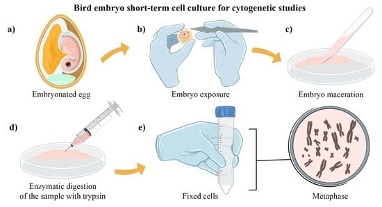
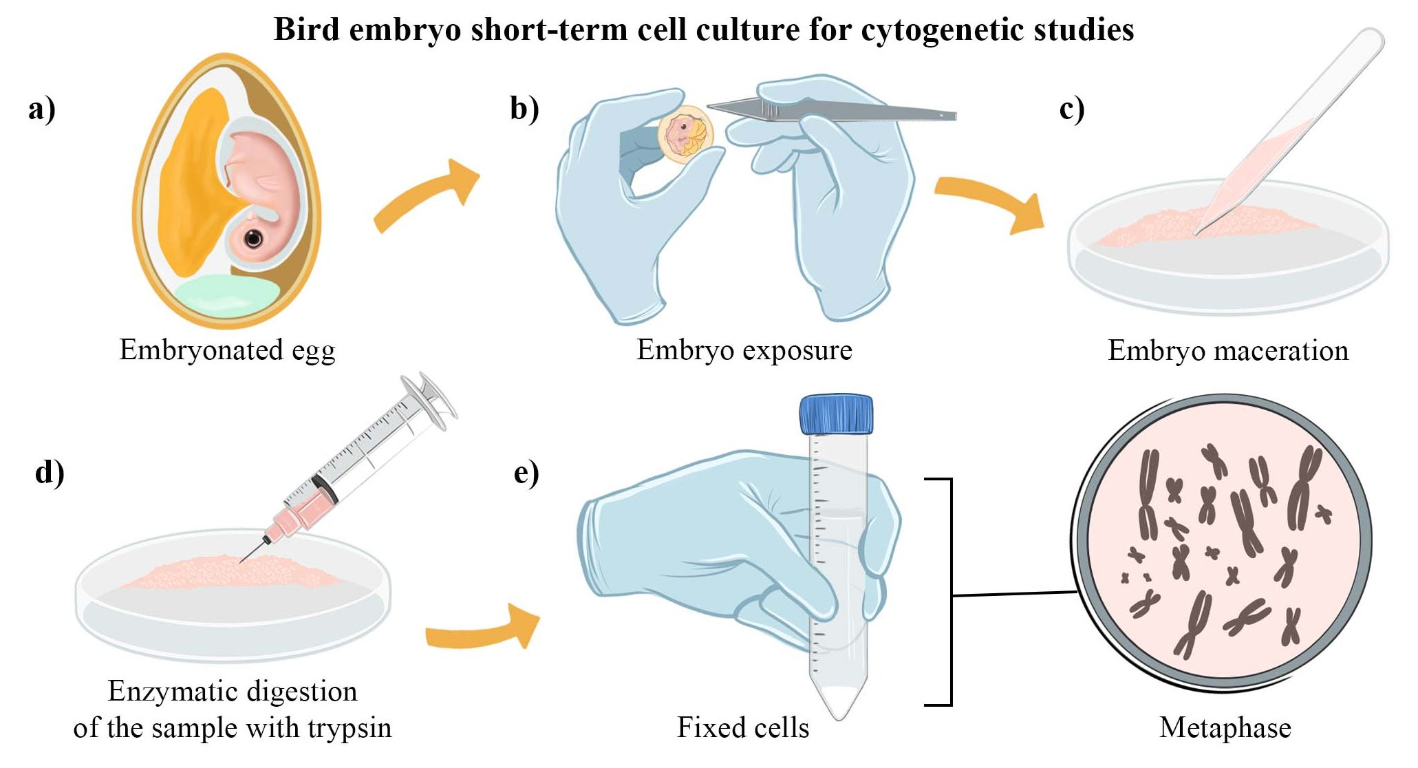
2.1. Bird Embryo Cell Culture Preparation

For this technique, 3 fertile bird eggs were used from individuals from distinct families of birds, i.e., Icteridae (Molothrus bonariensis), Columbidae (Columbina picui), Furnariidae (Synallaxis frontalis), Estrildidae (Estrilda astrild), Thraupidae (Sporophila caerulescens), Troglodytidae (Troglodytes musculus), and Ardeidae (Butorides striata), ranging from 10 to 15 days old. All samples were collected following the protocols approved by the Ethics Committee on the Use of Animals (CEUA 019/2020) and Biodiversity Authorization and Information System (SISBIO 61047-3/33860-2).
To begin with, the eggs were examined by candling, holding them in front of a bright light. Shells were cleaned by being wiped with 70% ethanol and were allowed it to air dry. Then, the egg air sac was found and marked with a pencil. Subsequently, the shell above the air sac was removed with sterile forceps, without touching the egg membrane. Then, the membrane was removed, exposing the embryo, which was placed into a sterile Petri dish with egg content. After that, the embryo was removed to another sterile Petri dish. To prepare the cell suspension, the embryo was washed with 2 mL of Hanks’ balanced salt solution 1x, and then, we dissected the embryo head using a sterile scissor. The Hanks’ solution was discarded, and the embryo was macerated (shown in Figure 1a). Then, a 5 mL syringe with a needle and pipette was used in order to help mix the embryo with 2 mL of trypsin 0.25% EDTA (shown in Figure 1b,c). Using a sterile pipette, the sample was placed into a 15 mL conical tube with 2 mL of trypsin 0.25% EDTA and mixed until complete tissue disaggregation was achieved, for approximately 10 min. Afterwards, 1 mL of fetal bovine serum was used to inactivate the trypsin solution. The content was placed into a new tube containing 10 mL of RPMI 1040 medium pre warmed at 37 °C and mixed. Then, 3 drops of colchicine 0.01% was added. The sample was incubated at 37 °C for 1 h, and then subsequently centrifuged for 10 min at 1000 rpm (room temperature). The supernatant was removed, and the cell pellet was suspended in 10 mL of hypotonic solution (0.075 M KCl) at 37 °C for 20 min. Finally, 1 mL of methanol and acetic acid (3:1) solution was added in order to fix the samples. The process was repeated 3 times with 10 mL of methanol and acetic acid (3:1), each at 1000 rpm for 10 min before storage at −20 °C (shown in Figure 1d). From start to finish, the whole protocol took less than 3 h.
Metaphases were stained with Giemsa 5% in phosphate buffer pH 6.8 for 5 min. Figures were photographed with an optical microscope (OLYMPUS DP53, Olympus Corporation, Ishikawa, Japan) and cellSens Imaging software (Olympus Corporation).

2.2. Fluorescent In Situ Hybridization (FISH)
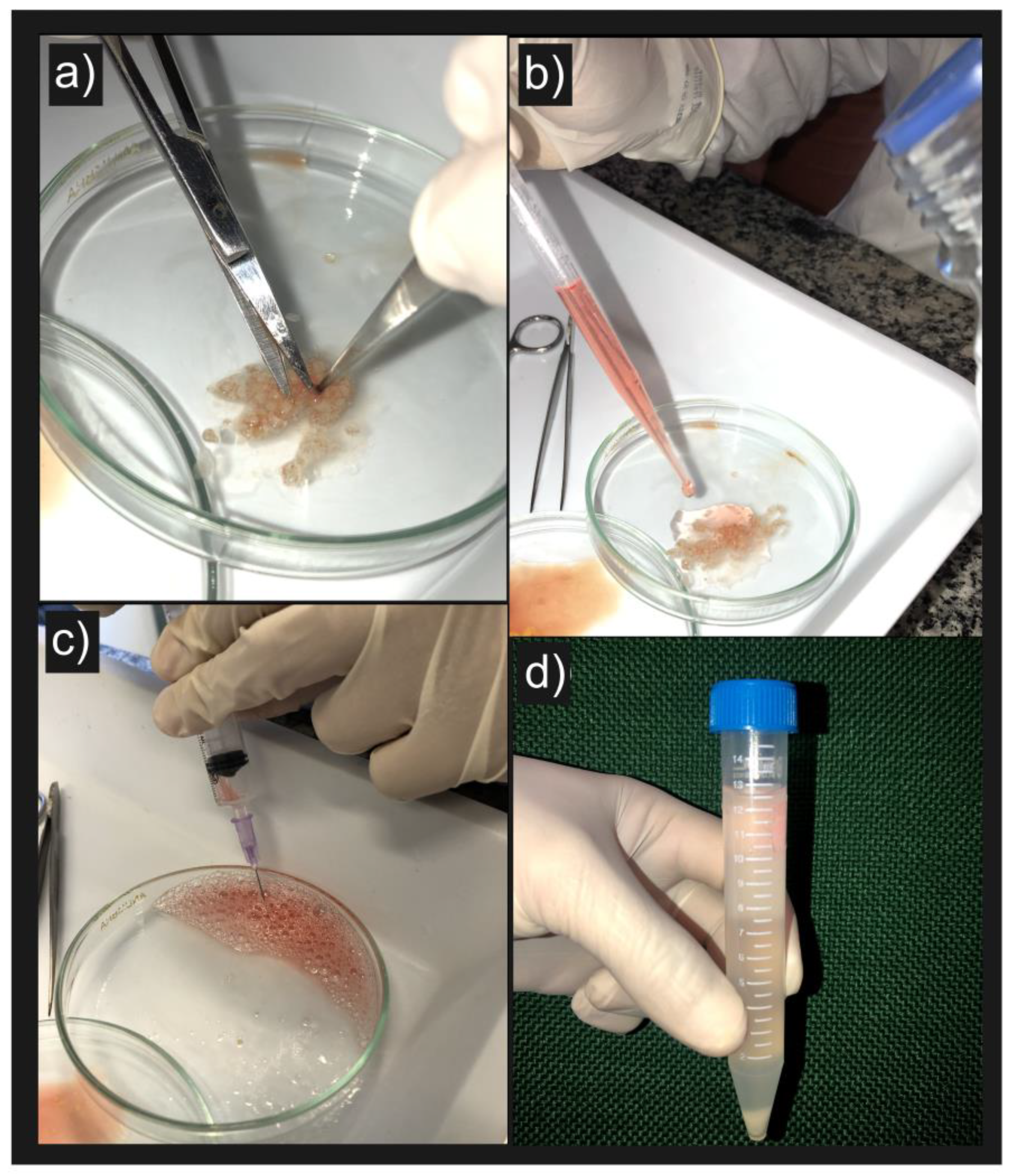
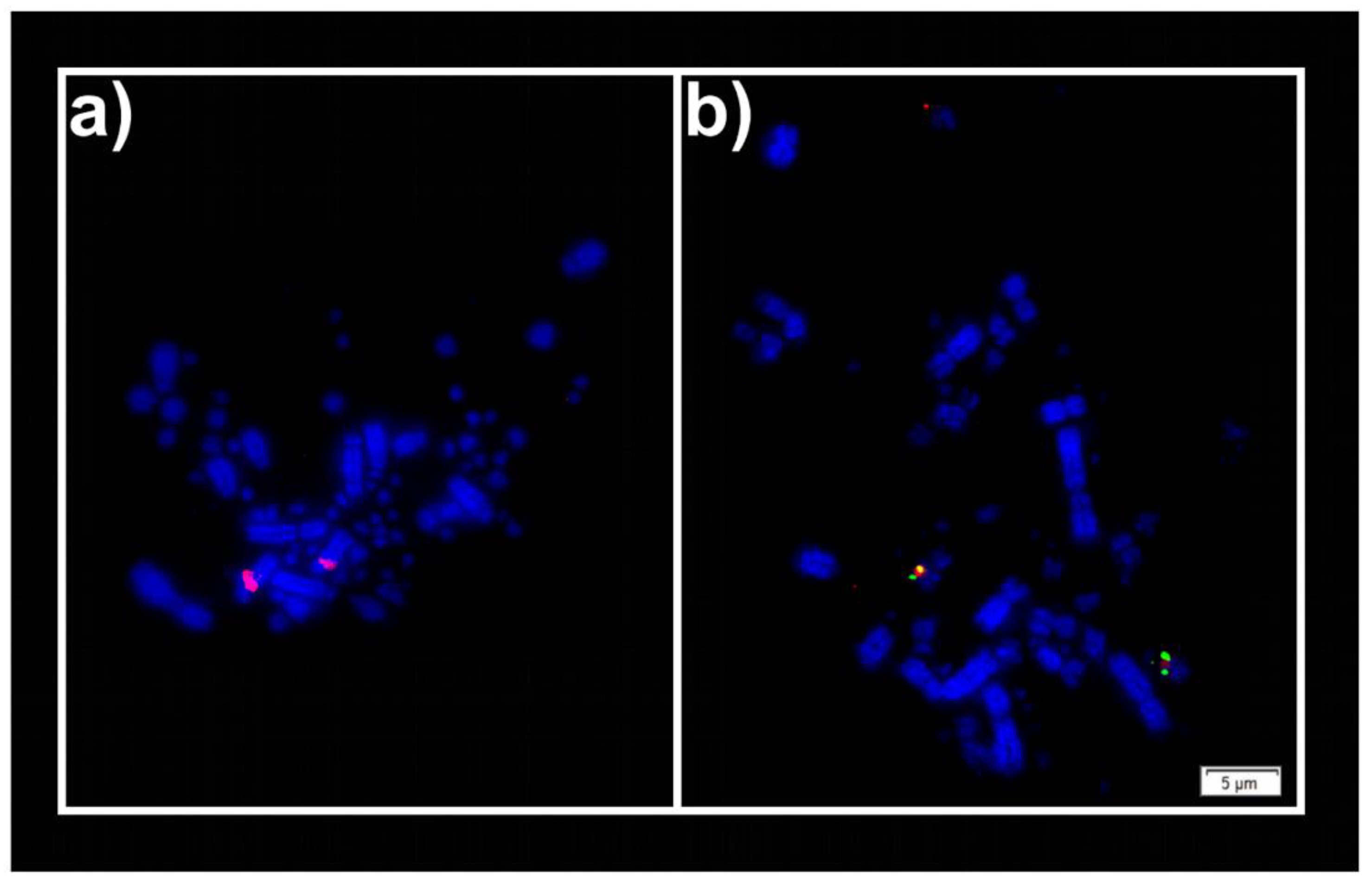
In order to test the effectiveness of FISH analysis in these chromosomal preparations, we chose two sets of probes. Bacterial artificial chromosomes (BACs) from G. gallus microchromosomes (GGA12) and the Z sex chromosome (GGAZ) were respectively applied in the B. striata and T. musculus species, respectively. FISH experiments were performed in accordance with O’Connor et al. [21]. The results were photographed using an epifluorescence microscope (Olympus BX61, Olympus Corporation).

3. Results

Overall, all bird species sampled using this new embryo short-term protocol presented high-quality metaphases. Figure 2 displays three individuals sampled using this protocol. The chromosomal number found in these species was the same as previously cited in the literature, S. caerulecens 2n = 78, E. astrild 2n = 78 and B. striata 2n = 60 [22,23,24,25,26].
Although we did not perform specific statistical analyses of mitotic indexes, they were nonetheless high in all samples and more than good enough for our purposes. Several metaphases were present in each field of view for all samples.
The estimated time for all processes from the embryo disassociation to slide preparation was less than 3 h, with extra time being necessary for staining, FISH and microscopy.
The BAC probes were successfully hybridized, GGA12 in one microchromosome pair of B. striata, as well as GGAZ in the Z chromosome of T. musculus. FISH results are shown in Figure 3.

4. Discussion

The generation of suitable-quality metaphases in a reasonable time frame is the basis for all cytogenetic experiments. In this study, we combined the benefits of bone marrow cultivation techniques with fibroblast culture.
Long-term fibroblast culture from embryonic and non-embryonic cells such as muscle, liver, skin and lungs have been the most commonly applied technique for obtaining bird chromosomes [7,8,27,28,29]. However, these protocols require extensive time cultivation, large quantities of reagents and specific equipment, for instance, a CO2 incubator [9,10].
The colchicinized embryo technique is also a good option for obtaining bird metaphases; however, as with the bone marrow cell culture, it contains residual cytoplasm, which can cause hybridization issues for FISH analysis [19,20,30,31].
This new method presents several benefits when compared to other types of cell cultures used for avian cytogenetics: (1) it is easier, cheaper and quicker than fibroblast cell culture protocols. Most of the previous fibroblast culture methods take more than 3 days, some of them more than 2 weeks, to obtain good quality metaphases [7,8,9,27,28]. By contrast, the method described here takes around 3 h in total; (2) one bird embryo provides numerous metaphases of high quality; (3) the method only requires a few items of equipment and reagents; (4) it has a low contamination rate. For fibroblast cultures, one of the major challenges is to avoid the contamination that can cause the loss of the samples. In contrast, our protocol can cope with this issue due to the short amount of cultivation time. A comparison table (Table 1) shows the advantages and disadvantages of the most common cell culture techniques in more detail.
Bone marrow or fibroblast cultures that use the organs of adult individuals need to sacrifice the bird, which can be problematic if these animals were in the reproductive season [8,9,10,11,12,13,14,15,16]. Considering that in more than 80% of all bird species, both parents provide care, if one of the parents were to be sacrificed, its progeny would probably die [29,32]. Furthermore, there is no guarantee that the offspring would survive until adulthood due to nest predation and all of the difficulties in the maturation process [33,34,35]. Despite the high energy expenditure during reproduction, most females are still able to re-lay eggs in the same season in case the first attempt did not succeed [23,35,36,37]. Thus, this is another advantage of the bird embryo short-term culture protocol, as it tries to mitigate the environmental impact. Furthermore, the metaphases present a better quality than those obtained via short-term bone marrow culture [18].
Avian cytogenetics is concerned with genome reconstruction and comparative genomics [6]. Molecular cytogenetics has to keep pace with the number of species that are being sequenced, and rapid methods for chromosome preparation such as the one proposed are increasingly essential [38,39]. High-quality preparations with longer chromosomes allow for predicted chromosome fragments (PCFs) to be mapped more accurately, and cleaner preparations mean that cross-species hybridizations are more likely to work.
To this end, the present study provided a new method for cell culture that can be widely applied for cytogenetic studies in bird species.

Author Contributions

Conceptualization, S.A.B.; methodology, S.A.B., M.S.d.S. and R.K.; software, S.A.B.; validation, S.A.B., R.J.G. and A.D.V.G.; formal analysis, S.A.B. and R.J.G.; investigation, S.A.B. and M.S.d.S.; resources, S.A.B., R.J.G. and A.D.V.G.; data curation S.A.B.; writing—original draft preparation, S.A.B.; writing—review and editing, S.A.B., M.S.d.S., V.T., L.R.P., R.K., R.J.G. and A.D.V.G.; visualization, S.A.B. and M.S.d.S.; supervision, R.J.G. and A.D.V.G.; project administration, R.J.G. and A.D.V.G.; funding acquisition, S.A.B., R.J.G. and A.D.V.G. All authors have read and agreed to the published version of the manuscript.

Funding

This study was financed in part by the Coordenação de Aperfeiçoamento de Pessoal de Nível Superior—Brazil (CAPES)—Finance Code 001.

Institutional Review Board Statement

The study was conducted according to the guidelines of the Brazilian Institute of Environment and Renewable Natural Resources (IBAMA/SISBIO 61047-1) and approved by the Ethics Committee on the use of animals of Universidade Federal do Pampa (CEUA 019/2020, 30 November 2020).

Informed Consent Statement

Not applicable.

Data Availability Statement

The data presented in this study are available in the article.

Acknowledgments

The authors would like to thank all members of the “Grupo de Pesquisa Diversidade Genética Animal” from UNIPAMPA and Darren K. Griffin, who kindly provided the BAC probes.

Conflicts of Interest

The authors declare no conflict of interest.

References

  1. Degrandi, T.M.; Barcellos, S.A.; Costa, A.L.; Garnero, A.D.V.; Hass, I.; Gunski, R.J. Introducing the Bird Chromosome Database: An Overview of cytogenetic studies in birds. Cytogenet. Genome Res. 2020, 160, 199–205. [Google Scholar] [CrossRef] [PubMed]
  2. Schartl, M.; Schmid, M.; Nanda, I. Dynamics of vertebrate sex chromosome evolution: From equal size to giants and dwarfs. Chromosoma 2016, 125, 553–571. [Google Scholar] [CrossRef] [PubMed]
  3. Schmid, M.; Nanda, I.; Guttenbach, M.; Steinlein, C.; Hoehn, M.; Schartl, M.; Haaf, T.; Weigend, S.; Fries, R.; Buerstedde, J.M.; et al. First report on chicken genes and chromosomes 2000. Cytogenet. Genome Res. 2000, 90, 169–218. [Google Scholar] [CrossRef] [PubMed]
  4. Romero, L.M.; Romero, R.C. Corticosterone responses in wild birds: The importance of rapid initial sampling. Condor 2002, 104, 129–135. [Google Scholar] [CrossRef]
  5. Brown, M.B.; Brown, C.R. Blood sampling reduces annual survival in cliff swallows (Petrochelidon pyrrhonota). Auk 2009, 126, 853–861. [Google Scholar] [CrossRef]
  6. Kretschmer, R.; Ferguson-Smith, M.A.; de Oliveira, E.H.C. Karyotype evolution in birds: From conventional staining to chromosome painting. Genes 2018, 9, 181. [Google Scholar] [CrossRef] [Green Version]
  7. Habermann, F.A.; Cremer, M.; Walter, J.; Kreth, G.; von Hase, J.; Bauer, K.; Wienberg, J.; Cremer, C.; Cremer, T.; Solovei, I. Arrangements of macro-and microchromosomes in chicken cells. Chromosome Res. 2001, 9, 569–584. [Google Scholar] [CrossRef]
  8. Nanda, I.; Schrama, D.; Feichtinger, W.; Haaf, T.; Schartl, M.; Schmid, M. Distribution of telomeric (TTAGGG) in sequences in avian chromosomes. Chromosoma 2002, 111, 215–227. [Google Scholar] [CrossRef]
  9. Itoh, Y.; Arnold, A.P. Chromosomal polymorphism and comparative painting analysis in the zebra finch. Chromosome Res. 2005, 13, 47–56. [Google Scholar] [CrossRef]
  10. Tsuda, Y.; Nishida-Umehara, C.; Ishijima, J.; Yamada, K.; Matsuda, Y. Comparison of the Z and W sex chromosomal architectures in elegant crested tinamou (Eudromia elegans) and ostrich (Struthio camelus) and the process of sex chromosome differentiation in palaeognathous birds. Chromosoma 2007, 116, 159–173. [Google Scholar] [CrossRef] [Green Version]
  11. Degrandi, T.M.; de Furo, I.O.; de Oliveira, E.H.C.; Costa, A.L.; Ferguson-Smith, M.A.; O’Brien, P.C.M.; Pereira, J.C.; Garnero, A.D.V.; Gunski, R.J.; Artoni, R.F. Comparative chromosome painting in hummingbirds (Trochilidae). Genet. Mol. Biol. 2020, 43, e20200162. [Google Scholar] [CrossRef] [PubMed]
  12. Rodrigues, B.S.; Kretschmer, R.; Gunski, R.J.; Garnero, A.D.V.; O’Brien, P.C.M.; Ferguson-Smith, M.; de Oliveira, E.H.C. Chromosome painting in Tyrant Flycatchers confirms a set of inversions shared by Oscines and Suboscines (Aves, Passeriformes). Cytogenet. Genome Res. 2017, 153, 205–212. [Google Scholar] [CrossRef] [PubMed]
  13. Pigozzi, M.I.; Solari, A.J. Germ cell restriction and regular transmission of an accessory chromosome that mimics a sex body in the zebra finch, Taeniopygia guttata. Chromosome Res. 1998, 6, 105–113. [Google Scholar] [CrossRef] [PubMed]
  14. Degrandi, T.M.; Gunski, R.J.; Garnero, A.D.V.; de Oliveira, E.H.C.; Kretschmer, R.; Santos, M.S.; Barcellos, S.A.; Hass, I. The distribution of 45S rDNA sites in bird chromosomes suggests multiple evolutionary histories. Genet. Mol. Biol. 2020, 43, e20180331. [Google Scholar] [CrossRef]
  15. Poignet, M.; Pokorná, M.J.; Altmanová, M.; Majtánová, Z.; Dedukh, D.; Albrecht, T.; Reif, J.; Osiejuk, T.S.; Reifová, R. Comparison of karyotypes in two hybridizing passerine species: Conserved chromosomal structure but divergence in centromeric repeats. Front. Genet. 2021, 12, 768987. [Google Scholar] [CrossRef]
  16. Pinheiro, M.L.S.; Nagamachi, C.Y.; Ribas, T.F.A.; Diniz, C.G.; O’Brien, P.C.M.; Ferguson-Smith, M.; Yang, F.; Pieczarka, J.C. Chromosomal painting of the sandpiper (Actitis macularius) detects several fissions for the Scolopacidae family (Charadriiformes). BMC Ecol. Evol. 2021, 21, 8. [Google Scholar] [CrossRef]
  17. Sasaki, M.; Ikeuchi, T.; Makino, S. A feather pulp culture technique for avian chromosomes, with notes on the chromosomes of the peafowl and the ostrich. Experientia 1968, 24, 1292–1293. [Google Scholar] [CrossRef]
  18. Garnero, A.D.V.; Gunski, R.J. Comparative analysis of the karyotype of Nothura maculosa and Rhynchotus rufescens (Aves: Tinamidae). A case of chromosomal polymorphism. Nucleus 2000, 43, 64–70. [Google Scholar]
  19. Bloom, S.E.; Buss, E.G. Triploid-diploid mosaic chicken embryo. Science 1966, 153, 759–760. [Google Scholar] [CrossRef]
  20. Bloom, S.E. Chromosome abnormalities in early chicken (Gallus domesticus) embryos. Chromosome 1969, 28, 357–369. [Google Scholar] [CrossRef]
  21. O’Connor, R.E.; Kiazim, L.; Skinner, B.; Fonseka, G.; Joseph, S.; Jennings, R.; Larkin, D.M.; Griffin, D.K. Patterns of microchromosome organization remain highly conserved throughout avian evolution. Chromosoma 2019, 128, 21–29. [Google Scholar] [CrossRef] [PubMed] [Green Version]
  22. Christidis, L. Chromosomal evolution within the family Estrildidae (Aves) III. The Estrildae (waxbill finches). Genetica 1987, 72, 93–100. [Google Scholar] [CrossRef]
  23. De Lucca, E.J. Cariótipo de oito espécies de Aves. Rev. Bras. Biol. 1974, 34, 387–392. (In Portuguese) [Google Scholar] [PubMed]
  24. Carvalho, M.W. Estudos Citogenéticos na Família Fringillidae (Passeriformes: Aves). Master’s Thesis, Universidade Federal de Rio Grande do Sul, Porto Alegre, Brazil, 1989. (In Portuguese). [Google Scholar]
  25. Christidis, L. Animal Cytogenetics 4: Chordata 3 B: Aves; Gebrüder Borntraeger: Berlin, Germany, 1990; pp. 20–59. [Google Scholar]
  26. Vielmo, P.G. Bandeamentos Cromossômicos em Butorides Striata (Aves: Pelecaniformes) Revelam um Padrão Distinto Para o Cromossomo W; Universidade Federal do Pampa: São Gabriel, Brazil, 2019. (In Portuguese) [Google Scholar]
  27. Griffin, D.K.; Haberman, F.; Masabanda, J.; O’Brien, P.; Bagga, M.; Sazanov, A.; Smith, J.; Burt, D.W.; Ferguson-Smith, M.; Wienberg, J. Micro- and macrochromosome paints generated by flow cytometry and microdissection: Tools for mapping the chicken genome. Cytogenet. Genome Res. 1999, 87, 278–281. [Google Scholar] [CrossRef] [PubMed]
  28. Ahlroth, M.K.; Kola, E.H.; Ewald, D.; Masabanda, J.; Sazanov, A.; Fries, R.; Kulomaa, M.S. Characterization and chromosomal localization of the chicken avidin gene family. Anim. Genet. 2000, 31, 367–375. [Google Scholar] [CrossRef] [PubMed]
  29. Cockburn, A. Prevalence of different modes of parental care in birds. Proc. R. Soc. B 2006, 273, 1375–1383. [Google Scholar] [CrossRef] [PubMed] [Green Version]
  30. Bloom, S.E. Haploid chicken embryos: Evidence for diploid and triploid cell populations. Heredity 1970, 61, 147–150. [Google Scholar] [CrossRef]
  31. Bloom, S.E. Chromosome abnormalities in the chicken (Gallus domesticus) embryos: Types, frequencies and phenotypic effects. Chromosome 1972, 37, 309–326. [Google Scholar] [CrossRef] [PubMed]
  32. Goymann, W.; Safari, I.; Muck, C.; Schwabl, I. Sex roles, parental care and offspring growth in two contrasting coucal species. R. Soc. Open Sci. 2016, 167, 413–425. [Google Scholar] [CrossRef] [Green Version]
  33. Martin, T.E. Avian life history evolution in relation to nest sites, nest predation and food. Ecol. Monogr. 1995, 65, 101–127. [Google Scholar] [CrossRef]
  34. Cox, W.A.; Thompson, R.F.; Cox, A.S.; Faaborg, J. Post fledging survival in passerine birds and the value of post fledging studies to conservation. J. Wildl. Manag. 2014, 78, 183–193. [Google Scholar] [CrossRef]
  35. Ibáñez-Álamo, J.D.; Magrath, R.D.; Oteyza, J.C.; Chalfoun, A.D.; Haff, T.M.; Schmidt, K.A.; Thomson, R.L.; Martin, T.E. Nest predation research: Recent findings and future perspectives. J. Ornithol. 2015, 156, 247–262. [Google Scholar] [CrossRef] [Green Version]
  36. Burger, L.W.; Ryan, M.R.; Dailey, T.V.; Kurzejeski, E.W. Reproductive strategies, success, and mating systems of northern bobwhite in Missouri. J. Wildl. Manag. 1995, 59, 417–426. [Google Scholar] [CrossRef]
  37. Thompson, B.C.; Knadle, G.E.; Brubaker, D.L.; Brubaker, K.S. Nest Success Is Not an Adequate Comparative Estimate of Avian Reproduction (El Éxito al Anidar no es un Estimado Comparativo Adecuado de la Reproduccion en Aves). J. Field Ornithol. 2001, 72, 527–536. [Google Scholar] [CrossRef]
  38. Feng, S.; Stiller, J.; Deng, Y.; Armstrong, J.; Fang, Q.; Reeve, A.H.; Xie, D.; Chen, G.; Guo, C.; Faircloth, B.C.; et al. Dense sampling of bird diversity increases power of comparative genomics. Nature 2020, 587, 252–257. [Google Scholar] [CrossRef] [PubMed]
  39. Rhie, A.; McCarthy, S.A.; Fedrigo, O.; Damas, J.; Formenti, G.; Koren, S.; Uliano-Silva, M.; Chow, W.; Fungtammasan, A.; Kim, J.; et al. Towards complete and error-free genome assemblies of all vertebrate species. Nature 2021, 592, 737–746. [Google Scholar] [CrossRef] [PubMed]
Figure 1. Some of the steps in preparing embryos for short-term culture. The embryo was macerated using sterile scissors and tweezers in a sterile Petri dish (a); 2 mL of trypsin 0.25% EDTA was transferred into the Petri dish using a sterile pipette (b); the embryo and the solution were mixed by using a 5 mL syringe with needle (c); after incubation and washes, the sample was ready to be stored (d).
Dna 02 00002 g001
Figure 2. Conventional staining with Giemsa 5%. Sporophila caerulescens (a); Estrilda astrild (b); Butorides striata (c). Bar = 5 μm.
Dna 02 00002 g002
Figure 3. Fluorescent in situ hybridization analysis using BAC probes. (a) GGAZ (TGMCBA-270I9 Texas red) in Troglodytes musculus. (b) GGA12 (CH261-60P3 Texas red and CH261-4M5 Fluorescein isothiocyanate) in B. striata. Bar = 5 μm.
Dna 02 00002 g003
Table 1. Comparison table between cell culture methods for bird cytogenetics.
TechniquesAdvantagesDisadvantages
Colchicinized embryo technique developed by Bloom and Buss [19]
  • Good-quality chromosomes
  • Short-term cultivation (approximately 2 h)
  • Only requires a few reagents
  • Accumulation of residual content in the metaphases
Fibroblast cell culture developed by Sasaki, Ikeuchi and Makino [17]
  • High-quality chromosomes
  • Metaphases free of cytoplasm
  • Long-term cultivation (weeks)
  • Requires many items of equipment and reagents
  • Risk of contamination
Colchicinized embryo technique developed by Bloom [20]
  • Good-quality chromosomes
  • Short-term cultivation (approximately 1 h)
  • Only requires a few reagents
  • Accumulation of residual content in the metaphases
  • Limited number of metaphases per embryo due to the absence of passages
Bone marrow developed by Garnero and Gunski [18]
  • Short-term cultivation (approximately 1 h)
  • Only requires a few reagents
  • Accumulation of residual content in the metaphases
  • Condensed chromosomes
  • Limited number of metaphases per individual due to the absence of passages
Fibroblast (skin and liver) developed by Itoh and Arnold [9]
  • High-quality chromosomes
  • Possibility of increasing the number of metaphases using the passages
  • Long-term cultivation (one week)
  • Requires many items of equipment and reagents
  • Risk of contamination
Cell culture developed by Tsuda and Umehara [10]
  • High-quality chromosomes
  • Possibility of increasing the number of metaphases using the passages
  • Long-term cultivation (more than 48 h)
  • Requires many items of equipment and reagents
  • Risk of contamination
Method described in this paper
  • Short-term cultivation (1 h)
  • Only requires a few reagents and items of equipment
  • High-quality chromosomes
  • Contamination issues do not affect the quality of the culture
  • Limited number of metaphases per embryo due to the absence of passages
Publisher’s Note: MDPI stays neutral with regard to jurisdictional claims in published maps and institutional affiliations.

