Entry

SARS-CoV-2 Associated Pulmonary Pathology

1
Department of General and Clinical Pathology, Forensic Medicine and Deontology, Faculty of Medicine, Medical University—Varna “Prof. Dr. Paraskev Stoyanov”, 9002 Varna, Bulgaria
2
Department of Anatomy and Cell Biology, Faculty of Medicine, Medical University—Varna “Prof. Dr. Paraskev Stoyanov”, 9002 Varna, Bulgaria
*
Author to whom correspondence should be addressed.
Encyclopedia 2022, 2(4), 1698-1709; https://doi.org/10.3390/encyclopedia2040116
Submission received: 30 July 2022 / Revised: 14 September 2022 / Accepted: 22 September 2022 / Published: 30 September 2022
(This article belongs to the Collection Encyclopedia of COVID-19)

Definition

:
Severe acute respiratory syndrome coronavirus 2 (SARS-CoV-2) is a novel entry in the betacoronaviridae group of coronaviruses. This is the second member of this group, and the third of the family overall to emerge in the last 20 years, which has caused significant health concerns due to the clinical severity and spread of the disease it causes—coronavirus disease identified in 2019 (COVID-19). While initially emerging as a respiratory disease, and while most cases experience symptoms predominantly from this system, SARS-CoV-2 has emerged as a multisystem pathogen. From a pathomorphological point of view, the severity of changes in the respiratory system can be summed up as diffuse alveolar damage—desquamation of the alveolar epithelium with exudative and proliferative changes—pulmonary hyaline membranes, Clara cell hyperplasia, squamous cell metaplasia, and fibrosis. The second most prominent way the disease affects the lung is through endotheliitis—damage to the endothelial cells of the pulmonary vasculature, predominantly affecting the medium and large caliber blood vessels that cause the well-established clinical phenomenon of thrombosis/thromboembolism of the pulmonary vasculature. As the spread of the disease continues with the emergence of new variants and the number of cases continues to grow, including a large percentage of recurrent cases, it is essential to remember that the viral effects are not only acute but, due to the proliferative phenomena, can produce chronic sequelae. Therefore, in the background of dwindling publication interest, it is critical to focus on the histopathological aspects of the pulmonary disease, with the goal of better understanding the effects of the virus on the organism and identifying probable future complications after infection.

1. Introduction

The novel representative of coronaviruses (SARS-CoV-2) and the clinical disease it causes (COVID-19), which emerged at the end of 2019, have led to healthcare and social consequences not seen in the last 100 years [1,2,3,4,5,6]. The rapid spread of the infection, the severe organ damage to multiple systems, and the lack of effective prevention forced the World Health Organization (WHO) to officially declare a pandemic of the disease [7,8,9]. Given the new nature of the infectious agent and the many unknowns surrounding it, the WHO implemented protocols developed during the most closely related infectious agents—MERS and SARS-CoV, namely, social distance, personal protective equipment, quarantine of healthy disease carriers and contacts of the sick, and strict monitoring of the epidemiological process [10,11,12]. The severe health challenges including the unknowns surrounding the course of the disease and the epidemiological process, the large number of healthy infection spreaders, the severe clinical course with high and sometimes sudden mortality in some patients, the lack of healthcare personnel, and effective prevention and misinformation (at the beginning of the pandemic due to lack of scientifically proven facts, and, in the later stages, for non-medical reasons) led to the mobilization of medical science in the study of SARS-CoV-2 not only towards the origin of the virus and the study of its clinical consequences and morphology, but also to the rapid development of effective vaccine prophylaxis, largely possible based on the experience gained in the previous two severe coronavirus diseases [9,13,14,15,16].
With the abatement of the pandemic in 2022 and given the mutational variants of the disease leading to a significantly milder clinical course and a decrease in overall mortality, as well as due to the availability of widespread effective vaccine prevention, not only the public but also the publication interest in the subject has gradually subsided [17,18,19]. The question remains open not only about the origin of the virus and, to a large extent, the characteristics regarding organ involvement, but above all about the chronic consequences of the illness, which the medical community has yet to face [20,21,22,23,24,25,26,27].
Historically, this is not the first time that medicine has encountered a similar situation; numerous pandemics, not only of viral origin but also of bacterial origin, have led to the permanent disability of those who have suffered, and subsequently to the development of new and, at first glance, unexpected long-term consequences [28]. A relatively recent example is the HIV-AIDS infection, which, in addition to immune deficiency, has been proven to lead to many severe consequences, including neoplasia and dementia [29,30,31,32]. In the history of medicine, there are many more such examples, such as von Economo’s disease (lethargic encephalitis), often confused as a consequence of the Spanish flu; although, the epidemic process began several years before it and led to postencephalitic parkinsonism [33,34,35].
It is for these reasons that it is necessary to retain the interest of medical science in the matter of the detailed study of the acute and chronic effects of the virus that are yet to be encountered and that can be used in combating future pandemic variants of coronavirus infections [36,37,38,39].
Autopsies of patients with COVID-19 have revealed a myriad of gross and especially histological changes in multiple internal organs, namely, the lungs, where the changes lead to the dominating clinical symptom of hypoxia, but also the liver, heart, and other organs [40,41,42,43,44]. It is important, however, that autopsies reveal only the most severe effects of the disease in patients who had either a severe protracted clinical course or those who expired suddenly. It is highly likely that in cases with a moderate clinical course, either a significantly less diffuse and developed set of complications is present or it did not manifest at all.
Furthermore, it is likely that in patients with mild clinical disease, a whole new spectrum of changes can be present, with all of these further developing and evolving into chronic changes in the spectrum of post-COVID syndrome. Post-COVID syndrome, or long COVID or persistent COVID, are a direct result of the persistent damage caused by the viral infection. While mainly focusing on the diverse pulmonary pathology, which leads to chronic complications, this spectrum also includes sequelae from other systems and organs, such as the kidneys, cardiovascular, gastrointestinal, and especially the central nervous system [21,45,46,47,48].

Safety Precautions during Autopsy—Determining the Hazard Group
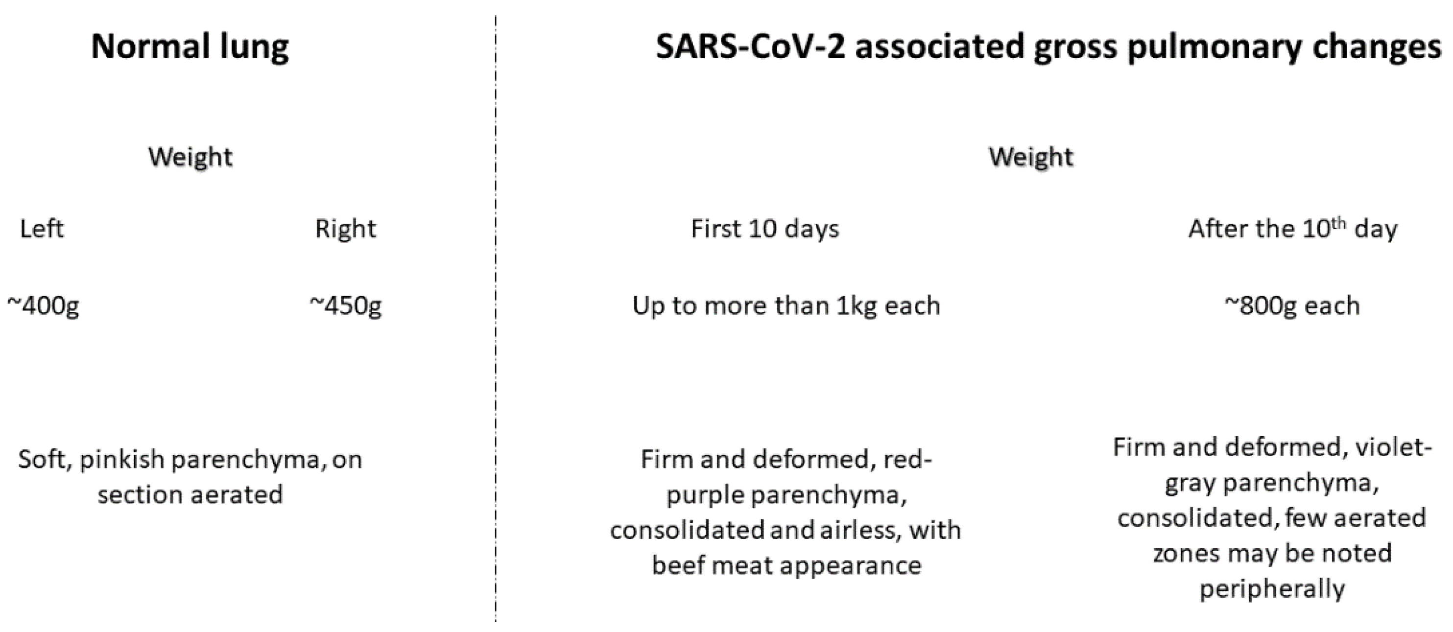
As the causative agent of the disease has been identified by the WHO as a pandemic entity, special precautions must be taken during the autopsy of suspected, probable, and identified cases [49]. Furthermore, as a general precautionary rule in the presence of diffusely spreading infectious pandemic entities, safety precautions should be taken in all cases, even if the autopsy case is not suspected or even has been ruled out to be a carrier of the disease.
For autopsy practice, especially for infectious disease entries, cases are separated into hazard groups (HG) based on the ability of the pathogens to infect new hosts, the severity of the disease it causes, and the presence of effective prevention, treatment, or the lack thereof [50]. HG1 refers to pathogens that are unlikely to cause human disease, cause minimal illness, or there is effective prevention and treatment; HG2 refers to pathogens that can lead to infection in the personnel but are unlikely to spread to the general population, and there is effective prevention and treatment; HG3 refers to pathogens that cause severe disease to the personnel and can spread to the community, but there is effective prevention and treatment; and HG4 refers to pathogens that cause severe diseases, can spread in the general population, and there is no effective prevention and treatment. It is essential to know that in many regions, based on their legislation, HG4 autopsies are counter-indicated, while in others, HG4 is mandated as it is viewed as a threat to national and regional security [49,51,52,53,54].
During the initial phases of the pandemic, there was a broad discussion regarding which HG COVID-19 should be placed in, with initial decisions putting it in HG4, and, hence, the number of autopsy cases was very low [49,51,55]. As data started to accumulate and there was a relatively low incidence of periautopsy infection, it was gradually moved down to HG3 and, in some areas after the introduction of vaccinations, even to HG2. Based, however, on the significant mutation frequency of the virus and the evasion of acquired immunity by the new variants, in the future, it would be sound to keep SARS-CoV-2 in HG3 to prevent the spreading of potentially deadly variants of the virus to healthcare personnel and the population in general [56].

2. Gross Findings

The respiratory system in patients who have died from COVID-19 shows a myriad of gross changes. The mucous membranes of the trachea and bronchi are moderate to severely erythematous, most prominently in the interchondral areas, and often have scattered mucosal hemorrhages and ulcerations (Figure 1) [57]. In the thoracic cavity, a large subset of patients had pleural effusions, varying from serous to hemorrhagic [41]. The characteristics of the pleural surface correspond to the presence and nature of pleural effusions, ranging from preserved pleural surfaces to edematous ones, diffusely scattered fibrin deposits, or hemorrhagic areas [58]. However, the lungs have the most striking changes to them. They are significantly enlarged, weighing more than a kilogram (i.e., more than twice their average weight), and can reach nearly two kilograms in the most severe cases. The parenchyma is firm to the touch, red-violet, and often deformed (Figure 1) [55,59,60,61]. On sectioning, the cut surface is diffusely consolidated, with a scant amount of mucohemmorhagic fluid flowing from it spontaneously when the parenchyma is compressed [60,61]. In cases with extremely severe parenchymal involvement, hepatization results in the bulging of the large and medium-sized blood vessels, giving the parenchyma a beef meat appearance (Figure 1). A large subset of patients also have a positive gross probe for pulmonary thromboembolism, and the vessel section reveals thrombi in smaller branches of the vessels, some in different stages of organization and/or areas of hemorrhagic infarction [44,62,63].
In patients with a more protracted clinical course (survival more than ten days from the onset of symptoms), the lungs are relatively lighter, regularly around 800–900 g, and grossly have a more grayish-red-violet color; they are firmer to palpation, deformed, and some peripheral areas may have reaeration resembling emphysematous deformation (Figure 1) [60]. When a secondary bacterial infection is superimposed, areas of suppuration and/or abscessation are visible in the parenchyma, and the bronchi are filled with purulent-hemorrhagic exudate [44,59,63]. In some patients, a fungal superinfection may be present, with aspergillus and mucormycosis being the most commonly isolated causative agents [64,65].
In chronically and severely ill patients and the elderly, the lungs may have significantly fewer changes, varying from focal and mild consolidation to nearly normal-looking lungs, with some cases showing only histological changes [58,59,60].
Histology largely depends on the innate resilience of the organism and the virulence and pathogenicity of the viral variant. In most cases, however, there are at least minimal signs of diffuse alveolar damage correlating to the clinical constellation of acute respiratory distress syndrome [44,55,59].

Practical Tips for Obtaining Respiratory System Specimens

In our practice, the authors have implemented a harvesting protocol for respiratory system specimens, intending to identify, even if minimally present, the changes induced by the virus on histology. When obtaining samples from the respiratory system, the authors open the ribcage with bone cutters, which is an important safety tip, as if opened with an electrical saw, as in some institutions, the vibrations produced can mobilize vital viral particles and spread them in the air, increasing the risk for the personnel present. Secondly, the respiratory system is sectioned only after in situ probing of the pulmonary artery for the presence of large thrombi/emboli. Once the respiratory system is eviscerated, the authors take at least three tissue specimens from every pulmonary lobe—one hilar, one central, and one peripheral, with other areas harvested as desired by the obductor. Furthermore, the authors take specimens from the trachea, bronchial bifurcation, and principal bronchi.

3. Histological Findings

The histological findings are largely non-specific, as similar changes can be observed in MERS, SARS-CoV, respiratory syncytial virus, pandemic variants of influenza and others, and some opportunistic viral infections to the lung and respiratory system [66,67,68,69,70,71].

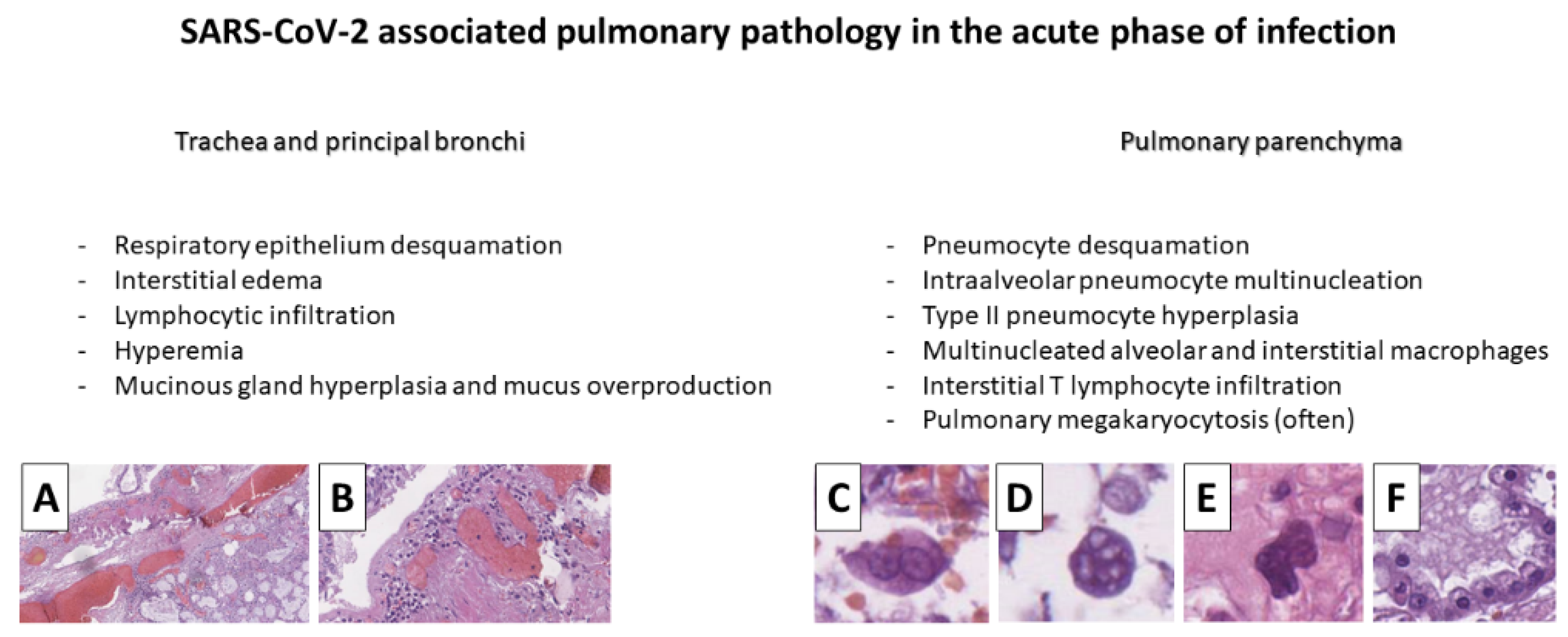
3.1. Trachea and Large Bronchi

The trachea and large bronchi in patients with COVID-19 generally have histologic findings of extensive desquamation of the respiratory epithelium, basal cell hyperplasia, scant interstitial infiltration by lymphocytes, and goblet cell hyperplasia in the submucosal glands (Figure 2) [57].

3.2. Pulmonary Histopathology

As already mentioned, pulmonary histopathology is non-specific and follows the pattern of diffuse alveolar damage and vascular endothelial damage—its two coexisting forms.
Diffuse alveolar damage consists of severe damage to the respiratory and alveolar epithelium, occurring in two primary forms—an exudative phase (acute), which is observed in patients who die within the first week of the onset of symptoms, and a proliferative (initial and advanced) phase, which is observed in patients with survival of more than ten days from the onset of symptoms [44,59,72]. The initial phase of proliferative diffuse alveolar damage combines the findings of the exudative and advanced proliferative phases [44,60,69].

3.2.1. Acute Phase of the Infection—Diffuse Alveolar Damage

The first histological changes observed in the exudative phase of diffuse alveolar damage (patients who died by the seventh day from the onset of symptoms) are the desquamation of the epithelium of the terminal bronchioles and alveoli, as well as the hyperplasia of type II pneumocytes, the formation of alveolar and interstitial multinucleated cells, scarce interstitial infiltration comprised by predominantly T lymphocytes, intra-alveolar and interstitial macrophage infiltration with multinucleation, and occasionally pulmonary megakaryocytes (Figure 2) [58,60,63]. The alveolar multinucleated cells originating from pneumocytes have characteristically ground glass opacity cytoplasm, peripheral inclusions, up to six nuclei (predominantly, these cells have two or three nuclei) with a central, sometimes ruby-red nucleoli, and a diameter varying between 20 and 40 μm (Figure 2) [60,61]. The second type of multinucleated cells, with macrophage origin, also presents intra-alveolarly, but the cells are mostly interstitially located, smaller (about 20 μm in diameter), primarily trinucleated, but may have up to seven nuclei, and their cytoplasm is more basophilic and positive for CD68 (Figure 2) [59,60,61,73].

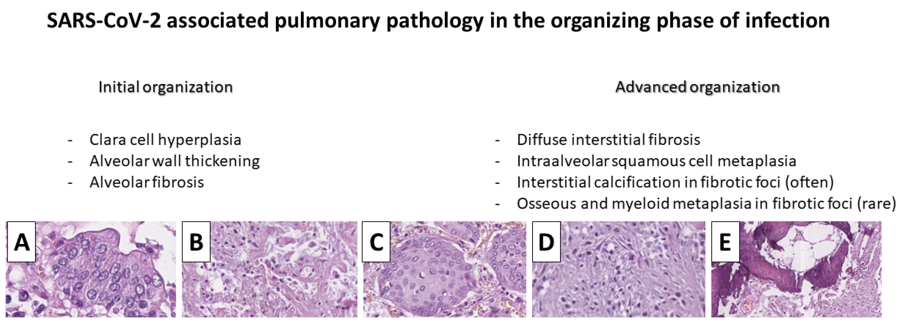
3.2.2. Organization Phase of Diffuse Alveolar Damage

In patients with a survival of about ten days from the onset of symptoms, a transformation of changes with an initial phase of organization is observed. Alveolar macrophages decrease in number, and in the terminal and respiratory bronchioles, there is hyperplasia of Clara-type cells—non-ciliated cells of the bronchioles—with a secretory function (Figure 3) [60]. Other than their secretory function, which plays a significant role in local homeostasis regulation and stress response, these cells also play a role in tissue repair as they serve as a regeneratory “reservoir” but are also susceptible to metaplastic change [74]. Fibrous changes also begin with the thickening of the alveolar walls and focal interstitial fibrosis (Figure 3) [41,44,58,63].
In patients who died 15 days after the onset of symptoms, advanced organization of the lung parenchyma can be noted, with pronounced fibrosis—diffuse interstitial, thickening of the alveolar septa to a bronchiolitis obliterans organizing pneumonia pattern with intra-alveolar fibroblastic proliferation and alveolar obliteration [47,60,75].
In this phase, several interesting phenomena can also be observed, the most striking of which is the transformation (metaplasia) of Clara-type cells into squamous epithelium that diffusely fills the alveoli and respiratory tracts without the involvement of the larger bronchi (Figure 3) [60,63]. Secondary to fibrosis, diffuse calcium deposition can also be observed in some cases with further osteoid and myeloid metaplasia (Figure 3) [43,60].

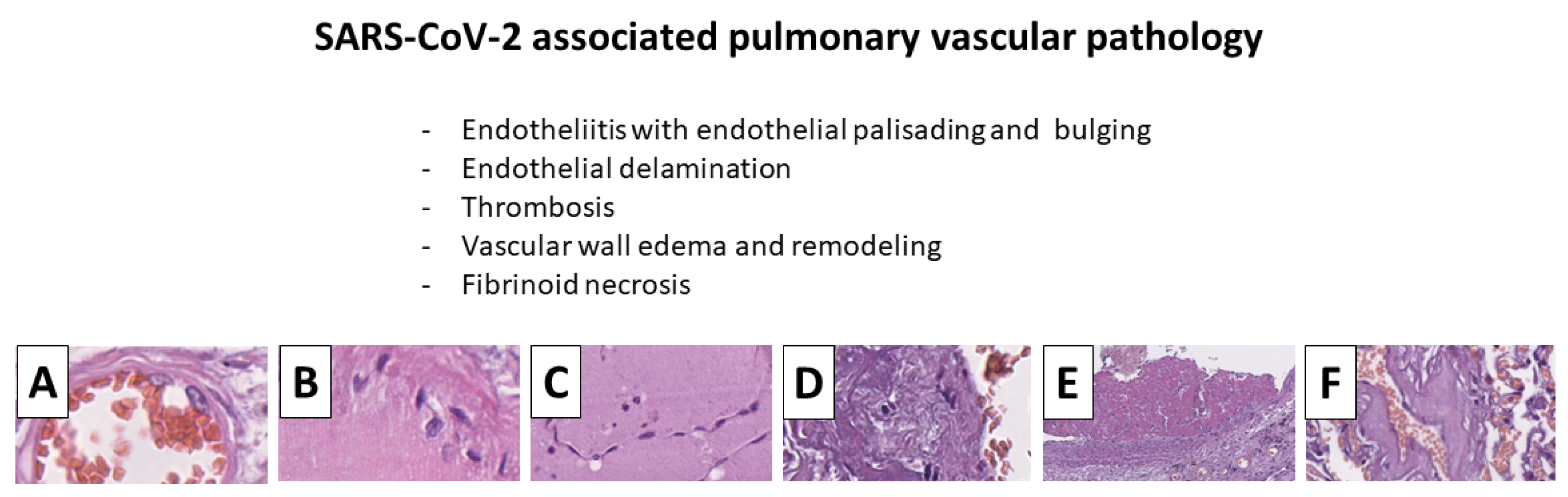
3.3. Vascular Changes

The second most prominent group of changes—those of the pulmonary blood vessels—is more monomorphic and does not undergo such great dynamics. These changes manifest with endotheliitis (endothelialitis)—swelling, palisading, and desquamation of the endothelium of the larger vessels with focal intravascular endothelial proliferation in the regeneration phase (Figure 4) [57,60,62,76,77]. CD34-labeled vessels show extensive areas of endothelial desquamation, with sometimes interconnected endothelial cells freely “hanging” in the lumens of large vessels like a curtain (Figure 4). Interestingly smaller blood vessels, especially capillaries, do not constitute such an endothelial change [60].
These described endothelial changes have two immediate effects—thrombosis, a well clinically manifested phenomenon, and degenerative changes in the vascular wall due to increased permeability [60,62,77,78].
Although pulmonary thrombosis is clinically comparable to thromboembolic disease, given the severe single-vessel involvement and advanced organization in some cases, it is more likely an autochthonous thrombosis of the pulmonary circulation (Figure 4) [44,62,77,78,79].
The changes in the vessel wall as a result of the increased permeability are represented by distortion of the cellular arrangement in the structure of the vascular wall and degeneration of the extracellular matrix—the walls of the vessels are eccentrically thickened, acellular and amorphic, akin to hyalinized, with focal fibrinoid necrosis, lymphocytic infiltrate in the vascular wall, and subintimal foamy macrophages may be observed in some cases (Figure 4) [60].
Special histochemical stains show that these changes develop due to simple extracellular matrix edema, with only focally positive proteinaceous deposits (PAS-positive), while Congo red, Phosphotungstic acid hematoxylin (PTAH) and Van Gieson stains, Mason trichrome, and Alcian blue are negative (Figure 4) [60]. Immunohistochemically, the larger vessels show a distortion of the smooth muscle cell arrangement and focal loss of fibroblasts (Figure 4). It is important to note that the changes described up to this point involve the vessels of mostly medium and small caliber, with the larger vessels having less pronounced changes, while in the microcirculation, these changes are hardly noticeable. Precisely because of this, as already mentioned, fibrin thrombi are present in these branches in most of the cases, while thrombi in the microcirculation are present in a significantly smaller proportion of patients.

3.4. Non-Time Associated Changes

Changes observed in the lung parenchyma, which are not associated with the time since the onset of infection, are again non-specific and associated with diffuse alveolar damage (pulmonary hyaline membranes) and secondary infections [41,58,59,63,72,73,80].

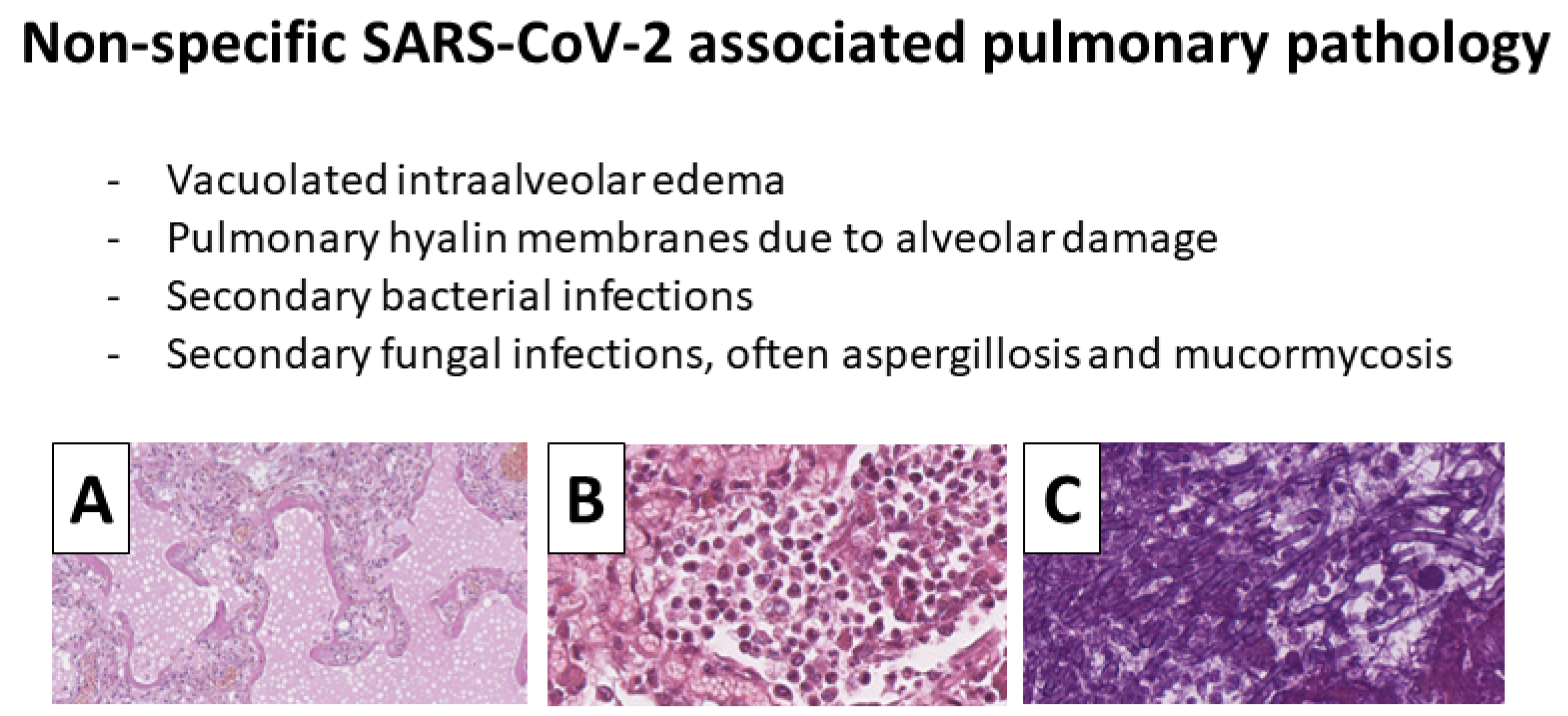
3.4.1. Pulmonary Hyaline Membranes

Pulmonary hyaline membranes (alveolar fibrinous exudate) present with classic morphology; however, the intra-alveolar edema in these areas has a more vacuolated appearance due to increased albumin (Figure 5) [58,60,63]. It is important to note that hyaline membranes are only seen in half of the patients who have expired from COVID-19 [43,57,59,60,63]. These reflect the severe direct alveolar epithelium damage and the dysregulation of alveolar homeostasis. The spectrum of vascular phenomena also plays a big part in it, as the alveolar walls are comprised of capillaries. At the onset of hyaline membranes, the alveoli are no longer able to carry out their gas exchanging function. As such, the condition of these patients deteriorates rapidly with a sudden drop in peripheral oxygen saturation [81,82,83]. Furthermore, as already stated, these changes do not appear in a time-related manner and can be seen in the exudative, early, and late organizational phases of diffuse alveolar damage [59,60]. In patients with more prolonged survival, mainly those on invasive lung ventilation, the hyaline membranes are thick and layered, in some areas resembling intra-alveolar globules.

3.4.2. Secondary Infections

Secondary infections, in the form of suppuration and abscessation in the lung parenchyma, as a time-independent complication, can develop in around one-third of patients, regardless of antibiotic prophylaxis, and can be not only bacterial but also fungal, with aspergillus and mucormycosis being the most prevalently isolated pathogens from this group (Figure 5) [64,65].

3.5. Practical Tips for Reporting Histopathology in Autopsy Protocols

If the patient tests positive antemortem, then COVID-19, as a socially significant disease, should be noted as the main/principal disease that unlocked the mechanisms of tanatogenesis [50]. In cases where the patient has multiple severe conditions, such as malignancy, other infectious diseases (e.g., tuberculosis), or immune suppression, all of these should be mentioned together as primary diseases, while COPD, CVDs, etc., should be specified as background diseases [50].
In cases where the patient was not tested antemortem or tested negative, but the clinical symptoms and morphology are compatible with COVID-19, then instead of COVID-19 being recognized as the primary disease, the main illness should be specified as an acute viral infection and its components listed, such as diffuse alveolar damage in organization phase with alveolar and interstitial multinucleated cells, scant lymphocytic interstitial infiltration, Clara cell hyperplasia, squamous cell metaplasia, fibrosis, and medium caliber pulmonary vessel degeneration and thrombosis. While many institutions allow for postmortem PCR tests and recognize them as valid, hence, for postmortem COVID-19 diagnosis, it is important to note that all commercially available tests have been validated for use only on the living, and none for postmortem evaluation. Immunohistochemistry on neutral formaldehyde-fixated tissue specimens, although a validated method for postmortem evaluation, should also be noted as not always being reliable depending on the antibody used, i.e., if the antibody targets a frequently mutated part of the antigenic structure of the virus, it may not detect a positive case. At the same time, cross-reactivity with other members of the coronaviridae family should not be underestimated, such as SARS-CoV-2, and MERS, which present with virtually identical morphology, as well as different non-pandemic variants of coronaviruses such as those causing seasonal common cold [44,51,68,73,84].

4. Conclusions

  • The pulmonary pathology induced by SARS-CoV-2 follows a relatively strict pattern of evolution with:
    Diffuse alveolar damage—desquamation of the alveolar epithelium with exudative and proliferative changes—pulmonary hyaline membranes, Clara cell hyperplasia, squamous cell metaplasia, and fibrosis;
    Endotheliitis—endothelial cell infection, edema, bulging, desquamation, and vascular thrombosis with vascular wall remodeling;
    Opportunity for secondary infections—bacterial and fungal.
  • These changes are not pathognomonic COVID-19, as multiple other viruses, including some other coronaviruses (SARS-CoV-2 and MERS), present with similar morphology.
  • The described acute changes have further implications to chronic sequele in the respiratory and cardiovascular systems.

Author Contributions

G.S.S. and L.P. conceptualized the study; G.S.S. and H.P. performed the initial literature review; G.S.S. and D.S. performed detailed review on available data of acute effects; L.P. and M.I. performed detailed review on chronic effects; D.S., M.I. and A.B.T. reviewed the mechanisms of viral biology and functional changes; G.S.S. and H.P. wrote the initial draft; G.S.S., D.S., M.I. and H.P. devised the figures; A.B.T., M.I., H.P. and L.P. revised the initial draft critically; A.B.T. performed the final revision and approved the manuscript for submission. All authors have read and agreed to the published version of the manuscript.

Funding

This research received no external funding.

Institutional Review Board Statement

Not applicable.

Informed Consent Statement

Not applicable.

Data Availability Statement

Not applicable.

Conflicts of Interest

The authors declare no conflict of interest.

References

  1. Li, X.; Zai, J.; Zhao, Q.; Nie, Q.; Li, Y.; Foley, B.T.; Chaillon, A. Evolutionary history, potential intermediate animal host, and cross-species analyses of SARS-CoV-2. J. Med. Virol. 2020, 92, 602–611. [Google Scholar] [CrossRef] [PubMed]
  2. Andersen, K.G.; Rambaut, A.; Lipkin, W.I.; Holmes, E.C.; Garry, R.F. The proximal origin of SARS-CoV-2. Nat. Med. 2020, 26, 450–452. [Google Scholar] [CrossRef] [PubMed]
  3. Wu, Y.-C.; Chen, C.-S.; Chan, Y.-J. The outbreak of COVID-19: An overview. J. Chin. Med. Assoc. 2020, 83, 217–220. [Google Scholar] [CrossRef] [PubMed]
  4. Liu, Q.; Luo, D.; Haase, J.E.; Guo, Q.; Wang, X.Q.; Liu, S.; Xia, L.; Liu, Z.; Yang, J.; Yang, B.X. The experiences of health-care providers during the COVID-19 crisis in China: A qualitative study. Lancet Glob. Health 2020, 8, e790–e798. [Google Scholar] [CrossRef]
  5. Sun, N.; Wei, L.; Shi, S.; Jiao, D.; Song, R.; Ma, L.; Wang, H.; Wang, C.; Wang, Z.; You, Y.; et al. A qualitative study on the psychological experience of caregivers of COVID-19 patients. Am. J. Infect. Control 2020, 48, 592–598. [Google Scholar] [CrossRef]
  6. De Brier, N.; Stroobants, S.; Vandekerckhove, P.; De Buck, E. Factors affecting mental health of health care workers during coronavirus disease outbreaks (SARS, MERS & COVID-19): A rapid systematic review. PLoS ONE 2020, 15, e0244052. [Google Scholar] [CrossRef]
  7. Wang, C.; Horby, P.W.; Hayden, F.G.; Gao, G.F. A novel coronavirus outbreak of global health concern. Lancet 2020, 395, 470–473. [Google Scholar] [CrossRef]
  8. Hu, B.; Guo, H.; Zhou, P.; Shi, Z.-L. Characteristics of SARS-CoV-2 and COVID-19. Nat. Rev. Microbiol. 2021, 19, 141–154. [Google Scholar] [CrossRef] [PubMed]
  9. Baloch, S.; Baloch, M.A.; Zheng, T.; Pei, X. The Coronavirus Disease 2019 (COVID-19) Pandemic. Tohoku J. Exp. Med. 2020, 250, 271–278. [Google Scholar] [CrossRef]
  10. Guarner, J. Three Emerging Coronaviruses in Two Decades the Story of SARS, MERS, and Now COVID-19. Am. J. Clin. Pathol. 2020, 153, 420–421. [Google Scholar] [CrossRef]
  11. Peeri, N.C.; Shrestha, N.; Rahman, M.S.; Zaki, R.; Tan, Z.; Bibi, S.; Baghbanzadeh, M.; Aghamohammadi, N.; Zhang, W.; Haque, U. The SARS, MERS and novel coronavirus (COVID-19) epidemics, the newest and biggest global health threats: What lessons have we learned? Int. J. Epidemiol. 2020, 49, 717–726. [Google Scholar] [CrossRef] [PubMed]
  12. Dagens, A.; Sigfrid, L.; Cai, E.; Lipworth, S.; Cheung, V.; Harris, E.; Bannister, P.; Rigby, I.; Horby, P. Scope, quality, and inclusivity of clinical guidelines produced early in the COVID-19 pandemic: Rapid review. BMJ 2020, 369, m1936. [Google Scholar] [CrossRef] [PubMed]
  13. Dzieciatkowski, T.; Szarpak, L.; Filipiak, K.J.; Jaguszewski, M.; Ladny, J.R.; Smereka, J. COVID-19 challenge for modern medicine. Cardiol. J. 2020, 27, 175–183. [Google Scholar] [CrossRef] [PubMed]
  14. Harris, C.; Carson, G.; Baillie, J.K.; Horby, P.; Nair, H. An evidence-based framework for priority clinical research questions for COVID-19. J. Glob. Health 2020, 10, 11001. [Google Scholar] [CrossRef] [PubMed]
  15. Khalifa, S.A.M.; Mohamed, B.S.; Elashal, M.H.; Du, M.; Guo, Z.; Zhao, C.; Musharraf, S.G.; Boskabady, M.H.; El-Seedi, H.H.R.; Efferth, T.; et al. Comprehensive Overview on Multiple Strategies Fighting COVID-19. Int. J. Environ. Res. Public Health 2020, 17, 5813. [Google Scholar] [CrossRef]
  16. Kuhn, S.A.K.; Lieb, R.; Freeman, D.; Andreou, C.; Zander-Schellenberg, T. Coronavirus conspiracy beliefs in the German-speaking general population: Endorsement rates and links to reasoning biases and paranoia. In Psychological Medicine; Cambridge University Press: Cambridge, UK, 2021; pp. 1–15. [Google Scholar] [CrossRef]
  17. Karim, S.S.A.; Karim, Q.A. Omicron SARS-CoV-2 variant: A new chapter in the COVID-19 pandemic. Lancet 2021, 398, 2126–2128. [Google Scholar] [CrossRef]
  18. Del Rio, C.; Omer, S.B.; Malani, P.N. Winter of Omicron—The Evolving COVID-19 Pandemic. JAMA 2022, 327, 319. [Google Scholar] [CrossRef]
  19. Hoffmann, M.; Krüger, N.; Schulz, S.; Cossmann, A.; Rocha, C.; Kempf, A.; Nehlmeier, I.; Graichen, L.; Moldenhauer, A.-S.; Winkler, M.S.; et al. The Omicron variant is highly resistant against antibody-mediated neutralization: Implications for control of the COVID-19 pandemic. Cell 2022, 185, 447–456.e11. [Google Scholar] [CrossRef]
  20. Phillips, N. The coronavirus is here to stay—here’s what that means. Nature 2021, 590, 382–384. [Google Scholar] [CrossRef]
  21. Torres-Castro, R.; Vasconcello-Castillo, L.; Alsina-Restoy, X.; Solis-Navarro, L.; Burgos, F.; Puppo, H.; Vilaró, J. Respiratory function in patients post-infection by COVID-19: A systematic review and meta-analysis. Pulmonology 2021, 27, 328. [Google Scholar] [CrossRef]
  22. Shaw, B.; Daskareh, M.; Gholamrezanezhad, A. The lingering manifestations of COVID-19 during and after convalescence: Update on long-term pulmonary consequences of coronavirus disease 2019 (COVID-19). Radiol. Med. 2021, 126, 40–46. [Google Scholar] [CrossRef] [PubMed]
  23. Zhao, Y.-M.; Shang, Y.-M.; Song, W.-B.; Li, Q.-Q.; Xie, H.; Xu, Q.-F.; Jia, J.-L.; Li, L.-M.; Mao, H.-L.; Zhou, X.-M.; et al. Follow-up study of the pulmonary function and related physiological characteristics of COVID-19 survivors three months after recovery. eClinicalMedicine 2020, 25, 100463. [Google Scholar] [CrossRef] [PubMed]
  24. Castagnoli, R.; Votto, M.; Licari, A.; Brambilla, I.; Bruno, R.; Perlini, S.; Rovida, F.; Baldanti, F.; Marseglia, G.L. Severe Acute Respiratory Syndrome Coronavirus 2 (SARS-CoV-2) Infection in Children and Adolescents: A Systematic Review. JAMA Pediatr. 2020, 174, 882–889. [Google Scholar] [CrossRef] [PubMed]
  25. Dehingia, N.; Raj, A. Sex differences in COVID-19 case fatality: Do we know enough? Lancet Glob. Heal. 2021, 9, e14–e15. [Google Scholar] [CrossRef]
  26. Lazzerini, M.; Putoto, G. COVID-19 in Italy: Momentous decisions and many uncertainties. Lancet Glob. Health 2020, 8, e641–e642. [Google Scholar] [CrossRef]
  27. Levin, A.T.; Hanage, W.P.; Owusu-Boaitey, N.; Cochran, K.B.; Walsh, S.P.; Meyerowitz-Katz, G. Assessing the age specificity of infection fatality rates for COVID-19: Systematic review, meta-analysis, and public policy implications. Eur. J. Epidemiol. 2020, 35, 1123–1138. [Google Scholar] [CrossRef]
  28. Huremović, D. Brief History of Pandemics (Pandemics Throughout History). In Psychiatry of Pandemics; Springer: Cham, Switzerland, 2019; pp. 7–35. [Google Scholar] [CrossRef]
  29. Quinn, T.C. HIV epidemiology and the effects of antiviral therapy on long-term consequences. AIDS 2008, 22, S7–S12. [Google Scholar] [CrossRef]
  30. Clutton, G.; Xu, Y.; Baldoni, P.L.; Mollan, K.R.; Kirchherr, J.; Newhard, W.; Cox, K.; Kuruc, J.D.; Kashuba, A.; Barnard, R.; et al. The differential short- and long-term effects of HIV-1 latency-reversing agents on T cell function. Sci. Rep. 2016, 6, 30749. [Google Scholar] [CrossRef]
  31. Lake, J.E.; Currier, J.S. Metabolic disease in HIV infection. Lancet Infect. Dis. 2013, 13, 964–975. [Google Scholar] [CrossRef]
  32. Degano, B.; Yaïci, A.; Le Pavec, J.; Savale, L.; Jaïs, X.; Camara, B.; Humbert, M.; Simonneau, G.; Sitbon, O. Long-term effects of bosentan in patients with HIV-associated pulmonary arterial hypertension. Eur. Respir. J. 2009, 33, 92–98. [Google Scholar] [CrossRef]
  33. Dickman, M.S. von Economo Encephalitis. Arch. Neurol. 2001, 58, 1696–1698. [Google Scholar] [CrossRef] [PubMed]
  34. Reid, A.; McCall, S.; Henry, J.M.; Taubenberger, J.K. Experimenting on the Past: The Enigma of von Economo′s Encephalitis Lethargica. J. Neuropathol. Exp. Neurol. 2001, 60, 663–670. [Google Scholar] [CrossRef] [PubMed]
  35. Hoffman, L.A.; Vilensky, J.A. Encephalitis lethargica: 100 years after the epidemic. Brain 2017, 140, 2246–2251. [Google Scholar] [CrossRef] [PubMed]
  36. Assaad, R.; El-Adaway, I.H. Guidelines for Responding to COVID-19 Pandemic: Best Practices, Impacts, and Future Research Directions. J. Manag. Eng. 2021, 37, 6021001. [Google Scholar] [CrossRef]
  37. Khanna, R.C.; Cicinelli, M.V.; Gilbert, S.S.; Honavar, S.G.; Murthy, G.S.V. COVID-19 pandemic: Lessons learned and future directions. Indian J. Ophthalmol. 2020, 68, 703–710. [Google Scholar] [CrossRef]
  38. Saqr, M.; Wasson, B. COVID-19: Lost opportunities and lessons for the future. Int. J. Health Sci. 2020, 14, 4. [Google Scholar]
  39. Lai, J.W.; Cheong, K.H. Superposition of COVID-19 waves, anticipating a sustained wave, and lessons for the future. BioEssays 2020, 42, e2000178. [Google Scholar] [CrossRef]
  40. Siripanthong, B.; Nazarian, S.; Muser, D.; Deo, R.; Santangeli, P.; Khanji, M.Y.; Cooper, L.T.; Chahal, C.A.A. Recognizing COVID-19–related myocarditis: The possible pathophysiology and proposed guideline for diagnosis and management. Hear. Rhythm 2020, 17, 1463–1471. [Google Scholar] [CrossRef]
  41. Buja, L.M.; Wolf, D.A.; Zhao, B.; Akkanti, B.; McDonald, M.; Lelenwa, L.; Reilly, N.; Ottaviani, G.; Elghetany, M.T.; Trujillo, D.O.; et al. The emerging spectrum of cardiopulmonary pathology of the coronavirus disease 2019 (COVID-19): Report of 3 autopsies from Houston, Texas, and review of autopsy findings from other United States cities. Cardiovasc. Pathol. 2020, 48, 107233. [Google Scholar] [CrossRef]
  42. Haider, A.; Siddiqa, A.; Ali, N.; Dhallu, M. COVID-19 and the Brain: Acute Encephalitis as a Clinical Manifestation. Cureus 2020, 12, e10784. [Google Scholar] [CrossRef]
  43. Menezes, R.G.; Rizwan, T.; Ali, S.S.; Hasan, W.; Khetpal, A.; Aqil, M.; Madadin, M.; Siddiqi, T.J.; Usman, M.S. Postmortem findings in COVID-19 fatalities: A systematic review of current evidence. Leg. Med. 2022, 54, 102001. [Google Scholar] [CrossRef]
  44. Montero-Fernandez, M.A.; Pardo-Garcia, R. Histopathology features of the lung in COVID-19 patients. Diagn. Histopathol. 2021, 27, 123–127. [Google Scholar] [CrossRef] [PubMed]
  45. Anaya, J.-M.; Rojas, M.; Salinas, M.L.; Rodríguez, Y.; Roa, G.; Lozano, M.; Rodríguez-Jiménez, M.; Montoya, N.; Zapata, E.; Monsalve, D.M.; et al. Post-COVID syndrome. A case series and comprehensive review. Autoimmun. Rev. 2021, 20, 102947. [Google Scholar] [CrossRef] [PubMed]
  46. Ceban, F.; Ling, S.; Lui, L.M.; Lee, Y.; Gill, H.; Teopiz, K.M.; Rodrigues, N.B.; Subramaniapillai, M.; Di Vincenzo, J.D.; Cao, B.; et al. Fatigue and cognitive impairment in Post-COVID-19 Syndrome: A systematic review and meta-analysis. Brain Behav. Immun. 2022, 101, 93–135. [Google Scholar] [CrossRef]
  47. Oronsky, B.; Larson, C.; Hammond, T.C.; Oronsky, A.; Kesari, S.; Lybeck, M.; Reid, T.R. A Review of Persistent Post-COVID Syndrome (PPCS). Clin. Rev. Allergy Immunol. 2021, 1, 1–9. [Google Scholar] [CrossRef]
  48. Ayoubkhani, D.; Khunti, K.; Nafilyan, V.; Maddox, T.; Humberstone, B.; Diamond, I.; Banerjee, A. Post-covid syndrome in individuals admitted to hospital with COVID-19: Retrospective cohort study. BMJ 2021, 372, n693. [Google Scholar] [CrossRef]
  49. Kim, M.-Y.; Cheong, H.; Kim, H.-S. Medicine TWG for SAG for C-19 from TKS for L: Proposal of the Autopsy Guideline for Infectious Diseases: Preparation for the Post-COVID-19 Era (abridged translation). J. Korean Med. Sci. 2020, 35, e310. [Google Scholar] [CrossRef]
  50. Lee, J.A. Guidelines on autopsy practice. In The Royal College of Pathologists; The Royal College of Pathologists: London, UK, 2002. [Google Scholar]
  51. Hanley, B.; Lucas, S.B.; Youd, E.; Swift, B.; Osborn, M. Autopsy in suspected COVID-19 cases. J. Clin. Pathol. 2020, 73, 239–242. [Google Scholar] [CrossRef]
  52. Schwartz, D.A.; Herman, C.J. Editorial Response: The Importance of the Autopsy in Emerging and Reemerging Infectious Dis-eases. Clin. Infect. Dis. 1996, 23, 248–254. [Google Scholar] [CrossRef] [Green Version]
  53. Buja, L.M.; Barth, R.F.; Krueger, G.R.; Brodsky, S.V.; Hunter, R.L. The Importance of the Autopsy in Medicine: Perspectives of Pathology Colleagues. Acad. Pathol. 2019, 6. [Google Scholar] [CrossRef]
  54. Nolte, K.B.; Muller, T.B.; Denmark, A.M.; Burstein, R.; Villalobos, Y.A. Design and Construction of a Biosafety Level 3 Autopsy Laboratory. Arch. Pathol. Lab. Med. 2021, 145, 407–414. [Google Scholar] [CrossRef] [PubMed]
  55. Barton, L.M.; Duval, E.J.; Stroberg, E.; Ghosh, S.; Mukhopadhyay, S. COVID-19 Autopsies, Oklahoma, USA. Am. J. Clin. Pathol. 2020, 153, 725–733. [Google Scholar] [CrossRef] [PubMed]
  56. Lacy, J.M.; Brooks, E.G.; Akers, J.; Armstrong, D.; Decker, L.; Gonzalez, A.; Humphrey, W.; Mayer, R.; Miller, M.; Perez, C.; et al. COVID-19: Postmortem Diagnostic and Biosafety Considerations. Am. J. Forensic Med. Pathol. 2020, 41, 143–151. [Google Scholar] [CrossRef]
  57. Bradley, B.T.; Maioli, H.; Johnston, R.; Chaudhry, I.; Fink, S.L.; Xu, H.; Najafian, B.; Deutsch, G.; Lacy, J.M.; Williams, T.; et al. Histopathology and ultrastructural findings of fatal COVID-19 infections in Washington State: A case series. Lancet 2020, 396, 320–332. [Google Scholar] [CrossRef]
  58. Arrossi, A.V.; Farver, C. The pulmonary pathology of COVID-19. Clevel. Clin. J. Med. 2020, 89. [Google Scholar] [CrossRef]
  59. Borczuk, A.C.; Salvatore, S.P.; Seshan, S.V.; Patel, S.S.; Bussel, J.B.; Mostyka, M.; Elsoukkary, S.; He, B.; DEL Vecchio, C.; Fortarezza, F.; et al. COVID-19 pulmonary pathology: A multi-institutional autopsy cohort from Italy and New York City. Mod. Pathol. 2020, 33, 1–13. [Google Scholar] [CrossRef] [PubMed]
  60. Stoyanov, G.S.; Yanulova, N.; Stoev, L.; Zgurova, N.; Mihaylova, V.; Dzhenkov, D.L.; Stoeva, M.; Stefanova, N.; Kalchev, K.; Petkova, L. Temporal Patterns of COVID-19-Associated Pulmonary Pathology: An Autopsy Study. Cureus 2021, 13, e20522. [Google Scholar] [CrossRef]
  61. Stoyanov, G.S.; Petkova, L.; Dzhenkov, D.L.; Sapundzhiev, N.R.; Todorov, I. Gross and Histopathology of COVID-19 With First Histology Report of Olfactory Bulb Changes. Cureus 2020, 12, e11912. [Google Scholar] [CrossRef]
  62. Ackermann, M.; Verleden, S.E.; Kuehnel, M.; Haverich, A.; Welte, T.; Laenger, F.; Vanstapel, A.; Werlein, C.; Stark, H.; Tzankov, A.; et al. Pulmonary Vascular Endothelialitis, Thrombosis, and Angiogenesis in Covid-19. N. Engl. J. Med. 2020, 383, 120–128. [Google Scholar] [CrossRef]
  63. Bösmüller, H.; Matter, M.; Fend, F.; Tzankov, A. The pulmonary pathology of COVID-19. Virchows Arch. 2021, 478, 137–150. [Google Scholar] [CrossRef]
  64. Bhatt, K.; Agolli, A.; Patel, M.H.; Garimella, R.; Devi, M.; Garcia, E.; Amin, H.; Domingue, C.; Del Castillo, R.G.; Sanchez-Gonzalez, M. High mortality co-infections of COVID-19 patients: Mucormycosis and other fungal infections. Discoveries 2021, 9, e126. [Google Scholar] [CrossRef] [PubMed]
  65. Zurl, C.; Hoenigl, M.; Schulz, E.; Hatzl, S.; Gorkiewicz, G.; Krause, R.; Eller, P.; Prattes, J. Autopsy Proven Pulmonary Mucormycosis Due to Rhizopus microsporus in a Critically Ill COVID-19 Patient with Underlying Hematological Malignancy. J. Fungi 2021, 7, 88. [Google Scholar] [CrossRef] [PubMed]
  66. Alsaad, K.O.; Hajeer, A.H.; Balwi, M.A.; Moaiqel, M.A.; Oudah, N.A.; Ajlan, A.A.; Aljohani, S.; Alsolamy, S.; Gmati, G.E.; Balkhy, H.; et al. Histopathology of Middle East respiratory syndrome coronovirus (MERS-CoV) infection—clinicopathological and ultrastructural study. Histopathology 2018, 72, 516–524. [Google Scholar] [CrossRef] [PubMed]
  67. Ng, D.L.; Hosani, F.A.; Keating, M.K.; Gerber, S.I.; Jones, T.L.; Metcalfe, M.G.; Tong, S.; Tao, Y.; Alami, N.N.; Haynes, L.M.; et al. Clinicopathologic, Immunohistochemical, and Ultrastructural Findings of a Fatal Case of Middle East Respiratory Syndrome Coronavirus Infection in the United Arab Emirates, April 2014. Am. J. Pathol. 2016, 186, 652–658. [Google Scholar] [CrossRef] [PubMed]
  68. Bradley, B.T.; Bryan, A. Emerging respiratory infections: The infectious disease pathology of SARS, MERS, pandemic influenza, and Legionella. Semin. Diagn. Pathol. 2019, 36, 152–159. [Google Scholar] [CrossRef]
  69. Ding, Y.; Wang, H.; Shen, H.; Li, Z.; Geng, J.; Han, H.; Cai, J.; Li, X.; Kang, W.; Weng, D.; et al. The clinical pathology of severe acute respiratory syndrome (SARS): A report from China. J. Pathol. 2003, 200, 282–289. [Google Scholar] [CrossRef]
  70. Nicholls, J.M.; Poon, L.L.; Lee, K.C.; Ng, W.F.; Lai, S.T.; Leung, C.Y.; Chu, C.M.; Hui, P.K.; Mak, K.L.; Lim, W.; et al. Lung pathology of fatal severe acute respiratory syndrome. Lancet 2003, 361, 1773–1778. [Google Scholar] [CrossRef]
  71. Lang, Z.-W.; Zhang, L.-J.; Zhang, S.-J.; Meng, X.; Li, J.-Q.; Song, C.-Z.; Sun, L.; Zhou, Y.-S.; Dwyer, D.E. A clinicopathological study of three cases of severe acute respiratory syndrome (SARS). Pathology 2003, 35, 526–531. [Google Scholar] [CrossRef]
  72. Calabrese, F.; Pezzuto, F.; Fortarezza, F.; Hofman, P.; Kern, I.; Panizo, A.; von der Thüsen, J.; Timofeev, S.; Gorkiewicz, G.; Lunardi, F. Pulmonary pathology and COVID-19: Lessons from autopsy. The experience of European Pulmonary Pathologists. Virchows Arch. 2020, 477, 359–372. [Google Scholar] [CrossRef]
  73. Hariri, L.P.; North, C.M.; Shih, A.R.; Israel, R.A.; Maley, J.H.; Villalba, J.A.; Vinarsky, V.; Rubin, J.; Okin, D.A.; Sclafani, A.; et al. Lung Histopathology in Coronavirus Disease 2019 as Compared with Severe Acute Respiratory Sydrome and H1N1 Influenza: A Systematic Review. Chest 2021, 159, 73–84. [Google Scholar] [CrossRef]
  74. Stripp, B.; Reynolds, S. CLARA CELLS. In Encyclopedia of Respiratory Medicine; Elsevier: Amsterdam, The Netherlands, 2006; pp. 471–478. [Google Scholar] [CrossRef]
  75. Vasarmidi, E.; Tsitoura, E.; Spandidos, D.; Tzanakis, N.; Antoniou, K. Pulmonary fibrosis in the aftermath of the Covid-19 era (Review). Exp. Ther. Med. 2020, 20, 2557–2560. [Google Scholar] [CrossRef] [PubMed]
  76. Varga, Z.; Flammer, A.J.; Steiger, P.; Haberecker, M.; Andermatt, R.; Zinkernagel, A.S.; Mehra, M.R.; Schuepbach, R.A.; Ruschitzka, F.; Moch, H. Endothelial Cell Infection and Endotheliitis in COVID-19. Lancet 2020, 395, 1417–1418. [Google Scholar] [CrossRef]
  77. Vrints, C.J.M.; Krychtiuk, K.A.; Van Craenenbroeck, E.M.; Segers, V.F.; Price, S.; Heidbuchel, H. Endothelialitis plays a central role in the pathophysiology of severe COVID-19 and its cardiovascular complications. Acta Cardiol. 2020, 76, 109–124. [Google Scholar] [CrossRef] [PubMed]
  78. Becker, R.C. COVID-19-associated vasculitis and vasculopathy. J. Thromb. Thrombolysis 2020, 50, 499–511. [Google Scholar] [CrossRef]
  79. Nishiga, M.; Wang, D.W.; Han, Y.; Lewis, D.B.; Wu, J.C. COVID-19 and cardiovascular disease: From basic mechanisms to clinical perspectives. Nat. Rev. Cardiol. 2020, 17, 543–558. [Google Scholar] [CrossRef] [PubMed]
  80. Suess, C.; Hausmann, R. Gross and histopathological pulmonary findings in a COVID-19 associated death during self-isolation. Int. J. Legal Med. 2020, 134, 1285–1290. [Google Scholar] [CrossRef] [PubMed]
  81. Jefferies, A.L.; Coates, G.; O′Brodovich, H. Pulmonary Epithelial Permeability in Hyaline-Membrane Disease. N. Engl. J. Med. 1984, 311, 1075–1080. [Google Scholar] [CrossRef]
  82. Capers, T.H.; Minden, B. Pulmonary fibrinolysins in hyaline membrane formation in adults. Am. J. Med. 1964, 36, 377–381. [Google Scholar] [CrossRef]
  83. Capers, T.H. Pulmonary hyaline membrane formation in the adult: A clinicopathologic study. Am. J. Med. 1961, 31, 701–710. [Google Scholar] [CrossRef]
  84. Rockx, B.; Kuiken, T.; Herfst, S.; Bestebroer, T.; Lamers, M.M.; Munnink, B.B.O.; de Meulder, D.; van Amerongen, G.; van den Brand, J.; Okba, N.M.A.; et al. Comparative pathogenesis of COVID-19, MERS, and SARS in a nonhuman primate model. Science 2020, 368, 1012–1015. [Google Scholar] [CrossRef] [Green Version]
Figure 1. Comparison between gross features of normal lung and SARS-CoV-2 infected lung.
Encyclopedia 02 00116 g001
Figure 2. Histological features of SARS-CoV-2-induced damage in the trachea, principal bronchi, and the acute phase of pulmonary involvement: (A) respiratory epithelium desquamation, edema, lymphocytic infiltration, and mucinous gland hyperplasia in the trachea; (B) lymphocytic infiltrate in the tracheal submucosa around hyperemic blood vessels; (C) alveolar multinucleated cell of pneumocyte origin; (D) alveolar multinucleated cell of macrophage origin; (E) pulmonary megakaryocyte; (F) type II pneumocyte hyperplasia. Note: Histology sections adapted from ref. [60].
Encyclopedia 02 00116 g002
Figure 3. Organizational changes in COVID-19: (A) Clara cell hyperplasia in alveolar tracts and alveoli; (B) fibrosis with thickening of the alveolar septa; (C) intra-alveolar squamous cell metaplasia; (D) diffuse fibrosis; (E) osteoid and myeloid metaplasia in fibrotic foci. Note: Histology sections adapted from ref. [60].
Encyclopedia 02 00116 g003
Figure 4. SARS-CoV-2 associated pulmonary vasculature pathology: (A) endothelial cell edema; (B) endothelial cell palisading; (C) endothelial cell desquamation in groups; (D) fibrinoid necrosis; (E) fibrin thrombus with organization partially attached to the wall of a large vessel; (F) homogenization and thickening of the vascular wall. Note: Histology sections adapted from ref. [60].
Encyclopedia 02 00116 g004
Figure 5. Non-specific pulmonary changes associated with SARS-CoV-2 infection, Hyaline membranes, and edema in COVID-19: (A) pulmonary hyaline membranes and vacuolated intra-alveolar edema; (B) secondary bacterial infection (purulent bronchopneumonia); (C) secondary fungal infection.
Encyclopedia 02 00116 g005
Publisher’s Note: MDPI stays neutral with regard to jurisdictional claims in published maps and institutional affiliations.

