Case Report

PAX 2 Mutation in an Indian Family with Renal Coloboma Syndrome

1
Department of Nephrology, Sir Ganga Ram Hospital, New Delhi 110060, India
2
Department of Nephrology, Dialysis and Transplantation, San Bortolo Hospital, 36100 Vicenza, Italy
3
IRRIV-International Renal Research Institute, 36100 Vicenza, Italy
4
Department of Medical Sciences, University di Torino, 10126 Torino, Italy
5
Department of Nephrology, Federal University of Sào Paulo Hospital, Sào Paulo 9003500, Brazil
6
Department of Medicine, Guilan University of Medical Sciences, Rasht 4314637758, Iran
7
Functional Genomics, CSIR-Institute of Genomics and Integrative Biology, New Delhi 110025, India
8
Department of Medical Oncology, All India Institute of Medical Sciences, New Delhi 110029, India
*
Authors to whom correspondence should be addressed.
Kidney Dial. 2023, 3(3), 255-264; https://doi.org/10.3390/kidneydial3030023
Submission received: 8 February 2023 / Revised: 1 May 2023 / Accepted: 25 May 2023 / Published: 4 July 2023
(This article belongs to the Collection Teaching Cases in Nephrology, Dialysis and Transplantation)

Abstract

:
The transcription factor encoded by the PAX2 gene plays a significant role in the development of the urogenital tract, eyes, ears, and central nervous system. Heterozygous mutations in the PAX2 gene cause renal coloboma syndrome, a rare autosomal dominant disorder characterized by optic nerve coloboma and renal anomalies. In this study, two siblings with chronic kidney disease (CKD) receiving regular dialysis therapy were investigated. DNA sequencing was performed on blood samples from both patients, which revealed four novel heterozygous variations in the PAX2 gene in both patients. Sequencing analysis showed a C to G transversion at position c.352 of the PAX2 gene in a heterozygous state.

1. Introduction

Papillorenal syndrome, also known as renal coloboma syndrome (RCS), is a rare autosomal dominant condition with variable clinical phenotypic expression, including optic nerve dysplasia or coloboma, renal hypoplasia, and a reduced number of nephrons [1,2,3,4]. The condition may lead to end-stage kidney failure in both children and adults. Mutations in the PAX2 gene located on chromosome 10q24.3–10q25 are responsible for RCS. In 50% of RCS cases, mutations can be found in the PAX2 transcription factor gene [1]. This gene encodes a protein that plays a crucial role in the development of the urogenital tract, eyes, ears, and central nervous system [5]. Therefore, any alterations in the PAX2 gene can have a significant impact on the function and development of these organ systems.
Optic nerve dysplasia, also known as coloboma, is a common feature of papillorenal syndrome, and it results from the abnormal development of the optic fissure during embryogenesis [2]. This abnormality affects the optic nerve, and it can lead to visual impairments, including blindness [6]. Renal hypoplasia, another characteristic feature of RCS, refers to the small size of the kidneys, which is caused by a reduction in the number of nephrons [3]. The reduced number of nephrons can lead to chronic kidney disease and end-stage renal disease [7].
Studies have shown that mutations in the PAX2 gene can cause a wide range of clinical phenotypes, including isolated coloboma, isolated renal hypoplasia, and optic nerve dysplasia with renal hypoplasia [2]. The severity of the symptoms varies among individuals, even among those with the same mutation. This variation is attributed to the interaction between the PAX2 gene and other genetic and environmental factors [8].
Papillorenal syndrome is a rare genetic condition that can lead to optic nerve dysplasia, renal hypoplasia, and end-stage kidney failure. The condition is caused by mutations in the PAX2 gene, which plays a critical role in the development of the urogenital tract, eyes, ears, and central nervous system. Early diagnosis and management of the symptoms are essential for improving the quality of life of affected individuals.
Despite the highly variable clinical presentation and progression of papillorenal syndrome (PRS), genotype–phenotype correlations have yet to be identified [9]. Research suggests that identical mutations in the PAX2 gene may result in highly variable clinical features due to a combination of epigenetic factors, environmental factors, and modifier genes [10]. The first case of PRS was reported by Weaver et al. in 1988, describing two siblings with end-stage renal disease, optic nerve colobomas, and interstitial nephritis [11]. In 1995, a two-generation family with PRS featuring optic nerve colobomas, vesicoureteral reflux, and renal dysplasia was diagnosed with autosomal dominant mutations in the PAX2 transcriptional regulator gene [9]. The same year, Weaver et al. confirmed the association between PAX2 and PRS [12].
Recent studies have sought to update and expand upon our understanding of PRS genetics. Bower et al. (2012) established a locus-specific database and provided updates on PAX2 mutations in PRS, with a particular focus on expanding the understanding of the wide range of mutations in the PAX2 gene [2]. Meanwhile, Favor et al. (1996) used the molecular characterization of PAX2 mutants to suggest the contribution of neural crest cells to the development of the urogenital system, further advancing our understanding of the pathogenesis of PRS [8].
In the present report, we delineate a fourth Indian lineage manifesting the signs of renal coloboma syndrome (RCS), where a heterozygous mutation in exon 3 (c.352 C > G, Leu118Val) of the PAX2 gene was confirmed. The kinfolk members exhibit bilateral renal hypoplasia that accompanies a gradual loss of renal function, strabismus, and decreased visual acuity. This instance supplements the current knowledge of RCS and offers innovative observations regarding the phenotypic spectrum and PAX2 mutation analysis in this Indian kindred. Prior to our research, two instances of PAX2 mutations were recorded in Indian families [13,14], and a contemporary survey by Ammayappan et al. (2023) similarly identified a case of RCS in an Indian clan with a PAX2 gene mutation [15].

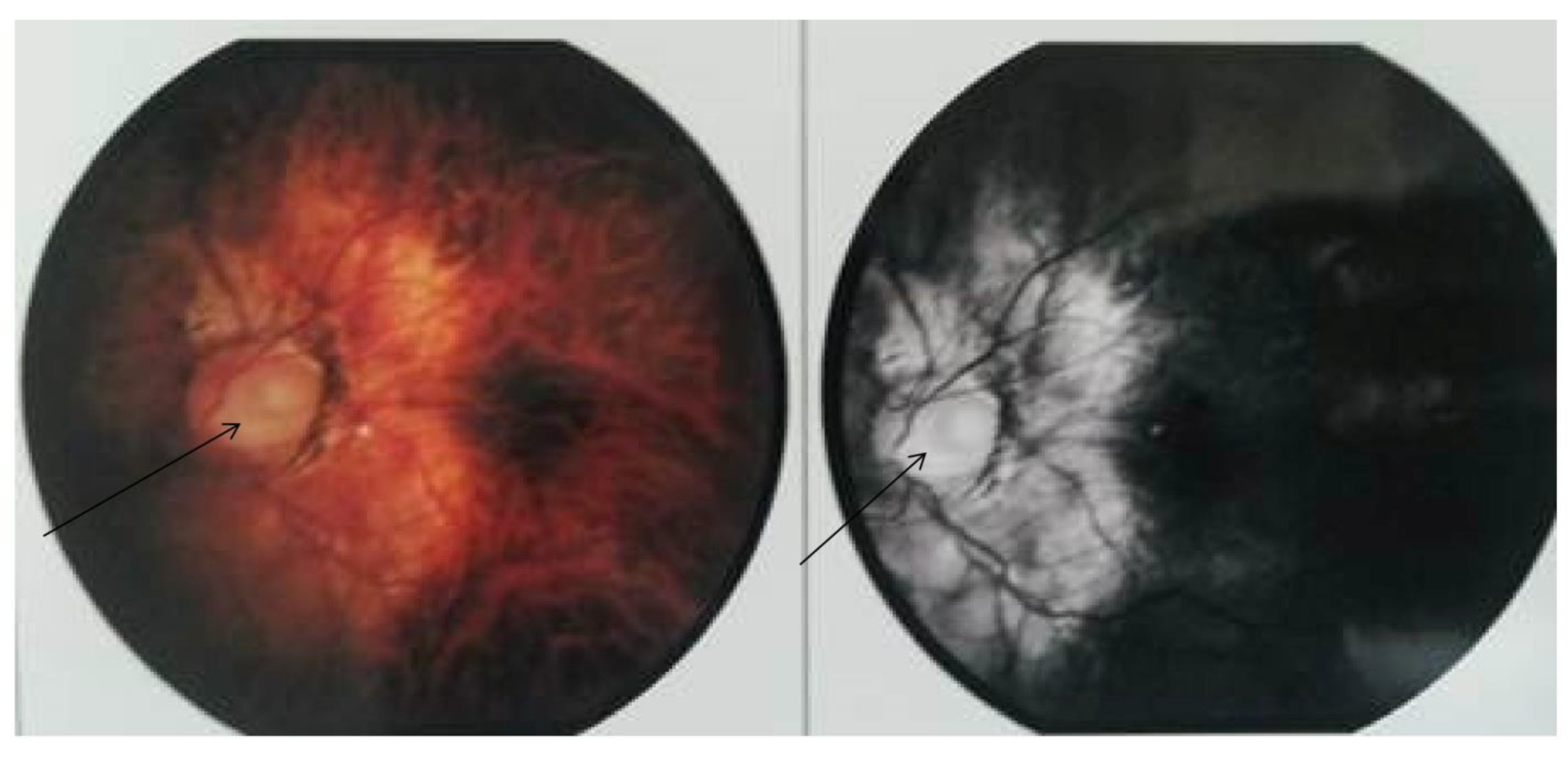
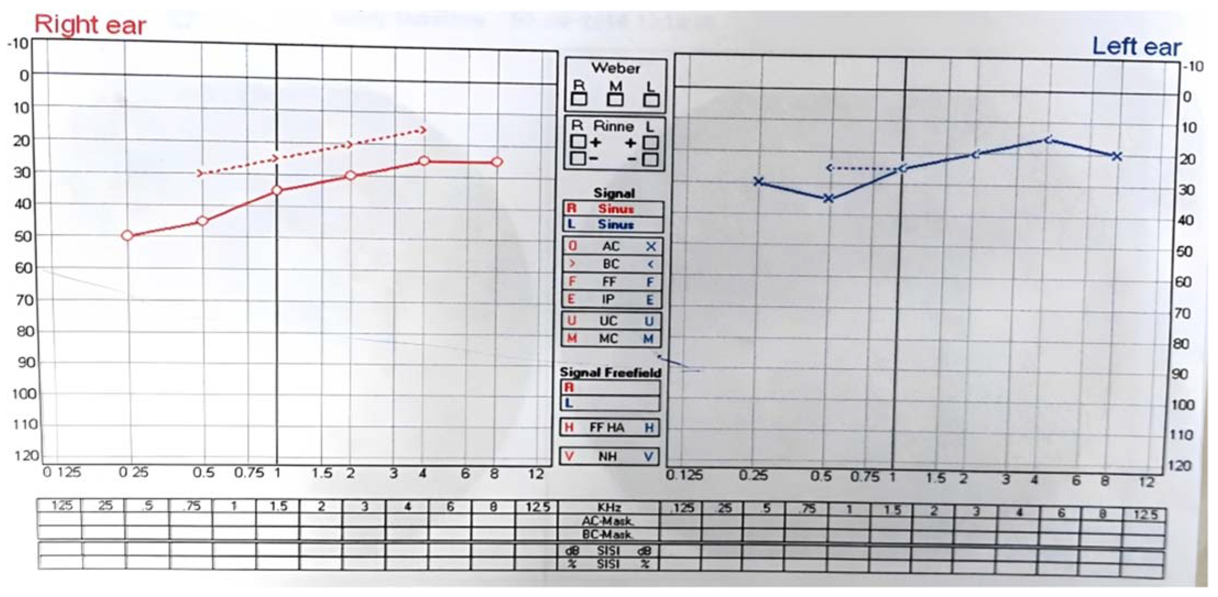
2. Case Report/Case Presentation

In this investigation, we examined a duo of siblings who had a background of bilateral hypoplastic kidneys. Both patients endured recurring dialysis therapy and exhibited a decline in renal function (22 mg/dL and 11 mg/dL, correspondingly). The older sibling (II:2) underwent a renal transplant and subsequently experienced chronic allograft nephropathy (CAN). The younger sibling has suffered from strabismus in the left eye from an early age and a reduction in vision. A qualified ophthalmologist scrutinized both siblings, and the younger brother was diagnosed with an optic disc coloboma in the left eye and the presence of conductive hearing loss in the right ear and sensorineural hearing loss in the left ear.

3. Materials and Methods
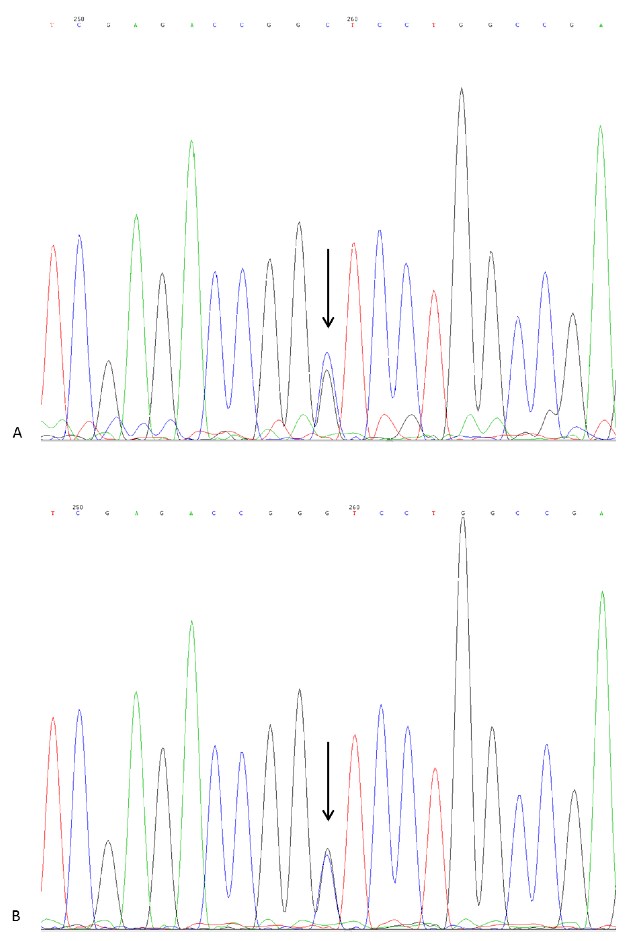
DNA Sequencing

In order to determine the genetic sequence of the PAX2 gene, PCR amplification and DNA sequencing were performed. The primers used for sequencing PAX2 exon and intron–exon boundaries, along with the specified genetic analyzer used, are detailed in Table 1. The resulting sequences were compared to the reference sequence for PAX2 in the NCBI GenBank database using BLAST analysis to identify any genetic variants or mutations.
Genomic DNA was extracted from the blood samples collected from both patients using the QIAamp DNA mini kit (Qiagen, Hilden, Germany). The coding regions and intron–exon junctions of the PAX2 gene (NM_000278) were screened for mutations using PCR amplification with a dye-terminator cycle-sequencing kit (BD Ready Reaction kit, Perkin Elmer, Branchburg, NJ, USA). The PCR products were purified using the QIAquick PCR purification kit (Qiagen, Hilden, Germany) and subjected to direct fluorescent sequencing. The cycle-sequencing product was then outsourced for further genetic or automated sequencing (Thermo Fisher Scientific, Gurgaon, India). The resulting sequence data were analyzed using the CHROMAS software and compared to reference sequences and NM_000278.
Sequence variants were analyzed in reference to the American College of Medical Genetics and Genomics and Association for Molecular Pathology guidelines [16] to determine their potential pathogenicity in the PAX2 gene. The results showed four novel heterozygous variations in PAX2 in both patients, including a C to G transversion at position c.352 in the PAX2 gene in a heterozygous state.

4. Results and Discussion
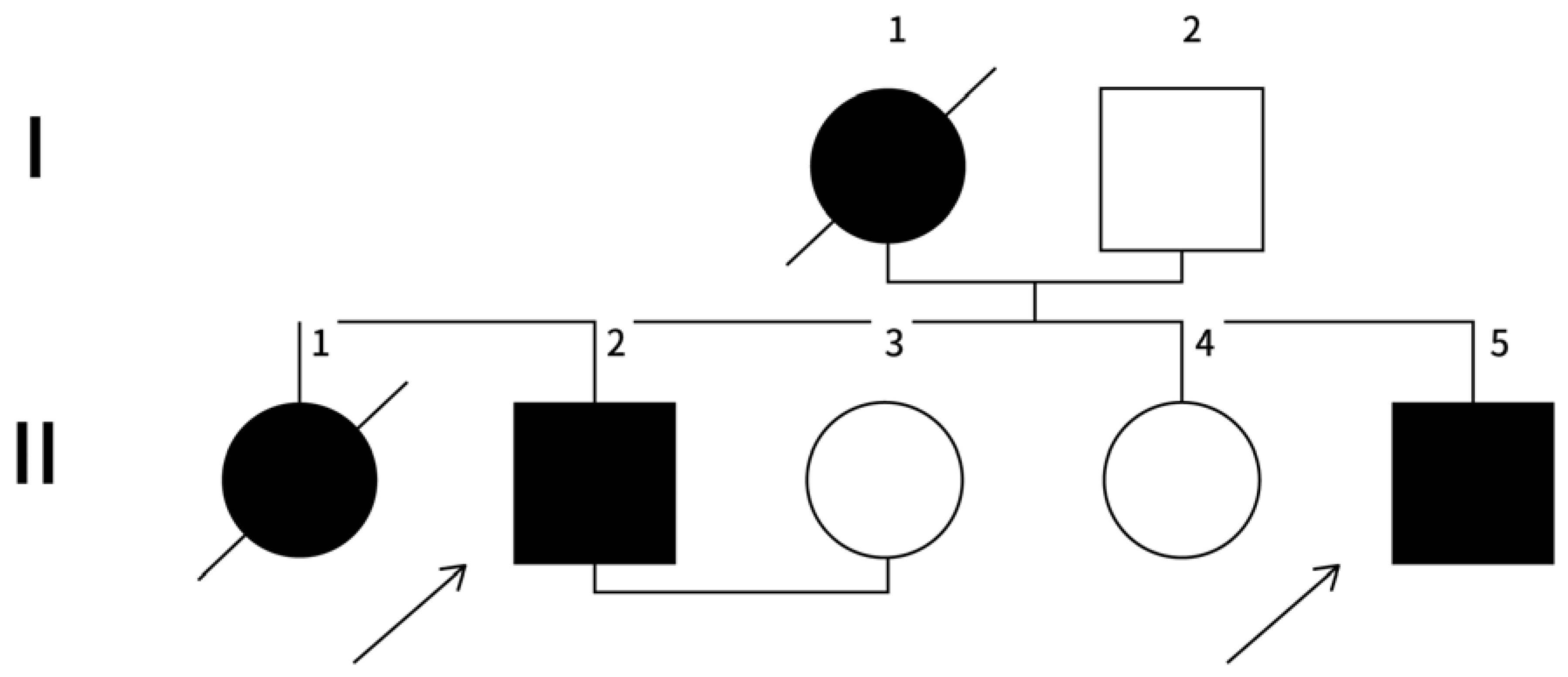
Family History

In this study, we evaluated two siblings (patients II:2 and II:5) with a family history of chronic kidney disease (CKD). A thorough ophthalmologic examination revealed that the younger sibling had amblyopia with a left eye optical disc coloboma, while the other sibling had a very mild, asymptomatic coloboma. Information on the family pedigree was gathered and is presented in Figure 1 (genetic tree).
The proband’s father (I:2), who is 57 years old and has no comorbidities, was found to have a normal creatinine level (0.58 mg/dL) and no history of ocular or auditory problems in his family on the paternal side. He is currently being evaluated as a potential kidney donor for his son. The genetic variations (c.617-19 T > A, c.617-70 C > A, and c.617-41 T > A) detected in the affected individual may not have been inherited from the father as he has not been diagnosed with any renal, ocular, or auditory problems.
The mother (I:1) had a history of squint and vision loss due to a left optic nerve coloboma and was diagnosed with kidney disease at the age of 39. She was on maintenance hemodialysis and passed away at the age of 47. The elder sister (II:1) had squint and CKD, was on maintenance hemodialysis, and died at the age of 18. The younger sister (II:4), 30 years old, has no renal, visual, or auditory complaints and her serum creatinine level is 0.58 mg/dL.
Patient 1 (II:2) is a 32-year-old man who was diagnosed with CKD in 2010 with a serum creatinine level of 8.8 mg/dL and an estimated glomerular filtration rate (eGFR) of 7 mL/min. A urine analysis revealed 2+ proteinuria with no signs of hematuria, and an ultrasound indicated contracted kidneys on both sides. He received a renal transplant from his paternal grandmother and was put on a triple-drug-immunosuppression regimen. However, in April 2013, after getting married, he discontinued all medications and experienced a gradual increase in his serum creatinine level. As a result, he has been on maintenance hemodialysis since then.
Patient 2 (II:5) is a 23-year-old man who was found to have raised creatinine levels (1.69 mg/dL, eGFR: 57.68 mL/min) during a routine evaluation. An ultrasound showed bilateral contracted kidneys. His progressive worsening of renal function within 3 years led to him being put on maintenance hemodialysis. He has had a squint in his left eye since childhood with decreased vision, and he underwent surgery at the age of 16 but saw no improvement in his vision. An ophthalmic evaluation showed amblyopia with an optic disc coloboma in his left eye (as depicted in Figure 2).
Additionally, we obtained an audiogram report which indicated conductive hearing loss in the right ear and sensorineural hearing loss in the left ear (as depicted in Figure 3). Conductive hearing loss occurs when sound waves cannot pass through the outer or middle ear, while sensorineural hearing loss is caused by damage to the inner ear or the auditory nerve.

5. Genetic Analysis and Discussion

The genetic study conducted on both patients (II:2 and II:5) revealed the presence of four novel heterozygous variations in the PAX2 gene. Notably, both patients shared the same heterozygous PAX2 mutation, c.352 C > G, as depicted in Figure 4.
This nucleotide change was localized in exon 3 of the PAX2 gene and resulted in the substitution of Leucine amino acid with Valine at position 118 of the protein (p.Leu118Val).
PAX2 gene mutations have been linked to various developmental abnormalities in the kidney and optic nerve, including papillorenal syndrome, renal coloboma syndrome, and optic nerve coloboma. The mutation identified in both patients (II:2 and II:5) has been previously reported in patients with papillorenal syndrome, characterized by optic nerve coloboma and renal abnormalities.
The p.Leu118Val mutation has been shown to have a dominant negative effect on the PAX2 protein function, impairing its binding to DNA and leading to a reduction in transcriptional activity. This, in turn, may contribute to the development of renal and ocular abnormalities observed in the patients.
Further genetic testing and counseling may be necessary to determine the potential risk of passing the PAX2 mutation to offspring, as well as to identify any other genetic factors that may contribute to the development of the patients’ clinical features [17].
The sequencing analysis manifested a C to G transversion at position c.352 in the PAX2 gene, which was discovered to be in a heterozygous state. This nucleotide change was localized in exon 3, resulting in the replacement of the highly conserved Leucine amino acid with Valine at position 118 of the protein (p.Leu118Val).
A computer-based comparison of this missense variation was conducted using three distinct bioinformatics tools: Mutation Taster, Provean, and SIFT. All three tools projected a pathological impact for this alteration. It is worth noting that the PAX2 genes are comprised of 12 exons, with most mutations transpiring in exons 2-4 (paired domain); however, there are also recorded cases of mutations described in other domains [9].
The novel heterozygous variations in intron 5 of the PAX2 gene found in both patients (II:2 and II:5) are depicted in the sequence chromatograms (Figure 5), with the location of the variations marked by arrows (c.617-70 C > A, c.617-41 T > A, c.617-19 T > A). Patient 2 (II:2) is represented in chromatogram J and Patient 5 (II:5) is represented in chromatogram M.
The occurrence of the three newly identified heterozygous variations in intron 5 of the PAX2 gene in both patients indicates that these variations may have a role in the development of CKD and ocular abnormalities. Nonetheless, additional investigation and validation are needed to determine the precise function of these variations. These discoveries augment the current understanding of the genetic mechanism underlying CKD and ocular and hearing abnormalities and lay the groundwork for future genetic counseling of this family.
Using computer-based bioinformatics tools, we analyzed the changes in intron 5 by comparing it with the wild-type sequence. The variants 617-70 C > A and c.617-41 T > A were deemed polymorphisms, with all bioinformatics programs predicting a benign effect. However, only one of the tools used suggested a pathogenic effect for 617-19 T > A.
Therefore, it can be concluded that 617-19 T > A is most likely a variant of uncertain significance, which does not provide any informative insight into the cause of the patients’ clinical features.
In brief, we have reported the identification of the fourth Indian family to exhibit genetic confirmation of RCS. All affected individuals for whom DNA samples were available had the identical heterozygous mutation in exon 3 (c. 352 C > G, Leu118Val) of the PAX2 gene, supporting an autosomal dominant mode of inheritance. We deduced that the mother (I:1) and the probands’ sister (II:1) had the identical genetic change in PAX2, which was consistent with the transmission of this change to her elder son (II:2). Clinical manifestations varied within the family, as is often seen in RCS cases. Interestingly, all previously reported patients with PAX2 mutations had marked ophthalmic dysplasia [18,19,20]. Patient 2 (II:5) underwent a thorough ophthalmologic examination which revealed the presence of optic disc coloboma, renal hypoplasia, and optic nerve coloboma. The patient was also diagnosed with conductive hearing loss in the right ear and sensorineural hearing loss in the left ear, as indicated in the audiogram report. It is worth noting that the patient and other family members also had a history of renal hypoplasia and optic nerve coloboma. These findings highlight the need for a comprehensive evaluation of patients with similar symptoms and a thorough examination of their family history.
Furthermore, we identified a new mutation and three novel variations in the PAX2 gene that had not been previously described in the scientific literature. The PAX2 gene is responsible for coding a protein that plays a critical role in the development of the kidney and the eye. Mutations in this gene have been associated with a rare genetic disorder called renal coloboma syndrome (RCS), which is characterized by kidney abnormalities and eye defects.
The identification of these new mutations and variations has expanded the current understanding of the spectrum of PAX2 mutations and variations known in the literature. Unfortunately, RCS remains a rare condition and may be underdiagnosed due to its variable phenotype, which means that the symptoms can vary greatly from person to person. This makes it challenging to recognize and diagnose the condition, particularly in individuals with milder subclinical phenotypes.
We highlighted the importance of recognizing this rare condition and the need for collaboration between nephrologic and ophthalmologic services for the proper diagnosis and management of patients. They recommend that individuals at risk of RCS undergo genetic testing, which can facilitate appropriate management and follow-up for patients with milder subclinical phenotypes.
In conclusion, the identification of new mutations and variations in the PAX2 gene has expanded the current understanding of RCS. However, the rarity and variability in the condition make it challenging to recognize and diagnose. Therefore, collaboration between medical specialties and genetic testing can be invaluable in providing proper diagnosis and management for patients with RCS.

Author Contributions

K.D., G.M.V. and D.P.M.—detailed analysis and editing and writing of the manuscript. L.G.d.L. and M.M.T.—drafting initial paper. A.G. (Ashwani Gupta), D.S.R. and M.M.—drafting the final paper. A.G. (Anurag Gupta) and V.B.—interpretation of clinical data. M.V.—PCR and primary analysis of the results. C.R. and A.K.B.—designing the study and final approval. All authors have read and agreed to the published version of the manuscript.

Funding

This research received no external funding.

Institutional Review Board Statement

As it was a diagnostic test, no approval from an ethics committee was needed. Both of the subjects gave written informed consent for the investigation.

Informed Consent Statement

Written informed consent was obtained from both the subjects for this study and to publish this work which includes images.

Data Availability Statement

The data that support the findings of this study are included as supplementary material files. Further enquiries about the PAX2 sequences can be directed to the corresponding authors and could be available on request.

Acknowledgments

We express our gratitude towards IRRIV for collaboration and allowing the use of bio-informatics software, and detailed analysis of the results.

Conflicts of Interest

No conflict of interest, financial or otherwise, for all authors.

References

  1. Sanyanusin, P. Papillorenal Syndrome. In GeneReviews®; Adam, M.P., Ardinger, H.H., Pagon, R.A., Eds.; University of Washington: Seattle, WA, USA, 1993. [Google Scholar]
  2. Bower, M.; Salomon, R.; Allanson, J.; Antignac, C.; Benedicenti, F.; Benetti, E.; Binenbaum, G.; Jensen, U.B.; Cochat, P.; DeCramer, S.; et al. Update of PAX2 mutations in renal coloboma syndrome and establishment of a locus-specific database. Hum. Mutat. 2012, 33, 457–466. [Google Scholar] [CrossRef] [PubMed]
  3. Sanyanusin, P.; Schimmenti, L.A.; McNoe, L.A.; Ward, T.A.; Pierpont, M.E.; Sullivan, M.J.; Dobyns, W.B.; Eccles, M.R. Mutation of the PAX2 gene in a family with optic nerve colobomas, renal anomalies and vesicoureteral reflux. Nat. Genet. 1995, 9, 358–364. [Google Scholar] [CrossRef] [PubMed]
  4. Nishimoto, K.; Iijima, K.; Shirakawa, T.; Kitagawa, K.; Satomura, K.; Nakamura, H.; Yoshikawa, N. Papillorenal syndrome: The frequency of clinical symptoms and PAX2 mutations in Japanese pediatric patients. Pediatr. Nephrol. 2005, 20, 1723–1728. [Google Scholar]
  5. Schedl, A.; Hastie, N.D. Genetics of kidney development. Nat. Genet. 2000, 24, 210–211. [Google Scholar]
  6. Singh, A.D.; De Potter, P.; Fijal, B.A.; Shields, C.L.; Shields, J.A.; Elston, R.C. Lifetime prevalence of uveal melanoma in white patients with oculo(dermal) melanocytosis. Ophthalmology 1998, 105, 1955–1958. [Google Scholar] [CrossRef] [PubMed]
  7. Winyard, P.J.; Risdon, R.A.; Sams, V.R.; Dressler, G.R.; Woolf, A.S. The PAX2 transcription factor is expressed in cystic and hyperproliferative dysplastic epithelia in human kidney malformations. J. Clin. Investig. 1996, 98, 451–459. [Google Scholar] [CrossRef] [PubMed]
  8. Favor, J.; Peters, H.; Hermann, T. Molecular characterization of Pax2 mutants suggest that neural crest cells contribute to the development of the urogenital system. Dev. Dyn. 1996, 206, 169–179. [Google Scholar]
  9. Sanyanusin, P.; McNoe, L.A.; Sullivan, M.J.; Weaver, R.G.; Eccles, M.R. Mutation of PAX2 in two siblings with renal-coloboma syndrome. Hum. Mol. Genet. 1995, 4, 2183–2184. [Google Scholar] [CrossRef] [PubMed]
  10. Eccles, M.R.; Schimmenti, L.A. Renal-coloboma syndrome: A multi-system developmental disorder caused by PAX2 mutations. Clin. Genet. 1999, 56, 1–9. [Google Scholar] [CrossRef] [PubMed]
  11. Weaver, R.G., Jr.; Torczynski, E.; Mao, J.I. Renal-coloboma syndrome: A report of 2 cases and review of the literature. Am. J. Kidney Dis. 1988, 12, 406–410. [Google Scholar]
  12. Weaver, R.G., Jr.; Hopper, K.; Torczynski, E.; Scharre, J. Familial renal-coloboma syndrome: Report of a second family. Am. J. Kidney Dis. 1995, 26, 498–506. [Google Scholar]
  13. Lakshmanaiah, L.; Rangarajan, T.; Chaitanya Ramana Reddy, V. Renal Coloboma Syndrome: A Novel PAX2 Mutation Involving An Indian Family. Indian J. Nephrol. 2016, 26, 327–331. [Google Scholar]
  14. Yovagood, S.; Vuppala, S.; Jalali, S. Phenotypic Spectrum and PAX2 Mutation Analysis in a Second Indian Family with Renal Coloboma Syndrome. Indian J. Nephrol. 2018, 28, 310–313. [Google Scholar]
  15. Ammayappan, S.K.; Rajagopalan, A.; Arunachalam, J.; Prasath, A.; Durai, R. A case of renal coloboma syndrome. J. Nephrol. 2023, 36, 233–235. [Google Scholar] [CrossRef] [PubMed]
  16. Richards, S.; Aziz, N.; Bale, S.; Bick, D.; Das, S.; Gastier-Foster, J.; Grody, W.W.; Hegde, M.; Lyon, E.; Spector, E.; et al. Standards and guidelines for the interpretation of sequence variants: A joint consensus recommendation of the American College of Medical Genetics and Genomics and the Association for Molecular Pathology. ACMG Laboratory Quality Assurance Committee. Genet. Med. 2015, 17, 405–424. [Google Scholar] [CrossRef] [PubMed] [Green Version]
  17. Safieh, L.; Vithana, E.N.; Chan, W.M. Genetics of primary inherited optic neuropathies. Asia-Pac. J. Ophthalmol. 2015, 4, 318–325. [Google Scholar]
  18. Schimmenti, L.A.; Cunliffe, H.E.; McNoe, L.A.; Ward, T.A.; French, M.C.; Shim, H.H.; Zhang, Y.H.; Proesmans, W.; Leys, A. Further delineation of renal-coloboma syndrome in patients with extreme variability of phenotype and identical PAX2 mutations. Am. J. Hum. Genet. 1997, 60, 869–878. [Google Scholar] [PubMed]
  19. Fletcher, J.; Hu, M.; Berman, Y.; Collins, F.; Grigg, J.; McIver, M.; Jüppner, H.; Alexander, S.I. Multicystic dysplastic kidney and variable phenotype in a family with a novel deletion mutation of PAX2. J. Am. Soc. Nephrol. 2005, 16, 2754–2761. [Google Scholar] [CrossRef] [PubMed] [Green Version]
  20. Thomas, R.; Sanna-Cherchi, S.; Warady, B.A.; Furth, S.L.; Kaskel, F.J.; Gharavi, A.G. HNF1B and PAX2 mutations are a common cause of renal hypodysplasia in the CKiD cohort. Pediatr. Nephrol. 2011, 26, 897–903. [Google Scholar] [CrossRef] [PubMed] [Green Version]
Figure 1. The family pedigree, where males are represented by squares and females by circles. Family members who underwent genetic testing are marked with an arrow.
Kidneydial 03 00023 g001
Figure 2. The results of the ophthalmic evaluation of Patient 2 (II:5). The evaluation revealed myopic changes and amblyopia with an optic disc coloboma in the left eye. The arrows in the figure indicate an enlarged optic disc with blood vessels emerging from the side of the disc, rather than from the middle.
Kidneydial 03 00023 g002
Figure 3. The findings of the audiogram report, which show that the patient had different types of hearing loss in each ear. Specifically, the report indicated conductive hearing loss in the right ear and sensorineural hearing loss in the left ear.
Kidneydial 03 00023 g003
Figure 4. The sequence chromatograms of the affected patients, with chromatogram (A) belonging to Patient II:2 and chromatogram (B) belonging to Patient II:5. The arrows in the figure highlight the heterozygous variation c.352 C > G (Leu 118 Val) present in both patients.
Kidneydial 03 00023 g004
Figure 5. Both patients showed new heterozygous changes in intron 5 of the PAX2 gene, specifically, c.617-70 C > A, c.617-41 T > A, and c.617-19 T > A. The affected patients’ sequence chromatograms demonstrated these variations, with Patient 2 (II:2) represented by chromatogram J and Patient 5 (II:5) represented by chromatogram M. The heterozygous variation c.617-19 T > A was visible in both patients, as indicated by the arrows in the figure. The variations were presented as follows: (a,d) demonstrate c.617-19 T > A, (b,e) demonstrate c.617-41 T > A, and (c,f) demonstrate c.617-70 C > A.
Kidneydial 03 00023 g005
Table 1. The primers used to sequence PAX2 exon and intron–exon boundaries and the genetic analyzer used.
Primer NamePrimer Sequence (5′ to 3′)Exon/Intron BoundaryGenetic Analyzer Used
PaxF1CCCTCCCTTTTCTCCTCAAGIntron 1/Exon 23500xl
PaxR1GGTGACAGAAGGCAGGAGAGIntron 1/Exon 23500xl
PaxF2CTTCTCAAGCTCGGGAACATIntron 2/Exon 33500xl
PaxR2GCTGGACTTTTAGCCACGTCIntron 2/Exon 33500xl
PaxF3CACCCTCAGGAAGTCAGCTCIntron 3/Exon 43500xl
PaxR3CTCTCTGCCTCACAGGTTCCIntron 3/Exon 43500xl
PaxF4GACACAGGGAGCAGATGGATExon 4/Intron 43500xl
PaxR4GCTCAGGTGAGAGGAGTTGGExon 4/Intron 43500xl
PaxF5GGCTCCTCATCCCTCCTTATIntron 4/Exon 53500xl
PaxR5GGACCTGGGCTTTGCTACTAIntron 4/Exon 53500xl
PaxF6GGCCCAAGACTAGGGAGACTExon 5/Intron 53500xl
PaxR6GAGCCAACTCCTCCTTTTCCExon 5/Intron 53500xl
PaxF7CTCTTTGCCCTGCACTGTTCIntron 5/Exon 63500xl
PaxR7TGGCTATGCATGTGGTGTTTIntron 5/Exon 63500xl
PaxF8ACTACCCGCACAAAACTTGCExon 6/Intron 63500xl
PaxR8CAGGAAGCACCCTGGTTTTAExon 6/Intron 63500xl
PaxF9TTTCCTCTCCGTGCAGTACCIntron 6/Exon 73500xl
PaxR9GAGGGGCCATGACATACAGTIntron 6/Exon 73500xl
PaxF10ATGCCTCCTAGAACCGGAGTExon 7/Intron 73500xl
PaxR10CTGCACTAACAAGCCTGTCCExon 7/Intron 73500xl
PaxF11CAGGGGAGGTCTTTTCTGTGIntron 7/Exon 83500xl
PaxR11GGGGTGATGTGAAGGGTTGIntron 7/Exon 83500xl
Disclaimer/Publisher’s Note: The statements, opinions and data contained in all publications are solely those of the individual author(s) and contributor(s) and not of MDPI and/or the editor(s). MDPI and/or the editor(s) disclaim responsibility for any injury to people or property resulting from any ideas, methods, instructions or products referred to in the content.

