Article

Evaluating Serum RBP4 as an Auxiliary Biomarker for CKDu Diagnosis

by
Hannah L. F. Swa
1,†,‡,
Buddhi N. T. Fernando
2,†,
Shakila Premarathna
3,
Asfa Alli-Shaik
1,
Zeid Badurdeen
3,
Jayantha Gunarathna
1,4 and
Nishantha Nanayakkara
3,*
1
Translational Biomedical Proteomics Group, Institute of Molecular & Cell Biology, Agency for Science, Technology and Research (A*STAR), Singapore 138632, Singapore
2
Department of Medical Laboratory Science, Faculty of Allied Health Sciences, University of Ruhuna, Galle 81000, Sri Lanka
3
Transplant and Dialysis Unit, National Hospital Kandy, Kandy 20000, Sri Lanka
4
Yong Loo Lin School of Medicine, National University of Singapore, Singapore 119077, Singapore
*
Author to whom correspondence should be addressed.
These authors contributed equally to this work.
Current address: Diagnostic Development (DxD) Hub, Agency for Science, Technology and Research (A*STAR), Singapore 138632, Singapore.
Kidney Dial. 2022, 2(4), 576-587; https://doi.org/10.3390/kidneydial2040052
Submission received: 22 September 2022 / Revised: 26 October 2022 / Accepted: 31 October 2022 / Published: 3 November 2022

Abstract

:
Background: A chronic interstitial disease, chronic kidney disease of uncertain etiology (CKDu), has emerged as a notable contributor to the CKD burden in rural Sri Lanka. Most therapeutic and diagnostic approaches to CKD focus on glomerular diseases, and thus are not fully applicable to CKDu. Serum proteins, specifically those with the profile of markers representing different facets of a disease, are beneficial for a comprehensive evaluation of diseases, and hence in CKD. Our aim was to identify the role of serum-retinol-binding protein 4 (RBP4), a marker of the proximal tubule, in the diagnosis of CKDu. Methods: Definite CKDu cases were recruited from the renal clinic in Girandurukotte and Wilgamuwa (endemic regions). Healthy controls were recruited from Mandaramnuwara (nonendemic area). The levels of RBP4 and creatinine in serum were measured. An immunoassay (ELISA) was performed on the serum samples. The stages of CKD/ CKDu were classified according to eGFR. Results: Serum RBP4 was significantly increased in CKDu patients compared to CKD patients and healthy controls. The results show that the ratio of normalized serum RBP4 to serum creatine (S.cr) acts as a better competitive marker for CKDu (AUC 0.762, sensitivity 0.733) than CKD (AUC 0.584, sensitivity 0.733) when compared against healthy controls. Furthermore, the RBP4:S.cr ratio showed higher discriminating power (AUC 0.743) between CKDu and CKD, suggesting that the RBP4: S.cr ratio has potential as a serum marker to differentiate CKDu from CKDu. Conclusion: The RBP4: S.cr ratio was identified as a plausible indicator for differentiating CKDu from CKD with >70% sensitivity and specificity. Therefore, it could be used in the evaluation of the tubular interstitial involvement of CKD.

1. Introduction

Chronic kidney disease of uncertain etiology (CKDu) is a term coined to describe endemic chronic kidney disease (CKD) that has not been found or associated with its conventional predominant risk factors such as hypertension and diabetes [1,2]. This novel form of CKD is a major health issue that has gained public attention due to its high mortality [3,4] in several tropical regions, including Sri Lanka, India, Latin America and Egypt [5,6,7,8]. As this health condition mainly afflicts young and middle-aged males from low-socioeconomic-status agricultural communities [1,9], hypotheses regarding the etiologies of CKDu have focused on occupational triggers, including environmental toxins that might have leached from pesticides and aluminum vessels into drinking water and been ingested by these farmers. Despite many investigations on arsenic, cadmium, lead, fluoride, chromium, aluminum and agrochemical contamination in geographical resources [10,11,12,13,14,15], causative factors of CKDu remain inconclusive. This lack of understanding with regard to the etiology of CKDu has therefore prompted many relevant studies to seek a holistic comprehension of this disease.
CKDu has been observed to be a distinct form within the categorical diagnosis of CKD in regard to both clinical and pathological presentations. Whilst the majority of CKD patients present with high serum creatinine and proteinuria, the latter is minimal in CKDu patients, especially in early stage 1. This is probably attributed to the fact that CKD manifests predominantly as glomerular and vascular damage [16], whereas CKDu is hallmarked by the pathology of the tubular interstitial compartment of the kidney [17,18,19]. Thus, evaluation of tubulointerstitial lesions in CKDu remains the current “gold standard” for diagnosing as well classifying the severity [17]. Most of the current treatments for renal diseases are based on glomerular pathologies; hence, the serum-creatinine- and proteinuria-based approaches are appropriate. In contrast, renal tubules are neglected, at least to a certain extent, beyond their involvement in a diverse array of functions carried out to maintain the body physiology. More specific, reliable and sensitive markers are required to evaluate the specific functions and severity of tubular interstitial diseases, including CKDu.
Tubular markers are highly plausible as indicators for CKDu as the disease has been characterized with renal tubular damage as a first and early sign [20]. Indeed, urinary alpha-1-microglobulin (A1M), a marker of proximal tubular dysfunction, is probably the best single biomarker used to identify CKDu/CKD at the earliest stages. A1M has been reported to be elevated in CKDu patients in comparison to healthy controls, and the quantity increases as the disease progresses [20,21]. There have been several studies investigating the effectiveness of different tubular markers, such as kidney injury molecule 1 (KIM1) and neutrophil-gelatinase-associated lipocalin (NGAL), for the screening and diagnosis of CKDu [22,23,24]. Interestingly, one study reported elevated urinary KIM1 and NGAL were found not only in CKDu patients but also in healthy cohorts [23]. Findings strongly suggest subclinical kidney disease unless otherwise proven by a kidney biopsy or by follow up. Fernando et al., in 2019, proposed that using a combination of biomarkers leads to a better performance than using a single marker in distinguishing among CKD/CKDu and healthy controls [21]. This study demonstrated that a combination of A1M, KIM 1 and RBP4 as a marker panel could be used for distinguishing CKD/CKDu from healthy controls, while a panel consisting of urinary Osteopontin (OPN), KIM1 and RBP4 was used to differentiate CKDu from CKD with high sensitivity and specificity [21].
Preliminary data from a multiplexed immunoassay motivated us to explore the effectiveness of serum-retinol-binding protein 4 (RBP4) in the evaluation of tubular functions, more specifically the proximal tubular functions of CKDu. We propose the concomitant use of glomerular and tubular markers would provide a more realistic picture of chronic kidney disease, opening up new avenues for treatment. RBP4 is a small protein of 21kDa that is freely filtered through the glomeruli upon the release of retinol. It is then readily reabsorbed by the proximal tubule and catabolized [25,26]. As such, it has been regarded as an excellent marker candidate for tubular renal impairment [27,28]. Moreover, there is the possibility that leakage of RBP4 through tubular damage, and thereby an increase in urinary RBP4 can be observed before glomerular markers [28]. This would be extremely relevant in the context of CKDu. Some of the early studies on CKD reported a significant positive correlation of serum RBP4 with serum creatinine [29,30,31]. Xun et al. (2018) showed that serum RBP4 is useful in diagnosing CKD as the concentration of serum RBP4 has an association with renal functions [29]. They confirmed that combined detection of serum RBP4 and routine renal function biomarkers such as serum creatinine, urea and cystatin C would improve diagnostic accuracy for CKD [29].
Along these lines, concerted efforts have been made to assess the applicability of current CKD diagnostic tools for CKDu diagnostics. For example, the urine dipstick test, a cost-effective, sensitive screening test for CKD, has been proven to have a limited role in minimally proteinuric early stages of CKDu [32]. Further, estimated GFR (eGFR) is regarded as the most popular indicator of kidney dysfunction, and this index takes into account variables including S.Cr, age, weight and gender [33]. Though eGFR (<60 mL/min/1.73 m2) is an essential criterion in the diagnosis of CKDu, there is a strong possibility for the existence of significant kidney damage, even among so-called “healthy” individuals with an eGFR of more than 60 mL/min/1.73 m2 [34]. This indicates the unknown bona fide impact the diagnosis of CKDu, a primary tubular interstitial disease, has on eGFR. Hence, RBP4, a tubular marker, in combination with creatinine or eGFR, is complementary to the evaluation of the severity of tubular interstitial diseases.

2. Materials and Methods

2.1. Patient Information and Study Design

This cross-sectional study was conducted in definite CKDu patients (n = 44), CKD patients (n = 45) and healthy controls (n = 45). CKDu participants (biopsy-proven) were enrolled from renal clinics in a divisional hospital in Girandurukotte and Wilgamuwa, a region endemic for CKDu according to national guidelines for the diagnosis of CKDu. People with known causes for CKD (hypertension, diabetes mellitus, heart diseases, polycystic kidney disease, systemic lupus erythematosus) were enrolled from a renal clinic, National Hospital Kandy, in a nonendemic area for CKDu. All the CKD cases were diagnosed by a consultant nephrologist. Urinary-albumin-negative but otherwise healthy volunteers with an eGFR of more than 90 and normal serum creatinine were recruited from Mandaramnuwara, a nonendemic region located in the central province of the country, and selected as controls. The ethical clearance was obtained from the ethical review committee of the Faculty of Medicine, University of Peradeniya (2016/EC/28). All enrolled participants provided informed consent.

2.2. Clinical Sample Collection

Blood samples were taken, and the serum was separated immediately. Serum samples were transferred to sterile cryovials and transported in liquid N2 to the laboratory. Then, the samples were stored at −80 °C until analysis. Sample collection was carried out in Sri Lanka, and the samples were sent under the appropriate conditions to Singapore for the analysis.

2.3. Luminex X-MAP Screening

In the pilot study, serum RBP4 was measured using Luminex X-MAP technology. All procedures were performed in accordance with the manufacturer’s instructions. The assay platforms and methodologies were specifically selected, and great attention was paid to quality-control issues, including calibrating and adherence to well-annotated standard operating procedures. According to the results of the pilot study, serum RBP4 was found to be beyond the detectable range of the assay.

2.4. ELISA Assay

Serum creatinine (Scr) was measured using an Indiko Plus biochemical analyzer (Thermo ScientificTM, Vantaa, Finland). Estimated glomerular filtration rate (eGFR) was calculated using the Chronic Kidney Disease Epidemiology Collaboration (CKD-EPI) equation [35]. Normalized values, in this case normalizing against total protein concentration, are used to control for intra-assay and interpatient variation. Such a method has also been employed by ELISA studies [36].
An immunoassay (Human-Retinol-binding protein 4 ELISA Kit (RBP4); Abcam) was performed on the selected serum samples in three groups (CKDu, CKD, NEC) to confirm if the serum RBP4 level was indeed high in CKDu patients.

2.5. Statistical Analysis

The differential levels of measured serum RBP4 among the three different groups, control, CKD and CKDu, were assessed for significance using the nonparametric Mann–Whitney U test, and a p-value < 0.05 was considered significant. Marker performances for sensitivity, specificity and area under curve (AUC) were assessed using receiver operating characteristic (ROC) analysis as implemented in the statistical XLSTAT plug-in for Microsoft Excel.

3. Results

3.1. Demographic and Clinical Characteristics

Out of 134 recruited patients, 44 patients belonged to the CKDu group and 45 patients belonged to both the NECKD and NEC groups. Demographic data, life style and health characteristics of the three groups are shown in Table 1. Out of the three groups, the CKDu group had a more prevalent family history of CKD (50%) compared to the other two groups. Smoking (47.7%), betel chewing (81.8%) and alcohol consumption (43.2%) were higher in CKDu compared to the NECKD and NEC patients. History of malaria (40.9%) also had a higher percentage in the CKDu group compared to the other groups.

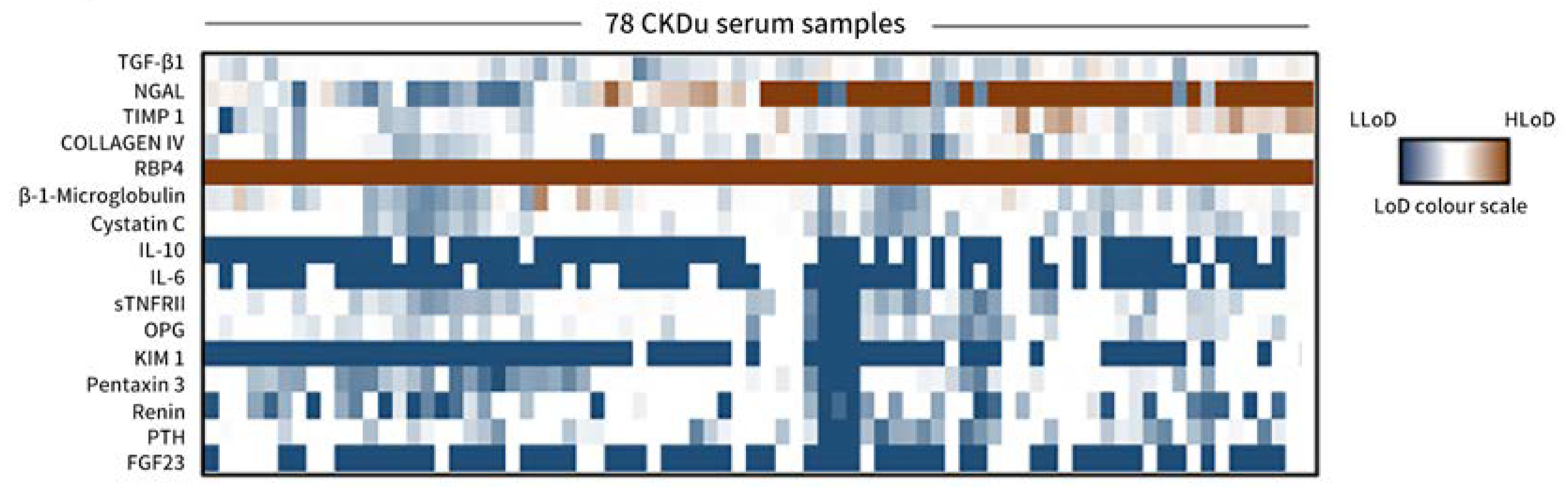
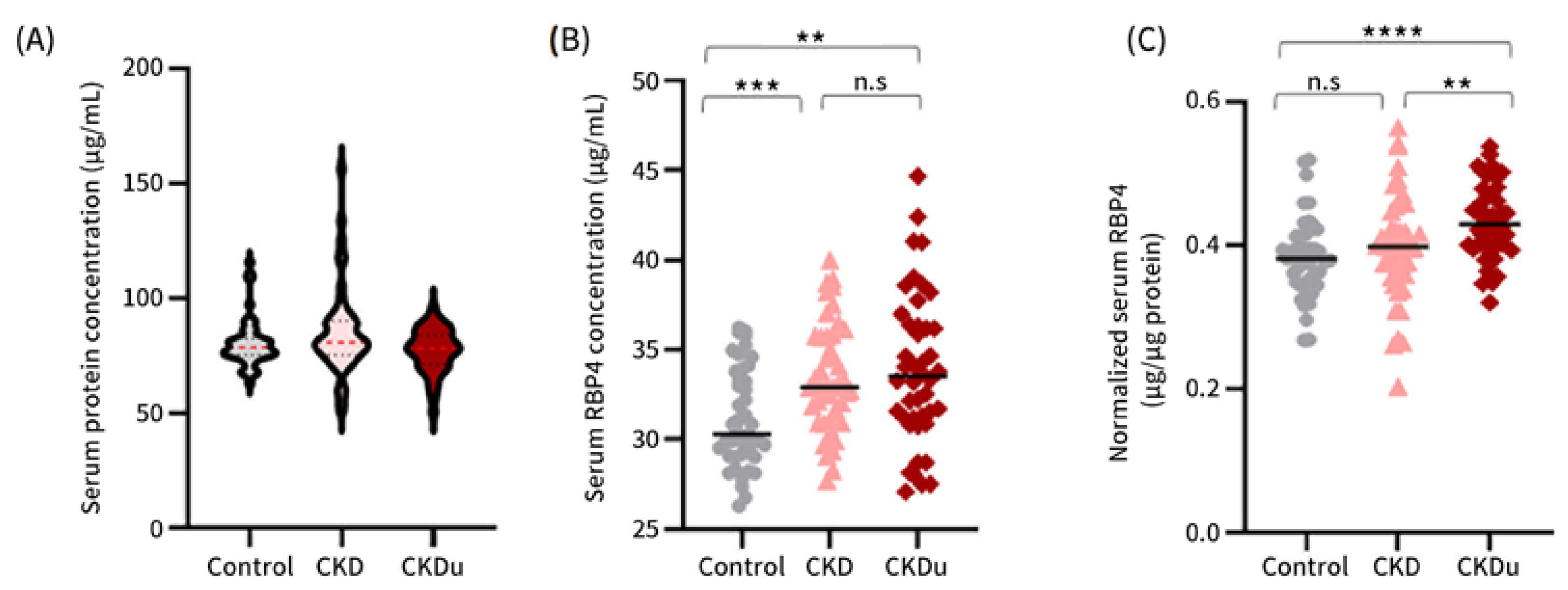
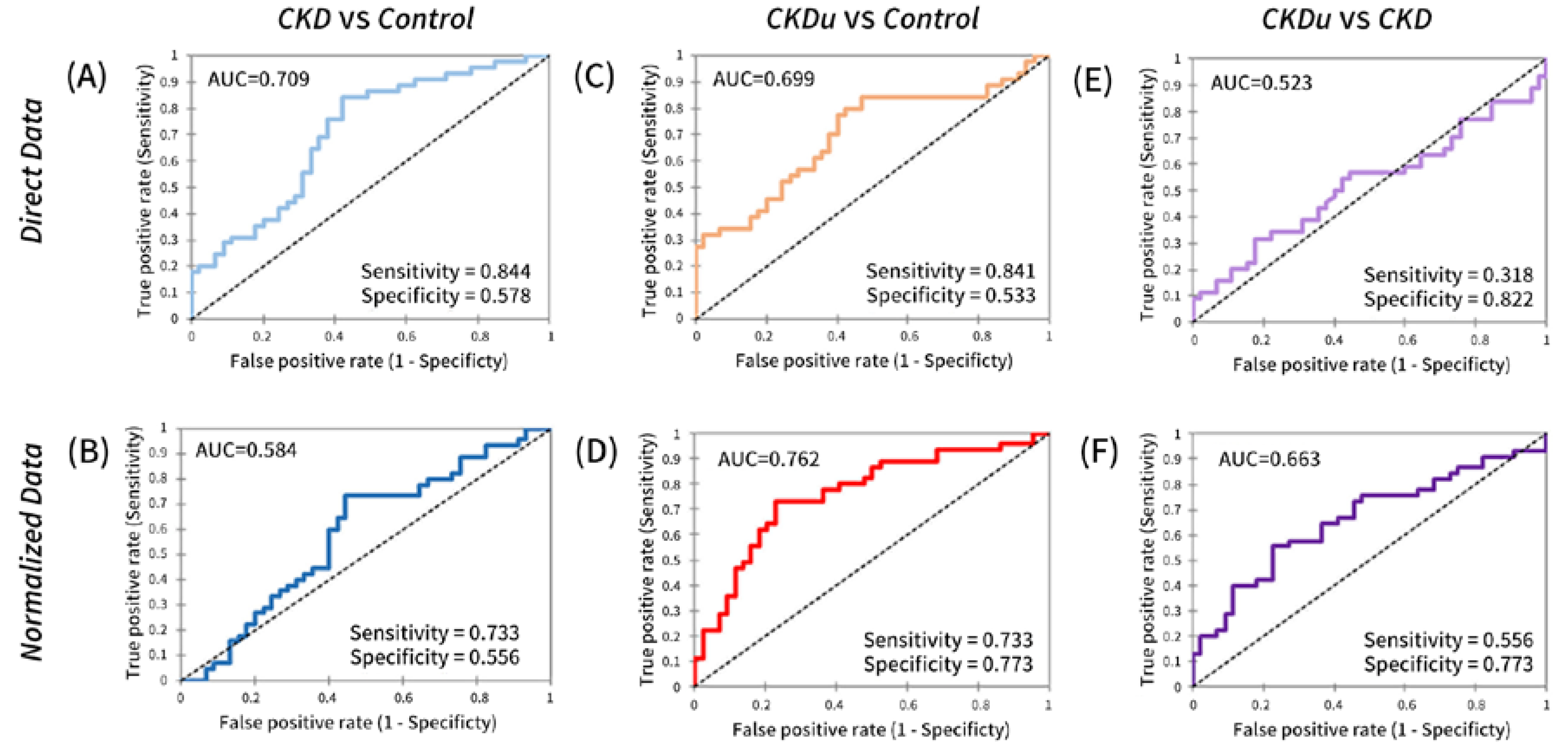
3.2. Serum RBP4 as a Potential Marker for Distinguishing between CKD and CKDu

In the pilot study, the serum of 78 CKDu patients was screened for a selected 16 serum marker candidates (Figure 1) based on their potential of applicability in CKDu using Luminex X-MAP technology.
Whilst many of the candidate markers fell within the detectable range of the assay, RBP4 readouts in all the serum samples exceeded the detection limit of the assay along with an exceedingly high level of NGAL in at least half of the samples. Based on this peculiar behaviour of RBP4, we were motivated to identify the prevalence of RBP4 in CKDu patients under controlled experimental settings [29].

3.3. Circulating RBP4 Levels and Significance in Study Cohorts

In the current study, we performed an RBP4-targeted immunoassay on a small cohort of serum samples from healthy controls (n = 45), CKD patients (n = 45) and CKDu patients (n = 44). It is interesting to note that there was a vast range of protein concentrations across the samples, with as much as a three-fold difference between the highest and lowest sera concentrations (Figure 2A). Such drastic variances motivated us to investigate if there would be any difference in our analysis outcomes based on direct data or after normalization against protein concentration. Total concentration of protein is likely to be decided by primary renal disease as well as nonrenal causes. Normalization of RBP4 to the protein concentration would diminish such artifacts in the results.
We observed that the serum RBP4 level was higher among CKDu patients than both healthy controls and CKD patients regardless of whether direct data or normalized data were considered (Figure 2B,C). The significance of serum RBP4 as a CKDu marker against healthy controls is obvious. It is interesting to report that serum RBP4 is capable of differentiating the CKD group from healthy groups and normalized RBP4 is capable of discriminating CKDu from CKD.

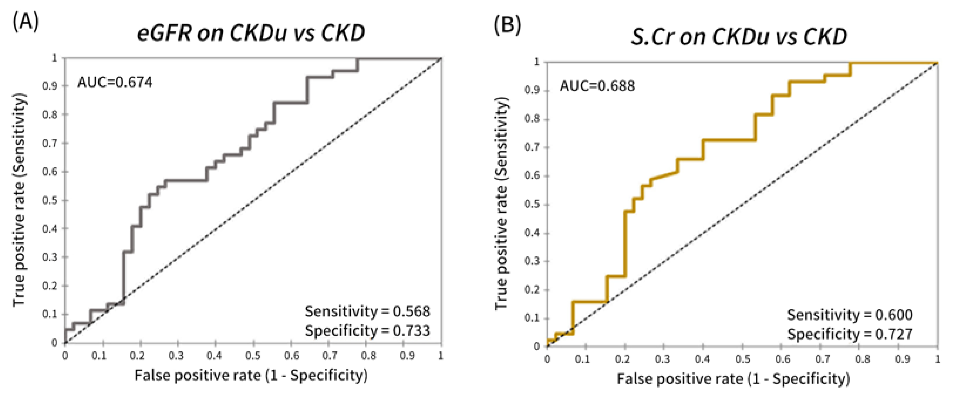
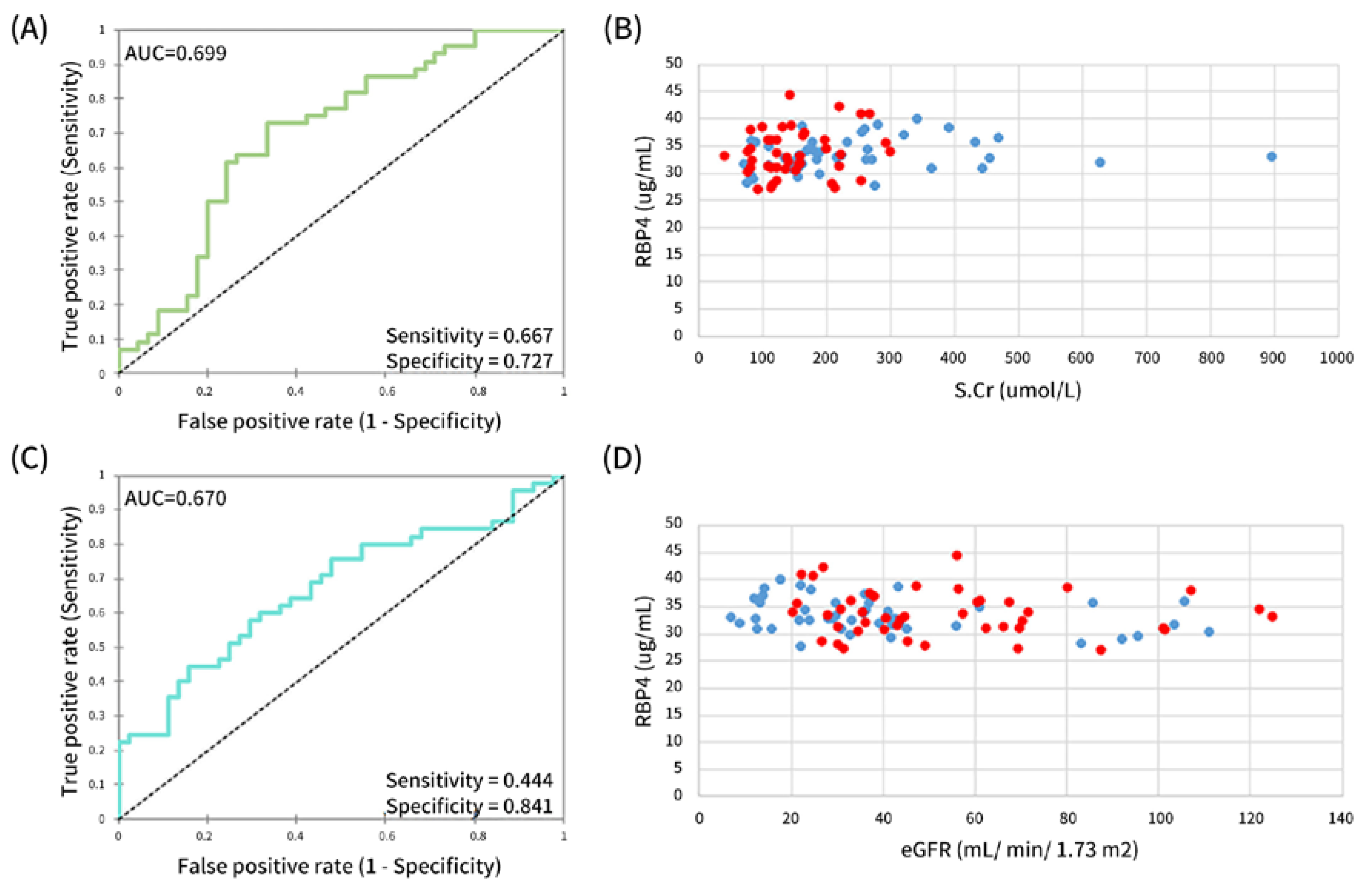
3.4. Performance of Serum RBP4 in Identifying Proximal Tubular Disease

To better understand if there was any impact in our findings, we looked at the overall performance of serum RBP4 in the evaluation of the extent of tubular interstitial involvement in CKDu.
The performance difference between serum RBP4 and creatinine, as a marker for differentiating CKDu (Figure 3A) from CKD with known causes (Figure 3C) or from healthy controls, was minimal (AUC 0.709 versus AUC 0.699). Moreover, their sensitivities (0.844 versus 0.841) and specificities (0.578 versus 0.533) were very similar. Despite the higher median of serum RBP4 level seen in CKDu (Figure 2B), its insignificant discriminatory power between CKD and CKDu is reflected in Figure 3E (AUC 0.523). In contrary, after normalization against protein serum, RBP4 appeared to be a more competitive marker for CKDu (Figure 3D; AUC 0.762, sensitivity 0.733) than CKD patients (Figure 3B; AUC 0.584, sensitivity 0.733, specificity 0.556) and healthy controls. Correspondingly, the discriminatory power of serum RBP4 for the two CKDs was also augmented in the normalized data (Figure 3F; AUC 0.663).

3.5. Synergy in Glomerular and Tubular Markers for Distinguishing between CKD and CKDu

As expected, serum creatinine was not able to be used to differentiate CKDu from known CKD (Figure 4A,B). The overall AUC, sensitivities and specificities for both did not differ much, suggesting they had comparable performances. Next, we looked at how serum RBP4 would perform with these two associated factors of CKD. Both direct and normalized serum RBP4 values were considered for this.
Figure 5 and Figure 6 (using direct and normalized serum RBP4 values, respectively) show the performances of ratio indices from glomerular and tubular indicators as discriminatory markers between known CKD and CKDu. For use of direct values, the RBP4:S.Cr (Figure 5A) index seemed to perform better than RBP4: eGFR (Figure 5C) as a discriminatory marker between the two CKDs despite similar AUCs (RBP4:S.Cr AUC 0.699 versus RBP4: eGFR AUC 0.670). This is reflected by the more balanced acceptable sensitivity and specificity (RBP4:S.Cr with sensitivity 0.727; specificity 0.667 versus RBP4: eGFR with sensitivity 0.444; specificity 0.841). Figure 5B,D depict the association between RBP4 and S.Cr or the eGFR, respectively, in CKD and CKDu patients. It is striking that there was a distinctive difference in the distribution of the direct relationship of these associated markers. Prominently, the compact distribution of RBP4 and S.Cr association (Figure 5B) as compared to that of RBP4 and eGFR (Figure 5D) in the CKDu group versus the CKD group puts forward the notion that the RBP4:S.Cr ratio is the more robust marker in discriminating CKDu from CKD.
A similar trend was observed when normalized serum RBP4 values were used. However, the overall performance of the RBP4:S.Cr index was elevated with an AUC of 0.743, sensitivity of 0.778 and specificity of 0.682 (Figure 6A). This robustness way surpasses that of the RBP4: eGFR ratio with an AUC 0.619 despite having a sensitivity of 0.864, as the specificity was only 0.444 (Figure 6C). The distribution pattern for the direct relationship of these associated factors remained robust when using normalized serum RBP4 values (Figure 6B,D), further affirming that the RBP4: S.Cr index is a robust putative discriminatory marker between CKD and CKDu.

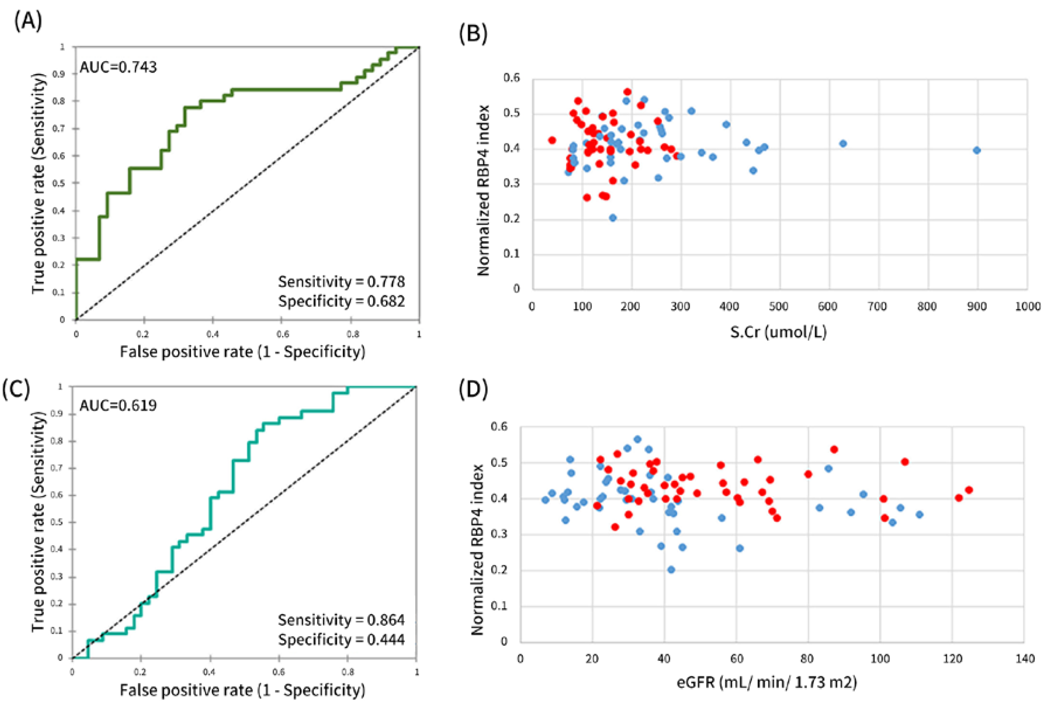
3.6. A Robust Putative Discriminatory Marker—Normalized RBP4:S.Cr Index

Table 2 shows an overview of performances of the associated markers. It is intriguing to observe distinct differences between using direct and normalized serum RBP4 values. Based on the analysis, the RBP4:S.Cr index using normalized RBP4 values emerged as the best-performing indicator for differentiating between CKD and CKDu.

4. Discussion

The term CKD categorically defines diverse kidney diseases with different pathophysiological manifestations under one umbrella using markers of kidney impairment, damage or imaging abnormalities. Broadly, CKDs can be further subdivided into glomerular, tubular interstitial and renovascular diseases based on the primary or predominant site of involvement. Even though it is important, there are no competent traditional markers able to confidently identify the severity or extent of damage to those specific compartments. More precisely, new biomarkers, which are promising candidates for this purpose, have to be evaluated intensively to identify their exact role in clinical practice. In our pilot study, we selected 16 serum biomarkers to evaluate their usefulness in CKDu. Most of the RBP4 concentrations were above the upper limit of quantification of the assay compared to the rest of the analytes in the panel. As this was unexpected, we conducted a case–control study to evaluate the performance of serum RBP4, a tubular maker, in combination with well-established markers, such as serum creatinine, eGFR and serum protein, in the evaluation of CKDu, a predominantly tubular interstitial disease.
According to our results, all three markers behaved independently in CKDu and known CKDs, affirming that our direct data do reflect what has been reported through ELISA studies: that serum RBP4 is a significant marker in discriminating CKD patients from healthy controls [29,37]. Furthermore, the authors used direct data to reach this conclusion. It is therefore intriguing to see that the significance diminished when normalized data were used. More interestingly, the normalized data showed that serum RBP4 behaves differently in CKD and CKDu (Figure 2C). As expected, there was a wide variation in serum protein levels in known CKDs, which is likely decided by the amount of glomerular protein leak. In contrast, in CKDu, a nonproteinuric tubular interstitial disease, serum protein levels were distributed within a narrow range and more comparable to the pattern in healthy individuals. As a result, normalized RBP4 against protein became a useful marker in differentiating CKDu from known CKDs. Further, mean RBP4 levels were significantly high in CKDu patients in comparison to both CKD patients and healthy controls, indicating the representativeness of serum RBP4 for the predominant site of involvement in CKDu, the proximal tubule [38]. Several previous studies have shown that elevated serum creatinine levels are associated with high serum RBP4 [29,30,31]. According to some studies [39,40], the association between serum creatinine and serum RBP4 is due to the loss of functional tissue in renal failure, which leads to decreased filtration and abnormal survival of small serum proteins [39,40]. According to our findings, high levels of RBP4 in CKDu can be explained by both reduced filtration and abnormal survival in proximal tubular disease.
RBP4 is a low-molecular-weight protein (21kDa) that is freely filtered through the glomeruli upon the release of retinol and subsequently reabsorbed readily by the proximal tubule and catabolized [25,26]. Theoretically, in glomerular sclerosis, tubules are atrophied; therefore, serum levels should represent glomerular functions. In tubular dysfunctions, either absorption or catabolism or both can produce disproportionate levels to the degree of kidney function. Hence, this would be extremely relevant in the context of primary tubulointerstitial diseases such as CKDu. Xun et al. (2018) showed that serum RBP4 is useful in the diagnosis of CKD as the concentration of serum RBP4 has an association with renal functional biomarkers. They suggested that combined detection of serum RBP4 and routine renal function biomarkers such as serum creatinine, urea and cystatin C would improve diagnostic accuracy for CKD [29].
Histological patterns are considered as the gold standard of diagnosis in most renal diseases. The absence of a biopsy in the diagnosis of CKD and relatively small sample size are limitations of this study. All the cases and controls were recruited according to the clinical and demographic data only. Occupational data were not considered. The absence of proteinuria results is another limitation of the current study.

5. Conclusions

There was evidence indicating the association between serum RBP4 and a reduced glomerular filtration rate. Our results not only support RBP4 as a renal biomarker, but also warrant further investigation of the capability of RBP4 in the evaluation of proximal tubular function. Further, we denote the significance of evaluating the complementary role of new biomarkers with traditional markers in complicated diseases such as CKD/CKDu.

Author Contributions

Conceptualization, B.N.T.F., A.A.-S., Z.B., J.G. and N.N.; Data curation, H.L.F.S. and A.A.-S.; Formal analysis, H.L.F.S., B.N.T.F., A.A.-S. and Z.B.; Funding acquisition, Z.B. and J.G.; Investigation, B.N.T.F., S.P., Z.B. and N.N.; Methodology, B.N.T.F., S.P. and A.A-S.; Project administration, J.G. and N.N.; Resources, B.N.T.F., S.P., J.G. and N.N.; Software, H.L.F.S., A.A.-S. and J.G.; Supervision, J.G. and N.N.; Validation, H.L.F.S.; Visualization, B.N.T.F. and N.N.; Writing—original draft, H.L.F.S., B.N.T.F. and A.A.-S.; Writing—review and editing, H.L.F.S., B.N.T.F., S.P., A.A.-S., Z.B., J.G. and N.N. All authors have read and agreed to the published version of the manuscript.

Funding

This research was funded by the Ministry of Health and National Research Council (Grant No. TO 14-05), Sri Lanka. Hannah L.F. Swa, Asfa Alli-Shaik and Jayantha Gunarathna are supported by the Institute of Molecular and Cell Biology, Agency for Science, Technology, and Research (A*STAR), Singapore. There are currently no funding sources for publication.

Institutional Review Board Statement

The study was conducted in accordance with the Declaration of Helsinki, and approved by the Ethic Review Committee of Faculty of Medicine, University of Peradeniya (2016/EC/28).

Informed Consent Statement

Written informed consent was obtained from all subjects involved in the study.

Data Availability Statement

Not applicable.

Acknowledgments

The authors are thankful to Lishantha Gunarathna and the staff of the Renal Center at Girandurukotte, Sri Lanka, and the staff of the Renal Unit, National Hospital Kandy, Sri Lanka. We also thank the President’s Task Force for their contribution in facilitating the research. This work was funded by the Ministry of Health, National Research Council (Grant No. TO 14-05) Sri Lanka, Presidential Task Force for CKDu prevention in Sri Lanka and Institute of Molecular and Cell Biology under the collaborative research agreement between Kandy National Hospital Sri Lanka and Institute of Molecular and Cell Biology Singapore.

Conflicts of Interest

The authors declare no conflict of interest. The funders had no role in the design of the study; in the collection, analyses or interpretation of data; in the writing of the manuscript; or in the decision to publish the results.

References

  1. Weaver, V.M.; Fadrowski, J.J.; Jaar, B.G. Global dimensions of chronic kidney disease of unknown etiology (CKDu): A modern era environmental and/or occupational nephropathy? BMC Nephrol. 2015, 16, 145. [Google Scholar] [CrossRef] [PubMed] [Green Version]
  2. Gooneratne, I.; Ranaweera, A.; Liyanarachchi, N.; Gunawardane, N.; Lanerolle, R. Epidemiology of chronic kidney disease in a Sri Lankan population. Int. J. Diabetes Dev. Ctries 2008, 28, 60–64. [Google Scholar] [CrossRef] [PubMed] [Green Version]
  3. Martin-Cleary, C.; Ortiz, A. CKD hotspots around the world: Where, why and what the lessonsare. A CKJ review series. Clin. Kidney J. 2014, 7, 519–523. [Google Scholar] [CrossRef] [PubMed] [Green Version]
  4. Tatapudi, R.R.; Rentala, S.; Gullipalli, P.; Komarraju, A.L.; Singh, A.K.; Tatapudi, V.; Goru, K.B.; Bhimarasetty, D.M.; Narni, H. High Prevalence of CKD of Unknown Etiology in Uddanam, India. Kidney Int. Rep. 2019, 4, 380–389. [Google Scholar] [CrossRef] [Green Version]
  5. Brooks, D.R.; Ramirez-Rubio, O.; Amador, J.J. CKD in Central America: A hot issue. Am. J. Kidney Dis. 2012, 59, 481–484. [Google Scholar] [CrossRef]
  6. Reddy, D.V.; Gunasekar, A. Chronic kidney disease in two coastal districts of Andhra Pradesh, India: Role of drinking water. Environ. Geochem. Health 2013, 35, 439–454. [Google Scholar] [CrossRef]
  7. Minshawy, E.I. End-stage renal disease in the El-Minia Governorate, upper Egypt: An epidemiological study. Saudi J. Kidney Dis. Transpl. 2011, 22, 1048–1054. [Google Scholar]
  8. Jayatilake, N.; Mendis, S.; Maheepala, P.; Mehta, F.R. Chronic kidney disease of uncertain aetiology: Prevalence and causative factors in a developing country. BMC Nephrol. 2013, 14, 180. [Google Scholar] [CrossRef] [Green Version]
  9. Wanigasuriya, K.P.; Peiris-John, R.J.; Wickremasinghe, R.; Hittarage, A. Chronic renal failure in North Central Province of Sri Lanka: An environmentally induced disease. Trans. R Soc. Trop. Med. Hyg. 2007, 101, 1013–1017. [Google Scholar] [CrossRef]
  10. Bandara, J.M.R.S.; Senevirathna, D.M.A.N.; Dasanayake, D.M.R.S.B.; Herath, V.; Abeysekara, T.; Rajapaksha, K.H. Chronic renal failure among farm families in cascade irrigation systems in Sri Lanka associated with elevated dietary cadmium levels in rice and freshwater fish (Tilapia). Environ. Geochem. Health 2008, 30, 465–478. [Google Scholar] [CrossRef]
  11. Edirisinghe, E.A.N.V.; Manthrithilake, H.; Pitawala, H.M.T.G.A.; Dharmagunawardhane, H.A.; Wijayawardane, R.L. Geochemical and isotopic evidences from groundwater and surface water for understanding of natural contamination in chronic kidney disease of unknown etiology (CKDu) endemic zones in Sri Lanka. Isotopes Environ. Health Stud. 2018, 54, 244–261. [Google Scholar] [CrossRef] [PubMed]
  12. Thammitiyagodage, M.G.; Gunatillaka, M.M.; Ekanayaka, N.; Rathnayake, C.; Horadagoda, N.U.; Jayathissa, R.; Gunaratne, U.K.; Kumara, W.G.; Abeynayake, P. Ingestion of dug well water from an area with high prevalence of chronic kidney disease of unknown etiology (CKDu) and development of kidney and liver lesions in rats. Ceylon Med. J. 2017, 62, 20–24. [Google Scholar] [CrossRef] [PubMed] [Green Version]
  13. Wasana, H.M.S.; Aluthpatabendi, D.; Kularatne, W.M.T.D.; Wijekoon, P.; Weerasooriya, R.; Bandara, J. Drinking water quality and chronic kidney disease of unknown etiology (CKDu): Synergic effects of fluoride, cadmium and hardness of water. Environ. Geochem. Health 2016, 38, 157–168. [Google Scholar] [CrossRef] [PubMed]
  14. Wasana, H.; Perera, G.D.; Gunawardena, P.D.S.; Fernando, P.S.; Bandara, J. WHO water quality standards Vs Synergic effect(s) of fluoride, heavy metals and hardness in drinking water on kidney tissues. Sci. Rep. 2017, 7, 42516. [Google Scholar] [CrossRef] [PubMed]
  15. Kulathunga, M.R.D.L.; Wijayawardena, M.A.A.; Naidu, R.; Wijeratne, A.W. Chronic kidney disease of unknown aetiology in Sri Lanka and the exposure to environmental chemicals: A review of literature. Environ. Geochem. Health 2019, 41, 2329–2338. [Google Scholar] [CrossRef]
  16. Fogo, A.B. Mechanisms of progression of chronic kidney disease. Pediatr. Nephrol. 2007, 22, 2011–2022. [Google Scholar] [CrossRef] [Green Version]
  17. Anand, S.; Montez-Rath, M.E.; Adasooriya, D. Prospective Biopsy-Based Study of Chronic Kidney Disease of Unknown Etiology in Sri Lanka. Clin. J. Am. Soc. Nephrol. 2019, 14, 224–232. [Google Scholar] [CrossRef] [Green Version]
  18. Nanayakkara, S.; Komiya, T.; Ratnatunga, N.; Senevirathna, S.T.M.L.D.; Harada, K.H.; Hitomi, T.; Gobe, G.; Muso, E.; Abeysekera, T.; Koizumi, A. Tubulointerstitial damage as the major pathological lesion in endemic chronic kidney disease among farmers in North Central Province of Sri Lanka. Environ. Health Prev. Med. 2012, 17, 213–221. [Google Scholar] [CrossRef] [Green Version]
  19. Wijkström, J.; Leiva, R.; Elinder, C.-G.; Leiva, S.; Trujillo, Z.; Trujillo, L.; Söderberg, M.; Hultenby, K.; Wernerson, A. Clinical and pathological characterization of Mesoamerican nephropathy: A new kidney disease in Central America. Am. J. Kidney Dis. 2013, 62, 908–918. [Google Scholar] [CrossRef]
  20. Nanayakkara, S.; Senevirathna, L.; Karunaratne, U.; Chandrajith, R.; Harada, K.H.; Hitomi, T.; Watanabe, T.; Abeysekara, T.; Aturaliya, T.N.C.; Koizumi, A. Evidence of tubular damage in the very early stage of chronic kidney disease of uncertain etiology in the North Central Province of Sri Lanka: A cross-sectional study. Environ. Health Prev. Med. 2012, 17, 109–117. [Google Scholar] [CrossRef] [Green Version]
  21. Fernando, B.N.T.W.; Alli-Shaik, A.; Hemage, R.K.D.; Badurdeen, Z.; Hettiarachchi, T.W.; Abeysundara, H.T.K.; Abeysekara, T.D.J.; Wazil, A.; Rathnayake, S.; Gunaratne, J.; et al. Pilot Study of Renal Urinary Biomarkers for Diagnosis of CKD of Uncertain Etiology. Kidney Int. Rep. 2019, 4, 1401–1411. [Google Scholar] [CrossRef] [PubMed]
  22. Sayanthooran, S.; Magana-Arachchi, D.N.; Gunerathne, L.; Abeysekera, T. Potential diagnostic biomarkers for chronic kidney disease of unknown etiology (CKDu) in Sri Lanka: A pilot study. BMC Nephrol. 2017, 18, 31. [Google Scholar] [CrossRef] [PubMed] [Green Version]
  23. De Silva, P.M.C.S.; Abdul, K.S.M.; Eakanayake, E.M.D.V.; Jayasinghe, S.S.; Jayasumana, C.; Asanthi, H.B.; Perera, H.S.D.; Chaminda, G.G.T.; Chandana, E.P.S.; Siribaddana, S.H. Urinary Biomarkers KIM-1 and NGAL for Detection of Chronic Kidney Disease of Uncertain Etiology (CKDu) among Agricultural Communities in Sri Lanka. PLoS Negl. Trop. Dis. 2016, 10, e0004979. [Google Scholar] [CrossRef] [PubMed] [Green Version]
  24. Wanigasuriya, K.; Jayawardene, I.; Amarasiriwardena, C.; Wickremasinghe, R. Novel urinary biomarkers and their association with urinary heavy metals in chronic kidney disease of unknown aetiology in Sri Lanka: A pilot study. Ceylon Med. J. 2017, 62, 210–217. [Google Scholar] [CrossRef] [Green Version]
  25. Paci, E.; Greene, L.H.; Jones, R.M.; Smith, L.J. Characterization of the molten globule state of retinol-binding protein using a molecular dynamics simulation approach. FEBS J. 2005, 272, 4826–4838. [Google Scholar] [CrossRef]
  26. Goodman, D.S. 8 Plasma Retinol-Binding Protein. In The Retinoids; Sporn, M.B., Roberts, A.B., Goodman, D.S., Eds.; Academic Press: New York, NY, USA, 1984; pp. 41–88. [Google Scholar]
  27. Klisic, A.; Kavaric, N.; Ninic, A. Retinol-binding protein 4 versus albuminuria as predictors of estimated glomerular filtration rate decline in patients with type 2 diabetes. J. Res. Med. Sci. 2018, 23, 44. [Google Scholar] [CrossRef] [PubMed]
  28. Abbasi, F.; Moosaie, F.; Khaloo, P.; Firouzabadi, F.D.; Abhari, S.M.F.; Atainia, B.; Ardeshir, M.; Nakhjavani, M.; Esteghamati, A. Neutrophil Gelatinase-Associated Lipocalin and Retinol-Binding Protein-4 as Biomarkers for Diabetic Kidney Disease. Kidney Blood Press Res. 2020, 45, 222–232. [Google Scholar] [CrossRef]
  29. Xun, C.; Zhao, Y.; Wang, W.; Chen, C. Circulating RBP4 Increase and Its Diagnosis of Chronic Kidney Disease. Ann. Clin. Lab. Sci. 2018, 48, 205–207. [Google Scholar]
  30. Smith, F.R.; Goodman, D.S. The effects of diseases of the liver, thyroid, and kidneys on the transport of vitamin A in human plasma. J. Clin. Investig. 1971, 50, 2426–2436. [Google Scholar] [CrossRef]
  31. Stewart, W.K.; Fleming, L.W. Plasma retinol and retinol binding protein concentrations in patients on maintenance haemodialysis with and without vitamin A supplements. Nephron 1982, 30, 15–21. [Google Scholar] [CrossRef]
  32. Jayasekara, K.B.; Dissanayake, D.M.; Sivakanesan, R.; Ranasinghe, A.; Karunarathna, R.H.; Kumara, G.W.G.P. Epidemiology of chronic kidney disease, with special emphasis on chronic kidney disease of uncertain etiology, in the north central region of Sri Lanka. J. Epidemiol. 2015, 25, 275–280. [Google Scholar] [CrossRef] [PubMed] [Green Version]
  33. Levey, A.S.; Stevens, L.A.; Hostetter, T. Automatic reporting of estimated glomerular filtration rate--just what the doctor ordered. Clin. Chem. 2006, 52, 2188–2193. [Google Scholar] [CrossRef] [PubMed]
  34. Redmon, J.H.; Levine, K.E.; Lebov, J.; Harrington, J.; Kondash, A. A comparative review: Chronic Kidney Disease of unknown etiology (CKDu) research conducted in Latin America versus Asia. Environ. Res. 2021, 192, 110270. [Google Scholar] [CrossRef] [PubMed]
  35. Levey, A.S.; Stevens, L.A.; Schmid, C.H.; Zhang, Y.L.; Castro, A.F., 3rd; Feldman, H.I.; Kusek, J.W.; Eggers, P.; Van Lente, F.; Greene, T.; et al. A new equation to estimate glomerular filtration rate. Ann. Intern Med. 2009, 150, 604–612. [Google Scholar] [CrossRef] [PubMed]
  36. Behrens, G.A.; Brehm, M.; Groß, R.; Heider, J.; Sauter, J.; Baier, D.M.; Wehde, T.; Castriciano, S.; Schmidt, A.H.; Lange, V. Non-invasive determination of CMV serostatus from dried buccal swab samples: Assay development, validation and application to 1.2 million samples. J. Infect Dis. 2021, 224, 1152–1159. [Google Scholar] [CrossRef] [Green Version]
  37. Jing, J.; Isoherranen, N.; Robinson-Cohen, C.; Petrie, I.; Kestenbaum, B.; Yeung, C. Chronic Kidney Disease Alters Vitamin A Homeostasis via Effects on Hepatic RBP4 Protein Expression and Metabolic Enzymes. Clin. Transl. Sci. 2016, 9, 207–215. [Google Scholar] [CrossRef]
  38. Vervaet, B.A.; Nast, C.C.; Jayasumana, C.; Schreurs, G.; Roels, F.; Herath, C.; Kojc, N.; Samaee, V.; Rodrigo, S.; Gowrishankar, S.; et al. Chronic interstitial nephritis in agricultural communities is a toxin-induced proximal tubular nephropathy. Kidney Int. 2020, 97, 350–369. [Google Scholar] [CrossRef] [Green Version]
  39. Vahlquist, A.; Peterson, P.A.; Wibell, L. Metabolism of the vitamin A transporting protein complex. I. Turnover studies in normal persons and in patients with chronic renal failure. Eur. J. Clin. Investig. 1973, 3, 352–362. [Google Scholar] [CrossRef]
  40. Waldmann, T.A.; Strober, W.; Mogielnicki, R.P. The renal handling of low molecular weight proteins. II. Disorders of serum protein catabolism in patients with tubular proteinuria, the nephrotic syndrome, or uremia. J. Clin. Investig. 1972, 51, 2162–2174. [Google Scholar] [CrossRef] [Green Version]
Figure 1. Heatmap showing expression level of serum analytes with respect to limit of detection.
Kidneydial 02 00052 g001
Figure 2. Sera protein and serum RBP4 level. (A) Violin plot showing distribution of sera protein levels in control and diseased cohorts. (B,C) Boxplots showing distribution of RBP4 in control and diseased cohorts; ** p < 0.01, *** p < 0.001, **** p < 0.0001, n.s = not significant.
Kidneydial 02 00052 g002
Figure 3. Serum RBP4 performance as marker. (A,B) ROCs showing performance of RBP4 as a marker for CKD. (C,D) ROCs showing performance of RBP4 as a marker for CKDu. (E,F) ROCs showing performance of RBP4 as a discriminatory marker between the two chronic kidney diseases. Raw RBP4 values are used for (A,C,E), and normalized RBP4 indices are used for (B,D,F).
Kidneydial 02 00052 g003
Figure 4. Serum creatinine (S.Cr) and estimated glomerular filtration rate (eGFR) performance as a discriminatory marker between the 2 CKD diseases. (A) ROC showing performance of S.Cr as a discriminatory marker for CKD and CKDu. (B) ROC showing performance of eGFR as a discriminatory marker between the two chronic kidney diseases.
Kidneydial 02 00052 g004
Figure 5. Ratio index as discriminatory marker between the 2 CKD diseases using direct RBP4 values. ROCs showing performance of RBP4:S.Cr ratio (A) and RBP4:eGFR ratio (C) as discriminatory markers for CKD and CKDu. Distribution of RBP4:S.Cr (B) and RBP4:eGFR ratios (D) in CKD (blue) and CKDu (red). Direct values of RBP4 are used.
Kidneydial 02 00052 g005
Figure 6. Ratio index as discriminatory marker between the 2 CKD diseases using normalized RBP4 values. ROCs showing performance of RBP4:S.Cr (A) and RBP4:eGFR ratios (C) as discriminatory markers for CKD and CKDu. Distribution of RBP4:S.Cr (B) and RBP4:eGFR ratios (D) in CKD (blue) and CKDu (red). Normalized values of RBP4 are used.
Kidneydial 02 00052 g006
Table 1. Demographic and clinical characteristics of the study subjects.
VariableAverage ± SD or Cases (%)
CKDu
(n = 44)
NECKD
(n = 45)
NEC
(n = 45)
Age, years51.7 ± 9.349.62 ± 13.548.1 ± 12.1
Gender distribution, males40 (90.9)23 (51)18 (40)
Family history of CKD22 (50)7 (15.5)4 (8.9)
Smoking21 (47.7)11 (24.4)7 (15.5)
Chewing betel36 (81.8)6 (13.3)23 (51)
Alcohol consumption19 (43.2)8 (17.8)12 (26.7)
Hypertension14 (31.8)36 (80)0 (0)
Diabetes mellitus2 (4.5)21 (46.7)0 (0)
History of malaria18 (40.9)7 (15.6)0 (0)
SD: standard deviation, CKDu: chronic kidney disease of uncertain etiology, NECKD: nonendemic chronic kidney disease, NEC: nonendemic control.
Table 2. Overview of performances of the associated markers.
S.CreGFRRaw RBP4Normalized RBP4
RBP4RBP4:SCrRBP4:eGFRRBP4RBP4:SCrRBP4:eGFR
CKD vs. NECAUC0.9390.9170.7090.9340.9160.5840.9370.897
Sensitivity0.8000.8440.8440.8000.8440.7330.8440.800
Specificity1.0000.9780.578 1.0000.9780.5560.9330.978
CKDu vs. NECAUC0.9090.8830.6990.8800.8930.7620.8470.912
Sensitivity0.7500.8410.8410.7500.8410.7330.8670.841
Specificity0.9560.9110.5330.9330.8890.7730.7270.956
CKD vs. CKDuAUC0.6880.6740.5230.6990.6700.6630.7430.619
Sensitivity0.6000.5680.8220.6670.4440.5560.7780.864
Specificity0.7270.7330.3180.7270.8410.7730.6820.444
S.Cr, serum creatinine; eGFR, estimated glomerular filtration rate; RBP4, retinol-binding protein 4; AUC, area under the curve; CKD, chronic kidney disease; CKDu, chronic kidney disease of uncertain etiology; NEC, nonendemic control.
Publisher’s Note: MDPI stays neutral with regard to jurisdictional claims in published maps and institutional affiliations.

