Editorial

Renal Nutrition—Where It Has Been and Where It Is Going †

by
Joel D. Kopple
1,2,3,* and
Maryam Ekramzadeh
1
1
Division of Nephrology and Hypertension, The Lundquist Institute, Harbor-UCLA Medical Center, Torrance, CA 90502, USA
2
David Geffen School of Medicine, University of California, Los Angeles, CA 90095, USA
3
Fielding School of Public Health, University of California, Los Angeles, CA 90095, USA
*
Author to whom correspondence should be addressed.
Presented, in part, at the 67th Annual Meeting of the Japanese Society of Dialysis Therapy, Yokohama, Japan, 1–3 July 2022.
Kidney Dial. 2022, 2(4), 512-533; https://doi.org/10.3390/kidneydial2040046
Submission received: 24 September 2022 / Accepted: 29 September 2022 / Published: 11 October 2022

Abstract

:
This paper is a synopsis of an invited lecture entitled, The Future of Renal Nutrition, that was presented at the Japanese Society of Dialysis Therapy, July 2022. The purpose of this presentation is to suggest some of the advances in the field of renal nutrition that the authors think are likely to occur during the next several years. There will be continued development of methods for precisely diagnosing and classifying protein-energy wasting and developing methods to treat this disorder. Why weight loss commonly occurs when the GFR decreases to about 30–35 mL/min/1.73 m2 and why substantial weight loss (>5%/year) is associated with increased mortality will be investigated. Clinical consequences of the interactions between gut microbiota, nutrient intake and other environmental influences will continue to be examined. The clinical value of diets high in fruits and vegetables or other plants for chronic kidney disease (CKD) patients will continue to be studied. Our knowledge of how different diets and medicines affect intestinal absorption, metabolism and excretion of nutrients will expand. Precision medicine will be extended to precision nutrition. There will be more focus on the effects of nutritional disorders and dietary treatment on the emotional status and quality of life of people with kidney disease and their families. Nutritional centers that provide centralized nutritional assessment and dietary counselling for CKD patients may develop in more urban centers. More clinical trials will be conducted to test whether nutritional management improves clinical outcomes in people with kidney disease. It is hoped that the foregoing comments will encourage more research on these topics.

Graphical Abstract

1. Introduction

My task in this presentation is to predict future development in the field of renal nutrition. This is, at best, a hazardous exercise, as it is impossible to guess what new discoveries and novel ways of thinking may alter the direction of this field, which might be occurring even as this paper is being written. Intellectual history is rife with examples of how predictions regarding the future course of science and medicine have been erroneous, not uncommonly egregiously so. Nonetheless, since this is our assignment, we will, with humility and with the recognition that, at the least, some of our speculations are certain to be wrong, try to describe some of the future directions of this field. This paper will briefly describe the recent history of renal nutrition. We will then speculate as to what developments may occur in the near future. This paper will end by describing some of the more distant developments that possibly may occur in this discipline. Our hope is that these speculations may emphasize some of the areas in renal nutrition of great need of advancement and possibly stimulate some of the younger scientists and leaders in this field to solve some of the challenges that lie before us.

2. The History of Renal Nutrition Since the 19th Century Encompasses Several Periods

Since the 19th century, two different views have been advanced regarding the potential benefits of dietary treatment for people with chronic kidney disease (CKD) [1]. 1. Protection of the health and function of the diseased kidney. 2. Preservation or improvement of the overall health of the patient. For over 100 years, the thinking of workers in this field has often fluctuated widely between these two potential goals. Particular attention was initially focused on reduction in the quantity of protein and modification of the type of protein in the diet. From at least the last part of the 19th century until about the “teens” of the 20th century, dietary protein restriction was recommended to protect the kidney, primarily from what was considered to be overwork. It was thought that in people with kidney disease, a higher protein intake, by increasing the generation of urea which is excreted by the kidney, will increase the work of the diseased kidney and may thereby further injure the kidney. It is now understood, of course, that renal urea excretion requires little or no energy expenditure by the kidney [1].
From about the “teens” of the 1900s until the very late 1940s, low protein diets (LPDs) were still recommended to protect the diseased kidney from more rapid progression of kidney failure. However, unlike in rodents, some studies did not show that LPDs slowed progression in humans. In the 1950s, LPDs (about 40 g protein/day) and control of mineral intake (primarily sodium and potassium) were primarily recommended to maintain metabolically and clinically healthier patients with CKD [1].
In the 1960s, with the advent of chronic dialysis therapy and renal transplantation, the focus of renal nutrition changed to maintaining a healthy metabolic and nutritional status in advanced CKD patients with the use of protein, sodium and potassium restriction and provision of adequate calories. Recommended protein restriction by different renal nutrition workers was sometimes about 40 g per day and sometimes about 20 g per day (viz., the Giovannetti-Giordano Diet) [2,3,4,5,6]. It was contended that such diets could reduce symptoms and maintain advanced CKD patients in much healthier and asymptomatic states. Thus, if CKD patients eventually needed dialysis or transplantation, they would be physically better prepared for these treatments.
In the late 1960s and early 1970s, it became apparent that the 20 g protein/day diet provided inadequate amounts of protein [7,8]. Three alternatives to dietary protein restriction were developed, each of which was shown to be nutritionally adequate:
i.
A low protein diet (LPD) providing about 0.55–0.60 g protein/kg/day diet (~40 g protein/day) [7,8].
ii.
A very low protein diet (VLPD) providing about 20 g protein/day diet supplemented with about 16–20 g/day of essential amino acids (EAA). It was referred to as a EAA supplemented VLPD (SVLPD) [9,10,11].
iii.
A VLPD providing about 20 g protein/day diet supplemented with about 16–20 g/day of a mixture of ketoacid and hydroxyacid analogues (KAs) of some EAA plus other EAA; this was called a KA supplemented VLPD (SVLPD) [12,13].
Shortly thereafter, Walser observed that KA SVLPDs seemed to slow the rate of progression of kidney failure [13]. Walser’s observations precipitated a firestorm of clinical trials. Some, but not all, studies reported slowing of progressive CKD with the ketoacid SVLPD. These studies led, in turn, to the United States NIH-funded large scale MDRD Study [14]. The results of the MDRD Study were ambiguous, in part because of several inadvertent flaws that were incorporated into the design of the study [14,15]. Since the MDRD study, other clinical trials examined whether LPDs or SVLPDs can retard progression of CKD. Several of these trials showed effectiveness of SVLPDs in slowing progression [16,17,18,19]. A confounding factor is that certain medicines given to CKD patients may mimic some of the physiologic effects of LPDs on glomerular hemodynamics (e.g., angiotensin converting enzyme inhibition or blockade, or the use of sodium-glucose cotransporter-2 (SGLT2) inhibitors), thus making it more difficult to assess the independent effects of LPDs or SVLPDs on progression of CKD.
There were other advances concerning the nutritional management of renal disease that developed concurrently with the clinical trials of LPDs and SVLPDs for nondialyzed CKD patients. These advances included greater understanding and more effective treatments of the nutritional, physiological and metabolic processes concerning:
i.
Patients with acute kidney injury (AKI)
ii.
Maintenance hemodialysis (MHD) patients
iii.
Chronic peritoneal dialysis (CPD) patients
iv.
Kidney transplant recipients
v.
Children with CKD
vi.
Macrominerals, especially sodium, potassium and phosphorus, in kidney disease
vii.
Vitamins and trace elements, especially iron, in kidney disease
viii.
Furthermore, developed during this time was the description of the syndrome of protein-energy wasting (PEW) in CKD [20]
ix.
Identification of the relationships between PEW, inflammation and adverse clinical outcomes.

Precision Nutrition

Precision nutrition is based upon precision medicine and is an approach to nutritional assessment, care and research that is designed for individual patients or subgroups of patients, instead of a one-treatment-fits-all model [21]. Precision nutrition attempts to involve the complex set of factors that comprise an individual, including the person’s genotype, phenotype, psychodynamics, psychosocial and economic status and past and anticipated future life experiences, among other items. Precision nutrition can be considered a general goal of renal nutrition research and also of the nutritional management of people with or at increased risk for developing kidney disease [22].

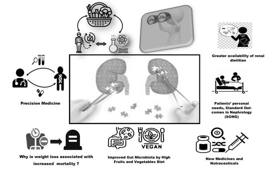
3. The Future of Renal Nutrition—The Immediate Future

3.1. Role of New Medicines That Modify Nutrient Biochemistry or Physiology

What may lie ahead in the immediate future regarding renal nutrition? Perhaps the following: Medicines that modify the intestinal absorption, urinary excretion or metabolism of nutrients will continue to be developed. The development of such pharmaceuticals has already been underway for over one century. These medicines include diuretics, intestinal binders of phosphate and potassium, HMG Coenzyme A inhibitors (statins), medicines that simulate (or replicate) the action of LPDs on glomerular hemodynamics (e.g., ACE inhibition or blockade, SGLT2 inhibitors), and purified vitamins or analogs of such vitamins as pyridoxine HCl, folate, cholecalciferol, and calcitriol (1,25-dihydroxycholecalciferol), which may have slightly different and more clinically desirable characteristics.

3.2. Reexamination of the Classification and Diagnostic Criteria for Protein-Energy Wasting (PEW) in Kidney Disease

The rationale for this reexamination is that PEW is a powerful risk factor for mortality [23,24,25]. As we have learned more about PEW syndrome, the classification of PEW and the current diagnostic criteria for PEW have turned out to be somewhat inadequate. There appears to be a need for more highly developed criteria. This process for more precise classification and diagnostic criteria is being conducted right now. It is led by Professor Denis Fouque and Dr. Laetitia Koppe from Lyon, France.

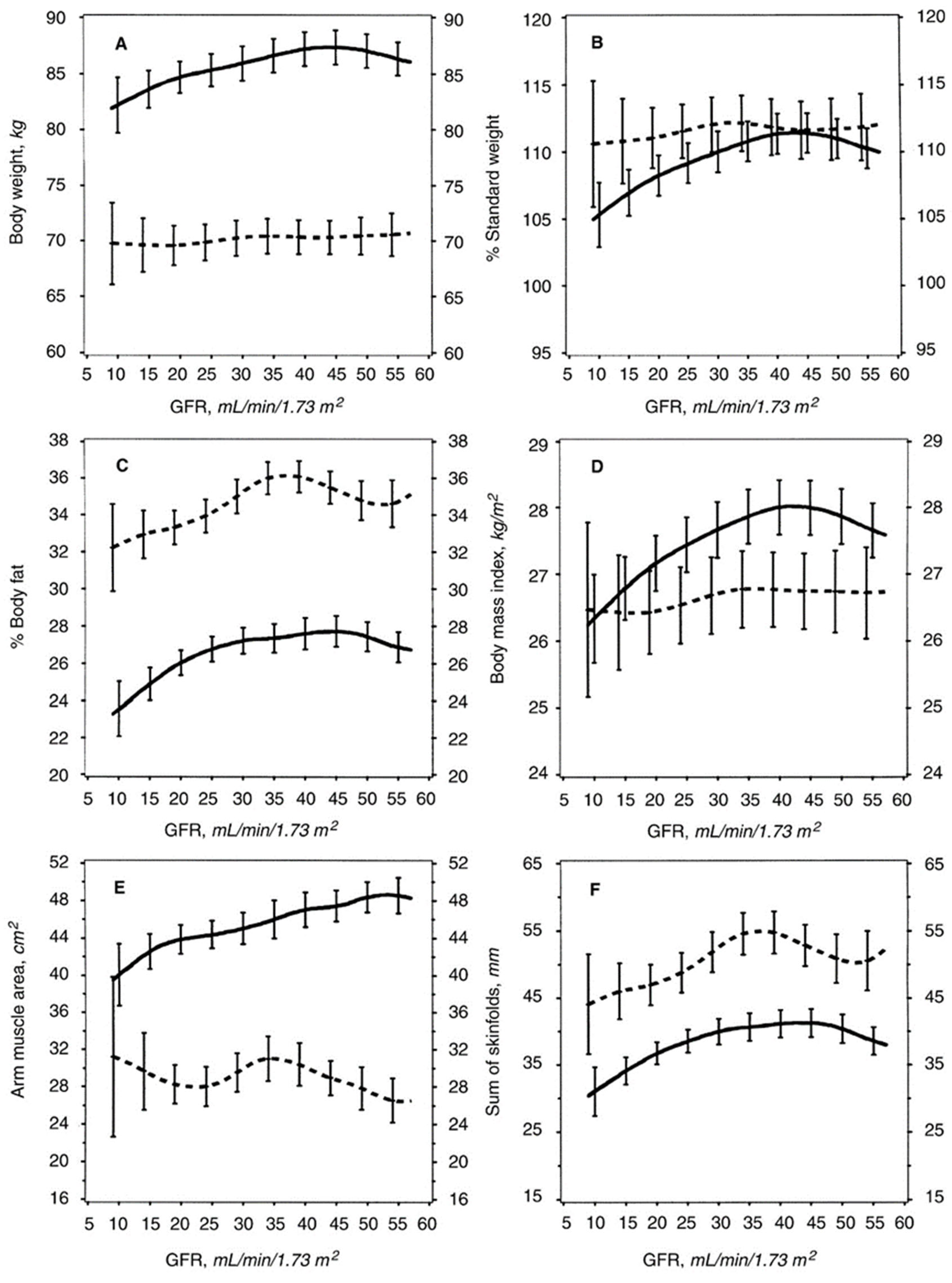
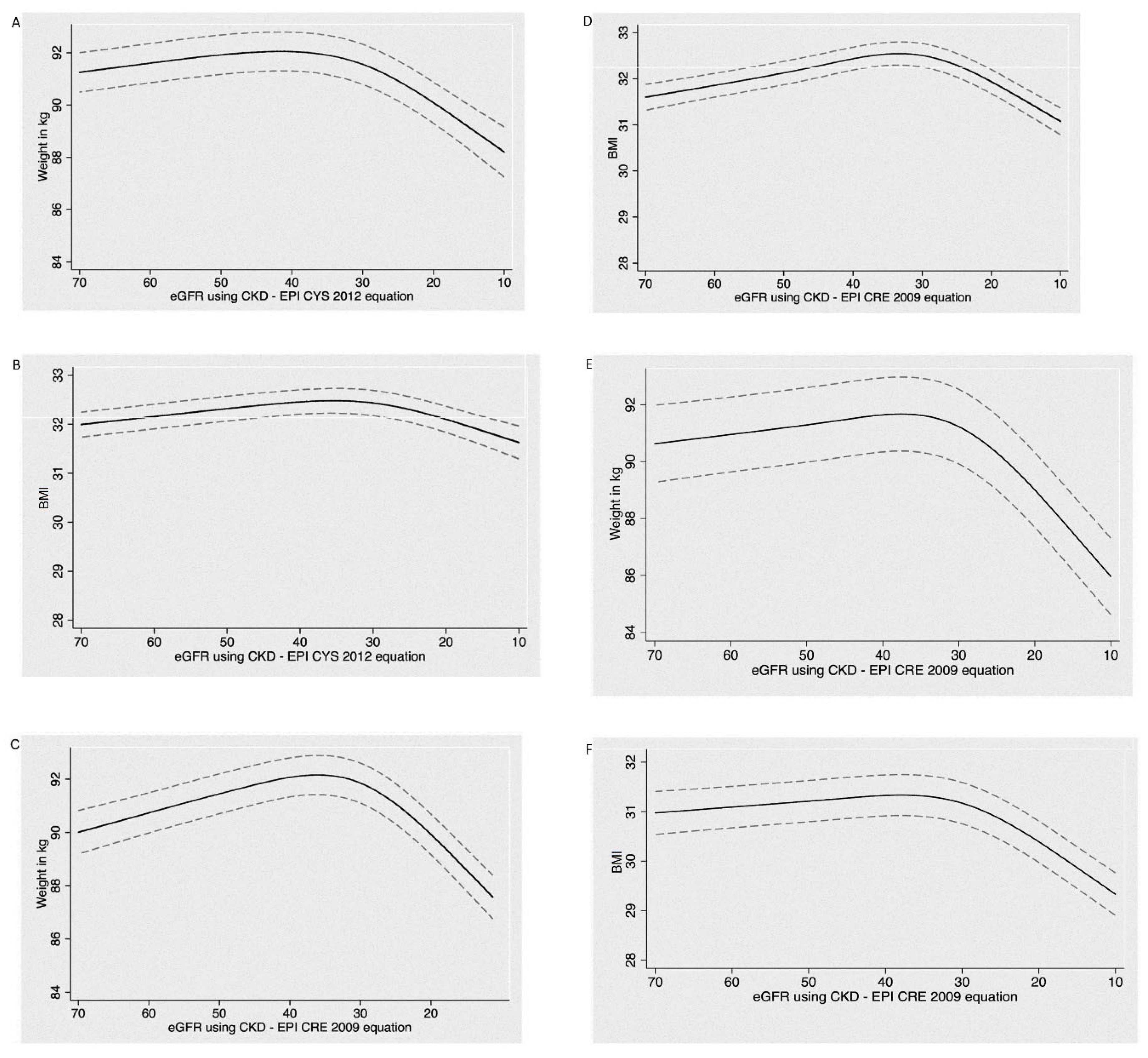
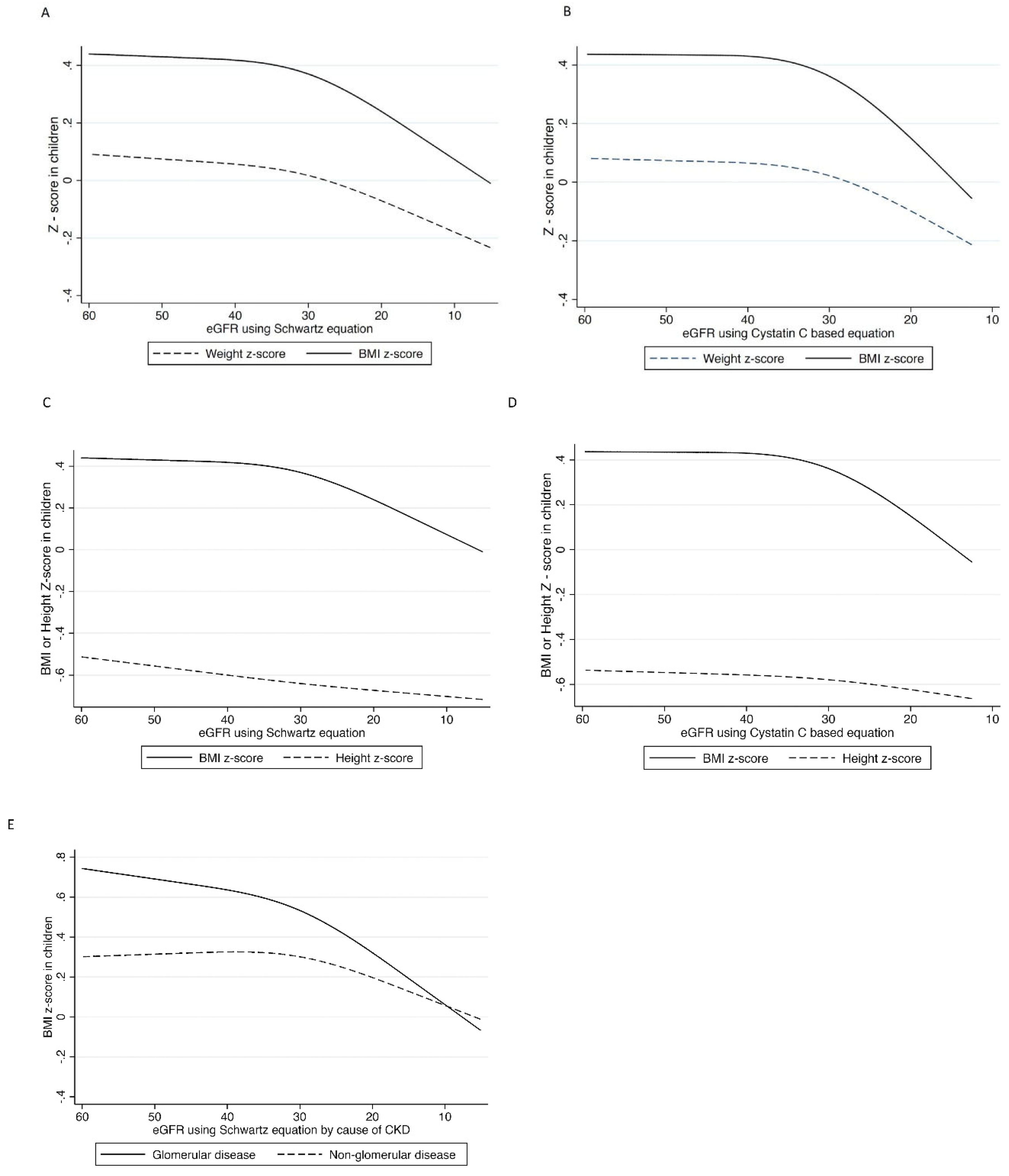
3.3. Examination of Why CKD Patients Often Lose Weight When the GFR Decreases to about 25–32 mL/min/1.73 m2, and Why a Large Weight Loss Is Associated with Increased Mortality?

The majority of CKD patients appear to lose edema-free weight as they approach end-stage kidney disease (ESKD) [26]. These latter patients not uncommonly have at least some mild signs or symptoms of uremia and may have anorexia from uremic toxicity. Accumulation of uremic toxins may contribute to this weight loss. Figure 1 indicates the results of a cross-sectional evaluation of the relationship between true GFR and body mass in 1760 CKD patients undergoing screening for the MDRD Study [26]. The solid lines indicate men, and the broken lines indicate women. When the GFR, determined by 125I-iothalamate clearances, reaches about 30–35 mL/min/1.73 m2, body weight, expressed either directly or as a percentage of the standardized weight of normal people of the same sex, age range, height and frame size as the patients, often begins to decrease or starts to decrease at a more rapid rate. The same findings are observed when body weight is expressed as a percentage of previous body weight or body mass index. Dietary protein intake began to decline, particularly in men but also in women, around a GFR of 30–35 mL/min/1.73 m2. This decline was also observed for dietary energy intake at about this GFR level in men and at a somewhat lower GFR level in women [26]. Also many pediatric and adult CKD patients lose weight when their GFR decreases to about 30–35 mL/min/1.73 m2 (Figure 2 and Figure 3) [27,28].
Similar results were obtained when the relationship between changes in estimated GFR and changes in weight and BMI were examined in almost 4000 people with CKD who participated in the CRIC study (Panels A–D) or in 1067 people with CKD in the AASK study (Panels E–F) (Figure 2) [27]. The data obtained from both groups of patients are longitudinal; the GFR and body weight in most of these patients were measured more than once. Again, when the GFR fell to about 30–35 mL/min/1.73 m2 in both the CRIC study and the AASK study there was a decline in body weight [27]. Similar results were obtained when 854 children under 16 years who had an eGFR between 30–90 mL/min/1.73 m2 and who were followed in the CKiD study in children [28] (Figure 3). At about the same eGFR level, their BMI began to decrease [28].
These data are not limited to the American experience. In the KNOW-CKD Study from South Korea, over 1800 patients with CKD stages 1–5 were evaluated [29]. None were undergoing dialysis. In this cross-sectional study, as the eGFR fell and the CKD stages increased starting with stage 3, the percentage of patients with serum albumin levels less than 3.8 g/dL began to increase. The percentage with low serum albumin levels jumped in the patients with stage 4 (eGFR 15–29 mL/min/1.73 m2) and stage 5 CKD (eGFR <15 mL/min/1.73 m2). Similarly, the number of patients diagnosed with PEW was rather low in South Korean patients with CKD stages 1 and 2, increased modestly in those with stages 3A and 3B, and then increased rather abruptly in stage 4 CKD patients, and rose even higher in stage 5 CKD. In stage 5 CKD, one out of four patients was diagnosed with PEW [29].
We have observed that adult CKD patients with eGFR of about 30–35 mL/min/1.73 m2 and greater weight loss (>5% per year) had a 54% increase in mortality rate [27]. Children with an eGFR of about 30–35 mL/min/1.73 m2 and greater weight loss (a decline in the z-score of BMI of > 0.2 kg/m2 per year) had a 3.28 times greater increase in the rate of development of ESKD. In addition, weight loss that occurs in advanced CKD patients shortly after they commence chronic dialysis therapy is also associated with increased mortality [30].

3.4. Why Do CKD Patients Often Lose Weight When GFR Falls to about 30–35 mL/min/1.73 m2?

CKD patients who lose weight when their GFR decreases to about 30–35 mL/min/1.73 m2 may look normal unless they are suffering from the consequences of a systemic disease. The cause of their weight loss is unknown. Reduced energy intake is a likely cause of the weight loss, but the mechanism responsible for a reduction in energy intake in these patients is not clear. The potentially toxic metabolites that are normally excreted in urine and often accumulate in advanced CKD as the GFR, and hence urinary toxin excretion, decreases would not be expected to be sufficiently high in the body to cause anorexia in people with an eGFR of about 30–35 mL/min/1.73 m2. This raises the question of whether the weight loss is due to anorexia caused by increased or reduced levels of one or more circulating hormones. Serum levels of many hormones may be increased at this level of reduced GFR [31,32] The paper by Kuro-O and Moe regarding bone mineral metabolism in kidney disease may be particularly illustrative in this regard [33,34].
Theoretically, anorexia and weight loss could be due to elaboration of proinflammatory cytokines in the kidney or elsewhere in the body. However, these patients seem to feel well and generally do not appear or act as if they are systemically inflamed. Another possibility is alterations in the gut microbiome that might alter energy metabolism or cause anorexia (see below).
The kidney is an endocrine organ that elaborates erythropoietin, renin and 1,25-dihydroxycholecalciferol. Is it possible that the kidney also normally elaborates an orexigenic (appetite stimulating) hormone, and perhaps when diseased or damaged, there is a reduction in the synthesis or release of this hormone by the kidney. Alternatively, could the kidney start to produce appetite suppressing hormones at this time? Although there is no evidence for these possibilities, they have never been systematically explored. The hypothesis is consistent with the weight loss occurring in CKD patients with beginning stage 4 CKD, which is a point at which the renal parenchyma can be expected to be extensively damaged. Research will be necessary to answer this question.

3.5. Why Is Large Weight Loss Associated with Increased Mortality in These CKD Patients?

In the CRIC study, CKD patients who had a GFR <35 mL/min/1.73 m2 and who lost greater than 5% of their body weight per year, the risk of death was increased by 54% in comparison to people with no weight change. In the AASK study, CKD patients who had a GFR <35 mL/min/1.73 m2 and who had an annualized percent weight loss greater than 5% also displayed a 56% increase in their adjusted mortality [27]. Interestingly, those people who gained weight also had increased mortality. The cause for this latter finding is particularly obscure; this weight gain might reflect accumulation of edema fluid in these patients [27].
In the CKiD study, using two different models of adjusted odds ratios, children with CKD and an eGFR <35 mL/min/1.73 m2 who had a decline in their BMI z-score >0.2 per year had a 3.28 times greater risk of developing ESRD in comparison to children who had a stable BMI (0–0.1 z-score per year) [28]. Children who displayed a more moderate degree of weight loss or who gained body weight also showed a trend toward increased risk of ESRD in comparison to children with little or no weight change, although this trend was not statistically significant [28].

3.6. The Human Microbiome

What is the microbiome in humans? The adult human body is composed of about 30–40 trillion cells. Vastly greater amounts of microbial organisms, about one hundred trillion microbial cells, live in or on the human body [35]. Most of these microbes live in the gut, but they are also found in or on the mouth, vagina, auditory canals, skin, etc. [35]. In humans, there are ≥100 times more different genes in the gut microbiome than in the entire human genome [36,37]. Most of these microbes cannot be cultured and are identified by their DNA or RNA. However, they elaborate enzymes which may be bioactive in humans. The gut microbiota have the three domains of life: Archaea, Eubacteria, and Eukaryotes [36], and normally are rather similar in different people. However, diet, antibiotics, other medicines (such as metformin), hormones, aging, and many illnesses including CKD and acute kidney injury (AKI), can alter the gut microbiome [38]. Animal studies suggest that altered gut microbiome activity may injure the kidney and also promote conversion of a person with AKI to CKD [39]. No information is available as to whether other, non-gut, microbiomes may injure the kidney or affect the health of CKD patients. Whether nutritional intervention can modify the relation between the gut microbiome, and the risk of developing CKD or the progression of AKI and CKD and thereby protect the human kidney needs to be investigated [38,40,41].
Differences have been observed in the microbiota and the gut wall when normal people ingest a diet poor in quality versus a healthy diet. A healthy diet, predominantly composed of fruits, vegetables, fibers, plant-derived proteins, monounsaturated fatty acids (MUFAs) and n-3 polyunsaturated fatty acids (n3-PUFAs), has been associated with high microbial diversity, resistance to colonization of gut with pathogenic bacteria, immune homeostasis, a healthy mucus layer adherent to the gut wall, and a healthy gut barrier [41]. In contrast, a less healthy diet that contains more animal-derived protein (meat, including processed meat), saturated fats, refined grains, sugar, salt, alcohol and corn-derived fructose has been associated observationally with reduced microbial diversity, increased likelihood of colonization by other pathogenic microbes, inflammation, erosion of the protective mucus layer, and increased intestinal permeability [41].

3.7. The Gut Microbiome in CKD

In people with advanced CKD, several factors involving the diseased kidney and the gut predispose to increased concentrations of potentially toxic compounds:
i.
Microbiota in the gut become altered (dysbiosis). The gut microbes may synthesize increased amounts or different types of compounds [42]
ii.
These newly formed metabolites may be absorbed from the gut into the circulation [42,43,44].
iii.
Blood levels of some compounds may increase due, in part, to reduced renal excretion and probably to increased synthesis [42,44].
There is degradation of the protective mucous layer adjacent to the gut wall and altered intestinal permeability. These changes may facilitate passage of intestinal bacterial matter and chemicals into the blood stream. Among the most studied of the altered chemicals produced in the CKD gut are trimethylamine and trimethylamine-N-oxide (TMAO) derived from carnitine and choline, p-cresyl sulfate and P-cresyl-glucuronide from tyrosine, and indoxyl sulfate, and indole-3-aldehyde produced from tryptophan. The gut microbiota participate in the synthesis of these compounds, and the liver usually also contributes to their formation. All of these metabolites are normally excreted by the kidney. In the presence of impaired GFR, they accumulate. Animal studies indicate that many of these gut-derived uremic toxins may induce chronic inflammation and cause nephrotoxicity in CKD [42,43,44].

3.8. Plant-Based Diets for CKD Patients

Diets that are high in plant-based sources of protein can generate less acid in the body, reduce the acid burden and/or decrease intestinal phosphate absorption. A more appropriate name may be High Fruit and Vegetable Diet (HFVD). As conceived by Donald Wesson, MD, who has published extensively on the acid-base ramifications of this diet, the HFVD is high in fruits and vegetables but is not very low in protein. By Dr. Wesson’s design, the total protein content of this diet is about 0.8–0.9 g/kg/day, including the protein provided by the fruit and vegetables (Wesson, personal communication). One reason we prefer to not just call this a plant dominant (PLAYDO) diet is that grains, which include such foods as bread, rolls, bagels, cakes and donuts, are also plants, and they may provide an acid load and therefore do not contribute to the alkalizing properties of this diet.
Table 1 from Wesson [45] shows the calculated potential renal acid load of selected relevant foods, per 100 g of an edible portion, and whole diets. Meat and seafood generate about 13.6 mmoles of acid. Dairy products also engender an acid load. In contrast, an equivalent amount of vegetables provide an alkali (negative acid) load (−24.9 mmoles acid), and fruits provide a smaller amount of alkali (−1.81 mmoles). Oils are virtually neutral with regard to acid or base production (+0.01 mmoles). As indicated in the table, grain-based foods produce a modest acid load (+6.3 mmoles) [45]. Table 1 also indicates the average intake of these acid generating foods in a rather typical United States diet, expressed in terms of acid production per each serving of a meal. As can be seen there is a generation of about 27 mmoles of acid per meal per day from a typical diet, and grains contribute about 6.4 mmoles to this acid load. A typical Mediterranean diet, which is particularly high in vegetables and fruits, low in meat/seafood, and contains a lot of grains, is essentially neutral in acid production. The DASH diet, which is high in vegetables, fruits, and low-fat dairy products, generates more acid due to the high meat or seafood content in the diet. A vegan diet provides a smaller acid load, which could be even lower if the content of grain-based foods is reduced (Table 1) [45].

3.9. The Problem of Adherence to High Fruit and Vegetable Diets (HFVDs)

In the experience of one author (JDK), roughly 15% of CKD patients will adhere to a LPD (0.60 g protein/kg/d). Dr Wesson estimates that around one-third of his CKD patients will adhere to an HFVD for an extended period of time; i.e., for at least one year (personal communication). However, these latter patients tend to have low incomes, and the fruit and vegetable portion of their diet is given to them at no cost, even for as long as one year or more. It is not known what degree of long-term adherence to a HFVD can be expected if patients must personally pay for their fruits and vegetables and also ingest a LPD; for example a total daily protein intake closer to 0.60 g protein/kg/day rather than the 0.8–0.9 g protein/kg/day that CKD patients were apparently eating with the HFVD. For individuals who may have difficult adhering to the HFVD, the alkalizing benefits of this diet can be attained, without focusing so intensely on fruit and vegetable intake, by providing alkali, such as sodium bicarbonate or a sodium citrate/citric acid solution, or an alkali binding resin (veverimer). Another issue is the degree to which a HFVD or a high plant food diet will suppress intestinal absorption of phosphate. In this regard, the role for grain- and nut-based foods for CKD patients’ needs to be investigated. Let us examine these questions.

3.10. What Type of Plant-Based Diets May Lower Dietary Phosphorus Uptake?

Grain-Based Foods

There has been much interest in phytates recently because they bind avidly to phosphate and reduce intestinal phosphate absorption. Table 2 shows the phytate content of various plant foods. Nuts and grain-based foods contain rather large amounts of phytate, in contrast to coconut, corn, strawberries, and polished rice that have rather small amounts [46,47,48,49]. On the other hand, grains provide an acid load (Table 1) [45]. So it would seem that the plant foods that may reduce intestinal phosphate absorption by binding phosphate with phytate may not be as effective at providing an alkaline load. Some potential benefits and limitations of plant-based diets are summarized in Table 3.

3.11. Effect of the Source of Dietary Phosphorus on Urinary Phosphorus Excretion

There have been several publications regarding the different degrees of intestinal phosphate absorption of organically bound phosphate in plant vs. animal foods and of inorganic phosphate salts [50,51,52]. We are of the opinion that the role of phytate in reducing phosphorus absorption and of the inorganic form of phosphate in enhancing phosphate absorption may be somewhat overstated (A. Shah et al.; unpublished observations). In most studies, the magnitude of intestinal phosphate absorption was estimated from the changes in urinary phosphate excretion. To assess more precisely the effects of these different forms of phosphorus on intestinal phosphate absorption, it would be helpful to measure net phosphorus absorption more directly in these studies; for example, by measuring dietary phosphorus intake minus fecal phosphorus excretion. These studies have yet to be done in CKD patients.

3.12. Can Medicines Substitute for Foods to Reduce Acidosis-Induced Progression of CKD or to Prevent or Treat Hyperphosphatemia?

Randomized clinical trials (RCTs) demonstrate that alkaline salts or solutions, such as sodium bicarbonate, sodium citrate and solutions of sodium citrate and citric acid, prevent or treat acidosis and slow the progression of CKD [53,54,55,56,57,58]. Other alkaline preparations, such as calcium citrate [59], potassium citrate and citric acid [60], potassium bicarbonate [61] and calcium bicarbonate [62], in rodent models of renal injury progression also slow the progression of chronic renal injury. Of course, many of these latter alkaline preparations may be contraindicated in CKD patients, because the associated cations may be toxic.
There appear to be at least two mechanisms responsible for the slowing of progression of kidney disease with alkalinization. Acidification is associated with increased synthesis and excretion of ammonium ion in the kidney which appears to stimulate complement deposition in the kidney with consequent development of interstitial fibrosis [63,64]. Acidification of the kidney also promotes the synthesis and release of endothelin 1 in the kidney which also stimulates interstitial fibrosis in the kidney [58,65,66].
With regard to the use of phosphate lowering medicines rather than diet to prevent or correct hyperphosphatemia, there are innumerable studies that show the effectiveness of these medicines at preventing or correcting hyperphosphatemia [67,68,69,70]. Although dietary phosphorus restriction is generally a necessary component of treatment to prevent or correct hyperphosphatemia in both far advanced CKD and chronic dialysis patients, the use of phosphate binders is almost always necessary as well. It is not known whether inclusion in the diet of large amounts of plants rich in phytate will enable some patients to completely avoid use of phosphate binders.

3.13. Summary of Discussion on the Use of High Plant Diets to Control Acidosis and Hyperphosphatemia in Advanced CKD and Chronic Dialysis Patients

  • A HFVD, as defined by Wesson and colleagues, is documented to reduce or prevent acidosis in CKD patients [71,72]. Since the PLAYDO diet may contain lower amounts of fruits and vegetables than the HFVD described by Wesson et al. [72] and may contain substantial animal-based protein, it is not clear how effective the PLAYDO diets are collectively at reducing the acid load and therefore, decreasing net body protein catabolism and slowing progression of CKD. It should be emphasized that grain-based foods are also plant foods, but still provide some acid (Table 1) [45].
  • Any diet, vegan or omnivorous, that provides 0.60 g protein/kg/day is likely to be deficient in calcium and certain essential micronutrients and may require supplements to prevent calcium and micronutrient deficiencies. Such LPDs that are composed entirely or almost entirely of plant foods may also be deficient in some essential amino acids, especially methionine and lysine [73]. It is therefore important that the primarily plant-based LPDs prescribed for CKD patients should be designed with the assistance of an experienced renal dietitian.
  • According to the tastes and preferences of the CKD patient, the potential benefits of plant dominant diets on alkalinization, decreased constipation, reduced intestinal phosphate absorption can be replicated with omnivorous LPDs that are augmented with alkali supplements, supplemental fiber, and intestinal phosphate binders.
  • To our knowledge, with the exception of the HFVD described by Wesson et al., there are no RCTs that demonstrate beneficial clinical outcomes (e.g., reduced rate of loss of GFR, less adverse cardiovascular events, decreased mortality) with PLADO diets as compared to similar LPDs that contain less plant protein but that have the same protein content and contain the medicines or supplements necessary to control blood pH, serum phosphate, bone-mineral disease, and fecal flow.
  • The difficulty with the term, PLAYDO Diet, is the amount of plant foods present in the diet is not well-specified. There is a similar concern with the HFVD. Perhaps it would be helpful if the amount of plant food or plant food protein in these diets was defined more precisely.

3.14. Renal Nutrition in the More Distant Future?

The following predictions can be considered highly speculative and could easily be disproven by future developments.
There will be increased development and use of medicines to facilitate the body’s handling of nutrients. The following are examples of these types of medicines that are already in use:
1.
Surveys of chronic dialysis patients indicate that food and fluid restriction are not uncommonly onerous [74,75,76]. In this regard, there are intestinal binders for potassium [77,78,79] and phosphorus [67,68,69,70]. An inhibitor of the Na/H exchanger iso-form 3 (NHE3) in the small intestine may suppress intestinal phosphate absorption [80]. Inhibitors of sodium absorption are under development [81,82,83,84]. Veverimer may bind hydrogen ion in the gut [85]. Diuretics, especially loop diuretics, may enhance the renal excretion of sodium, chloride and potassium, even in chronic dialysis patients who are not anuric.
2.
Sodium-glucose co-transporter-2 (SGLT2) inhibitors stimulate tubular glomerular feedback to reduce intraglomerular hypertension and protect the kidney [86,87,88]. Glucagon-like peptide-1 (GLP-1) receptor agonists may improve serum glucose control [89,90,91]. Selective mineral corticoid antagonists may decrease blood pressure, improve glomerular hemodynamics and reduce renal fibrosis [92,93,94]. Hypoxia-inducible factor-proline hydroxylase inhibitors (HIF PHIs) enhance intestinal iron absorption and may improve anemia of CKD [95,96]. Calcimimetic medicines (cinecalcet, etalcalcetide) are used to treat hyperparathyroidism [97,98]. Oral, enteral or parenteral nutritional supplements are given to improve nutritional intake and prevent malnutrition in CKD patients. Dialysis treatments may also become more nutritionally relevant. For example, dialysate solutions may provide additional nutrients [99,100].
3.
There will be more sophisticated methods for assessment of the patient’s nutritional status of micronutrients. For example, more effective methods may be developed for:
-
Simple measurements of serum or blood cell concentrations that can accurately indicate the body burden of an individual micronutrient.
-
Measurements may be developed to indicate the functionality, as well as the blood and individual tissue concentrations, of a micronutrient and also the presence of inhibitors or other modifiers of the physiology, metabolism or actions of micronutrients. As examples, in the uremic state, retained endogenous metabolites, increased hormone levels, medicines or metabolites of medicines might inhibit or enhance actions of micronutrients [101,102,103].
-
Altered Vitamin Function or Metabolism in CKD/ESKD.
Examples of altered vitamin function in CKD and ESKD include:
  • Erythrocyte transketolase (ETK) activity which requires the presence of the vitamin, thiamine pyrophosphate. However, low ETK activity has been found in patients who have normal blood thiamine levels [104].
  • There is an increased daily need for pyridoxine hydrochloride (vitamin B6) in advanced CKD and chronic dialysis patients that exceeds the Recommended Dietary Allowance of vitamin B6 for normal adults [105].
  • Membrane tetrahydrofolate (THF) transport is reported to be inhibited in advanced CKD [106].
  • There is decreased vitamin B12 uptake by blood monocytes in advanced CKD patients [107].
  • There is a reduced synthesis of 1,25(OH)2D3 by the diseased kidney [108]. In rats with CKD, impaired intestinal absorption of a number of vitamins, including riboflavin [109], folate [110] and vitamin D [111], have been described.
  • Many medicines or other compounds, such as hydralazine, isoniazid and methotrexate, may inhibit or impair the actions of vitamin B6 [101,102] or folate [112].
These considerations have a number of implications should stimulate further research:
  • How do these alterations affect dietary needs for the respective vitamins or, for that matter, trace elements?
  • How can we assess the daily vitamin and trace element needs and the state of vitamin and trace element nutriture of CKD and chronic dialysis patients? By function tests? By blood levels?
  • How can we know whether CKD or chronic dialysis patients are receiving adequate amounts of every essential micronutrient?
4.
There will be more definitive investigations of the role of the microbiota, and particularly the gut microbiome, as contributing causes of AKI or CKD, of the clinical manifestations of these disease states, and of the general health of AKI and CKD patients. The potential role of nutrition and nutrients for modifying the microbiome and its pathogenetic or health-enhancing effects in AKI and CKD patients will continue to be investigated.

3.15. Is Nutritional Care Clinically Valuable?

We are of the opinion that a major reason why nutritional care is not as widely practiced for CKD patients is that many nephrologists are unconvinced that the benefits of nutritional prescription to the patient outweigh its negative effects. Even the use of LPDs and VLPDs to slow progression of renal failure in nondialyzed CKD patients can be considered controversial [14,15,113,114,115]. Previous reports suggest that nutritional support reduces the mortality risk in chronic dialysis patients with PEW [116]. However, these reports are almost all retrospective analyses of studies that were not randomized prospective controlled clinical trials (RPCTs). Although some of these studies were very well analyzed, it is hoped that in the future, there will be more RPCTs that test whether various types of nutritional management for CKD, chronic dialysis and AKI patients or renal transplant recipients are beneficial. It is likely that such trials will also inform the medical community of more effective ways to deliver nutritional care to people with kidney disease.

3.16. The SONG (Standard Outcomes in Nephrology) Initiative May Become Operative in Renal Nutrition

This initiative (viz., SONG) “aims to establish a set of core outcomes and outcome measures across the spectrum of kidney disease for trials and other forms of research. The outcomes will be developed based upon the shared priorities of patients, caregivers, clinicians, researchers, policy makers, and other relevant stakeholders. This will help to ensure that research is reporting outcomes that are meaningful and relevant to patients with kidney disease, their family, and their clinicians to support decisions about treatment” [117].

3.17. What May Be the Effects of SONG on Renal Nutrition?

The effects that SONG may have on renal nutrition are uncertain. It is clear that most CKD and chronic dialysis patients do not like to eat their prescribed diets [74,75,76]. We suspect that in the future there will be a greater focus on improving the comfort and enjoyment that CKD and chronic dialysis patients derive from their dietary intake. Will the combination of medicines (e.g., inhibitors of intestinal absorption of certain mineral or compounds, drugs that increase renal excretion) and chemically modified yet tasteful foods (e.g., low in various minerals and protein and higher in calorie content) enable CKD and chronic dialysis patients to eat more enjoyably? There may be increased publication of renal nutrition research on social media, and this may stimulate more interest and greater sophistication concerning renal nutrition among patients and their families.

3.18. Major Challenges to the Nutritional Treatment in CKD

Finally, there will continue to be logistical challenges to instituting nutritional therapy for advanced CKD and chronic dialysis patients. A major challenge to dietary therapy for CKD and chronic dialysis patients is the fact that many nephrologists are not very knowledgeable concerning the potential benefits of nutritional management and lack enthusiasm for prescribing renal nutrition for their patients. The time needed for a physician to introduce to CKD patients the concept of dietary therapy with its major ramifications, including explaining the importance of good adherence to the prescribed diet, can provide a disincentive for the physician to prescribe nutritional therapy. Furthermore, effective renal nutritional management requires the involvement of knowledgeable dietitians, and there is a paucity of dietitians who are experienced and sophisticated in renal nutrition therapy, especially for CKD patients who are not undergoing chronic dialysis. Moreover, many dietitians have only limited time that they can allocate to an individual patient. Many CKD patients have malaise, and they and their families or significant others may lack the motivation to acquire, prepare and eat specialized diets. The dietary prescription is restrictive of the types, variety and amounts of foods that many patients desire. Healthier foods and food supplements often cost more. Unfortunately, the less expensive foods tend to be the less healthy foods.
Figure 4 and Figure 5 are from a study published by Angela Wang and coworkers in 2022 [118]. These figures summarize data obtained from a survey from representatives from 155 countries that responded to a series of questionnaires. This survey showed that the availability for CKD patients of renal dietitian support, or even the access of CKD patients to dietitians who do not have special expertise in renal nutrition, is actually quite low in the great majority of countries and particularly in low-income countries. The availability of dietetic counseling even by a person with some training in dietetics or nutrition and who is not necessarily a dietitian or nutritionist was only identified in 14% of low income countries. Among lower-middle income countries, this number rises to 31%. People surveyed described availability of dietetic counselling for CKD patients in 71% of upper-middle income countries and 84% of high-income countries (Figure 5). In contrast, in low-income countries for the great majority of CKD patients, consultations with a dietitian is either usually not available or never available. Some low-income countries, which may be rather populous, do not have a single university program to train students to become dietitians.
The availability of dietitians or renal dietitians for nutritional care of non-dialyzed CKD and chronic dialysis patients varies enormously according to the patients’ income status. The availability of renal dietitians for CKD and chronic dialysis patients is less than 10% for low-income patients, about 17–20% for lower-middle income patients, about 39–54% for upper-middle income patients, and about 72–77% for high income patients (Figure 5). Approximately one quarter of kidney disease patients have no access to a renal dietitian or any dietitian for their nutritional care in low income countries (Figure 4) [118]. It is the authors’ experience that even in high income countries, or among CKD patients with high income, it is often difficult to access a knowledgeable dietitian who is experienced in the nutritional management of CKD patients who are not undergoing chronic dialysis.

3.19. A Different Institutional System for Nutritionally Managing People with Kidney Disease

Recently, a novel and potentially effective institution was developed in Mexico for managing the nutritional needs of non-hospitalized CKD and chronic dialysis patients and kidney transplant recipients: the Fresenius Kabi Nutritional Care Centers (CEANs). Most patients have CKD and are not receiving dialysis, but some are chronic dialysis patients or kidney transplant recipients. Patients are referred to the CEAN for nutritional care by any nephrologist in the metropolitan area. When the patient comes to this center, a renal dietitian conducts a nutritional, social and pharmaceutical history, performs anthropometry, bioelectric impedance (BIA) studies, assesses handgrip strength and reviews the patient’s biochemical data. A nutritional diagnosis is then made, and a plan for personalized nutritional care is formulated. A consultation letter is sent to the referring nephrologist, usually within several workdays. The patient is counselled on his/her prescribed diet by the renal dietitian. Nutritional counseling with the patient is performed monthly until dietary adherence becomes acceptable. Patients are then seen every 3, 4 or 6 months depending upon the level of their dietary adherence and their clinical condition. Dietary adherence is assessed by both 24-h dietary records and sometimes by urine collection and measurement of protein nitrogen appearance.
Currently there are eight such CEAN centers located in different urban areas in Mexico. Attending the CEAN clinic is at no cost to the patient. If this model is to be applied widely, a method for funding these clinics would have to be devised. Possibly, funds from government or insurance companies might be allocated to support the renal nutrition centers, since these former agencies often pay for renal dietitian support in the hospital or dialysis center, and presumably the need for these latter dietitians might diminish when the renal nutrition centers become operative. These centers may also delay the need for chronic dialysis therapy with its attendant costs, thereby further defraying the overall costs to the government or other third party payers for these centers.
These renal nutrition centers may remove some of the key barriers to nephrologists offering nutritional therapy. As with a dialysis unit, much of the orientation and rationale for treatment could be provided to patients by dietitians or other non-physician workers in the renal nutrition centers. This would save nephrologists the very substantial amount of work and time that they might normally spend explaining to the patients their need for dietary therapy. We suspect that this might remove one of the major disincentives that physicians may have to starting patients on dietary therapy.

3.20. Summary: Future of Renal Nutrition May Include

  • Development of new medicines to help control absorption in the intestinal tract and facilitate removal of unwanted nutrients and their metabolites.
  • The refinement of the classification and the criteria for diagnosis of PEW.
  • Examine why people commonly lose weight when GFR decreases to 30–35 mL/min/1.73 m2 and why this weight loss is associated with increased mortality, or in children with CKD, an increased risk of developing ESRD.
  • Continue the investigation of the gut microbiome, particularly with regard to how it influences human physiology and metabolism in CKD and ESRD and how it can be modified to make it more health enhancing.
  • Continue to investigate the most effective uses of high fruit and vegetable diets and other plant-based diets vs. medicinal intake.
  • Continued to explore the interactions and potential treatments of nutrient-medicine-hormonal interactions; for example, with regard to bone-mineral disorders.
  • Develop and refine better methods for identifying altered vitamin and trace element metabolism, nutritional needs and deficiencies in kidney disease and kidney failure.
  • Continue to develop more patient and family friendly nutritional therapy. The needs and feelings of the patients will become more central with regard to planning and implementing nutritional therapy.
  • Explore the possible development of widely disseminated renal nutritional care centers, particularly in urban areas.

Author Contributions

Conceptualization and design of the editorial, J.D.K.; original draft preparation, M.E.; writing—review and editing, J.D.K.; supervision, J.D.K. All authors have read and agreed to the published version of the manuscript.

Funding

This research received no external funding.

Conflicts of Interest

The authors declare no conflict of interest.

References

  1. Kopple, J.D.B.; Jerrilynn, D. History of Dietary Protein Treatment for Non-dialyzed Chronic Kidney Disease Patients. In Nutrition in Kidney Disease, 3rd ed.; Burrowes, J., Kovesdy, C.P., Byham-Gray, L.D., Eds.; Humana Press: Totowa, NJ, USA, 2022. [Google Scholar]
  2. Giovannetti, S.; Maggiore, Q. A low-nitrogen diet with proteins of high biological value for severe chronic uræmia. Lancet 1964, 283, 1000–1003. [Google Scholar] [CrossRef]
  3. Monasterio, G.; Giovannetti, S.; Maggiore, Q. The place of the low protein diet in the treatment of chronic uraemia. Panminerva Medica 1965, 7, 479–484. [Google Scholar] [PubMed]
  4. Giovannetti, S. Dietary Treatment of Chronic Renal Failure: Why Is It Not Used More Frequently? Nephron 1985, 40, 1–12. [Google Scholar] [CrossRef] [PubMed]
  5. Giordano, C.; Pluvio, M.; Di Guida, G.; Savoia, S.; Di Serfino, A. Modulated nitrogen intake for patients on low-protein diets. Am. J. Clin. Nutr. 1980, 33, 1638–1641. [Google Scholar] [CrossRef] [Green Version]
  6. Giordano, C. The Role of Diet in Renal Disease. Hosp. Pract. 1977, 12, 113–119. [Google Scholar] [CrossRef]
  7. Kopple, J.D.; Shinaberger, J.H.; Coburn, J.W.; Sorensen, M.K.; Rubini, M.E. Evaluating modified protein diets for uremia. J. Am. Diet. Assoc. 1969, 54, 481–485. [Google Scholar] [CrossRef]
  8. Kopple, J.D.; Coburn, J.W. Metabolic studies of low protein diets in uremia. Medicine 1973, 52, 583–595. [Google Scholar] [CrossRef]
  9. Alvestrand, A.; Furst, P.; Bergstrom, J. Plasma and muscle free amino acids in uremia: Influence of nutrition with amino acids. Clin. Nephrol. 1982, 18, 297–305. [Google Scholar]
  10. Bergström, J.; Ahlberg, M.; Alvestrand, A.; Fürst, P. Amino Acid Therapy for Patients with Chronic Renal Failure. Transfus. Med. Hemotherapy 1987, 14, 8–11. [Google Scholar] [CrossRef]
  11. Fürst, P. Amino acid metabolism in uremia. J. Am. Coll. Nutr. 1989, 8, 310–323. [Google Scholar] [CrossRef]
  12. Walser, M. Keto Acid Therapy in Chronic Renal Failure. Nephron Exp. Nephrol. 1978, 21, 57–74. [Google Scholar] [CrossRef] [PubMed]
  13. Mitch, W.E.; Walser, M.; Steinman, T.I.; Hill, S.; Zeger, S.; Tungsanga, K. The Effect of a Keto Acid–Amino Acid Supplement to a Restricted Diet on the Progression of Chronic Renal Failure. N. Engl. J. Med. 1984, 311, 623–629. [Google Scholar] [CrossRef] [PubMed]
  14. Klahr, S.; Levey, A.S.; Beck, G.J.; Caggiula, A.W.; Hunsicker, L.; Kusek, J.W.; Striker, G. The Effects of Dietary Protein Restriction and Blood-Pressure Control on the Progression of Chronic Renal Disease. N. Engl. J. Med. 1994, 330, 877–884. [Google Scholar] [CrossRef] [PubMed]
  15. Menon, V.; Kopple, J.D.; Wang, X.; Beck, G.J.; Collins, A.J.; Kusek, J.W.; Greene, T.; Levey, A.S.; Sarnak, M.J. Effect of a Very Low-Protein Diet on Outcomes: Long-term Follow-up of the Modification of Diet in Renal Disease (MDRD) Study. Am. J. Kidney Dis. 2009, 53, 208–217. [Google Scholar] [CrossRef] [PubMed] [Green Version]
  16. Mircescu, G.; Gârneaţă, L.; Stancu, S.H.; Căpuşă, C. Effects of a Supplemented Hypoproteic Diet in Chronic Kidney Disease. J. Ren. Nutr. 2007, 17, 179–188. [Google Scholar] [CrossRef] [PubMed]
  17. Aparicio, M.; Bellizzi, V.; Chauveau, P.; Cupisti, A.; Ecder, T.; Fouque, D.; Garneata, L.; Lin, S.; Mitch, W.E.; Teplan, V.; et al. Protein-Restricted Diets Plus Keto/Amino Acids—A Valid Therapeutic Approach for Chronic Kidney Disease Patients. J. Ren. Nutr. 2012, 22, S1–S21. [Google Scholar] [CrossRef]
  18. Garneata, L.; Stancu, A.; Dragomir, D.; Stefan, G.; Mircescu, G. Ketoanalogue-Supplemented Vegetarian Very Low–Protein Diet and CKD Progression. J. Am. Soc. Nephrol. 2016, 27, 2164–2176. [Google Scholar] [CrossRef] [Green Version]
  19. Fouque, D.; Chen, J.; Chen, W.; Garneata, L.; Hwang, S.J.; Kalantar-Zadeh, K.; Kopple, J.D.; Mitch, W.E.; Piccoli, G.; Teplan, V.; et al. Adherence to ketoacids/essential amino acids-supplemented low protein diets and new indications for patients with chronic kidney disease. BMC Nephrol. 2016, 17, 63. [Google Scholar] [CrossRef] [Green Version]
  20. Fouque, D.; Kalantar-Zadeh, K.; Kopple, J.; Cano, N.; Chauveau, P.; Cuppari, L.; Franch, H.; Guarnieri, G.; Ikizler, T.A.; Kaysen, G.; et al. A proposed nomenclature and diagnostic criteria for protein–energy wasting in acute and chronic kidney disease. Kidney Int. 2008, 73, 391–398. [Google Scholar] [CrossRef] [Green Version]
  21. Stover, P.J.; King, J.C. More Nutrition Precision, Better Decisions for the Health of Our Nation. J. Nutr. 2020, 150, 3058–3060. [Google Scholar] [CrossRef]
  22. Kalantar-Zadeh, K.; Moore, L.W. Precision Nutrition and Personalized Diet Plan for Kidney Health and Kidney Disease Management. J. Ren. Nutr. 2020, 30, 365–367. [Google Scholar] [CrossRef] [PubMed]
  23. Carrero, J.J.; Stenvinkel, P.; Cuppari, L.; Ikizler, T.A.; Kalantar-Zadeh, K.; Kaysen, G.; Mitch, W.E.; Price, S.R.; Wanner, C.; Wang, A.Y.; et al. Etiology of the Protein-Energy Wasting Syndrome in Chronic Kidney Disease: A Consensus Statement from the International Society of Renal Nutrition and Metabolism (ISRNM). J. Ren. Nutr. 2013, 23, 77–90. [Google Scholar] [CrossRef] [PubMed] [Green Version]
  24. Beddhu, S.; Chen, X.; Wei, G.; Raj, D.; Raphael, K.L.; Boucher, R.; Chonchol, M.B.; Murtaugh, M.A.; Greene, T. Associations of Protein−Energy Wasting Syndrome Criteria with Body Composition and Mortality in the General and Moderate Chronic Kidney Disease Populations in the United States. Kidney Int. Rep. 2017, 2, 390–399. [Google Scholar] [CrossRef] [PubMed] [Green Version]
  25. Moreau-Gaudry, X.; Jean, G.; Genet, L.; Lataillade, D.; Legrand, E.; Kuentz, F.; Fouque, D. A Simple Protein–Energy Wasting Score Predicts Survival in Maintenance Hemodialysis Patients. J. Ren. Nutr. 2014, 24, 395–400. [Google Scholar] [CrossRef]
  26. Kopple, J.D.; Greene, T.; Chumlea, W.C.; Hollinger, D.; Maroni, B.J.; Merrill, D.; Scherch, L.K.; Schulman, G.; Wang, S.-R.; Zimmer, G.S. Relationship between nutritional status and the glomerular filtration rate: Results from the MDRD Study. Kidney Int. 2000, 57, 1688–1703. [Google Scholar] [CrossRef] [Green Version]
  27. Ku, E.; Kopple, J.D.; Johansen, K.L.; McCulloch, C.E.; Go, A.S.; Xie, D.; Lin, F.; Hamm, L.L.; He, J.; Kusek, J.W.; et al. Longitudinal Weight Change During CKD Progression and Its Association with Subsequent Mortality. Am. J. Kidney Dis. 2018, 71, 657–665. [Google Scholar] [CrossRef] [Green Version]
  28. Ku, E.; Kopple, J.D.; McCulloch, C.E.; Warady, B.A.; Furth, S.L.; Mak, R.H.; Grimes, B.A.; Mitsnefes, M. Associations Between Weight Loss, Kidney Function Decline, and Risk of ESRD in the Chronic Kidney Disease in Children (CKiD) Cohort Study. Am. J. Kidney Dis. 2018, 71, 648–656. [Google Scholar] [CrossRef]
  29. Hyun, Y.Y.; Lee, K.-B.; Han, S.H.; Kim, Y.H.; Kim, Y.-S.; Lee, S.W.; Oh, Y.K.; Chae, D.W.; Ahn, C. Nutritional Status in Adults with Predialysis Chronic Kidney Disease: KNOW-CKD Study. J. Korean Med Sci. 2017, 32, 257–263. [Google Scholar] [CrossRef]
  30. Chang, T.I.; Ngo, V.; Streja, E.; Chou, J.A.; Tortorici, A.R.; Kim, T.H.; Soohoo, M.; Gillen, D.; Rhee, C.M.; Kovesdy, C.P.; et al. Association of body weight changes with mortality in incident hemodialysis patients. Nephrol. Dial. Transplant. 2017, 32, 1549–1558. [Google Scholar] [CrossRef] [Green Version]
  31. Mehta, R.; Cai, X.; Lee, J.; Xie, D.; Wang, X.; Scialla, J.; Anderson, A.H.; Taliercio, J.; Dobre, M.; Chen, J.; et al. Serial Fibroblast Growth Factor 23 Measurements and Risk of Requirement for Kidney Replacement Therapy: The CRIC (Chronic Renal Insufficiency Cohort) Study. Am. J. Kidney Dis. 2020, 75, 908–918. [Google Scholar] [CrossRef]
  32. Isakova, T.; Cai, X.; Lee, J.; Mehta, R.; Zhang, X.; Yang, W.; Nessel, L.; Anderson, A.H.; Lo, J.; Porter, A.; et al. Longitudinal Evolution of Markers of Mineral Metabolism in Patients With CKD: The Chronic Renal Insufficiency Cohort (CRIC) Study. Am. J. Kidney Dis. 2020, 75, 235–244. [Google Scholar] [CrossRef] [PubMed]
  33. Hu, M.C.; Shiizaki, K.; Kuro-O, M.; Moe, O.W. Fibroblast Growth Factor 23 and Klotho: Physiology and Pathophysiology of an Endocrine Network of Mineral Metabolism. Annu. Rev. Physiol. 2013, 75, 503–533. [Google Scholar] [CrossRef] [PubMed] [Green Version]
  34. Kuro-O, M.; Moe, O.W. FGF23-αKlotho as a paradigm for a kidney-bone network. Bone 2017, 100, 4–18. [Google Scholar] [CrossRef] [PubMed]
  35. Sender, R.; Fuchs, S.; Milo, R. Revised Estimates for the Number of Human and Bacteria Cells in the Body. PLoS Biol. 2016, 14, e1002533. [Google Scholar] [CrossRef] [Green Version]
  36. Qin, J.; Li, R.; Raes, J.; Arumugam, M.; Burgdorf, K.S.; Manichanh, C.; Nielsen, T.; Pons, N.; Levenez, F.; Yamada, T.; et al. A human gut microbial gene catalogue established by metagenomic sequencing. Nature 2010, 464, 59–65. [Google Scholar] [CrossRef] [Green Version]
  37. Zhu, B.; Wang, X.; Li, L. Human gut microbiome: The second genome of human body. Protein Cell 2010, 1, 718–725. [Google Scholar] [CrossRef] [Green Version]
  38. Forslund, K.; Hildebrand, F.; Nielsen, T.; Falony, G.; Le Chatelier, E.; Sunagawa, S.; Prifti, E.; Vieira-Silva, S.; Gudmundsdottir, V.; Krogh Pedersen, H.; et al. Disentangling type 2 diabetes and metformin treatment signatures in the human gut microbiota. Nature 2015, 528, 262–266. [Google Scholar] [CrossRef] [Green Version]
  39. Jang, H.R.; Gandolfo, M.T.; Ko, G.J.; Satpute, S.; Racusen, L.; Rabb, H. Early exposure to germs modifies kidney damage and inflammation after experimental ischemia-reperfusion injury. Am. J. Physiol.-Ren. Physiol. 2009, 297, F1457–F1465. [Google Scholar] [CrossRef] [Green Version]
  40. Noel, S.; Martina-Lingua, M.N.; Bandapalle, S.; Pluznick, J.; Hamad, A.R.A.; Peterson, D.A.; Rabb, H. Intestinal Microbiota-Kidney Cross Talk in Acute Kidney Injury and Chronic Kidney Disease. Nephron Exp. Nephrol. 2014, 127, 139–143. [Google Scholar] [CrossRef] [Green Version]
  41. Mills, S.; Stanton, C.; Lane, J.; Smith, G.; Ross, R. Precision Nutrition and the Microbiome, Part I: Current State of the Science. Nutrients 2019, 11, 923. [Google Scholar] [CrossRef] [Green Version]
  42. Noce, A.; Marchetti, M.; Marrone, G.; Di Renzo, L.; Di Lauro, M.; Di Daniele, F.; Albanese, M.; Di Daniele, N.; De Lorenzo, A. Link between gut microbiota dysbiosis and chronic kidney disease. Eur. Rev. Med. Pharmacol. Sci. 2022, 26, 2057–2074. [Google Scholar] [CrossRef] [PubMed]
  43. Vaziri, N.D. CKD impairs barrier function and alters microbial flora of the intestine. Curr. Opin. Nephrol. Hypertens. 2012, 21, 587–592. [Google Scholar] [CrossRef] [PubMed] [Green Version]
  44. Vaziri, N.D.; Wong, J.; Pahl, M.; Piceno, Y.M.; Yuan, J.; DeSantis, T.Z.; Ni, Z.; Nguyen, T.-H.; Andersen, G.L. Chronic kidney disease alters intestinal microbial flora. Kidney Int. 2013, 83, 308–315. [Google Scholar] [CrossRef] [PubMed] [Green Version]
  45. Wesson, D.E. The Continuum of Acid Stress. Clin. J. Am. Soc. Nephrol. 2021, 16, 1292–1299. [Google Scholar] [CrossRef]
  46. Macfarlane, B.J.; Bezwoda, W.R.; Bothwell, T.H.; Baynes, R.D.; Bothwell, E.J.; MacPhail, A.P.; Lamparelli, R.D.; Mayet, F. Inhibitory effect of nuts on iron absorption. Am. J. Clin. Nutr. 1988, 47, 270–274. [Google Scholar] [CrossRef]
  47. Helfrich, A. Determination of phytic acid and its degradation products by ion-pair chromatography (IPC) coupled to inductively coupled plasma-sector field-mass spectrometry (ICP-SF-MS). J. Anal. At. Spectrom. 2004, 19, 1330–1334. [Google Scholar] [CrossRef]
  48. Wyatt, C.J.; Triana-Tejas, A. Soluble and Insoluble Fe, Zn, Ca, and Phytates in Foods Commonly Consumed in Northern Mexico. J. Agric. Food Chem. 1994, 42, 2204–2209. [Google Scholar] [CrossRef]
  49. Liang, J.; Han, B.-Z.; Nout, M.R.; Hamer, R.J. Effects of soaking, germination and fermentation on phytic acid, total and in vitro soluble zinc in brown rice. Food Chem. 2008, 110, 821–828. [Google Scholar] [CrossRef]
  50. Moe, S.M.; Zidehsarai, M.P.; Chambers, M.A.; Jackman, L.A.; Radcliffe, J.S.; Trevino, L.L.; Donahue, S.E.; Asplin, J.R. Vegetarian Compared with Meat Dietary Protein Source and Phosphorus Homeostasis in Chronic Kidney Disease. Clin. J. Am. Soc. Nephrol. 2011, 6, 257–264. [Google Scholar] [CrossRef] [Green Version]
  51. Moorthi, R.N.; Armstrong, C.L.; Janda, K.; Ponsler-Sipes, K.; Asplin, J.R.; Moe, S.M. The Effect of a Diet Containing 70% Protein from Plants on Mineral Metabolism and Musculoskeletal Health in Chronic Kidney Disease. Am. J. Nephrol. 2014, 40, 582–591. [Google Scholar] [CrossRef] [Green Version]
  52. Vorland, C.J.; Lachcik, P.J.; Aromeh, L.O.; Moe, S.M.; Chen, N.X.; Gallant, K.M.H. Effect of dietary phosphorus intake and age on intestinal phosphorus absorption efficiency and phosphorus balance in male rats. PLoS ONE 2018, 13, e0207601. [Google Scholar] [CrossRef] [PubMed]
  53. Mannon, E.C.; O’Connor, P.M. Alkali supplementation as a therapeutic in chronic kidney disease: What mediates protection? Am. J. Physiol.-Ren. Physiol. 2020, 319, F1090–F1104. [Google Scholar] [CrossRef] [PubMed]
  54. Melamed, M.L.; Horwitz, E.J.; Dobre, M.A.; Abramowitz, M.K.; Zhang, L.; Lo, Y.; Mitch, W.E.; Hostetter, T.H. Effects of Sodium Bicarbonate in CKD Stages 3 and 4: A Randomized, Placebo-Controlled, Multicenter Clinical Trial. Am. J. Kidney Dis. 2020, 75, 225–234. [Google Scholar] [CrossRef] [PubMed]
  55. Raphael, K.L.; Greene, T.; Wei, G.; Bullshoe, T.; Tuttle, K.; Cheung, A.K.; Beddhu, S. Sodium Bicarbonate Supplementation and Urinary TGF-β1 in Nonacidotic Diabetic Kidney Disease. Clin. J. Am. Soc. Nephrol. 2020, 15, 200–208. [Google Scholar] [CrossRef] [PubMed]
  56. Dubey, A.K.; Sahoo, J.; Vairappan, B.; Haridasan, S.; Parameswaran, S.; Priyamvada, P.S. Correction of metabolic acidosis improves muscle mass and renal function in chronic kidney disease stages 3 and 4: A randomized controlled trial. Nephrol. Dial. Transplant. 2020, 35, 121–129. [Google Scholar] [CrossRef]
  57. Mahajan, A.; Simoni, J.; Sheather, S.J.; Broglio, K.R.; Rajab, M.; Wesson, D.E. Daily oral sodium bicarbonate preserves glomerular filtration rate by slowing its decline in early hypertensive nephropathy. Kidney Int. 2010, 78, 303–309. [Google Scholar] [CrossRef] [PubMed] [Green Version]
  58. Phisitkul, S.; Khanna, A.; Simoni, J.; Broglio, K.; Sheather, S.; Rajab, M.H.; Wesson, D.E. Amelioration of metabolic acidosis in patients with low GFR reduced kidney endothelin production and kidney injury, and better preserved GFR. Kidney Int. 2010, 77, 617–623. [Google Scholar] [CrossRef] [Green Version]
  59. Gadola, L.; Noboa, O.; Márquez, M.N.; Rodriguez, M.J.; Nin, N.; Boggia, J.; Ferreiro, A.; García, S.; Ortega, V.; Musto, M.L.; et al. Calcium citrate ameliorates the progression of chronic renal injury. Kidney Int. 2004, 65, 1224–1230. [Google Scholar] [CrossRef] [Green Version]
  60. Tanner, G.A. Potassium citrate/citric acid intake improves renal function in rats with polycystic kidney disease. J. Am. Soc. Nephrol. 1998, 9, 1242–1248. [Google Scholar] [CrossRef]
  61. Torres, V.E.; Mujwid, D.K.; Wilson, D.M.; Holley, K.H. Renal cystic disease and ammoniagenesis in Han:SPRD rats. J. Am. Soc. Nephrol. 1994, 5, 1193–1200. [Google Scholar] [CrossRef]
  62. Wesson, D.E.; Simoni, J. Acid retention during kidney failure induces endothelin and aldosterone production which lead to progressive GFR decline, a situation ameliorated by alkali diet. Kidney Int. 2010, 78, 1128–1135. [Google Scholar] [CrossRef] [PubMed]
  63. Nath, K.A.; Hostetter, M.K.; Hostetter, T.H. Increased Ammoniagenesis as a Determinant of Progressive Renal Injury. Am. J. Kidney Dis. 1991, 17, 654–657. [Google Scholar] [CrossRef]
  64. Clark, E.C.; Nath, K.A.; Hostetter, M.K.; Hostetter, T.H. Role of ammonia in tubulointerstitial injury. Miner. Electrolyte Metab. 1990, 16, 315–321. [Google Scholar] [PubMed]
  65. Wesson, D.E.; Dolson, G.M. Endothelin-1 increases rat distal tubule acidification in vivo. Am. J. Physiol.-Ren. Physiol. 1997, 273, F586–F594. [Google Scholar] [CrossRef]
  66. Khanna, A.; Simoni, J.; Hacker, C.; Duran, M.J.; Wesson, D.E. Increased endothelin activity mediates augmented distal nephron acidification induced by dietary protein. Trans. Am. Clin. Climatol. Assoc. 2005, 116, 239–256. [Google Scholar] [CrossRef] [Green Version]
  67. Qunibi, W.Y.; Hootkins, R.E.; McDowell, L.L.; Meyer, M.S.; Simon, M.; Garza, R.O.; Pelham, R.W.; Cleveland, M.V.; Muenz, L.R.; He, D.Y.; et al. Treatment of hyperphosphatemia in hemodialysis patients: The Calcium Acetate Renagel Evaluation (CARE Study). Kidney Int. 2004, 65, 1914–1926. [Google Scholar] [CrossRef] [Green Version]
  68. Hervás, J.G.; Prados, D.; Cerezo, S. Treatment of hyperphosphatemia with sevelamer hydrochloride in hemodialysis patients: A comparison with calcium acetate. Kidney Int. 2003, 63, S69–S72. [Google Scholar] [CrossRef] [Green Version]
  69. Chen, N.; Wu, X.; Ding, X.; Mei, C.; Fu, P.; Jiang, G.; Li, X.; Chen, J.; Liu, B.; La, Y.; et al. Sevelamer carbonate lowers serum phosphorus effectively in haemodialysis patients: A randomized, double-blind, placebo-controlled, dose-titration study. Nephrol. Dial. Transplant. 2014, 29, 152–160. [Google Scholar] [CrossRef] [Green Version]
  70. Chiang, S.-S.; Chen, J.-B.; Yang, W.-C. Lanthanum carbonate (Fosrenol®) efficacy and tolerability in the treatment of hyperphosphatemic patients with end-stage renal disease. Clin. Nephrol. 2005, 63, 461–470. [Google Scholar] [CrossRef]
  71. Goraya, N.; Simoni, J.; Jo, C.-H.; Wesson, D.E. Treatment of metabolic acidosis in patients with stage 3 chronic kidney disease with fruits and vegetables or oral bicarbonate reduces urine angiotensinogen and preserves glomerular filtration rate. Kidney Int. 2014, 86, 1031–1038. [Google Scholar] [CrossRef] [Green Version]
  72. Wesson, D.E.; Kitzman, H.; Montgomery, A.; Mamun, A.; Parnell, W.; Vilayvanh, B.; Tecson, K.M.; Allison, P. A population health dietary intervention for African American adults with chronic kidney disease: The Fruit and Veggies for Kidney Health randomized study. Contemp. Clin. Trials Commun. 2020, 17, 100540. [Google Scholar] [CrossRef]
  73. Khor, B.-H.; Tallman, D.A.; Karupaiah, T.; Khosla, P.; Chan, M.; Kopple, J.D. Nutritional Adequacy of Animal-Based and Plant-Based Asian Diets for Chronic Kidney Disease Patients: A Modeling Study. Nutrients 2021, 13, 3341. [Google Scholar] [CrossRef] [PubMed]
  74. Kopple, J.D.; Shapiro, B.B.; Feroze, U.; Kim, J.C.; Zhang, M.; Li, Y.; Martin, D.J. Hemodialysis treatment engenders anxiety and emotional distress. Clin. Nephrol. 2017, 88, 205–217. [Google Scholar] [CrossRef] [PubMed]
  75. Gibson, E.L.; Held, I.; Khawnekar, D.; Rutherford, P. Differences in Knowledge, Stress, Sensation Seeking, and Locus of Control Linked to Dietary Adherence in Hemodialysis Patients. Front. Psychol. 2016, 7, 1864. [Google Scholar] [CrossRef] [PubMed] [Green Version]
  76. Lambert, K.; Mullan, J.; Mansfield, K. An integrative review of the methodology and findings regarding dietary adherence in end stage kidney disease. BMC Nephrol. 2017, 18, 318. [Google Scholar] [CrossRef] [Green Version]
  77. Bakris, G.L.; Pitt, B.; Weir, M.R.; Freeman, M.W.; Mayo, M.R.; Garza, D.; Stasiv, Y.; Zawadzki, R.; Berman, L.; Bushinsky, D.A. Effect of Patiromer on Serum Potassium Level in Patients With Hyperkalemia and Diabetic Kidney Disease. JAMA 2015, 314, 151–161. [Google Scholar] [CrossRef]
  78. Agarwal, R.; Rossignol, P.; Romero, A.; Garza, D.; Mayo, M.R.; Warren, S.; Ma, J.; White, W.B.; Williams, B. Patiromer versus placebo to enable spironolactone use in patients with resistant hypertension and chronic kidney disease (AMBER): A phase 2, randomised, double-blind, placebo-controlled trial. Lancet 2019, 394, 1540–1550. [Google Scholar] [CrossRef]
  79. Rastogi, A.; Hanna, R.M.; Mkrttchyan, A.; Khalid, M.; Yaqoob, S.; Shaffer, K.; Dhawan, P.; Nobakht, N.; Kamgar, M.; Goshtaseb, R.; et al. Sodium zirconium cyclosilicate for the management of chronic hyperkalemia in kidney disease, a novel agent. Expert Rev. Clin. Pharmacol. 2021, 14, 1055–1064. [Google Scholar] [CrossRef]
  80. Rieg, J.D.; Chavez, S.D.L.M.; Rieg, T. Novel developments in differentiating the role of renal and intestinal sodium hydrogen exchanger 3. Am. J. Physiol. Integr. Comp. Physiol. 2016, 311, R1186–R1191. [Google Scholar] [CrossRef]
  81. Spencer, A.G.; Greasley, P.J. Pharmacologic inhibition of intestinal sodium uptake. Curr. Opin. Nephrol. Hypertens. 2015, 24, 410–416. [Google Scholar] [CrossRef]
  82. Linz, B.; Hohl, M.; Reil, J.C.; Böhm, M.; Linz, D. Inhibition of NHE3-mediated Sodium Absorption in the Gut Reduced Cardiac End-organ Damage Without Deteriorating Renal Function in Obese Spontaneously Hypertensive Rats. J. Cardiovasc. Pharmacol. 2016, 67, 225–231. [Google Scholar] [CrossRef] [PubMed]
  83. Pergola, P.E.; Rosenbaum, D.P.; Yang, Y.; Chertow, G.M. A Randomized Trial of Tenapanor and Phosphate Binders as a Dual-Mechanism Treatment for Hyperphosphatemia in Patients on Maintenance Dialysis (AMPLIFY). J. Am. Soc. Nephrol. 2021, 32, 1465–1473. [Google Scholar] [CrossRef] [PubMed]
  84. Markham, A. Tenapanor: First Approval. Drugs 2019, 79, 1897–1903. [Google Scholar] [CrossRef] [PubMed]
  85. Wesson, D.E.; Mathur, V.; Tangri, N.; Stasiv, Y.; Parsell, D.; Li, E.; Klaerner, G.; Bushinsky, D.A. Long-term safety and efficacy of veverimer in patients with metabolic acidosis in chronic kidney disease: A multicentre, randomised, blinded, placebo-controlled, 40-week extension. Lancet 2019, 394, 396–406. [Google Scholar] [CrossRef]
  86. Sarafidis, P.; Ortiz, A.; Ferro, C.J.; Halimi, J.-M.; Kreutz, R.; Mallamaci, F.; Mancia, G.; Wanner, C. Sodium—Glucose co-transporter-2 inhibitors for patients with diabetic and nondiabetic chronic kidney disease: A new era has already begun. J. Hypertens. 2021, 39, 1090–1097. [Google Scholar] [CrossRef]
  87. Herrington, W.G.; Preiss, D.; Haynes, R.; Von Eynatten, M.; Staplin, N.; Hauske, S.J.; George, J.T.; Green, J.B.; Landray, M.J.; Baigent, C.; et al. The potential for improving cardio-renal outcomes by sodium-glucose co-transporter-2 inhibition in people with chronic kidney disease: A rationale for the EMPA-KIDNEY study. Clin. Kidney J. 2018, 11, 749–761. [Google Scholar] [CrossRef] [Green Version]
  88. Sen, T.; Heerspink, H.J. A kidney perspective on the mechanism of action of sodium glucose co-transporter 2 inhibitors. Cell Metab. 2021, 33, 732–739. [Google Scholar] [CrossRef]
  89. Sattar, N.; Lee, M.M.Y.; Kristensen, S.L.; Branch, K.R.H.; Del Prato, S.; Khurmi, N.S.; Lam, C.S.P.; Lopes, R.D.; McMurray, J.J.V.; Pratley, R.E.; et al. Cardiovascular, mortality, and kidney outcomes with GLP-1 receptor agonists in patients with type 2 diabetes: A systematic review and meta-analysis of randomised trials. Lancet Diabetes Endocrinol. 2021, 9, 653–662. [Google Scholar] [CrossRef]
  90. Nauck, M.A.; Quast, D.R.; Wefers, J.; Meier, J.J. GLP-1 receptor agonists in the treatment of type 2 diabetes—State-of-the-art. Mol. Metab. 2020, 46, 101102. [Google Scholar] [CrossRef]
  91. Greco, E.V.; Russo, G.; Giandalia, A.; Viazzi, F.; Pontremoli, R.; De Cosmo, S. GLP-1 Receptor Agonists and Kidney Protection. Medicina 2019, 55, 233. [Google Scholar] [CrossRef] [Green Version]
  92. Agarwal, R.; Kolkhof, P.; Bakris, G.; Bauersachs, J.; Haller, H.; Wada, T.; Zannad, F. Steroidal and non-steroidal mineralocorticoid receptor antagonists in cardiorenal medicine. Eur. Heart J. 2021, 42, 152–161. [Google Scholar] [CrossRef] [PubMed]
  93. Barrera-Chimal, J.; Girerd, S.; Jaisser, F. Mineralocorticoid receptor antagonists and kidney diseases: Pathophysiological basis. Kidney Int. 2019, 96, 302–319. [Google Scholar] [CrossRef] [PubMed]
  94. Patel, V.; Joharapurkar, A.; Jain, M. Role of mineralocorticoid receptor antagonists in kidney diseases. Drug Dev. Res. 2021, 82, 341–363. [Google Scholar] [CrossRef] [PubMed]
  95. Singh, A.K.; Carroll, K.; Perkovic, V.; Solomon, S.; Jha, V.; Johansen, K.L.; Lopes, R.D.; Macdougall, I.C.; Obrador, G.T.; Waikar, S.S.; et al. Daprodustat for the Treatment of Anemia in Patients Undergoing Dialysis. N. Engl. J. Med. 2021, 385, 2325–2335. [Google Scholar] [CrossRef] [PubMed]
  96. Mima, A. Hypoxia-inducible factor-prolyl hydroxylase inhibitors for renal anemia in chronic kidney disease: Advantages and disadvantages. Eur. J. Pharmacol. 2021, 912, 174583. [Google Scholar] [CrossRef]
  97. Block, G.A.; Bushinsky, D.A.; Cheng, S.; Cunningham, J.; Dehmel, B.; Drueke, T.B.; Ketteler, M.; KewalRamani, R.; Martin, K.J.; Moe, S.M.; et al. Effect of Etelcalcetide vs Cinacalcet on Serum Parathyroid Hormone in Patients Receiving Hemodialysis with Secondary Hyperparathyroidism. JAMA 2017, 317, 156–164. [Google Scholar] [CrossRef] [Green Version]
  98. Friedl, C.; Zitt, E. Role of etelcalcetide in the management of secondary hyperparathyroidism in hemodialysis patients: A review on current data and place in therapy. Drug Des. Dev. Ther. 2018, 12, 1589–1598. [Google Scholar] [CrossRef] [Green Version]
  99. Kopple, J.D.; Bernard, D.; Messana, J.; Swartz, R.; Bergström, J.; Lindholm, B.; Lim, V.; Brunori, G.; Leiserowitz, M.; Bier, D.M.; et al. Treatment of malnourished CAPD patients with an amino acid based dialysate. Kidney Int. 1995, 47, 1148–1157. [Google Scholar] [CrossRef] [Green Version]
  100. Feinstein, E.I.; Collins, J.F.; Blumenkrantz, M.J.; Roberts, M.; Kopple, J.D. Nutritional hemodialysis. In Progress in Artificial Organs, 1st ed.; ISAO Press: Cleveland, OH, USA, 1984; Volume 1, pp. 421–426. [Google Scholar]
  101. Raskin, N.H.; Fishman, R.A. Pyridoxine-Deficiency Neuropathy Due to Hydralazine. N. Engl. J. Med. 1965, 273, 1182–1185. [Google Scholar] [CrossRef]
  102. Levy, L.; Higgins, L.J.; Burbridge, T.N. Isoniazid-induced vitamin B6 deficiency. Metabolic studies and preliminary vitamin B6 excretion studies. Am. Rev. Respir. Dis. 1967, 96, 910–917. [Google Scholar] [CrossRef]
  103. Bastl, C.; Finkelstein, F.O.; Sherwin, R.; Hendler, R.; Felig, P.; Hayslett, J.P. Renal extraction of glucagon in rats with normal and reduced renal function. Am. J. Physiol.-Ren. Physiol. 1977, 233, F67–F71. [Google Scholar] [CrossRef] [PubMed]
  104. Descombes, E.; Hanck, A.B.; Fellay, G. Water soluble vitamins in chronic hemodialysis patients and need for supplementation. Kidney Int. 1993, 43, 1319–1328. [Google Scholar] [CrossRef] [Green Version]
  105. Kopple, J.D.; Mercurio, K.; Blumenkrantz, M.J.; Jones, M.R.; Tallos, J.; Roberts, C.; Card, B.; Saltzman, R.; Casciato, D.A.; Swendseid, M.E. Daily requirement for pyridoxine supplements in chronic renal failure. Kidney Int. 1981, 19, 694–704. [Google Scholar] [CrossRef] [Green Version]
  106. Jennette, J.C.; Goldman, I.D. Inhibition of the membrane transport of folates by anions retained in uremia. J. Lab. Clin. Med. 1975, 86, 834–843. [Google Scholar] [PubMed]
  107. Obeid, R.; Kuhlmann, M.; Kirsch, C.-M.; Herrmann, W. Cellular Uptake of Vitamin B12 in Patients with Chronic Renal Failure. Nephron Exp. Nephrol. 2005, 99, c42–c48. [Google Scholar] [CrossRef]
  108. DeLuca, H.F. The kidney as an endocrine organ involved in the function of vitamin D. Am. J. Med. 1975, 58, 39–47. [Google Scholar] [CrossRef]
  109. Vaziri, N.; Said, H.; Hollander, D.; Barbari, A.; Patel, N.; Dang, D.; Kariger, R. Impaired Intestinal Absorption of Riboflavin in Experimental Uremia. Nephron Exp. Nephrol. 1985, 41, 26–29. [Google Scholar] [CrossRef]
  110. Said, H.M.; Vaziri, N.D.; Kariger, R.K.; Hollander, D. Intestinal absorption of 5-methyltetrahydrofolate in experimental uremia. Acta Vitaminol. Et Enzymol. 1984, 6, 339–346. [Google Scholar]
  111. Vaziri, N.D.; Hollander, D.; Hung, E.K.; Vo, M.; Dadufalza, L. Impaired intestinal absorption of vitamin D3 in azotemic rats. Am. J. Clin. Nutr. 1983, 37, 403–406. [Google Scholar] [CrossRef]
  112. Alqarni, A.M.; Zeidler, M.P. How does methotrexate work? Biochem. Soc. Trans. 2020, 48, 559–567. [Google Scholar] [CrossRef]
  113. Rhee, C.M.; Ahmadi, S.-F.; Kovesdy, C.P.; Kalantar-Zadeh, K. Low-protein diet for conservative management of chronic kidney disease: A systematic review and meta-analysis of controlled trials. J. Cachex Sarcopenia Muscle 2018, 9, 235–245. [Google Scholar] [CrossRef] [PubMed]
  114. Hamidianshirazi, M.; Shafiee, M.; Ekramzadeh, M.; Jahromi, M.T.; Nikaein, F. Diet therapy along with Nutrition Education can Improve Renal Function in People with Stages 3-4 chronic kidney disease who do not have diabetes. (A randomized controlled trial). Br. J. Nutr. 2022, 1–36. [Google Scholar] [CrossRef] [PubMed]
  115. Bellizzi, V.; Signoriello, S.; Minutolo, R.; Di Iorio, B.; Nazzaro, P.; Garofalo, C.; Calella, P.; Chiodini, P.; De Nicola, L.; Torraca, S.; et al. No additional benefit of prescribing a very low-protein diet in patients with advanced chronic kidney disease under regular nephrology care: A pragmatic, randomized, controlled trial. Am. J. Clin. Nutr. 2022, 115, 1404–1417. [Google Scholar] [CrossRef] [PubMed]
  116. Lodebo, B.T.; Shah, A.; Kopple, J.D. Is it Important to Prevent and Treat Protein-Energy Wasting in Chronic Kidney Disease and Chronic Dialysis Patients? J. Ren. Nutr. 2018, 28, 369–379. [Google Scholar] [CrossRef] [Green Version]
  117. Evangelidis, N.; Sautenet, B.; Madero, M.; Tong, A.; Ashuntantang, G.; Sanabria, L.C.; de Boer, I.H.; Fung, S.; Gallego, D.; Levey, A.S.; et al. Standardised Outcomes in Nephrology—Chronic Kidney Disease (SONG-CKD): A protocol for establishing a core outcome set for adults with chronic kidney disease who do not require kidney replacement therapy. Trials 2021, 22, 1–8. [Google Scholar] [CrossRef]
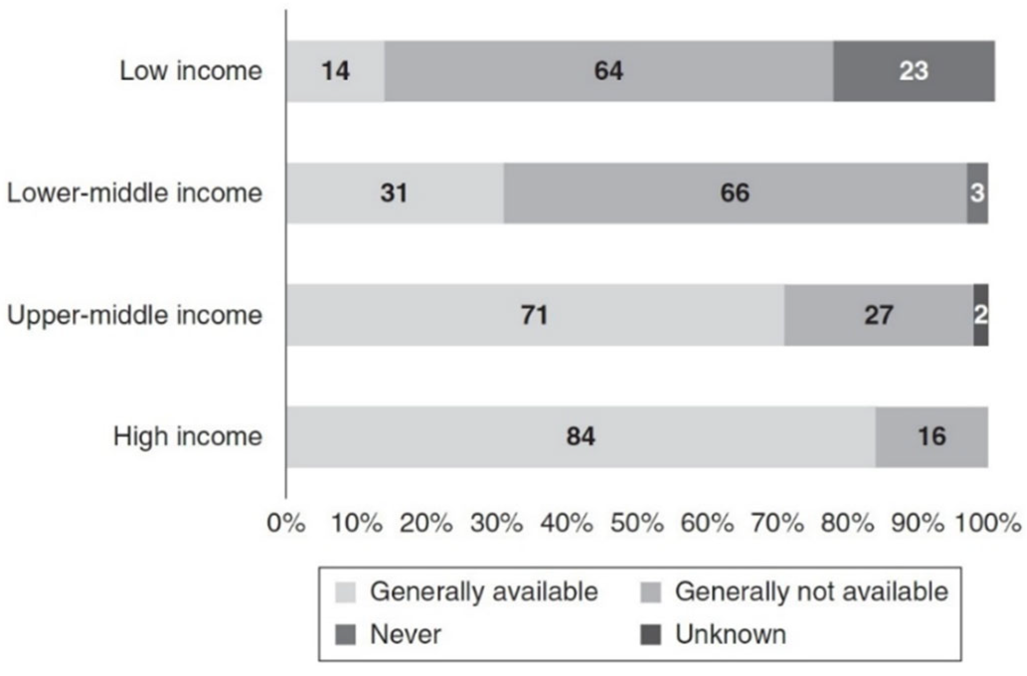
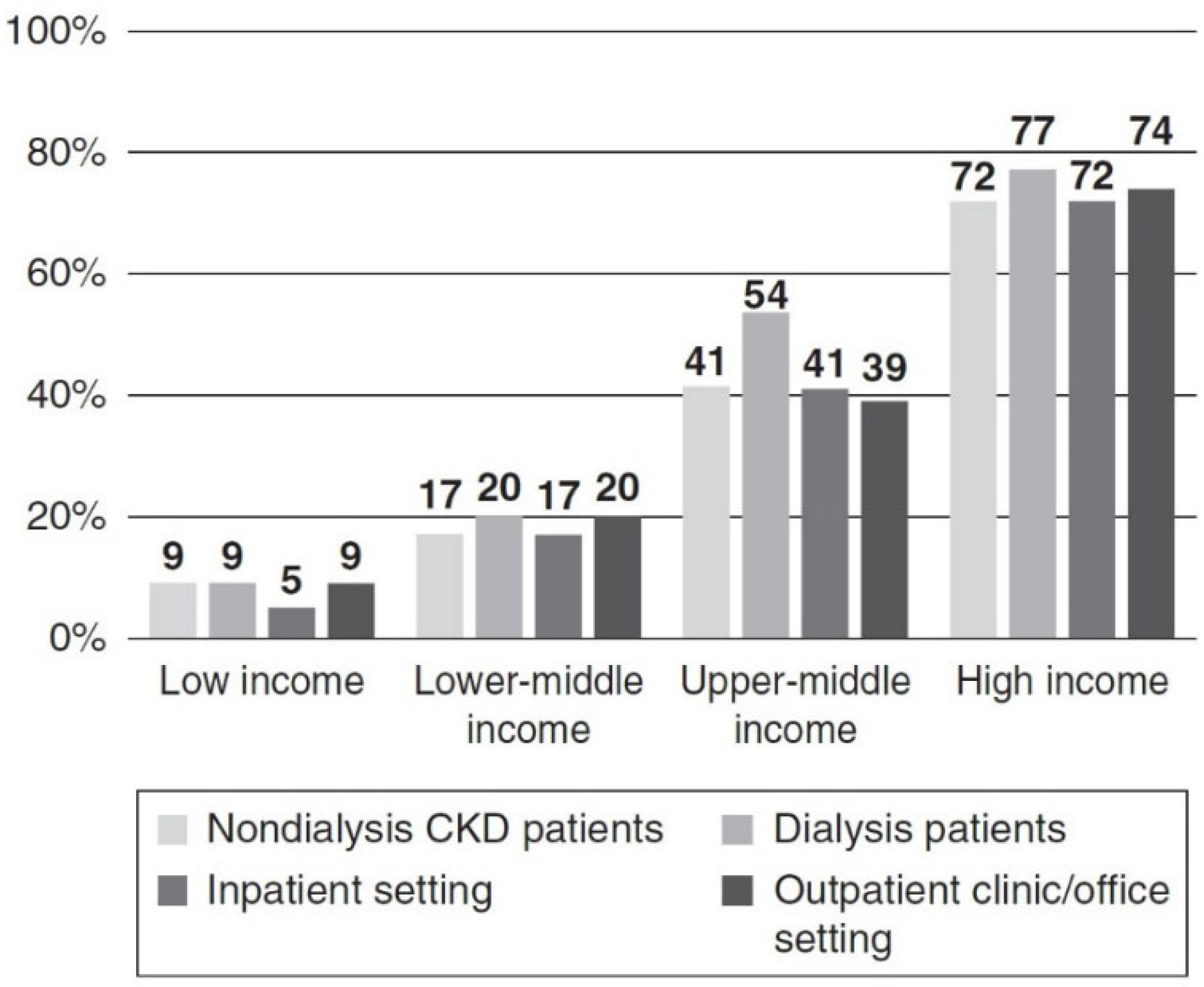
  118. Wang, A.Y.-M.; Okpechi, I.G.; Ye, F.; Kovesdy, C.P.; Brunori, G.; Burrowes, J.D.; Campbell, K.; Damster, S.; Fouque, D.; Friedman, A.N.; et al. Assessing Global Kidney Nutrition Care. Clin. J. Am. Soc. Nephrol. 2022, 17, 38–52. [Google Scholar] [CrossRef]
Figure 1. Mean levels of some anthropometric measures of nutritional status as a function of GFR. The estimated mean levels with 95% confidence limits of anthropometric measures of nutritional status are shown as a function of GFR (males, solid line; females, dashed line) controlling for age, race and use of protein and energy restricted diets. (A) Males, N = 1077 (p = 0.009); females, N = 702 (p = 0.61). (B) Males, N = 1077 (p < 0.001); females, N = 702 (p = 0.62). (C) Males, N = 649 (p < 0.001); females, N = 414 (p = 0.057). (D) Males, N = 1069 (p = 0.002); females, N = 701 (p = 0.67). (E) Males, N = 695 (p < 0.001); females, N = 435 (p = 0.26). (F) Males, N = 648 (p < 0.001); females, N = 410 (p = 0.11). Reprinted from Kidney International, Vol. 57, J. D. Kopple et.al., Relationship between nutritional status and the glomerular filtration rate: results from the MDRD study, Pages 1688–1703, Copyright (2000), with permission from International Society of Nephrology [26].
Kidneydial 02 00046 g001
Figure 2. Repeated measures of weight and body mass index with advancing CKD. (A) Longitudinal repeated measures of weight with repeated measures of eGFR by cystatin C in CRIC. (B) Longitudinal repeated measures of BMI with repeated measures of eGFR by cystatin C in CRIC. (C) Longitudinal repeated measures of weight with repeated measures of eGFR by creatinine in CRIC. (D) Longitudinal repeated measures of BMI with repeated measures of eGFR by creatinine in CRIC. (E) Longitudinal repeated measures of weight with repeated measures of eGFR by creatinine in AASK. (F) Longitudinal repeated measures of BMI with repeated measures of eGFR by creatinine in AASK. Reprinted from Am J Kidney Dis., Vol. 71, Ku, E. et.al., Longitudinal Weight Change During CKD Progression and Its Association With Subsequent Mortality, Pages 657–665, Copyright (2018), with permission from the National Kidney Foundation, Inc., New York, NY, USA [27].
Kidneydial 02 00046 g002
Figure 3. Association between longitudinal repeated measures of body mass index, weight, and height z score with repeated measures of kidney function over time in the Chronic Kidney Disease in Children study in the overall cohort, and by cause of CKD (glomerular versus non-glomerular disease). (A) Repeated measures of BMI and weight z scores with repeated measures of eGFR by serum creatinine. (B) Repeated measures of BMI and weight z scores with repeated measures of eGFR by cystatin C. (C) Repeated measures of BMI and height z scores with repeated measures of eGFR by serum creatinine. (D) Repeated measures of BMI and height z scores with repeated measures of eGFR by cystatin C. (E) Repeated measures of BMI with repeated measures of eGFR by serum creatinine by glomerular versus non-glomerular causes of CKD. Reprinted from Am J Kidney Dis., Vol. 71, Ku, E. et.al., Associations Between Weight Loss, Kidney Function Decline, and Risk of ESRD in the Chronic Kidney Disease in Children (CKiD) Cohort Study, Pages 648–656, Copyright (2018), with permission from the National Kidney Foundation, Inc., New York, NY, USA [28].
Kidneydial 02 00046 g003
Figure 4. Global availability and capacity of kidney nutrition care. Dietitians/renal dietitians practice settings in outpatient and inpatient settings and nutrition care availability for nondialyzed CKD patients versus those undergoing maintenance dialysis for different World Bank income groups. Used with permission of the American Society of Nephrology, from Assessing Global Kidney Nutrition Care, Wang, A.Y. et.al., 17, 2022 of copyright [118].
Kidneydial 02 00046 g004
Figure 5. Global availability and capacity of kidney nutrition care. Dietitians/renal dietitians practice settings in outpatient and inpatient settings and nutrition care availability for nondialyzed CKD patients versus those undergoing maintenance dialysis for different World Bank income groups. Used with permission of the American Society of Nephrology, from Assessing Global Kidney Nutrition Care, Wang, A.Y. et.al., 17, 2022 of copyright [118].
Kidneydial 02 00046 g005
Table 1. Calculated potential renal acid load of selected relevant diets. Used with permission of the American Society of Nephrology, from The continuum of acid stress, Wesson, D.E., 16, 2021 of copyright [45].
FoodUSDA
Recommended
Average Intake in the
United States
Study
Participants
Study
Participants Given F + V
Mediterranean DietDASH DietVegan Diet
Meat/seafood13.6122.5527.4525.216.0322.830
Vegetables−24.91−12.46− 5.75−13.96−22.63−20.19−6.52
Fruit−1.81−0.916.23−18.68−10.04−5.91−13.72
Grains6.346.4310.649.5718.268.1527.6
Dairy10.1611.2123.3123.317.995.770
Oils0.010.010.010.010.010.010
Total3.426.8361.8925.46−0.3910.637.36
All values are shown in millimoles per day. USDA, United States Department of Agriculture; F + V, fruits and vegetables; DASH, dietary approaches to stop hypertension.
Table 2. Phytate content of different plant foods (derived from [46,47,48,49]).
FoodPhytate (In Milligrams per 100 g of Dry Weight)
Brazil nuts 1719
Cocoa powder 1684–1796
Brown rice 1250
Oat flakes 1174
Almond 1138–1400
Walnut 982
Peanut roasted 952
Peanuts ungerminated 821
Lentils 779
Peanuts germinated 610
Hazel nuts 648–1000
Wild rice flour 634–752.5
Yam meal 637
Refried beans 622
Corn tortillas 448
Coconut 357
Corn 367
Entire coconut meat 270
White flour 258
White flour tortillas 123
Polished rice 11.5–66
Strawberries 12
Table 3. Potential Benefits of High Plant Diets for People with CKD.
Potential Benefit Potential Limitations
1. Reduces intestinal phosphorus absorption.The reduction in intestinal phosphate absorption by plant foods is rather modest. Such medicines as phosphate binders or tenapanor also reduce phosphate absorption. LPDs are usually lower in phosphorus and therefore cause less intestinal phosphorus absorption.
2. Diets high in vegetables and fruits can alkalize blood, urine and, potentially, the kidneys. Sodium bicarbonate, solutions of sodium citrate and citric acid, and the resin veverimer also can alkalinize, neutralize acid or bind protons.
3. High dietary fiber may enhance GI motility and reduce risk of hyperkalemia in advanced CKD.Constipation usually is not a problem in CKD patients and often can be prevented with fiber supplements or other changes in the diet.
4. Animal studies indicate high plant diets may improve the microbiome and reduce renal inflammation and oxidative stress.There are no randomized prospective clinical trials (RCTS) in humans with CKD that demonstrate these beneficial outcomes.
5. Animal studies show high plant diets produce less trimethylamine oxide, p-cresyl phosphate, p-cresyl sulfate and indoxyl acetic acid.There are no RCTS that demonstrate beneficial clinical outcomes in humans with CKD from any such changes in production.
Publisher’s Note: MDPI stays neutral with regard to jurisdictional claims in published maps and institutional affiliations.

