
Diatom Biosilica in Targeted Drug Delivery and Biosensing Applications: Recent Studies


Department of Chemistry, University of Fribourg, Chemin du Musée 9, 1700 Fribourg, Switzerland
Micro 2022, 2(2), 342-360; https://doi.org/10.3390/micro2020023
Submission received: 18 May 2022
/
Revised: 10 June 2022
/
Accepted: 14 June 2022
/
Published: 20 June 2022
(This article belongs to the Section Microscale Materials Science)
Abstract
:Diatoms are single-cell algae encased in a cell wall (named frustule) composed of transparent, biogenic (or opaline) silica with intricate and strikingly regular patterns. Over the past 30 years, these microorganisms have proven to be a valuable replacement for synthetic silica that satisfies numerous pharmaceutical requirements for the realization of drug delivery vectors, biosensing supports and photonic crystals. It is not only the structural features of the diatoms, but also the possibility of chemically modifying the frustule that permits the relatively straightforward transformation of the biosilica into potential devices for biomedical applications. In this short review, we explore the applications of diatoms-derived biosilica in the drug delivery and biosensing fields. Specifically, we consider the use of diatoms for the targeted delivery of anticancer and antibiotic drugs and how the same microalgae are employed in the fabrications of biosensors whose analyte signal response is evaluated via fluorescence and surface-enhanced Raman scattering techniques. We limit our discussion to studies published in the last seven years, with the intention of minimizing possible redundancy with respect to previously published contributions.
1. Introduction
Diatoms are unicellular organisms that populate virtually all aquatic environments on the planet. They are vastly abundant, having successfully thrived on Earth at least since the early Jurassic [1,2] and make up a substantial percentage of the planet’s biomass. These microalgae are fundamental for our ecosystem, being responsible for generating 20–50% of the oxygen and contributing to ca. 20% of the global CO2 fixation in the biosphere into organic matter [3,4]. One striking feature of these organisms is that they are the only living entities encased in a cell wall composed of transparent, biogenic silica. In their reproductive cycles, diatoms convert annually ca. seven billion metric tons of silicon from the waters that they populate [5]. Sediments of the silica shells of dead diatoms now form mountains of the soft, siliceous sedimentary rock known as diatomite, and the same sediments can reach 800 m deep on the ocean floor [5].
Over the past 30 years, these microorganisms have proven to be a valuable replacement for synthetic silica that satisfies several pharmaceutical requirements for the realization of drug delivery vectors, biosensing supports and photonic crystals [6,7,8,9,10,11,12,13,14,15]. Indeed, silica-based micro- and nano-materials continue to play an important role in the technological development of our society, but one disadvantage of these materials is associated with their synthesis and manufacture. Synthetic silica production requires expensive reagents, time-consuming and fine-tuned reactions in addition to costly energy requirements for high-temperature synthetic procedures and, above all, it results in toxic and highly polluting waste. Conversely, diatom biosilica can be harvested in large quantities from diatomite in an environmentally sustainable manner, is recognized as safe for foods and pharmaceuticals production by the Food and Drug Administration (FDA) [16] and, crucially, satisfies physical, chemical and optical characteristics for technological and scientific applications. Increasing research interest in the exploitation of diatom biosilica comes from its varied 3D architecture currently unmatched by human engineering, mechanical and thermal inertness, relative straightforward chemical surface modifications, biocompatibility, stability, high specific surface area and regular pours structures. All these features have rendered diatoms very attractive as materials in a variety of applications, including wastewater treatment [17,18,19], bioremediation [19,20,21], tissue engineering [22,23] or catalysis [24]. The periodically nanostructured pore arrays of the diatoms, in particular, are responsible for the optical signal enhancement in fluorescence and Raman biosensing applications. These signal enhancements are principally ascribed to the electromagnetic field induced by localized surface plasmon resonance (LSPR) as well as chemical interactions between the analyte and the diatom substrate [25,26]. The combination of these effects results in an enhanced excitation and emission of fluorophores immobilized on the diatom surface [27] and localized ‘hot spots’ with large, surface-enhanced Raman scattering (SERS) enhancement factors [28,29,30,31]. As detailed in the following sections, this allows the fabrication of diatom-based sensors capable of detecting specific analytes in nM or even pM concentrations.
In this short review, we explore the applications of diatom-derived biosilica in drug delivery and biosensing. We have limited our discussion to studies published in the last seven years with the intention of minimizing possible redundancy with respect to previously published contributions. Indeed, several excellent reviews addressing the subjects prior to our selected timeframe are available to the reader [6,7,8,9,10,11,12,13,14,15,24]. More recently, other authors have also discussed different aspects of the fields in terms, e.g., of the applications of diatoms or microalgae in bio-sensing and -technology [18,32,33,34], the discovery of active compounds [35,36,37], surface chemistry [38], material science [39,40,41] and as drug carries [42,43,44]. We begin with a short overview of the diatoms’ structure and we then move specifically to the use of diatoms for the targeted delivery of anticancer and antibiotic drugs, and how the same microalgae are employed in the fabrications of biosensors whose analyte signal response is evaluated via fluorescence and surface-enhanced Raman scattering techniques.
2. Structure of Diatom Biosilica
Conservative estimates predict nearly half a million different species of diatoms, all varying in their 3D structure [45]. However, diatoms share common characteristics in the makeup of their silicified cell wall, called frustule. As shown in Figure 1 (top), the frustule is comprised of two overlapping halves, the epitheca and hypotheca, each composed of a valve and a girdle band. As it will become apparent in the following sections, virtually all chemical surface functionalization of diatoms occurs at the these sections. The frustule is made of SiO2, which, within the scope of this contribution, can be chemically modified either by magnesiothermic reduction, followed by further processing to obtain, e.g., Si replicas of the frustule, or by silanization chemistry. In silica bonding technology, the most common reagent used is 3-aminopropyltriethoxysilane (APTES), which allows, via normal synthetic procedures, further chemical modification of the diatom surface by exploiting the free surface-exposed amino functionalities of APTES (Scheme 1). These groups are used for chemical immobilization of, e.g., antibodies, enzymes and DNA aptamers in the realization of biosensors, receptor targeting molecules for targeted drug delivery, or stimuli-responsive coatings that allow, e.g., for controlled degradation, cellular uptake and drug loading/release. It should be mentioned here that APTES is a toxic reagent, but following its chemical immobilization on the diatoms’ surfaces, and further derivatization with selected biomolecules, the resulting constructs show no toxicity as demonstrated by several studies [46,47,48,49,50]. Thus, despite the inherent toxicity of APTES, the reagent has, and continues to play, a fundamental role in the preparation of diatom biosilica in the selected applications.
Frustules are further characterized by hundreds and thousands of micro- or nano-scale multilevel pore substructures (Figure 1, bottom) [51]. Foramen are the larger, first-level pores regularly arranged on the valves. Under the foramen, diatoms can develop circular or hexagonal chambers, called areola, which house hundreds of second-level, nano-scale blind holes (with different nm diameters, depending on the species). These second-level holes further cover thousands of sub-level sieve pores (diameter of ca. 20–60 nm). In addition, the mantle and girdle band of many diatom species show scores of lateral nanometers pores. These multilevel pores are of great importance for the application of diatom-derived biosilica. They are important for the chemisorption and infiltration of drugs (although the same drug may be chemisorbed anywhere on the frustule) and their hierarchical micro- and nano-scale periodicity confers photonic, crystal-like characteristics to diatoms [52], allowing the enhancement of localized, surface-plasmon resonance and leading to near-field optical amplification of signal applications. The latter is actually true for valve pores, as pores on the girdle bands tend to have an asymmetric structure [51], possibly involved in the Brownian motion of particles [53].
3. Diatom Biosilica in Targeted Drug Delivery
3.1. Anticancer Organic Drugs Delivery
Within the timeframe of this review, an early prominent benchmark example of the use of natural diatom biosilica in the delivery of anticancer compounds came from the study of the groups of Kröger and Voelcker [54]. The groups used genetically engineered biosilica derived from the diatom Thalassiosira pseudonana to specifically deliver the poorly water-soluble anticancer drugs camptothecin and its more potent derivative 7-ethyl-10-hydroxy- camptothecin to neuroblastoma and Burkitt’s lymphoma HR1K cells. Genetic engineering of Thalassiosira pseudonana was achieved by incorporation into the diatoms’ genome of the immunoglobulin G (IgG)-binding domain of protein G (GB1-bearing fusion protein). In the resulting construct, the GB1 fusion protein was found stably attached to the biosilica surface and accessible for IgG binding, thus enabling the attachment of cell-targeting antibodies. Treatment of mice via intraperitoneal injection of the same drug-loaded biosilica lead to tumour growth regression in mice in a xenograft mouse model of neuroblastoma [54].
This study is a unique example of genetically modified diatoms in the drug-delivery field. All other reports deal rather with chemical alterations of the diatoms’ surfaces in order to enhance cellular target, adhesion and/or drug loading/releasing efficiency. Terracciano et al. functionalized diatomite nanoparticles with polyethylene glycol and a cell-penetrating peptide and studied the construct for its hemocompatibility, toxicity, intracellular uptake in cancer cells and its ability to deliver to the MCF-7 and MDA-MB-231 breast cancer cells the poorly water-soluble anticancer drug, sorafenib [48]. The authors showed that the biofunctionalized nanoparticles exhibit low toxicity, optimal cellular uptake, and both drug-loading and -releasing properties. Sasirekha et al. chemically modified the surface of Amphora subtropica (AS) diatoms with chitosan (Chi), demonstrating that Chi-grafted AS can be used to encapsulate the chemotherapeutic drug doxorubicin (DOX) in the porous compartments of the diatoms (Figure 2) [55]. The construct, used as a drug delivery platform for anticancer therapy, shows high drug loading, strong luminescence, biodegradability and biocompatibility, and, importantly, sustained drug delivery and lower toxicity compared to free DOX. Kabir et al. employed a comparable chemical procedure (based on classic silanization) for the surface functionalization of diatomaceous earth microparticles, realizing core shell materials capable of encapsulating two different chemotherapeutic drugs (namely hydrophobic curcumin or paclitaxel and hydrophilic DOX) at constant molar ratios, in different compartments of the single drug delivery vehicle [56]. Such constructs were realized by cyclodextrin encapsulation (via host–guest complexation) of covalently surface-conjugated adamantane, followed by drug loading of the diatoms’ core versus the outer cyclodextrin shell.
Although prevalently used in the field, chemical modification of the surface of diatoms is not the only strategy employed in the synthesis of drug delivery carries. A few studies have detailed the use of diatom frustules as templates, or scaffolds, for the preparation of biocompatible materials that retain the same ordered structure morphology of the microparticles. Maher et al. have reported the synthesis of luminescent self-reporting degradable silicon diatom replicas for the therapeutic delivery of daunorubicin (DNR) [57] and DOX [58]. The Si-based microparticles (SiNPs) were prepared by the adaptation of the existing magnesiothermic reduction process, which essentially entails the reaction of magnesium with silica, resulting in an interwoven composite product of MgO and silicon [59]. The MgO layer is removed by acid treatment, leaving a silicon replica behind with a high surface area. The authors found that the silicon diatom replicas exhibit high crystallinity and improved degradation rates compared to the diatom precursors, allowing, in vitro, for the prolonged and sustained release of DNR, up to 30 days [57], and DOX. In addition, the replicas display red luminescence with a single emission peak at 682 nm, when excited under confocal microscopy at 458 nm [57]. Drug loading masks this red luminescence, which in turn is revealed again after drug discharge, thus characterizing the replicas as self-reporting carriers for monitoring drug release. The in vitro release results from SiNPs loaded with DOX show that the sustained drug release is a pH-dependent process, and that the construct exhibits a significantly enhanced cytotoxicity against cancer cells compared with free drug [58].
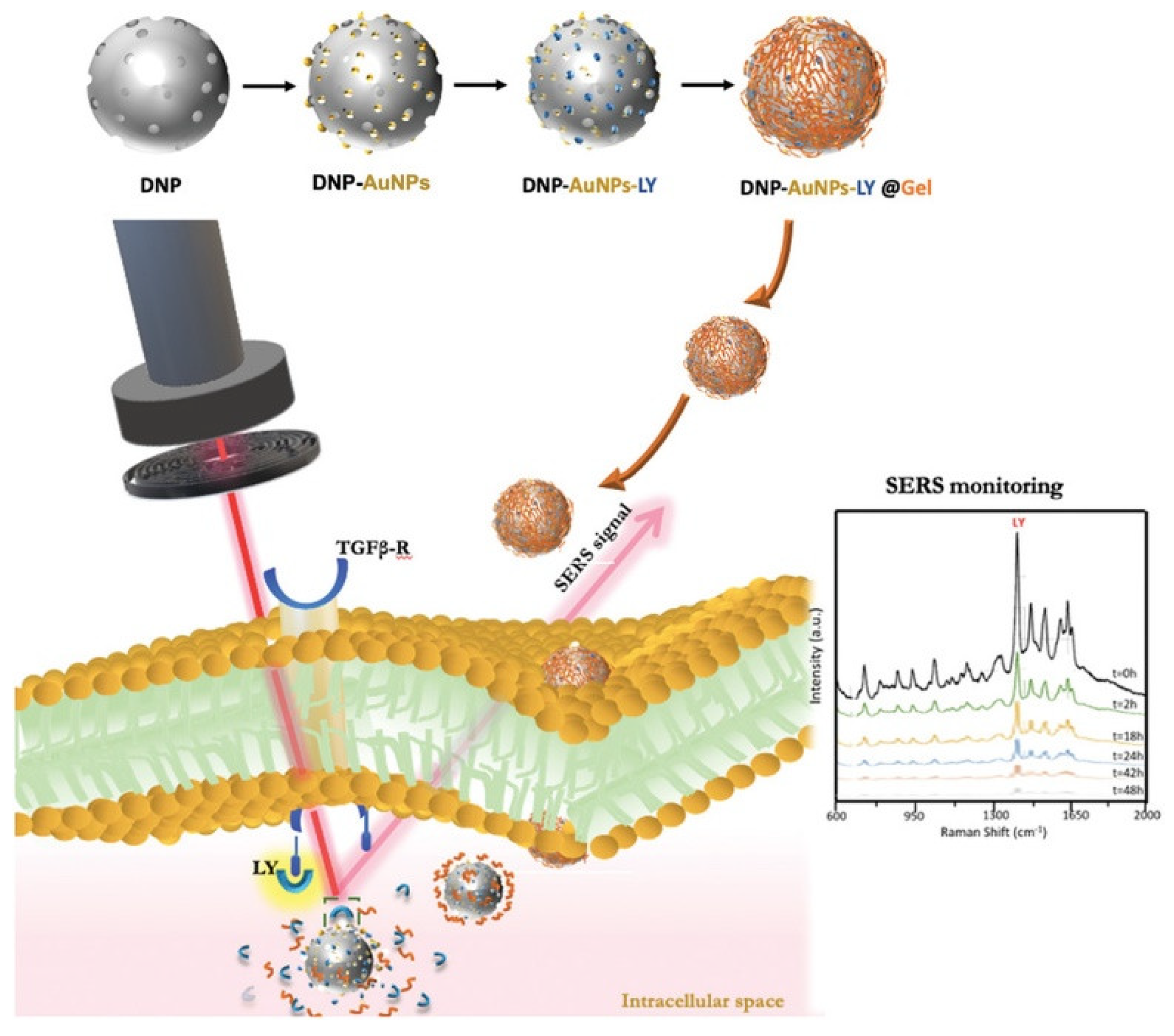
Tramontano, Manago et al. have used gold-coated, gelatin-capped diatomite nanoparticles to improve the drug-loading capacity of the same [60,61]. The authors demonstrated that that the drug-loading capacity of these diatomite-based carriers can be enhanced and modulated as a function of the gelatin shells around the biosilica surface. Furthermore, the gold coating confers remarkable properties to the carriers [62,63], allowing real-time monitoring of the drug release via near-field optical amplification of label-free sensing, with excellent specificity and sensitivity [61]. Full optical theoretical modeling of the hybrid system allowed the predicting and monitoring of gelatin generation, degradation, and drug release by surface plasmon resonance shifts of the coated gold nanoparticles [60]. In their studies, the authors selected the anticancer drug galunisertib, which acts by blocking the transforming growth factor-β1 receptor [64]. This growth factor-β1 is responsible for the epithelial-to-mesenchymal transition (EMT) by which colorectal cancer (CRC) cells acquire the metastatic phenotype [65,66]. Galunisertib release from the diatomite nanoparticles (via gelatin shell degradation) was pH sensitive and promoted by the CRC acidic microenvironment, with the multifunctional particles showing their antimetastatic effect by halting the EMT of CRC (Figure 3) [61].
Javalkote et al. have prepared magnetically active and responsive diatom frustules from Nitzschia sp. by the incorporation of iron oxide nanoparticles (FeONPs) via the ferrofluid or the in situ NPs synthesis methods [67]. These magnetic diatoms were then loaded with curcumin (as the model chemotherapeutic drug) and shown to also have potential as drug delivery carriers. Although magnetically responsive, the authors did not study the effect of an applied external magnetic field on the drug-releasing properties of the diatoms, but the concept remains an interesting one. Similarly, Uthappa et al. loaded curcumin in polydopamine (PDA) surface-modified diatoms and studied the effect of PDA on the drug-loading and -releasing properties of the material [68]. The authors found that PDA acts as a barrier (or gatekeeper), capable of modulating the drug discharge rate (slower by ca. 15–20% when compared to pristine, unmodified diatoms), suggesting that PDA surface modifications of the microparticles could be envisioned as a potential strategy for the preparation of drug delivery systems for targeted tumour therapy. Finally, Vona et al. have explored the use of unmodified diatomaceous earth for the incorporation and delivery of the fungal phytotoxic-derived compound Ophiobolin A (OphA) [69] as a broad-spectrum chemotherapeutic agent effective against cancer cell lines, such as lung, melanoma, brain and ovarian cancers [70]. Although somewhat limited in scope, the study suggests that diatomite activated by acid-oxidative treatment may be considered as a loading/delivery system for OphA due to the observation of the extension of drug releasing time and the increase in the total absolute drug released amount, comparable to the IC50 of OphA.
3.2. Anticancer Inorganic Drugs Delivery
Since the discovery of cisplatin, the investigation of metal-based complexes as anticancer agents continues to be a thriving field of research. There are, however, only limited examples of inorganic drugs loaded in diatoms, due to the realization that biomaterials are designed for the targeted delivery of poorly water-soluble, inorganic anticancer complexes. Delasoie et al. described the first such example. The authors functionalized the surface of diatomaceous earth microparticles vitamin B12 (acting as a tumor-targeting agent) and studied the loading and releasing efficiency of the same with the anticancer agents cisplatin, 5-fluorouracil (5-FU), and a polypyridyl ruthenium(II) complex [71]. The authors found that, while cisplatin and 5-FU are rapidly discharged from the material, the lipophilic ruthenium complex shows an unprecedented release profile. This particular compound is retained in the diatoms up to five days in aqueous media, but is rapidly released in lipophilic environments, such as the cell membrane. Of particular relevance for applications of the material was the increased adherence of the vitamin B12-coated particles to colorectal cancer cells (HT-29), which correlates with the increased transcobalamin II (TC(II)) and transcobalamin II receptor (TC(II)-R) expression of the targeted tissues [71,72].
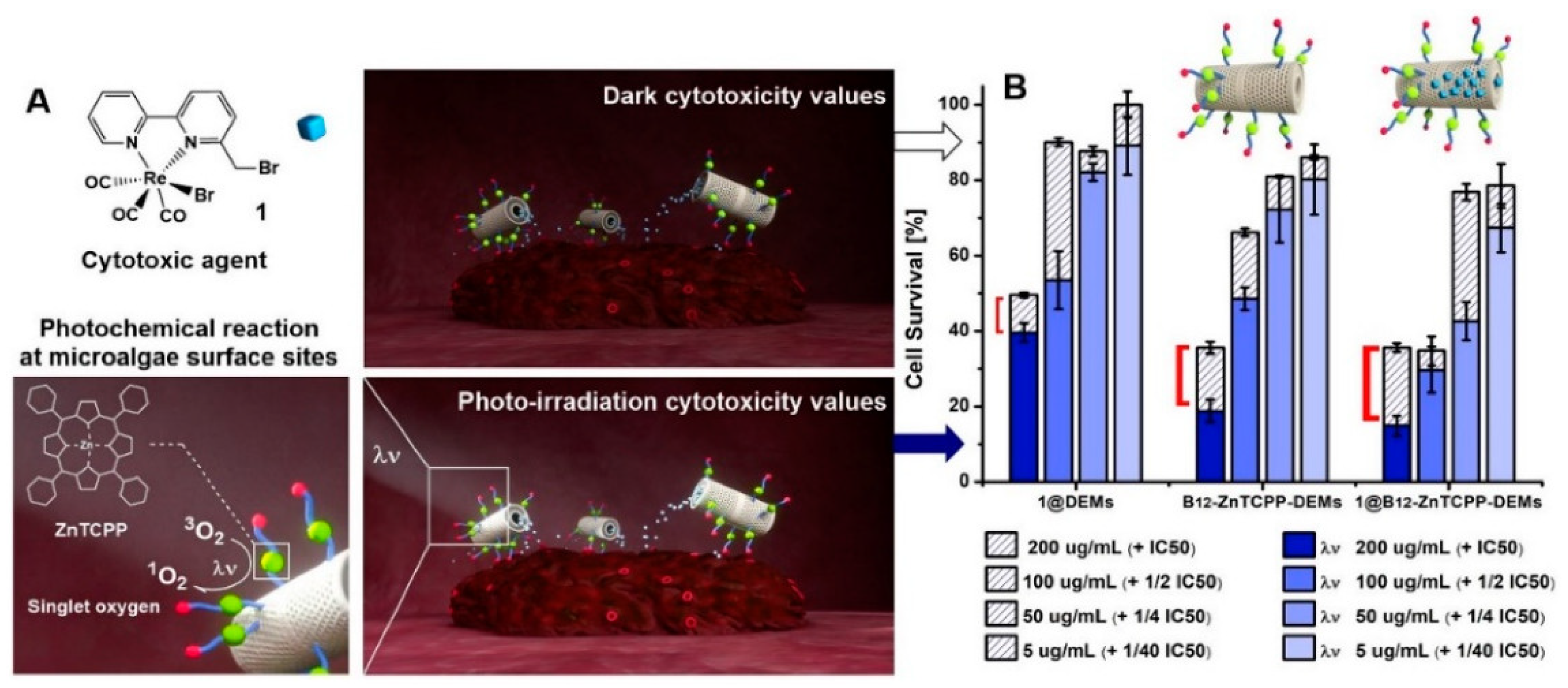
The same group later expanded on the concept by realizing photoactivatable surface-functionalized diatom microalgae promoting enhanced the cytotoxicity of selected anticancer complexes (Figure 4) [73]. The authors selected for the study highly effective rhenium complexes active in vivo against microbes [74,75] and colorectal carcinomas [76], and showed that the overall toxicity of the hybrid multifunctional drug delivery system could be further enhanced by the photoactivation of the microalgae surface (with up to a two-fold increase in the cytotoxic efficacy of the loaded drug). Delasoie et al. also described the neovascularization effects of carbon monoxide-releasing drugs chemisorbed on Coscinodiscus diatoms carriers [77]. When applied in vivo (zebrafish model) at doses ≥25 µM, the rhenium complexes significantly reduce intersegmental and subintestinal vessels development in zebrafish, revealing a high anti-angiogenic potential. Finally, Shi et al. have recently described diatom-like silica-protein nanocomposites for the sustained drug delivery of polypyridyl ruthenium(II) complexes [78]. The nanocomposites, inspired by the diatom structure, were prepared from core–satellite small silica nanoparticles coating human serum albumin, allowing the delivery of the metal complexes to cells. The material afforded the controlled release of the inorganic drug, which accumulates intracellularly via the clathrin-mediated endocytosis of lysosomes [79]. The authors showed that the composite material can be activated by light irradiation towards ROS generation, achieving an excellent photodynamic therapy efficiency, and concluded that the diatom-like nanostructure can not only function as a sustained drug delivery nanocarrier, but also for bioimaging and photodynamic therapy.
3.3. Antibiotics Delivery
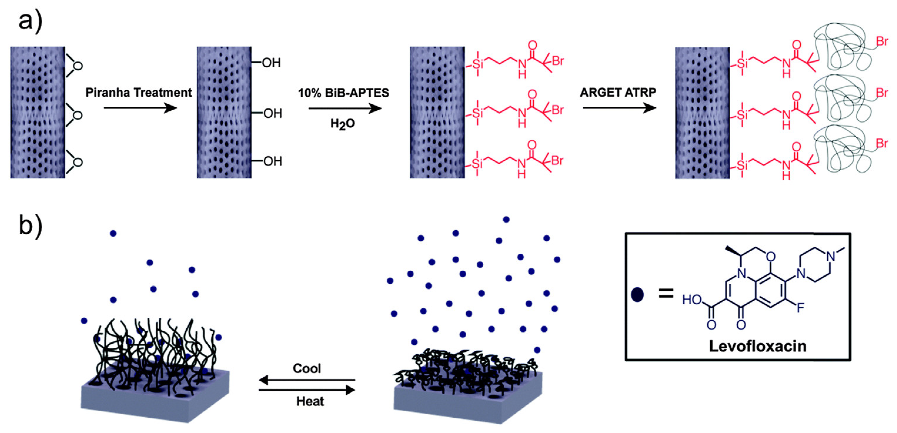
In comparison to studies detailing investigations of the use of diatoms in experimental cancer therapy, only limited examples are available on the use of diatoms as antibiotic delivery systems. In 2015, Vasani et al. presented the fabrication of stimulus-responsive diatom biosilica microcapsules for the delivery of the antimicrobial drug levofloxacin [80]. The thermo-responsive Aulacoseira sp. diatoms were prepared by grafting oligo(ethylene glycol) methacrylate copolymers on the surface of 4–5 μm diameter particles via silanization chemistry (Figure 5). The authors showed that kinetics of drug release from the copolymer-modified microcapsules are significantly influenced by temperature, allowing for the controlled discharge of antibiotics in, e.g., inflamed areas of infection. The modified levofloxacin–loaded diatoms showed thermo-responsive antibacterial activity against Staphylococcus aureus and Pseudomonas aeruginosa [80].
The same year, Cicco et al. described the chemical modification of Thalassiosira weissflogii diatoms for the combined drug delivery of the antibiotic ciprofloxacin and the concomitant anti-inflammatory protection against the adverse side effects of the same antibiotic [81]. The latter property of the biosilica was realized by the grafting the diatoms’ surface with cyclic nitroxide 2,6,6-tetramethylpiperidine-N-oxyl (TEMPO), which served as the efficient reactive oxygen species (ROS) scavenger in the tested biological systems. Typically, surface modification was realized by the derivatization of 4-carboxy-TEMPO with 3-aminopropyl triethoxy silane, via carbodiimide-activated esterification. Drug loading also followed a well-established procedure, but interestingly, in the formulation of the authors, it was observed that only about 17–24% of the initially loaded ciprofloxacin was released after 7 days, in saline or PBS media. When diatoms are used as drug-delivery systems, a burst release of the drug is generally observed within the first 24 h [71], but in this case, for reasons unclear, a large amount of the antibiotic remained adsorbed into the functionalized biosilica. Cell growth and biocompatibility experiments with MG63 osteoblast-like cells revealed the possibility of using this engineered biosilica as a support material in regenerative medicine for bone cells [81].
Uthappa et al. have more recently reported hybrid materials of diatom biosilica and metal–organic frameworks (MOFs) as porous scaffolds for the delivery of the anti-tuberculosis (TBC) drug isoniazid (INH) [82]. The engineered MOF–diatom hybrids materials (“MIL-100(Fe)-DE”) were prepared by reaction iron (II) chloride tetrahydrate in the presence of the diatom biosilica followed by trimesic acid treatment. MIL-100(Fe)-DE shows overall an enhanced drug-loading capacity of 9.6 ± 1.6% and in vitro protracted, controlled drug release performances over 23 days. As pointed out by the authors, such a prolonged and sustained drug release is a significant improvement over the direct use of INH, due to the relatively short biological half-life of the drug (1–4 h) that limits its full therapeutic benefits for treatment of TBC. Briceno et al. have instead decorated diatoms of the Aulacoseria genus with gold nanoparticles (AuNPs) and studied the in vitro gentamicin release in simulated body fluid [83]. The composite materials (Au/Diatom) were prepared by two different methods, namely “in situ” where AuNPs were formed in the presence of the diatoms, and “ex situ” where preformed AuNPs were added to natural diatoms. The authors found that diatoms decorated with AuNPs using the ex situ method are characterized by a faster drug release (93% of gentamicin release in 10 days) and a lower drug-loading efficiency, whereas Au/Diatom prepared via the in situ methodology show a longer and slower gentamicin release behavior. The study highlights the advantage of using diatoms in these formulations, as controlled chemical surface modifications of the same allow controlling and tailoring properties for potential application in drug delivery.
Finally, Cong et al. [84] and Sherief et al. [85] have described the antimicrobial activity of metal-coated diatoms. These last two examples do not rigorously fall within the drug delivery category, but are illustrative of the tremendous versatility of diatom biosilica. Cong et al. described the deposition of copper on Cyclotella cryptica diatoms and the realization of biomaterilas for infected wound therapy [84]. The deposition of copper onto C. cryptica diatoms significantly improved the photothermal and photodynamic performance of material by increasing the yield of ROS under near-infrared light irradiation (Figure 6). This, in turn, resulted in an improved sterilization effect and nearly 100% antibacterial efficiency against S. aureus- and E. coli-infected wounds. In vivo experiments not only confirmed the antibacterial properties of the material, but also revealed that the same could fast track wound healing by inhibiting inflammation, promoting angiogenesis as well as collagen deposition, and modulating type-III to type-I collagen ratio [84]. Sherief et al. used a similar approach, coating diatomite particles with AgNPs, and showed that the material is effective against five different pathogenic organisms [85].
3.4. Delivery of Other Drugs
Within the timeframe of this review, we are aware of only two studies dealing with the use of diatomite for the release of drugs not belonging to the previous two classes. Uthappa et al. have prepared xerogel-modified diatomaceous earth microparticles (DE-XER) for the controlled release of the anti-inflammatory and pain reliever drug diclofenac sodium [86]. DE-XER acts as a pH-sensitive micro carrier for the drug, allowing the control and modulation of the drug release mechanism. The covalent xerogel coating also influences the drug absorption of the material, enhancing the same over natural, unmodified DE. In terms of drug release, the presence of the xerogel coating appears to increase the overall surface area and pore volume of the material (from 18 to 173 m2 g−1 and 0.021 to 0.199 cm3 g−1, respectively), suggesting that the drug release mechanism (after the initial burst) is mainly due to the diffusive transport of the drug through the fine-tuned porous structures, and completely based on surface-dependent phenomena. The second example was described by Bonifacio et al., who reported gellan gum-honey-diatom composite materials for the release of the anti-inflammatory and anti-oxidant drug resveratrol [87]. Specifically, the composite porous scaffolds were designed as a biomaterial for cartilage regeneration, showing suitable mechanical properties for articular load bearing, prominent antibacterial features, and allowing, at the same time, for the controlled release of resveratrol for hindering bacterial proliferation and supporting stem cell colonization and chondrogenic differentiation.
It should perhaps be mentioned here that diatoms formulations have also been recently described in tissue engineering applications [88,89], with some examples also envisioned as drug delivery systems, as the example of Bonifacio et al. [87] shows. Lee et al. described diatom silica/polysaccharide elastomeric hydrogels as water-resistive adhesive tissues showing shock-absorbing properties [88]. The material, prepared by oxdidative crosslinking of catechol and chitosan on the diatoms’ surface, promoted the healing process in the early stages of the ischemia-reperfusion-based pressure ulcer mouse model. Similarly, Feng et al. used chitosan-coated Coscinodiscus sp. diatoms as hemostatic agent for hemorrhage control, showing excellent results in the induction of erythrocyte absorption and aggregation, as well as activation of coagulation in a rat-tail amputation model [89]. Other authors instead have used diatom-inspired approaches for, e.g., the mineral skeletonisation of insulin [90], diverse pollen-like surface patterns [91], the study of mechanical response of diatom-inspired structures [92], and model tools to map and quantify the role of surface topography on cells’ fate [93].
4. Diatom Biosilica in Biosensing
4.1. Fluorescence Biosensing
Diatoms-derived biosilica has been used for several years as the scaffold for the chemical immobilization of, e.g., antibodies, enzymes, drugs, and DNA aptamers in the realization of biosensors. In biosensing applications of the materials, two main monitoring techniques have been described in the past years: fluorescence and surface-enhanced Raman scattering (SERS) [94]. We discuss first fluorescence biosensing, as it is perhaps the most common and established biosensing and bioimaging method. In general, fluorescence immunoassays based on diatom biosilica are developed by crosslinking, e.g., antibodies on the surface of the microparticles via well-established silanization chemistry. Rea et al. converted Aulacoseira sp. diatomite to silicon replicas via the magnesiothermic process and immobilized proteins and antibodies as biological probes on their surface [95]. The authors exploited the light emission properties of the semiconducting bioengineered silicon diatoms for the recognition of the His-tagged p53 protein. The biosensing response of the material was demonstrated by a strong increase in photoluminescence intensity of the same following biomolecular interactions with the analyte.
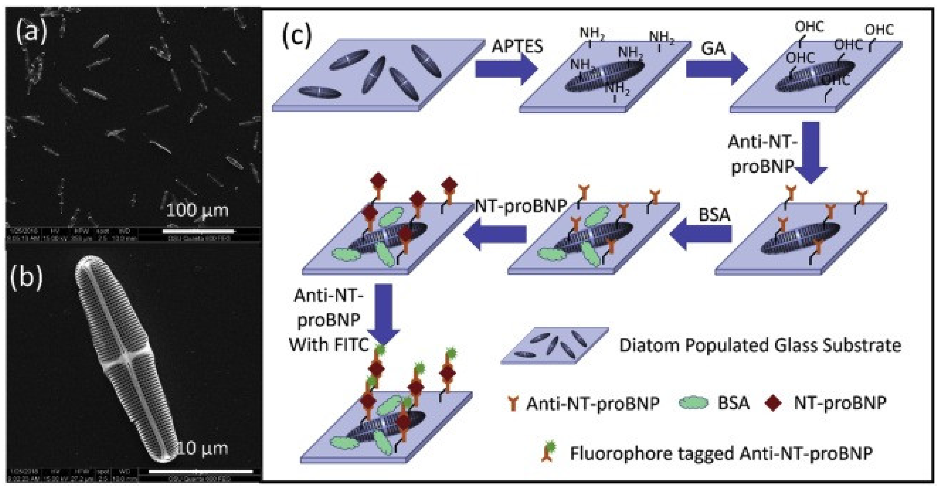
Squire et al. used pristine Pinnularia sp. diatoms in the preparation of photonic crystals showing enhanced fluorescence in an immunoassay for the detection of the mouse immunoglobulin G (IgG) protein [27]. Using a standard antibody-antigen-labeled antibody immunoassay protocol, the authors investigated the fluorescence enhancement effect of the diatom photonic biosilica with rhodamine 6G (R6G) as well as the R6G-labeled mouse IgG immunocomplex on the diatom frustules. By analyzing different antigen concentrations, Squire et al. demonstrated a 100-fold increase in the detection limit of fluorescence spectroscopy, with a limit of detection of the mouse IgG of 14 fg/mL, thereby proving the great ability of diatom fluorescence immunoassay to enhance fluorescence and achieve high sensitivity. Squire et al. later used the same diatoms to develop a fluorescence imaging immunoassay for the detection of cardiovascular disease biomarkers (Figure 7) [96]. Specifically, the surface of Pinnularia sp. diatoms was functionalized with fluorophore-labeled antibodies for the recognition of the clinically relevant N-terminal prohormone B-type natriuretic peptide (NT-proBNP). The diatom-based photonic crystals were capable of detecting low levels of NT-proBNP in solution (in a concentration range of 0–100 pg/mL), with an excellent specificity of 93%, as well as an accuracy and sensitivity of 78% and 65%, respectively, in human plasma. The material showed enhanced fluorescence signal intensity up to twice as high compared with those on the flat glass substrate, an increase attributed to the periodic, nanostructured pore arrays of diatom frustules. Specifically, the periodically-nanostructured pore arrays of the diatoms result in a dual field optical enhancement: enhanced excitation and enhanced emission [27]. Light incident on a diatom frustule induces enhanced electromagnetic fields on or near the surface of the structure [97,98], which in turn results in an enhanced excitation of the fluorophore and emission of more photons. Additionally, a fluorophore on the surface of a frustule experiences an increase in the density of optical states which results in enhanced emission due to the Purcell effect [99]. This dual-modal optical enhancement results in boosted fluorescence signals for easier detection of analytes.
Selvaraj et al. covalently immobilized Salmonella typhi antibody on the surface of glutaraldehyde crosslinked amine-functionalized Amphora sp. diatom substrates, and used the photonic crystals as optical biosensors for the detection of typhoid fever [100]. Photoluminescent studies showed that, when the S. typhi antibody-conjugated diatom frustule substrates were challenged with different concentrations of its S. typhi O and H antigens, or non-complementary E. coli antigens, the material was capable of distinguishing the two with good specificity. In particular, the immunocomplexed biosensor showed a calculated detection limit of 10 pg for the pathogen S. typhi. The excellent performance of biosensor was attributed by the authors to the large surface-to-volume ratio of functionalized diatoms, which also revealed good photoluminescence and biocompatibility. Finally, Esfandyari et al. prepared magnetic iron oxide nanoparticles-coated Chaetoceros sp. frustules and functionalized the same with Trastuzumab antibodies in order to biosense and separate in blood breast cancer cells from normal cells [101]. The diatoms showed remarkable fluorescence emission and magnetic properties, and were capable of selectively targeting and capturing SKBR3 breast cancer. Separation of the same cells from a mix of HER2 negative cells was achieved by application on an external magnetic field. Furthermore, the magnetic diatom conjugated with Trastuzumab antibodies displayed strong fluorescence emission with peaks centered at 493 and 650 nm.
4.2. Surface-Enhanced Raman Scattering (SERS) Biosensing
Surface-enhanced Raman scattering (SERS) refers to the inelastic scattering of photons that can take place when the elementary particles interact with matter [94]. Due to their unique structural features, diatoms are capable of enhancing the localized surface-plasmon resonance (LSPR) leading to near-field optical amplification of signal [52]. This is mainly due to the photonic, crystal-like, hierarchical, micro- and nano-scale periodicity of the frustules pores that allows for signal enhancement of the electromagnetic field induced by LSPR ‘hot spots’ with large SERS enhancement factors [28,29,30,31]. As a consequence, diatom biosilica has found wide applications in the fabrication of plasmonic devices in the fields of biochemical sensing, diagnostics and therapeutics. Numerous such examples have been described in the past [15,18,34,40,52], but within the constraints of this review, we highlight eight studies.
Kong et al. used Pinnularia sp. diatoms per the fabrication of photonic crystals that act as nano-plasmonic sensors for different chemical and biological analytes [102]. The sensing mechanism is based on surface-enhanced Raman scattering (SERS) of the surface localized in situ grown silver or self-assembled gold nanoparticles, whose plasmons optically couple with the guided-mode resonance of the diatom frustules. The enhanced sensitivity of the material also derives from the large surface-to-volume ratio of the diatoms that allows a greater concentration of analyte on the surface of the SERS substrates. This allows us, in turn, to detect even small concentrations of, e.g., biomolecules that interact or adsorb with the Ag/AuNPs. Kamińska et al. also exploited SERS for the realization of an ultrasensitive immunoassay for detection of interleukin IL-8 in blood plasma [103]. The biosensor was prepared by covalently linking specific antibodies on the surface of Pseudostaurosira trainorii diatoms, which allowed the construction of an immune substrate based on anti-IL-8-functionalized biosilica. The SERS tags were fabricated by functionalizing AuNPs with 5,5′-dithiobis(2-nitrobenzoic acid) that acted as the Raman reporter molecule. Notably, the SERS immunoassay described by the authors shows high specificity for the biological detection of IL-8 in complex fluids, and a detection limit for IL-8 in human blood plasma of 6.2 pg mL−1, a sensitivity clinically relevant for interleukin concentrations in body fluids (Figure 8). Similarly, Yang et al. prepared ultra-sensitive immunoassay biosensors by exploiting the enhanced SERS sensitivity of the nanostructured Pinnularia sp. diatoms—Ag NPs hybrid plasmonic material to detect the immune reactions between antigens and antibodies [104]. The biosensor was prepared by decorating glass slides with Pinnularia sp. diatoms frustules, which were fixed on the support by furnace annealing at 450 °C for 1 h. The diatom surface was then functionalized with a model goat anti-mouse immunoglobulin G (IgG) antibody following the standard APTES chemical protocol, and then, finally coated with Ag NPs allowed to self-assemble on the sensor. By challenging the biosensor with complementary antigen (mouse IgG) and non-complementary antigen (human IgG), the authors showed that this photonic crystal platform has a detection limit of mouse IgG of 10 pg/mL, ca. two orders of magnitude better than conventional colloidal SERS sensors on flat glass. Moreover, by mapping the Raman signals of the immobilized diatom frustules, the authors proved a strong correlation between the signal enhancement factors and the morphologies of the diatom frustules, thus substantiating that the hybrid plasmonic material can effectively eliminate from the analysis random signals affecting the measurement of low antigens concentration.
Korkmaz et al. prepared porous biosilica plasmonic composites on an inexpensive adhesive tape and showed the biosensing capability of the material in SERS-based applications with several bacteria as analytes [105]. The diatom–AgNP composite material was simply deposited on regular office-grade adhesive tape and characterized for its plasmon Raman resonance enhancement to identify three different types of bacteria (S. aureus, E. coli, and Bacillus spp.) and two different strains of S. aureus (ATCC6538 and ATCC29213). SERS spectra obtained from the bacteria on the strips showed a signal enhancement factor of ca. 105, demonstrating that the technique and inexpensive diatom–AgNP composite materials can be used to identify bacteria on surfaces. Manago et al. used Raman imaging to investigate the internalization kinetics and intracellular spatial distribution of functionalized diatomite microparticles conjugated with a non-targeting siRNA in human lung epidermoid carcinoma [106]. The authors showed that the highly specific Raman bands of the composite material allowed the direct label-free visualization of the particles in terms of both cellular uptake and localization. In particular, Raman spectra indicated co-localization of siRNA-diatomites with lipid structures when internalized, and with vesicle spectral features when not internalized. A kinetics analysis of the cellular uptake process revealed both intracellular and membrane-associated microparticles after 6 h of incubation, with the relative ratio of the two being constant over the successive 12 h of interaction with the cells. Cellular saturation of the material was observed after 18 h, with efficient distribution within the cell cytosol and a progressive increase in perinuclear localization. Interestingly, the authors found that cellular uptake depends on the diatomite particles’ orientations on the cell membrane.
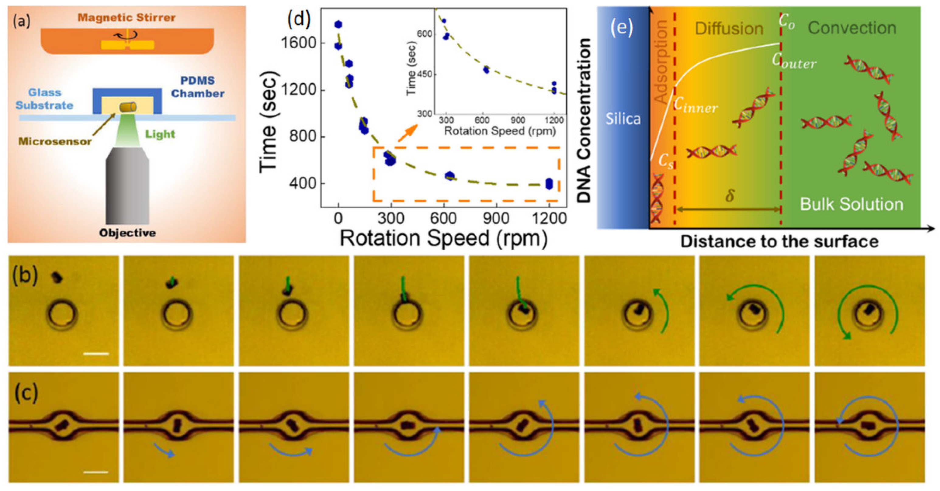
Finally, several authors have exploited the enhanced SERS sensitivity of diatoms in fluidic sensing system applications. Sivashanmugan et al. developed multiscale, hierarchical, photonic, crystal-enhanced plasmonic mesocapsules of Pinnularia sp. diatoms decorated with in situ synthesized Ag NPs for ultrasensitive optofluidic-SERS sensing of different analytes [107]. The construct was realized by exploiting the morphological features of the diatom frustules, which provided large reaction sites around the pores for in situ growth of Ag NPs with the surface silanol groups increasing the quantity of nucleation site and reaction rate. The authors showed that the plasmonic mesocapsules are capable of SERS detection of Rhodamine 6 G in microfluidic devices with a limit of detection of 0.1 pM, or virtually single-molecule SERS detection. Overall, diatom plasmonic mesocapsules showed 100× higher enhancement factors and more than 1000× better detection limits than traditional colloidal Ag NPs, an improvement attributed to the unique 3D structural properties of Pinnularia sp. the mesocapsules. The same mesocapsules were also used in the microfluidic devices to detect nM concentrations (0.05 µg L−1) of benzene and chlorobenzene compounds in tap water with near real-time response [107]. Guo et al. have reported a similar microfluidic application of bio-opto-plasmonic Raman microsensors based on diatom frustules (Figure 9) for the motorized mechanical accelerated capture and detection of DNA [108]. The devices, developed for the efficient detection, capture and extraction of small amounts of DNA for medical and forensic gene technologies, exploit plasmonic surface-deposited Ag hotspots for sensitive and quasi-real-time SERS detection of ribonucleic acid. A four-fold enhancement of DNA capture is realized when the diatoms are mechanically rotated at 1200 rpm, with the fundamental underlying mechanism of DNA capture attributed by the authors to the Nernst diffusion layer thinning effect induced by mechanical motions [109].
Kong et al. also reported the fluidic properties of plasmonic Au NPs-decorated diatomite biosilica immobilized on a chip for SERS biosensing [110]. The diatomite biosilica film was used as a photonic crystal matrix for a “lab-on-a-chip” device for the label-free biosensing chromatographic detection of small molecules in complex biological samples. As all applications described in this section, the photonic crystal effects of the diatoms were exploited to enhance the plasmonic resonances of metallic Au nanoparticles in the device. Specifically, the authors fabricated diatomite “chips” by spin coating diatomite on glass slides. These were then exposed to biological fluids (e.g., plasma), the analyte components were separated by chromatography and then, lastly, the chip was exposed to Au NPs for SERS analysis. The devices were capable of separating and detecting small molecules from mixture samples with an ultra-high detection sensitivity, down to 1 ppm. In particular, the authors showed the potential of the chips for biomedical applications by screening toxins in real biofluids, accomplishing concurrent label-free biosensing of phenethylamine and miR21cDNA in human plasma with limit of detections of 10 ppm and 5 μM, respectively [110].
5. Conclusions
Since the 1980s, diatoms have been exploited as a source of natural biosilica for the blueprint development of new generation of drug-delivery systems and biosensing materials. Several features render silica derived by these organisms unique. First, they have a unique and enormously varied 3D structure. Created by genetically controlled self-assembling processes, they are characterized by distinctive hierarchical porous structures that confer to the frustules specific optical and photoluminescent properties. They are harvested and purified economically and under environmentally friendly conditions, and their 3D architectures are currently unmatched by fully synthetic porous silica materials. Second, frustules, as nature-generated drug-delivery systems and sensing platforms, are fully biocompatible. Although continuous research needs to be conducted on the biological response of diatom silica in their developing pharmaceutical formulations for clinical use, several agencies have already approved the use of diatomaceous silica as additives in food in the pharmaceutical industry and in medicine [8]. Finally, frustules exhibit a high surface area and straightforward tailorable surface modification, permitting their use as low-cost scaffolds with broad biomedical applications. Chemical functionalization of the frustule surface is certainly a major advantage in using diatom-derived biosilica in the applications highlighted here. Their relatively mild conditions, in which, e.g., immobilization of (targeting) biomolecules can be achieved, translates often into insignificant denaturation of the same.
Within this context, we highlighted in this short review recent contributions dealing with different aspects of diatom technology, including their use as drug carriers, in the preparation of biosensors, in surface modifications, in drug loading, and in controllable delivery capability to transport therapeutic molecules. It should be noted that some authors already consider diatom sensors at technology readiness level (TRL) 8–9 [14]. Developed by NASA, TRL is a numerical metric for assessing the maturity of a technology. TRL 8 and 9 define, respectively, a system incorporated in commercial design, a system ready for full-scale deployment. Such an assessment for diatom-biosensors speaks of the tremendous opportunities that the material offers. Regardless of where the diatom technology readiness level may lie, we also believe that diatom biosilica is poised to influence and play a remarkable role in future micro and nano technology. In terms of their development as drug delivery vehicles, advancements may be achieved, e.g., by fully controlling drug release at specific locations within the body, and perhaps by considering several coating layers of the particles for the cascade degradation of the same as the carriers move through the gastro-intestinal track. In terms of the development in biosensing applications, solutions for single device calibration may remove one of the major obstacles for the large-scale production and commercialization of diatom biosensors.
Funding
This research received no external funding.
Conflicts of Interest
The author declares no conflict of interest.
References
- Kooistra, W.H.C.F.; Medlin, L.K. Evolution of the Diatoms (Bacillariophyta): IV. A Reconstruction of Their Age from Small Subunit rRNA Coding Regions and the Fossil Record. Mol. Phylogenet. Evol. 1996, 6, 391–407. [Google Scholar] [CrossRef] [PubMed]
- Schieber, J.; Krinsley, D.; Riciputi, L. Diagenetic origin of quartz silt in mudstones and implications for silica cycling. Nature 2000, 406, 981–985. [Google Scholar] [CrossRef] [PubMed]
- Armbrust, E.J.N. The life of diatoms in the world’s oceans. Nature 2009, 459, 185–192. [Google Scholar] [CrossRef] [PubMed]
- Bradbury, J.J.P.B. Nature’s nanotechnologists: Unveiling the secrets of diatoms. PLoS Biol. 2004, 2, e306. [Google Scholar] [CrossRef] [PubMed] [Green Version]
- Tréguer, P.; Nelson, D.M.; Bennekom, A.J.V.; DeMaster, D.J.; Leynaert, A.; Quéguiner, B. The Silica Balance in the World Ocean: A Reestimate. Science 1995, 268, 375–379. [Google Scholar] [CrossRef]
- Livage, J. Bioinspired nanostructured materials. Comptes Rendus Chim. 2018, 21, 969–973. [Google Scholar] [CrossRef]
- Uthappa, U.T.; Brahmkhatri, V.; Sriram, G.; Jung, H.-Y.; Yu, J.; Kurkuri, N.; Aminabhavi, T.M.; Altalhi, T.; Neelgund, G.M.; Kurkuri, M.D. Nature engineered diatom biosilica as drug delivery systems. J. Control Release 2018, 281, 70–83. [Google Scholar] [CrossRef]
- Maher, S.; Kumeria, T.; Aw, M.S.; Losic, D. Diatom Silica for Biomedical Applications: Recent Progress and Advances. Adv. Healthc. Mater. 2018, 7, 1800552. [Google Scholar] [CrossRef]
- Terracciano, M.; De Stefano, L.; Rea, I. Diatoms Green Nanotechnology for Biosilica-Based Drug Delivery Systems. Pharmaceutics 2018, 10, 242. [Google Scholar] [CrossRef] [Green Version]
- Terracciano, M.; Rea, I.; De Stefano, L.; Santos, H.A. Chapter 9—Diatoms: A Natural Source of Nanostructured Silica for Drug Delivery. In Diatom Nanotechnology: Progress and Emerging Applications; The Royal Society of Chemistry: London, UK, 2018; pp. 201–218. [Google Scholar] [CrossRef]
- Diab, R.; Canilho, N.; Pavel, I.A.; Haffner, F.B.; Girardon, M.; Pasc, A. Silica-based systems for oral delivery of drugs, macromolecules and cells. Adv. Colloid Interface Sci. 2017, 249, 346–362. [Google Scholar] [CrossRef]
- Albert, K.; Huang, X.C.; Hsu, H.Y. Bio-templated silica composites for next-generation biomedical applications. Adv. Colloid Interface Sci. 2017, 249, 272–289. [Google Scholar] [CrossRef]
- Mishra, M.; Arukha, A.P.; Bashir, T.; Yadav, D.; Prasad, G.B.K.S. All New Faces of Diatoms: Potential Source of Nanomaterials and Beyond. Front. Microbiol. 2017, 8, 1239. [Google Scholar] [CrossRef] [PubMed] [Green Version]
- Rea, I.; Terracciano, M.; De Stefano, L. Synthetic vs Natural: Diatoms Bioderived Porous Materials for the Next Generation of Healthcare Nanodevices. Adv. Healthc. Mater. 2017, 6, 1601125. [Google Scholar] [CrossRef] [PubMed]
- Leonardo, S.; Prieto-Simón, B.; Campàs, M. Past, present and future of diatoms in biosensing. TrAC Trend. Anal. Chem. 2016, 79, 276–285. [Google Scholar] [CrossRef]
- Chao, J.T.; Biggs, M.J.P.; Pandit, A.S. Diatoms: A biotemplating approach to fabricating drug delivery reservoirs. Expert Opin. Drug Deliv. 2014, 11, 1687–1695. [Google Scholar] [CrossRef] [PubMed] [Green Version]
- Khan, M.J.; Harish; Ahirwar, A.; Schoefs, B.; Pugazhendhi, A.; Varjani, S.; Rajendran, K.; Bhatia, S.K.; Saratale, G.D.; Saratale, R.G.; et al. Insights into diatom microalgal farming for treatment of wastewater and pretreatment of algal cells by ultrasonication for value creation. Environ. Res. 2021, 201, 111550. [Google Scholar] [CrossRef] [PubMed]
- Khan, M.J.; Rai, A.; Ahirwar, A.; Sirotiya, V.; Mourya, M.; Mishra, S.; Schoefs, B.; Marchand, J.; Bhatia, S.K.; Varjani, S.; et al. Diatom microalgae as smart nanocontainers for biosensing wastewater pollutants: Recent trends and innovations. Bioengineered 2021, 12, 9531–9549. [Google Scholar] [CrossRef] [PubMed]
- Marella, T.K.; López-Pacheco, I.Y.; Parra-Saldívar, R.; Dixit, S.; Tiwari, A. Wealth from waste: Diatoms as tools for phycoremediation of wastewater and for obtaining value from the biomass. Sci. Total Environ. 2020, 724, 137960. [Google Scholar] [CrossRef]
- Mandal, A.; Dutta, A.; Das, R.; Mukherjee, J. Role of intertidal microbial communities in carbon dioxide sequestration and pollutant removal: A review. Mar. Pollut. Bull. 2021, 170, 112626. [Google Scholar] [CrossRef]
- Furey, P.C.; Liess, A.; Lee, S. Substratum-associated microbiota. Water Environ. Res. 2019, 91, 1326–1341. [Google Scholar] [CrossRef] [Green Version]
- Reid, A.; Buchanan, F.; Julius, M.; Walsh, P.J. A review on diatom biosilicification and their adaptive ability to uptake other metals into their frustules for potential application in bone repair. J. Mater. Chem. B 2021, 9, 6728–6737. [Google Scholar] [CrossRef] [PubMed]
- Lowe, B.; Guastaldi, F.; Müller, M.-L.; Gootkind, F.; Troulis, M.J.; Ye, Q. Nanobiomaterials for Bone Tissue Engineering. In Marine-Derived Biomaterials for Tissue Engineering Applications; Choi, A.H., Ben-Nissan, B., Eds.; Springer: Singapore, 2019; pp. 81–97. [Google Scholar] [CrossRef]
- Diab, M.; Mokari, T. Bioinspired Hierarchical Porous Structures for Engineering Advanced Functional Inorganic Materials. Adv. Mater. 2018, 30, 1706349. [Google Scholar] [CrossRef] [PubMed]
- Kong, X.; Yu, Q.; Lv, Z.; Du, X. Tandem Assays of Protein and Glucose with Functionalized Core/Shell Particles Based on Magnetic Separation and Surface-Enhanced Raman Scattering. Small 2013, 9, 3259–3264. [Google Scholar] [CrossRef] [PubMed]
- Willets, K.A.; Duyne, R.P.V. Localized Surface Plasmon Resonance Spectroscopy and Sensing. Annu. Rev. Phys. Chem. 2007, 58, 267–297. [Google Scholar] [CrossRef] [Green Version]
- Squire, K.; Kong, X.; LeDuff, P.; Rorrer, G.L.; Wang, A.X. Photonic crystal enhanced fluorescence immunoassay on diatom biosilica. J. Biophotonics 2018, 11, e201800009. [Google Scholar] [CrossRef]
- Chen, G.; Wang, Y.; Yang, M.; Xu, J.; Goh, S.J.; Pan, M.; Chen, H. Measuring Ensemble-Averaged Surface-Enhanced Raman Scattering in the Hotspots of Colloidal Nanoparticle Dimers and Trimers. J. Am. Chem. Soc. 2010, 132, 3644–3645. [Google Scholar] [CrossRef]
- Gómez-Graña, S.; Pérez-Juste, J.; Alvarez-Puebla, R.A.; Guerrero-Martínez, A.; Liz-Marzán, L.M. Self-Assembly of Au@Ag Nanorods Mediated by Gemini Surfactants for Highly Efficient SERS-Active Supercrystals. Adv. Opt. Mater. 2013, 1, 477–481. [Google Scholar] [CrossRef]
- Pei, Y.; Wang, Z.; Zong, S.; Cui, Y. Highly sensitive SERS-based immunoassay with simultaneous utilization of self-assembled substrates of gold nanostars and aggregates of gold nanostars. J. Mater. Chem. B 2013, 1, 3992–3998. [Google Scholar] [CrossRef]
- Wu, L.; Wang, Z.; Zong, S.; Huang, Z.; Zhang, P.; Cui, Y. A SERS-based immunoassay with highly increased sensitivity using gold/silver core-shell nanorods. Biosens. Bioelectron. 2012, 38, 94–99. [Google Scholar] [CrossRef]
- Rabiee, N.; Khatami, M.; Jamalipour Soufi, G.; Fatahi, Y.; Iravani, S.; Varma, R.S. Diatoms with Invaluable Applications in Nanotechnology, Biotechnology, and Biomedicine: Recent Advances. ACS Biomater. Sci. Eng. 2021, 7, 3053–3068. [Google Scholar] [CrossRef]
- Tramontano, C.; Chianese, G.; Terracciano, M.; Napolitano, M.; De Stefano, L.; Rea, I. Nanostructured Biosilica of Diatoms: From Water World to Biomedical Applications. Appl. Sci. 2020, 10, 6811. [Google Scholar] [CrossRef]
- Rea, I.; De Stefano, L. Recent Advances on Diatom-Based Biosensors. Sensors 2019, 19, 5208. [Google Scholar] [CrossRef] [PubMed] [Green Version]
- Tiwari, A.; Melchor-Martínez, E.M.; Saxena, A.; Kapoor, N.; Singh, K.J.; Saldarriaga-Hernández, S.; Parra-Saldívar, R.; Iqbal, H.M.N. Therapeutic attributes and applied aspects of biological macromolecules (polypeptides, fucoxanthin, sterols, fatty acids, polysaccharides, and polyphenols) from diatoms—A review. Int. J. Biol. Macromol. 2021, 171, 398–413. [Google Scholar] [CrossRef] [PubMed]
- Khavari, F.; Saidijam, M.; Taheri, M.; Nouri, F. Microalgae: Therapeutic potentials and applications. Mol. Biol. Rep. 2021, 48, 4757–4765. [Google Scholar] [CrossRef]
- Hussein, H.A.; Abdullah, M.A. Anticancer Compounds Derived from Marine Diatoms. Mar. Drugs 2020, 18, 356. [Google Scholar] [CrossRef]
- Abdelhamid, M.A.A.; Pack, S.P. Biomimetic and bioinspired silicifications: Recent advances for biomaterial design and applications. Acta Biomater. 2021, 120, 38–56. [Google Scholar] [CrossRef]
- Panwar, V.; Dutta, T. Diatom Biogenic Silica as a Felicitous Platform for Biochemical Engineering: Expanding Frontiers. ACS Appl. Bio Mater. 2019, 2, 2295–2316. [Google Scholar] [CrossRef]
- Ragni, R.; Cicco, S.; Vona, D.; Leone, G.; Farinola, G.M. Biosilica from diatoms microalgae: Smart materials from bio-medicine to photonics. J. Mater. Res. 2017, 32, 279–291. [Google Scholar] [CrossRef]
- Lomora, M.; Shumate, D.; Rahman, A.A.; Pandit, A. Therapeutic Applications of Phytoplankton, with an Emphasis on Diatoms and Coccolithophores. Adv. Therap. 2019, 2, 1800099. [Google Scholar] [CrossRef]
- Krajišnik, D.; Daković, A.; Janićijević, J.; Milić, J. Chapter 8—Natural and Modified Silica-Based Materials as Carriers for NSAIDs. In Microsized and Nanosized Carriers for Nonsteroidal Anti-Inflammatory Drugs; Čalija, B., Ed.; Academic Press: Boston, MA, USA, 2017; pp. 219–258. [Google Scholar] [CrossRef]
- Phogat, S.; Saxena, A.; Kapoor, N.; Aggarwal, C.; Tiwari, A. Diatom mediated smart drug delivery system. J. Drug Deliv. Sci. Technol. 2021, 63, 102433. [Google Scholar] [CrossRef]
- Delasoie, J.; Zobi, F. Natural Diatom Biosilica as Microshuttles in Drug Delivery Systems. Pharmaceutics 2019, 11, 537. [Google Scholar] [CrossRef] [PubMed] [Green Version]
- Losic, D.; Rosengarten, G.; Mitchell, J.G.; Voelcker, N.H. Pore architecture of diatom frustules: Potential nanostructured membranes for molecular and particle separations. J. Nanosci. Nanotechnol. 2006, 6, 982–989. [Google Scholar] [CrossRef] [PubMed]
- Shahbazi, M.A.; Hamidi, M.; Makila, E.M.; Zhang, H.B.; Almeida, P.V.; Kaasalainen, M.; Salonen, J.J.; Hirvonen, J.T.; Santos, H.A. The mechanisms of surface chemistry effects of mesoporous silicon nanoparticles on immunotoxicity and biocompatibility. Biomaterials 2013, 34, 7776–7789. [Google Scholar] [CrossRef] [PubMed]
- Rea, I.; Martucci, N.M.; De Stefano, L.; Ruggiero, I.; Terracciano, M.; Dardano, P.; Migliaccio, N.; Arcari, P.; Tate, R.; Rendina, I.; et al. Diatomite biosilica nanocarriers for siRNA transport inside cancer cells. Biochim. Biophys. Acta 2014, 1840, 3393–3403. [Google Scholar] [CrossRef]
- Terracciano, M.; Shahbazi, M.A.; Correia, A.; Rea, I.; Lamberti, A.; De Stefano, L.; Santos, H.A. Surface bioengineering of diatomite based nanovectors for efficient intracellular uptake and drug delivery. Nanoscale 2015, 7, 20063–20074. [Google Scholar] [CrossRef] [Green Version]
- Zhang, H.; Shahbazi, M.A.; Makila, E.M.; da Silva, T.H.; Reis, R.L.; Salonen, J.J.; Hirvonen, J.T.; Santos, H.A. Diatom silica microparticles for sustained release and permeation enhancement following oral delivery of prednisone and mesalamine. Biomaterials 2013, 34, 9210–9219. [Google Scholar] [CrossRef]
- Terracciano, M.; De Stefano, L.; Tortiglione, C.; Tino, A.; Rea, I. In Vivo Toxicity Assessment of Hybrid Diatomite Nanovectors Using Hydra vulgaris as a Model System. Adv. Biosyst. 2019, 3, 1800247. [Google Scholar] [CrossRef]
- Zhang, D.; Wang, Y.; Cai, J.; Pan, J.; Jiang, X.; Jiang, Y. Bio-manufacturing technology based on diatom micro- and nanostructure. Chin. Sci. Bull. 2012, 57, 3836–3849. [Google Scholar] [CrossRef] [Green Version]
- De Tommasi, E.; De Luca, A.C. Diatom biosilica in plasmonics: Applications in sensing, diagnostics and therapeutics [Invited]. Biomed. Opt. Express 2022, 13, 3080–3101. [Google Scholar] [CrossRef]
- Matthias, S.; Müller, F. Asymmetric pores in a silicon membrane acting as massively parallel brownian ratchets. Nature 2003, 424, 53–57. [Google Scholar] [CrossRef]
- Delalat, B.; Sheppard, V.C.; Ghaemi, S.R.; Rao, S.; Prestidge, C.A.; McPhee, G.; Rogers, M.L.; Donoghue, J.F.; Pillay, V.; Johns, T.G.; et al. Targeted drug delivery using genetically engineered diatom biosilica. Nat. Commun. 2015, 6, 8791. [Google Scholar] [CrossRef] [PubMed] [Green Version]
- Sasirekha, R.; Sheena, T.S.; Sathiya Deepika, M.; Santhanam, P.; Townley, H.E.; Jeganathan, K.; Dinesh Kumar, S.; Premkumar, K. Surface engineered Amphora subtropica frustules using chitosan as a drug delivery platform for anticancer therapy. Mater. Sci. Eng. C 2019, 94, 56–64. [Google Scholar] [CrossRef] [PubMed]
- Kabir, A.; Nazeer, N.; Bissessur, R.; Ahmed, M. Diatoms embedded, self-assembled carriers for dual delivery of chemotherapeutics in cancer cell lines. Int. J. Pharm. 2020, 573, 118887. [Google Scholar] [CrossRef] [PubMed]
- Maher, S.; Alsawat, M.; Kumeria, T.; Fathalla, D.; Fetih, G.; Santos, A.; Habib, F.; Losic, D. Luminescent Silicon Diatom Replicas: Self-Reporting and Degradable Drug Carriers with Biologically Derived Shape for Sustained Delivery of Therapeutics. Adv. Funct. Mater. 2015, 25, 5107–5116. [Google Scholar] [CrossRef]
- Maher, S.; Kumeria, T.; Wang, Y.; Kaur, G.; Fathalla, D.; Fetih, G.; Santos, A.; Habib, F.; Evdokiou, A.; Losic, D. From the Mine to Cancer Therapy: Natural and Biodegradable Theranostic Silicon Nanocarriers from Diatoms for Sustained Delivery of Chemotherapeutics. Adv. Healthc. Mater. 2016, 5, 2667–2678. [Google Scholar] [CrossRef]
- Entwistle, J.; Rennie, A.; Patwardhan, S. A review of magnesiothermic reduction of silica to porous silicon for lithium-ion battery applications and beyond. J. Mater. Chem. A 2018, 6, 18344–18356. [Google Scholar] [CrossRef] [Green Version]
- Tramontano, C.; Miranda, B.; Chianese, G.; De Stefano, L.; Forestiere, C.; Pirozzi, M.; Rea, I. Design of Gelatin-Capped Plasmonic-Diatomite Nanoparticles with Enhanced Galunisertib Loading Capacity for Drug Delivery Applications. Int. J. Mol. Sci. 2021, 22, 755. [Google Scholar] [CrossRef]
- Manago, S.; Tramontano, C.; Delle Cave, D.; Chianese, G.; Zito, G.; De Stefano, L.; Terracciano, M.; Lonardo, E.; De Luca, A.C.; Rea, I. SERS Quantification of Galunisertib Delivery in Colorectal Cancer Cells by Plasmonic-Assisted Diatomite Nanoparticles. Small 2021, 17, e2101711. [Google Scholar] [CrossRef]
- Managò, S.; Zito, G.; Rogato, A.; Casalino, M.; Esposito, E.; De Luca, A.C.; De Tommasi, E. Bioderived Three-Dimensional Hierarchical Nanostructures as Efficient Surface-Enhanced Raman Scattering Substrates for Cell Membrane Probing. ACS Appl. Mater. Interfaces 2018, 10, 12406–12416. [Google Scholar] [CrossRef]
- Sivashanmugan, K.; Squire, K.; Tan, A.; Zhao, Y.; Kraai, J.A.; Rorrer, G.L.; Wang, A.X. Trace Detection of Tetrahydrocannabinol in Body Fluid via Surface-Enhanced Raman Scattering and Principal Component Analysis. ACS Sens. 2019, 4, 1109–1117. [Google Scholar] [CrossRef]
- Holmgaard, R.B.; Schaer, D.A.; Li, Y.; Castaneda, S.P.; Murphy, M.Y.; Xu, X.; Inigo, I.; Dobkin, J.; Manro, J.R.; Iversen, P.W.; et al. Targeting the TGFβ pathway with galunisertib, a TGFβRI small molecule inhibitor, promotes anti-tumor immunity leading to durable, complete responses, as monotherapy and in combination with checkpoint blockade. J. Immunother. Cancer. 2018, 6, 47. [Google Scholar] [CrossRef] [PubMed] [Green Version]
- Thiery, J.P. Epithelial–mesenchymal transitions in tumour progression. Nat. Rev. Cancer 2002, 2, 442–454. [Google Scholar] [CrossRef] [PubMed]
- Chen, X.-F.; Zhang, H.-J.; Wang, H.-B.; Zhu, J.; Zhou, W.-Y.; Zhang, H.; Zhao, M.-C.; Su, J.-M.; Gao, W.; Zhang, L.; et al. Transforming growth factor-β1 induces epithelial-to-mesenchymal transition in human lung cancer cells via PI3K/Akt and MEK/Erk1/2 signaling pathways. Mol. Biol. Rep. 2012, 39, 3549–3556. [Google Scholar] [CrossRef] [PubMed]
- Javalkote, V.S.; Pandey, A.P.; Puranik, P.R.; Deshmukh, P.K. Magnetically responsive siliceous frustules for efficient chemotherapy. Mater. Sci. Eng. C 2015, 50, 107–116. [Google Scholar] [CrossRef]
- Uthappa, U.T.; Kigga, M.; Sriram, G.; Ajeya, K.V.; Jung, H.-Y.; Neelgund, G.M.; Kurkuri, M.D. Facile green synthetic approach of bio inspired polydopamine coated diatoms as a drug vehicle for controlled drug release and active catalyst for dye degradation. Micropor. Mesopor. Mat. 2019, 288, 109572. [Google Scholar] [CrossRef]
- Vona, D.; Leone, G.; Ragni, R.; Palumbo, F.; Evidente, A.; Vurro, M.; Farinola, G.M.; Cicco, S.R. Diatoms Biosilica as Efficient Drug-Delivery System. MRS Adv. 2016, 1, 3825–3830. [Google Scholar] [CrossRef]
- Bury, M.; Novo-Uzal, E.; Andolfi, A.; Cimini, S.; Wauthoz, N.; Heffeter, P.; Lallemand, B.; Avolio, F.; Delporte, C.; Cimmino, A.; et al. Ophiobolin A, a sesterterpenoid fungal phytotoxin, displays higher in vitro growth-inhibitory effects in mammalian than in plant cells and displays in vivo antitumor activity. Int. J. Oncol. 2013, 43, 575–585. [Google Scholar] [CrossRef] [Green Version]
- Delasoie, J.; Rossier, J.; Haeni, L.; Rothen-Rutishauser, B.; Zobi, F. Slow-targeted release of a ruthenium anticancer agent from vitamin B-12 functionalized marine diatom microalgae. Dalton Trans. 2018, 47, 17221–17232. [Google Scholar] [CrossRef]
- Rossier, J.; Hauser, D.; Kottelat, E.; Rothen-Rutishauser, B.; Zobi, F. Organometallic cobalamin anticancer derivatives for targeted prodrug delivery via transcobalamin-mediated uptake. Dalton Trans. 2017, 46, 2159–2164. [Google Scholar] [CrossRef] [Green Version]
- Delasoie, J.; Schiel, P.; Vojnovic, S.; Nikodinovic-Runic, J.; Zobi, F. Photoactivatable Surface-Functionalized Diatom Microalgae for Colorectal Cancer Targeted Delivery and Enhanced Cytotoxicity of Anticancer Complexes. Pharmaceutics 2020, 12, 480. [Google Scholar] [CrossRef]
- Sovari, S.N.; Radakovic, N.; Roch, P.; Crochet, A.; Pavic, A.; Zobi, F. Combatting AMR: A molecular approach to the discovery of potent and non-toxic rhenium complexes active against C. albicans-MRSA co-infection. Eur. J. Med. Chem. 2021, 226, 113858. [Google Scholar] [CrossRef] [PubMed]
- Sovari, S.N.; Vojnovic, S.; Bogojevic, S.S.; Crochet, A.; Pavic, A.; Nikodinovic-Runic, J.; Zobi, F. Design, synthesis and in vivo evaluation of 3-arylcoumarin derivatives of rhenium(I) tricarbonyl complexes as potent antibacterial agents against methicillin-resistant Staphylococcus aureus (MRSA). Eur. J. Med. Chem. 2020, 205, 112533. [Google Scholar] [CrossRef] [PubMed]
- Delasoie, J.; Pavic, A.; Voutier, N.; Vojnovic, S.; Crochet, A.; Nikodinovic-Runic, J.; Zobi, F. Identification of novel potent and non-toxic anticancer, anti-angiogenic and antimetastatic rhenium complexes against colorectal carcinoma. Eur. J. Med. Chem. 2020, 204, 112583. [Google Scholar] [CrossRef] [PubMed]
- Delasoie, J.; Radakovic, N.; Pavic, A.; Zobi, F. Neovascularization Effects of Carbon Monoxide Releasing Drugs Chemisorbed on Coscinodiscus Diatoms Carriers Characterized by Spectromicroscopy Imaging. Appl. Sci. 2020, 10, 7380. [Google Scholar] [CrossRef]
- Shi, H.; Lou, J.; Lin, S.; Wang, Y.; Hu, Y.; Zhang, P.; Liu, Y.; Zhang, Q. Diatom-like silica–protein nanocomposites for sustained drug delivery of ruthenium polypyridyl complexes. J. Inorg. Biochem. 2021, 221, 111489. [Google Scholar] [CrossRef]
- Kaksonen, M.; Roux, A. Mechanisms of clathrin-mediated endocytosis. Nat. Rev. Mol. Cell Biol. 2018, 19, 313–326. [Google Scholar] [CrossRef]
- Vasani, R.B.; Losic, D.; Cavallaro, A.; Voelcker, N.H. Fabrication of stimulus-responsive diatom biosilica microcapsules for antibiotic drug delivery. J. Mater. Chem. B 2015, 3, 4325–4329. [Google Scholar] [CrossRef]
- Cicco, S.R.; Vona, D.; De Giglio, E.; Cometa, S.; Mattioli-Belmonte, M.; Palumbo, F.; Ragni, R.; Farinola, G.M. Chemically Modified Diatoms Biosilica for Bone Cell Growth with Combined Drug-Delivery and Antioxidant Properties. Chempluschem 2015, 80, 1104–1112. [Google Scholar] [CrossRef]
- Uthappa, U.T.; Sriram, G.; Arvind, O.R.; Kumar, S.; Ho Young, J.; Neelgund, G.M.; Losic, D.; Kurkuri, M.D. Engineering MIL-100(Fe) on 3D porous natural diatoms as a versatile high performing platform for controlled isoniazid drug release, Fenton’s catalysis for malachite green dye degradation and environmental adsorbents for Pb2+ removal and dyes. Appl. Surf. Sci. 2020, 528, 146974. [Google Scholar] [CrossRef]
- Briceño, S.; Chavez-Chico, E.A.; González, G. Diatoms decorated with gold nanoparticles by In-Situ and Ex-Situ methods for in vitro gentamicin release. Mater. Sci. Eng. C 2021, 123, 112018. [Google Scholar] [CrossRef]
- Cong, X.; Mu, Y.; Qin, D.; Sun, X.; Su, C.; Chen, T.; Wang, X.; Chen, X.; Feng, C. Copper deposited diatom-biosilica with enhanced photothermal and photodynamic performance for infected wound therapy. New J. Chem. 2022, 46, 2140–2154. [Google Scholar] [CrossRef]
- Sherief, M.A.; El-Bassyouni, G.T.; Gamal, A.A.; Esawy, M.A. Modification of diatom using silver nanoparticle to improve antimicrobial activity. Mater. Today Proc. 2021, 43, 3369–3374. [Google Scholar] [CrossRef]
- Uthappa, U.T.; Sriram, G.; Brahmkhatri, V.; Kigga, M.; Jung, H.-Y.; Altalhi, T.; Neelgund, G.M.; Kurkuri, M.D. Xerogel modified diatomaceous earth microparticles for controlled drug release studies. New J. Chem. 2018, 42, 11964–11971. [Google Scholar] [CrossRef]
- Bonifacio, M.A.; Cochis, A.; Cometa, S.; Gentile, P.; Scalzone, A.; Scalia, A.C.; Rimondini, L.; De Giglio, E. From the sea to the bee: Gellan gum-honey-diatom composite to deliver resveratrol for cartilage regeneration under oxidative stress conditions. Carbohydr. Polym. 2020, 245, 116410. [Google Scholar] [CrossRef]
- Lee, J.; Park, E.; Fujisawa, A.; Lee, H. Diatom Silica/Polysaccharide Elastomeric Hydrogels: Adhesion and Interlocking Synergy. ACS Appl. Mater. Interfaces 2021, 13, 21703–21713. [Google Scholar] [CrossRef]
- Feng, C.; Li, J.; Wu, G.S.; Mu, Y.Z.; Kong, M.; Jiang, C.Q.; Cheng, X.J.; Liu, Y.; Chen, X.G. Chitosan-Coated Diatom Silica as Hemostatic Agent for Hemorrhage Control. ACS Appl. Mater. Interfaces 2016, 8, 34234–34243. [Google Scholar] [CrossRef]
- Véliz, D.S.; Alam, C.; Nietzel, T.; Wyborski, R.; Rivero-Müller, A.; Alam, P. Diatom-inspired skeletonisation of insulin—Mechanistic insights into crystallisation and extracellular bioactivity. Colloids Surf. B 2015, 133, 140–147. [Google Scholar] [CrossRef]
- Liu, J.; Radja, A.; Gao, Y.; Yin, R.; Sweeney, A.; Yang, S. Mimicry of a biophysical pathway leads to diverse pollen-like surface patterns. Proc. Natl. Acad. Sci. USA 2020, 117, 9699–9705. [Google Scholar] [CrossRef]
- Gutiérrez, A.; Guney, M.G.; Fedder, G.K.; Dávila, L.P. The role of hierarchical design and morphology in the mechanical response of diatom-inspired structures via simulation. Biomater. Sci. 2018, 6, 146–153. [Google Scholar] [CrossRef]
- Walsh, P.J.; Clarke, S.A.; Julius, M.; Messersmith, P.B. Exploratory Testing of Diatom Silica to Map the Role of Material Attributes on Cell Fate. Sci. Rep. 2017, 7, 14138. [Google Scholar] [CrossRef] [Green Version]
- Pilot, R.; Signorini, R.; Durante, C.; Orian, L.; Bhamidipati, M.; Fabris, L. A Review on Surface-Enhanced Raman Scattering. Biosensors 2019, 9, 57. [Google Scholar] [CrossRef] [Green Version]
- Rea, I.; Terracciano, M.; Chandrasekaran, S.; Voelcker, N.H.; Dardano, P.; Martucci, N.M.; Lamberti, A.; De Stefano, L. Bioengineered Silicon Diatoms: Adding Photonic Features to a Nanostructured Semiconductive Material for Biomolecular Sensing. Nanoscale Res. Lett. 2016, 11, 405. [Google Scholar] [CrossRef] [Green Version]
- Squire, K.J.; Zhao, Y.; Tan, A.; Sivashanmugan, K.; Kraai, J.A.; Rorrer, G.L.; Wang, A.X. Photonic crystal-enhanced fluorescence imaging immunoassay for cardiovascular disease biomarker screening with machine learning analysis. Sens. Actuators B 2019, 290, 118–124. [Google Scholar] [CrossRef]
- Kong, X.; Xi, Y.; Le Duff, P.; Chong, X.; Li, E.; Ren, F.; Rorrer, G.L.; Wang, A.X. Detecting explosive molecules from nanoliter solution: A new paradigm of SERS sensing on hydrophilic photonic crystal biosilica. Biosens. Bioelectron. 2017, 88, 63–70. [Google Scholar] [CrossRef] [Green Version]
- Ren, F.; Campbell, J.; Wang, X.; Rorrer, G.L.; Wang, A.X. Enhancing surface plasmon resonances of metallic nanoparticles by diatom biosilica. Opt. Express 2013, 21, 15308–15313. [Google Scholar] [CrossRef]
- Purcell, E.M. Spontaneous Emission Probabilities at Ratio Frequencies. Phys. Rev. 1946, 69, 674–681. [Google Scholar] [CrossRef]
- Selvaraj, V.; Muthukumar, A.; Nagamony, P.; Chinnuswamy, V. Detection of typhoid fever by diatom-based optical biosensor. Environ. Sci. Pollut. Res. 2018, 25, 20385–20390. [Google Scholar] [CrossRef]
- Esfandyari, J.; Shojaedin-Givi, B.; Hashemzadeh, H.; Mozafari-Nia, M.; Vaezi, Z.; Naderi-Manesh, H. Capture and detection of rare cancer cells in blood by intrinsic fluorescence of a novel functionalized diatom. Photodiagn. Photodyn. Ther. 2020, 30, 101753. [Google Scholar] [CrossRef]
- Kong, X.; Squire, K.; Li, E.; Le Duff, P.; Rorrer, G.L.; Tang, S.; Chen, B.; McKay, C.P.; Navarro-Gonzalez, R.; Wang, A.X. Chemical and Biological Sensing Using Diatom Photonic Crystal Biosilica with In-Situ Growth Plasmonic Nanoparticles. IEEE Trans. Nanobiosci. 2016, 15, 828–834. [Google Scholar] [CrossRef]
- Kamińska, A.; Sprynskyy, M.; Winkler, K.; Szymborski, T. Ultrasensitive SERS immunoassay based on diatom biosilica for detection of interleukins in blood plasma. Anal. Bioanal. Chem. 2017, 409, 6337–6347. [Google Scholar] [CrossRef] [Green Version]
- Yang, J.; Zhen, L.; Ren, F.; Campbell, J.; Rorrer, G.L.; Wang, A.X. Ultra-sensitive immunoassay biosensors using hybrid plasmonic-biosilica nanostructured materials. J. Biophotonics 2015, 8, 659–667. [Google Scholar] [CrossRef]
- Korkmaz, A.; Kenton, M.; Aksin, G.; Kahraman, M.; Wachsmann-Hogiu, S. Inexpensive and Flexible SERS Substrates on Adhesive Tape Based on Biosilica Plasmonic Nanocomposites. ACS Appl. Nano Mater. 2018, 1, 5316–5326. [Google Scholar] [CrossRef]
- Manago, S.; Migliaccio, N.; Terracciano, M.; Napolitano, M.; Martucci, N.M.; De Stefano, L.; Rendina, I.; De Luca, A.C.; Lamberti, A.; Rea, I. Internalization kinetics and cytoplasmic localization of functionalized diatomite nanoparticles in cancer cells by Raman imaging. J. Biophotonics 2018, 11, e201700207. [Google Scholar] [CrossRef]
- Sivashanmugan, K.; Squire, K.; Kraai, J.A.; Tan, A.; Zhao, Y.; Rorrer, G.L.; Wang, A.X. Biological Photonic Crystal-Enhanced Plasmonic Mesocapsules: Approaching Single-Molecule Optofluidic-SERS Sensing. Adv. Opt. Mater. 2019, 7, 1900415. [Google Scholar] [CrossRef]
- Guo, J.; Kim, K.; Vandeventer, P. Mechanical Acceleration of Capture and Detection Rate of DNA Molecules by Motorizing Bio-Opto-Plasmonic Microsensors. arXiv 2018, arXiv:1811.06585. [Google Scholar]
- Eloul, S.; Compton, R.G. General Model of Hindered Diffusion. J. Phys. Chem. Lett. 2016, 7, 4317–4321. [Google Scholar] [CrossRef]
- Kong, X.; Li, E.; Squire, K.; Liu, Y.; Wu, B.; Cheng, L.J.; Wang, A.X. Plasmonic nanoparticles-decorated diatomite biosilica: Extending the horizon of on-chip chromatography and label-free biosensing. J. Biophotonics 2017, 10, 1473–1484. [Google Scholar] [CrossRef]
Figure 1.
Top: typical structure of a diatom frustule. Bottom: structure of multilevel pores in a 3D model (a) and SEM images of arrays of Coscinodiscus sp. foramen, scale bar 1 μm (b), sieve pores, scale bar 2 μm (c) and nanopores on the girdle band, scale bar 1 μm (d). Figures reproduced with permission from reference [51].

Scheme 1.
Schematic representation of the chemical immobilization process of, e.g., a biomolecule using 3-aminopropyltriethoxysilane (APTES). Typical conditions are: (i) 30% H2O2, 90 °C, 24 h; (ii) APTES, anhydrous toluene, 125 °C, 6 h; (iii) R-NH2 reactive biomolecule, controlled pH.
Scheme 1.
Schematic representation of the chemical immobilization process of, e.g., a biomolecule using 3-aminopropyltriethoxysilane (APTES). Typical conditions are: (i) 30% H2O2, 90 °C, 24 h; (ii) APTES, anhydrous toluene, 125 °C, 6 h; (iii) R-NH2 reactive biomolecule, controlled pH.

Figure 2.
Left: schematic representation of the surface functionalization of Amphora subtropica (AS) frustules with chitosan and loading of the same with the drug doxorubicin (DOX), as described by Sasirekha et al. Top right: field emission-scanning electron microscopy and fluorescent microscopy image of the fluorescein isothiocyanate-labelled diatoms (scale bars 20 mm). Bottom right: bright field and fluorescent microscopy image of A549 human lung cancer cells after exposure to Chi-grafted/DOX-loaded AS. The orange fluorescence indicates apoptotic cells. Figure reproduced with permission from reference [55].

Figure 3.
Schematic diagram of synthesis and internalization in CRC cell of gold-coated gelatin-capped diatomite nanoparticles described by Manago et al. The acidic tumor microenvironment promotes degradation of the gelatin layer covering the diatomite particles and drug release. The amount of the released drug is quantified via SERS. Figure reproduced with permission from reference [61].

Figure 4.
Conceptual representation of the photoactivatable surface-functionalized diatoms promoting enhanced cytotoxicity of selected anticancer complexes described by Delasoie et al. (A): structure of anticancer complex; detail of the photochemical reaction taking place at the diatoms surface. (B): histograms representing the effects of the material on HCT-116 colorectal cancer cells % survival at different doses when administered in the dark (empty bars) and following light activation of the microalgae surface (filled colored bars). The red square brackets on the graph highlight the enhanced cytotoxicity of the material upon photo-irradiation. Figure reproduced with permission from reference [73].

Figure 5.
Synthetic scheme for the diatom biosilica microcapsule functionalization (a) and drug release from thermo-responsive polymer-grafted biosilica frustule (b) described by Vasani et al. Figure reproduced with permission from reference [80].
Figure 5.
Synthetic scheme for the diatom biosilica microcapsule functionalization (a) and drug release from thermo-responsive polymer-grafted biosilica frustule (b) described by Vasani et al. Figure reproduced with permission from reference [80].

Figure 6.
The copper deposited diatom-biosilica with enhanced photothermal and photodynamic performance for infected wound healing applications described by Cong et al. The figure shows the treatment protocol (a) and the specific mechanism of wound healing (b). Figure reproduced with permission from reference [84].

Figure 7.
Left: SEM image of Pinnularia sp. diatoms-populated glass slide (a, and zoom in b) Right: schematic representation of diatom-based immunoassay for NT-proBNP detection described by Squire et al. (c). Figure reproduced with permission from reference [96].
Figure 7.
Left: SEM image of Pinnularia sp. diatoms-populated glass slide (a, and zoom in b) Right: schematic representation of diatom-based immunoassay for NT-proBNP detection described by Squire et al. (c). Figure reproduced with permission from reference [96].

Figure 8.
Schematic representation of the SERS-based immunoassay described by Kamińska et al. A glass slide modified with diatom frustules (a); SEM image of the modified slide (b); antibody capture and immobilization using the SERS immunosensor for interleukin 8 detection (c,d). Figure reproduced with permission from reference [103].

Figure 9.
(a) Schematic representation of the experimental setup for detection of DNA molecules from the motorized microsensors described by Guo et al. (b) A microsensor rotating and assembling into a microwell. The first five snapshots are taken every second. The following snapshots are taken every 0.05 s. (c) A microsensor rotating in a microfluidic channel; snapshots were taken at every 0.025 s. (d) Time needed to attain 95% of the saturation of DNA capture versus rotation speed of microsensors. (e) Schematic model based on the Nernst diffusion layer thinning effect for the description of DNA capture dynamics enhanced by mechanical rotation, including convection, diffusion and adsorption. Figure adapted with permission from reference [108].

Publisher’s Note: MDPI stays neutral with regard to jurisdictional claims in published maps and institutional affiliations. |
© 2022 by the author. Licensee MDPI, Basel, Switzerland. This article is an open access article distributed under the terms and conditions of the Creative Commons Attribution (CC BY) license (https://creativecommons.org/licenses/by/4.0/).
Share and Cite
MDPI and ACS Style
Zobi, F. Diatom Biosilica in Targeted Drug Delivery and Biosensing Applications: Recent Studies. Micro 2022, 2, 342-360. https://doi.org/10.3390/micro2020023
AMA Style
Zobi F. Diatom Biosilica in Targeted Drug Delivery and Biosensing Applications: Recent Studies. Micro. 2022; 2(2):342-360. https://doi.org/10.3390/micro2020023
Chicago/Turabian StyleZobi, Fabio. 2022. "Diatom Biosilica in Targeted Drug Delivery and Biosensing Applications: Recent Studies" Micro 2, no. 2: 342-360. https://doi.org/10.3390/micro2020023