Article

Assessment of the Spoilage Microbiota and the Growth Potential of Listeria monocytogenes in Minced Free-Range Chicken Meat Stored at 4 °C in Vacuum: Comparison with the Spoilage Community of Resultant Retail Modified Atmosphere Packaged Products

Dairy Research Department, Institute of Technology of Agricultural Products, General Directorate of Agricultural Research, Hellenic Agricultural Organization ‘DIMITRA’, Katsikas, 45221 Ioannina, Greece
*
Author to whom correspondence should be addressed.
Appl. Microbiol. 2023, 3(4), 1277-1301; https://doi.org/10.3390/applmicrobiol3040088
Submission received: 13 October 2023 / Revised: 23 November 2023 / Accepted: 24 November 2023 / Published: 28 November 2023
(This article belongs to the Special Issue Applied Microbiology of Foods 2.0)

Abstract

:
Although current diet and nutrition trends in developed countries led the poultry industry to shift to alternative breeding/production methods, such as organic and free-range, limited data on the microbiology of alternative compared to conventional poultry meat products exist. Therefore, this study assessed the evolution and composition of the spoilage microbiota and the growth potential of inoculated (3 log cfu/g) Listeria monocytogenes in freshly minced free-range chicken meat stored at 4 °C in vacuum packages (VP; four batches) for 0, 3, 5, 7, and 10 days. Additionally, two VP batches were compared with their resultant retail products stored in modified atmosphere packages (MAP 30:70 CO2/N2) at 4 °C to detect potential differences with the MAP spoilage community described previously. The initial pH of the VP minces was 6.0–6.1, except for one mince, designated VP + AA, which had initial pH 5.8 and was found to contain ‘external’ 1.26% L-lactate and 0.24% acetate associated with a vinegar smell during storage. The rest of the VP batches contained on average 0.75% L-lactate and 0.02% acetate on day 0. After 7 days at 4 °C, L-lactate decreased by at least 3-fold in VP and over 5-fold in VP + AA vs. minor decreases in MAP. Acetate increased 2-fold in all batches. D-lactate (ca. 0.02% on day 0) increased by 4-fold in VP batches only. Lactic acid bacteria (LAB) became the dominant spoilers in all samples. Only VP allowed a delayed 10-fold growth (>5.0 to 6.2 log cfu/g) of pseudomonads from day 7 to day 10 at 4 °C. Compared to VP, VP + AA and MAP retarded growth of LAB, pseudomonads, and enterobacteria by 1–2 log units, at final levels below 6.5, 4.5, and 3.0 log cfu/g, respectively. Enterococci, staphylococci, yeasts, and L. monocytogenes did not grow. Latilactobacillus sakei predominated in all spoiled VP batches (65.8% of 80 meat isolates) followed by Latilactobacillus fuchuensis (9.2%), Leuconostoc carnosum (6.6%), Carnobacterium divergens (6.6%), Latilactobacillus curvatus (5.3%), and Weissella koreensis (2.6%). VP + AA favored Latilactobacillus. Brochothrix thermosphacta was frequent in one VP batch. In conclusion, cold-stored (4 °C), minced, free-range chicken meat spoils more rapidly and offensively under VP than MAP or VP combined with acetate-containing (VP + AA) antimicrobial blends.

1. Introduction

Poultry meat consumption has faced an increasing trend globally during recent decades, due to its low cost, high nutritive value, low caloric density, and widely acceptable palatability [1,2,3]. Its worldwide consumption has reached 14.2 kg per capita per year, with the United States (49.8 kg per inhabitant per year) being the leading producing and consuming country, followed by Brazil, China, and the European Union [1,4]. Chicken meat, in particular, represents 89% of total poultry meat production, which in turn constitutes more than 30% of global meat production. Current diet and nutrition trends in developed countries led the industry to shift to alternative breeding/production methods, such as organic and free-range. The shift in free-range and organic housing systems of meat production is currently noticed in the consumers’ preferences, as they are associated with animal welfare and healthier products in terms of meat composition and potentially microbiological quality and safety [5,6].
Assuring the microbial quality and safety of poultry meat remains a challenge for the meat industry worldwide. In general, raw poultry meat spoils more rapidly than other animal meats (i.e., raw beef, pork, and lamb) due to several intrinsic and processing factors, including its overall higher pH > 6.0 and the closer location of the gastrointestinal tract to the muscle meat in birds compared to animals; hence, contamination with spoilage or pathogenic enteric bacteria during slaughter and evisceration increases [1,7,8]. Moreover, various parameters affect the types and counts of spoilage microbiota, including animal and environmental microbial load, farming and slaughtering practices, seasonal changes, type of packaging, and storage temperatures [7,9,10]. Depending on the composition of the spoilage microbiota, lower total viable counts than the 107 cfu/g ‘freshness’ threshold may be considered responsible for sensorial defects and quality degradation [7,11]. Poultry meat has also been linked with specific foodborne pathogens, such as Salmonella and Campylobacter, which may compromise its safety, in addition to its high microbial content, which makes it a rather perishable product [1,3,12].
Fresh poultry meat products have a quite short shelf-life, varying from 4 to 17 days depending on the type of packaging, i.e., air, vacuum (VP), or modified atmosphere (MAP) with CO2 and N2, and with low or high O2 [1,10,13]. Shelf-life is shorter than 5–7 days when whole poultry meat cuts (i.e., breast, thigh, wings) are stored aerobically under refrigeration and it is further reduced by mincing, which distributes microbial contaminants throughout the mince and increases the growth potential of aerobic bacteria. When stored in air, the spoilage microbiota of raw poultry is dominated by Pseudomonas spp. (mainly P. fragi, P. fluorescens, P. putida, P. psychrophile) and related psychrotrophic, Gram-negative aerobic genera (i.e., Aeromonas, Acinetobacter, Alcaligenes, Moraxella, Psychrobacter), whereas psychrotrophic Enterobacteriaceae, Brochothrix thermosphacta, and lactic acid bacteria (LAB) of the genera Lactobacillus, Carnobacterium, Leuconostoc, and Lactococcus are present at subdominant levels [7,14,15,16]. However, when stored in VP or MAP, the oxygen reduction causes a major shift of the poultry spoilage microbiota in favor of the aforementioned microaerophilic bacteria, among which LAB predominate; Pseudomonas are outnumbered but still may increase by 10–100 fold (i.e., >5 to 6 log cfu/g) in VP or MAP products with high residual or flushed oxygen, respectively [8,9,17,18,19]. Overall, compared to VP, MAP retards LAB growth, while it inhibits growth of Gram-negative bacteria and other non-LAB spoilers; thus, MAP selects more for LAB as a percent of the total spoilage microbiota on raw meats during chill storage [7,17,20]. At the end of shelf-life under MAP in fresh (ground) poultry meat, LAB dominate and cause several spoilage defects, such as pack blowing, offodors, slime formation, discoloration, and retail quality loss in general [1,4,21,22]. Similar defects are caused by the dominant spoilage LAB biota in spoiled fresh (ground) poultry meat under VP [7]; however, offodors associated with VP may often be more offensive than MAP due to the occurrence of putrefactive Gram-negative bacteria (mainly Shewanella putrefaciens) at high subdominant levels, especially when the pH is high [1,23,24].
Therefore, MAP is the widely used current method for maintaining the quality and extending the shelf-life of poultry meat under refrigeration [4,10,22,25,26], applied singly or combined with additional preservatives [1,27,28]. On the other hand, technologically, VP is a more convenient and less costly packaging method than MAP, still used widely in retail outlets and small meat packaging plants. A vacuum is also applied during processing (i.e., tumbling, stuffing) of whole or minced, cured or uncured, poultry meat products, when hygienic faults, such as improper sanitation, or delays in operations increase the bacterial spoilage load and may allow the psychrotrophic, microaerophilic, and resistant pathogen Listeria monocytogenes [29,30] to promote major growth and survive post-cooking in case the deli (poultry) meat product is cooked mildly (<70 °C; usually at 65–68 °C) [31]. A new delimeat with increasing popularity in the Greek market that may be cooked mildly, as above, is the Epirus country-style sausage from free-range chicken meat produced by a local poultry meat industry.
Compared to chicken meat coming from housed birds, organic-fed and free-range birds may provide chicken meat that displays potential differences in the pattern of spoilage, depending on the packaging and storage conditions. Overall, however, there are inconsistencies in the literature regarding the type of the dominant spoilage bacteria in MAP chicken meat [18], as well as between counterpart chicken meat products stored in MAP vs. VP conditions [19]. Moreover, published data regarding the spoilage processes of the minced chicken meat coming from free-range birds are quite limited. Therefore, we recently assessed the evolution and composition of the spoilage microbiota in minced, free-range chicken stored at 4 °C in retail MAP (30:70 CO2/N2) packages [32]. This follow-up study was undertaken to (i) assess the evolution and identify the terminal spoilage microbiota in minced free-range chicken meat during VP storage at 4 °C; (ii) compare the growth pattern of the primary spoilage microbial groups during storage and the species composition of the dominant LAB spoilage microbiota in the terminally spoiled VP products with those of the aforementioned resultant retail MAP products previously quantified, biochemically characterized and genotyped by Tsafrakidou et al. [32]; and (iii) assess, in parallel experiments, the growth potential of Listeria monocytogenes during storage at 4 °C in artificially contaminated VP trials of minced free-range chicken meat.

2. Materials and Methods

2.1. Chicken Meat Samples and Their Inoculation, Storage, and Sampling Conditions

Samples of freshly comminuted, free-range chicken meat mince (4 batches; 2 kg each) were obtained from the central retail outlet of the local poultry meat production and processing industry (‘PINDOS’ APSI, Rodotopi, Ioannina, Greece) and transported to the applied microbiology laboratory of the Dairy Research Department, Institute of Technology of Agricultural Products (Ioannina, Epirus, Greece), in insulated iced boxes within 30 min. Samples represented four industrial production runs of fresh free-range chicken meat processed from December 2019 to February 2020 (i.e., the last run was a few days before the onset of the COVID-19 pandemic in Greece). All minces were received air-sealed in 5 kg plastic trays and used in the experiments promptly. For the four VP trials (batches VP1-VP4), each tray was opened under a class II biological safety cabinet (Nuaire, Model No. NU-425-400E, Plymouth, MN, USA) and 50 g portions of fresh mince from each batch were aseptically transferred by weighing, with the aid of presterilized stainless steel spatulas, into two series (20 samples each) of clean vacuum bags of small size suitable for food storage (Cryovac BK3550; Food Care, Sealed Air Corporation, Milano, Italy). Next, the bags of the first series were vacuum-packaged directly (vacuum level: minus 1 bar; 99.9%) using a MiniPack-Torre, model MVS45L vacuum sealing machine (MiniPack-Torre S.p.A., Dalmine BG, Italy), whereas the second series of bags were inoculated with ca. 3 log cfu/g of a three-strain cocktail (Scott A, N-7143, No.10) of Listeria monocytogenes before vacuum packaging, as described above. Selection of the above L. monocytogenes strains as inocula in the present study was based on their effective use as target and/or indicator strains in previous challenge studies in respect of their meat origin, serotype, pathogenesis, low or moderate acid sensitivity, and high bacteriocin sensitivity [33]. The pathogen cell inocula were prepared from a fresh (24 h; 30 °C) 10 mL culture of each L. monocytogenes strain in brain heart infusion (BHI) broth (Neogen Culture Media; formerly Lab M, Heywood, UK). Each strain was activated by transferring 0.1% (v/v) of a frozen (−30 °C) stock culture (i.e., kept in BHI broth with 20% w/v glycerol) in 10 mL of BHI broth, and then subcultured twice as above; the final cultures were mixed and the three-strain composite was decimally diluted in sterile quarter-strength Ringer solution (Neogen, Lab M) to yield the desired cell density per gram of meat by adding 1 mL of inoculum in each bag.
All VP samples were stored in a refrigerated laboratory incubator (VelpScientifica FOC 225I, Usmate Velate, Italy) at 4.0 ± 0.1 °C for a maximum of 10 days. This storage period was greater than the retail shelf-life of the commercial minced chicken meat MAP (30:70 CO2/N2) products of ‘PINDOS’, estimated at seven days based on their labeled sell-by date under refrigeration [32]. Accordingly, all VP samples (batches VP1-VP4), with or without artificial L. monocytogenes contamination, were analyzed at 0, 3, 5, 7, and 10 days of 4 °C storage. On each sampling day, two individual VP samples were analyzed for each batch.

2.2. Microbiological Sampling and Analyses

Each VP sample was first inspected before opening for macroscopic spoilage defects, such as swelling, in-package purge accumulation, slime formation, and/or discoloration. Then the package was opened aseptically at its sealed edge using a flamed, stainless steel pair of scissors, and offodor release at opening was assessed by our experienced laboratory personnel. No sensory panel testing or adjective sensory quality analyses were performed for the purposes of this study. Next, 25 g of minced chicken meat from each pack, sampled aseptically by means of a flamed stainless spatula, were transferred to stomacher bags with 225 mL of sterile quarter-strength Ringer solution and homogenized in a stomacher (Lab Blender, Seward, London, UK) for 2 min at room temperature. The homogenate was serially diluted and 1 mL or 0.1 mL samples of appropriate dilutions were poured or spread in duplicate on total count and selective agar plates. Unless stated otherwise, all media and supplements were purchased from Lab M (Neogen, Lansing, MI, USA).
The quantification of the spoilage microbiota was performed as described by Tsafrakidou et al. [32]. Briefly, total mesophilic viable counts (TVCs) were determined on tryptone soy agar with 0.6% yeast extract (TSAYE), incubated at 30 °C for 72 h. Total LAB were enumerated on de Man, Rogosa, Sharpe (MRS) agar (Neogen/LAB223 MRS (ISO) agar formula, containing 1.08 g/L Tween 80 and 5 g/L sodium acetate, pH 5.7 ± 0.1), incubated at 25 °C for 72 h. Anaerobic incubation of the MRS agar plates was avoided to facilitate growth of all meat spoilage LAB types at 25 °C in comparison with the TVCs grown on the TSAYE plates at 30 °C. Pseudomonas spp. and other closely-related aerobic Gram-negative bacteria were enumerated on Pseudomonas agar base supplemented with cetrimide-fucidin-cephaloridine (CFC), incubated at 25 °C for 48 h. Enterobacteria were enumerated by pouring 1 mL samples into melted (45 °C) violet red bile dextrose (VRBD) agar, overlayed with 5 mL of the same medium and incubated at 37 °C for 24 h. Enterococci were enumerated on the selective Slanetz and Bartley (SB) agar, incubated at 37 °C for 48 h. Total staphylococci were determined on Baird–Parker agar base with egg yolk tellurite (BP), incubated at 37 °C for 48 h. Yeasts and molds were selectively enumerated on rose Bengal chloramphenicol (RBC) agar (Merck, Darmstadt, Germany), incubated at 25 °C for 5 days. When required, the media selectivity was checked by rapid testing of several colonies grown on the above agar plates [34]. Finally, the populations of the inoculated L. monocytogenes were counted on Palcam agar incubated at 30 °C for 48 h [31,33]. Also, 1 mL portions of the first dilution of two individual fresh (day 0) or spoiled (day 10) VP samples from each chicken meat batch without L. monocytogenes inoculation were distributed onto tetraplicate Palcam agar plates to determine whether the natural Listeria contamination, if any, was kept below 10 cells/g before and after storage at 4 °C.

2.3. Isolation and Biochemical Characterization of the Dominant Chicken Meat Spoilage Microbiota

The populations of the terminal spoilage microbiota in all VP samples after 7 to 10 days of storage at 4 °C were much more abundant on the TSAYE and MRS agar plates, suggesting that LAB dominated the spoilage community in all minced chicken meat batches, VP1 to VP4 (see Results). Accordingly, five random colonies were picked from one highest-dilution plate of the above media for each of the duplicate VP samples of each chicken meat batch. Hence, 80 colonies in total (20 from each batch) were isolated, 40 colonies from the TSAYE plates and another 40 colonies from the corresponding MRS agar plates, respectively. All isolates were considered as presumptive LAB, and thus, they were transferred for growth in 10 mL of MRS broth (pH 6.4 ± 0.2), incubated at 25 °C for 72 h, unless growth of the isolate was earlier. In case, however, growth of an isolated colony in MRS broth was weak or delayed, 0.1 mL of this culture or another similar colony was transferred for growth in 10 mL of BHI broth (pH 7.4 ± 0.2), incubated as above. Following growth, each isolate was checked for purity by streaking on MRS agar (pH 5.7 ± 0.1) or BHI agar (pH 7.4 ± 0.2), respectively, incubated at 25 °C for 72 h. Acetate-sensitive TSAYE isolates growing poorly on MRS agar were cultured in the BHI media onwards. The purified isolates were stored in MRS or BHI broth with 20% (w/v) glycerol at −30 °C. When required, each isolate was activated by transferring 100 μL of stock culture in 10 mL of MRS or BHI broth incubated at 25 °C for 24–48 h and subcultured twice in the same medium before being subjected to the biochemical characterization tests.
Presumptive LAB (TSAYE or MRS) isolates were confirmed for Gram-positive and catalase-negative reactions by the rapid 3% KOH and 3% H2O2 testing methods, respectively, and then, they were grouped according to basic taxonomic criteria at the genus or species level [34,35]. Unless stated otherwise, the incubation temperature was 25 °C for all biochemical tests. Grouping of the LAB isolates was based on cell morphology by phase contrast microscopy, growth at 37 °C and 45 °C in MRS or BHI broth, gas (CO2) production from glucose, ammonia (NH3) production from arginine, growth in the presence of 6.5, 8.0, and 10.0% NaCl, slime formation from sucrose, acetoin production from glucose, and fermentation of 13 key differentiating sugars (Merck or Serva), L arabinose, cellobiose, galactose, lactose, maltose, mannitol, melibiose, raffinose, ribose, sorbitol, sucrose, trehalose, and xylose, in 96-well miniplates [32,35]. One to five representative colonies of each LAB group (i.e., depending on the group size) were tested for hydrogen sulfide (H2S) formation on lead acetate (LA) agar with 0.1% (v/v) Tween 80 and 0.005% (w/v) MnSO4 4H2O, incubated aerobically and anaerobically (BD Gas Pack EZ Anaerobe Container System, Becton Dickinson Co., Sparks, MD, USA) for 5 days [32].

2.4. Physicochemical Analyses

All fresh chicken meat batch minces were analyzed for moisture before storage (day 0), according to the standard Association of the Official Chemists (AOAC) procedure [36]. The changes in pH values of all minced VP samples were determined throughout storage at 4 °C. The pH of each sample was measured after the microbiological sampling, by mixing 10 g of mince with 90 mL of distilled water in a Duran flask, agitated well by hand. A Jenway 3510 digital pH meter (Jenway, Essex, UK) equipped with a glass electrode was used for the measurement. Because commercial mixtures of organic acid salts may be injected as preservatives in chicken meat cuts before mincing, all samples were analyzed for lactate isomers and acetate on day 0 (fresh product) and day 7 (product at the sell-bydate or spoiled) of storage at 4 °C [32]. The concentrations of L-lactate, D-lactate, and acetate were determined by using the corresponding enzymatic kits of Boehringer Mannheim (R-Biopharm, Roche, Darmstadt, Germany), according to the instructions of the manufacturer for meat products.

2.5. Comparison of Minced Free-Range Chicken Meat VP Batch Samples with Resultant Retail MAP Products

According to the production manager of the ‘PINDOS’ poultry meat industry, who managed the provision and transportation of all samples to our laboratory, two of the freshly comminuted, free-range chicken meat batches of this study, namely batches VP3 and VP4, were used on the same production day to process two corresponding batches of retail modified atmosphere packaged (MAP 30:70 CO2/N2; ca. 400 g each) products, which were studied in parallel by Tsafrakidou et al. [32]. In that previous study, priority was given to assessing the spoilage microbiota of the resultant MAP samples (i.e., also stored at 4 °C for 0, 3, 5, 7, and 10 days and analyzed using the same methods) because minced, free-range chicken meat and most industrial/conventional whole-muscle or comminuted poultry products are preferably distributed in MAP in the Greek market to date. Hence, in this study, we considered it useful to compare the evolution of the spoilage microbial groups, the composition of the LAB biota prevailing at spoilage, and the associated changes in pH and organic acid content of the VP3 and VP4 batches with the corresponding data of the resultant MAP1 and MAP2 batches published by Tsafrakidou et al. ([32]; in Tables 1, 5 and 6), since the above products were directly comparable to each other.

2.6. Statistical Analysis

As mentioned in Section 2.1, four batches (i.e., different industrial production runs) of minced free-range chicken meat considered as independent replicates were studied under vacuum packaging (VP) and storage conditions at 4 °C for 10 days by analyzing two individual samples for each VP batch on each sampling day (n = 8). Data from microbiological counting were summarized by calculating mean values and standard deviations after a log10 transformation. Mean values among the enumeration agar media on each day and among storage days within each medium, as well as pH and organic acid measurements of the chicken meat samples during storage, were compared and subjected to the post hoc Duncan test at a predetermined significance level p < 0.05. Statistical analysis was performed with the IBM SPSS Statistics software for Windows, Version 25.0 (Armonk, NY, USA: IBM Corp.). Statistical comparisons were extended to the present VP data in correlation with the aforementioned previous MAP data [32], as appropriate.

3. Results

3.1. Changes in pH and Organic Acid Contents in Minced VP Free-Range Chicken Meat Stored at 4 °C

The measurement of the freshly minced, free-range chicken meat pH and an overall macroscopic evaluation of the samples by our experienced laboratory personnel were conducted promptly after opening of the four air-sealed 5 kg plastic trays that contained ca. 2 kg of fresh mince for each of the batches, VP1 to VP4, respectively. The above first observations are emphasized because they provided direct evidence that the fresh mince of batch VP2 differed from the fresh minces of the remaining three batches, VP1, VP3, and VP4, by having a reduced initial pH of 5.8 and a characteristic vinegar smell, suggesting the addition of an acetate-containing antimicrobial in VP2 meat before mincing. In contrast, the initial pH of the VP1, VP3, and VP4 batches was 6.0–6.1; furthermore, all had a typical fresh chicken meat smell before storage. Therefore, batch VP2 was designated VP + AA (n = 2) to be discriminated from the remaining three batches, VP1, V3, and VP4, whose data were pooled (VP; n = 6), as shown for the pH values and organic acid concentrations in Table 1.
From the organic acid results in Table 1, it was confirmed that batch VP + AA (pH 5.77 ± 0.03) contained a high acetate content (242.9 mg/100 g of meat) before storage (day 0), which was nearly 15-fold higher (p < 0.05) than the initial mean acetate content (17.4 mg/100 g of meat) of the VP batches (pH 6.04 ± 0.05). Additionally, the VP + AA batch was found to contain a significantly higher L-lactate content than the VP batches (p < 0.05), and consequently, total lactate in the fresh VP + AA mince was well above 1% on day 0 (Table 1). Thus, the VP + AA batch was probably treated with a commercial lactate/acetate blend before mincing, resulting in an excess of external L-lactate and acetate in the mince. In contrast, the concentration of the D-lactate isomer, solely formed by microorganisms, was fairly low (<30 mg/100 g of meat) and did not differ (p > 0.05) between all fresh minced free-range chicken meat samples on day 0 (Table 1). Overall, compared to the ‘pretreated’ VP + AA batch, the VP1, VP3, and VP4 batches were considered as ‘normal’ chicken meat, at least as regards their low acetate content before storage. However, the mean pH of 6.04 and L-lactate (ca. 750 mg/100 g) content of the fresh ‘untreated’ VP samples were lower and higher, respectively, than the expected pH value range and total lactate contents of fresh chicken breast and (mainly) thigh meat based on our previous literature survey [32]. Therefore, at first glance, we assumed that all minced, free-range chicken meat batches of this study might have been treated with L-lactate before mincing, an issue that will be addressed later in the Discussion section.
The pH of the VP samples decreased (p < 0.05) from day 0 to day 5, followed by reversal pH increases during an additional five days of refrigerated storage (Table 1). In contrast, the low initial pH of 5.8 of the VP + AA batch remained practically unchanged across storage; hence, its terminal pH of 5.7 was lower (p < 0.05) than the mean terminal pH of 5.9 of the three VP batches after 10 days at 4 °C (Table 1).
After 7 days of VP storage at 4 °C, the high initial L-lactate content declined greatly, i.e., by at least 3-fold (p < 0.05), in all minced, free-range chicken meat samples, irrespective of treatment (Table 1). This major change, which was prominent in all VP trials, was also reflected in the total lactate content of all batches. However, the L-lactate decrease was more pronounced and thus it was the highest (ca. 6-fold) in the VP + AA batch among all batches. Therefore, on day 7, the VP + AA and VP samples had similar L-lactate contents (p > 0.05). Conversely, D-lactate increased 4-fold (p < 0.05) in the VP samples, whereas it decreased to undetectable levels in the VP + AA samples (p < 0.05) after 7 days of storage at 4 °C (Table 1). Acetate also tended to increase from day 0 to day 7 in all samples, but the mean increases were not significant between batches. Unsurprisingly, the ‘external’ acetate content in the VP + AA batch remained around 7-fold to 8-fold higher than the ‘internal’ acetate content in the VP batches after one week of storage at 4 °C (Table 1). Of note, in addition to their differences in pH, L-lactate, and acetate (Table 1), the VP and the VP + AA chicken meat minces differed significantly in their moisture content, which was 73.3 ± 0.8% and 74.8% ± 0.4%, respectively, before storage.

3.2. Evolution of the Spoilage Microbiota in Minced VP Free-Range Chicken Meat during Storage at 4 °C

Mean initial (day 0) TVC levels of the VP and VP + AA samples were 5.3 and 5.5 log cfu/g, respectively, a result indicating that total contamination of the fresh chicken meat minces with mesophilic bacteria was similar (p > 0.05) in all batches (Table 2). Also, no major differences (p > 0.05) in the initial microbial contaminants enumerated on the other six (MRS, CFC, VRBG, SB, BP, RBC) agar media were found, although the VP + AA batch was by ca. 0.5 log unit more heavily contaminated than the VP batches, particularly with enterobacteria, enterococci, and yeasts (Table 2). The mean initial population levels of presumptive LAB, pseudomonad-like bacteria, and total staphylococci in the VP samples were lower by 1.2, 1.2, and 1.3 log units, respectively, than the mean TVC values; the respective population levels in the VP + AA samples were also lower by 1.2, 1.0, and 1.4 log units than the mean TVC values on day 0 (Table 2). Thus, each of the above main groups of bacterial contaminants accounted for ca. 30% of the TVCs in the fresh chicken meat minces before storage. Levels of spontaneous contaminating spoilage yeasts in the fresh minces ranged from 3.5 to 4.0 log cfu/g, whereas no molds were detected. Finally, initial contamination levels with enterobacteria and enterococci were below 3 and 4 log cfu/g, respectively, in all batch samples (Table 2). Altogether, the day 0 microbial quantification results indicated that the hygienic conditions of the chicken carcass and during subsequent carcass portioning and mincing operations in the industrial plant were quite high.
Major differences in the evolution of TVCs, LAB, and pseudomonad-like bacteria were noted between the VP and the VP + AA batches during storage at 4 °C for 10 days (Table 2). In the VP samples, TVCs and LAB increased by 1.0–1.5 log units (p < 0.05) after 5 days and continued to increase, finally reaching well above 7 log cfu/g after 10 days. Conversely, in the VP + AA samples, microbial growth was retarded until day 7; eventually, TVCs dominated by LAB could exceed 6 log cfu/g (p < 0.05) after 10 days of storage at 4 °C (Table 2). Meanwhile, in the VP samples, pseudomonad-like bacteria grew with an approximate five-day delay and managed to exceed 6 log cfu/g (p < 0.05) on the last day (day 10) only, whereas in the VP + AA samples, they failed to increase (p > 0.05) above their initial (day 0) contamination levels throughout storage at 4 °C (Table 2). Hence, the high lactate and mainly acetate contents present in the low-pH VP + AA batch before storage (Table 1) exerted strong growth-retarding effects against LAB and strong growth-inhibitory effects against pseudomonad-like bacteria during storage, compared to the VP samples, where respective effects were reduced or minimized. Therefore, after 7 to 10 days of storage at 4 °C, LAB prevailed in all batches; the prevalence of LAB was also reflected in the TVC increases. Conversely, the final population levels of Pseudomonas spp. and related aerobic Gram-negative bacteria were ca. 10-fold and 100-fold lower than the final LAB/TVC levels in the terminally spoiled VP and VP + AA samples, respectively (Table 2).
From the subdominant microbial groups, only enterobacteria managed an approximate 10-fold growth increase (p < 0.05) in the VP samples; however, their populations remained lower than 4 log cfu/g across storage. In contrast, enterobacteria showed progressive decreases and eventually declined 10-fold below their initial contamination levels in the VP + AA samples by the end of storage (Table 2), probably because they were sensitive to the high acetate content of this particular chicken meat mince. No major changes in the populations of enterococci, staphylococci, and yeasts occurred during storage; these non-psychrotrophic microorganisms survived without growth in all minced VP chicken meat samples and thus were not important at spoilage (Table 2). Their populations in the VP and VP + AA samples were similar (p > 0.05), indicating that surviving enterococci, staphylococci, and yeasts were unaffected by the high initial lactate and acetate content of the VP + AA batch throughout storage.
All VP samples neither swelled nor accumulated in-package slime, but they displayed putrid offodor-related spoilage defects, which occasionally started on day 5 (i.e., VP4) and were intensified in all VP samples on day 7, when the TVCs in the VP1 and VP4 batches had already exceeded 7 log cfu/g, i.e., the ‘freshness’ threshold above which microbial spoilage of raw meats is clearly manifested. Although the TVCs of the VP3 batch samples remained about 0.5 log units below the 7-log threshold on day 7, strong putrid offodors were released from those packages at opening, too. In general, all minced VP free-range chicken meat samples were considered unfit for human consumption after 7 days, and all of them became terminally spoiled by releasing offensive putrid smells on day 10 of storage at 4 °C. In contrast, spoilage defects in the VP + AA were retarded until the end of storage and were not associated with putrid offodors but with the primary acidic vinegar-like smell on day 0, which was intensified after day 3 and eventually turned to an unpleasant ‘malty’ offodor, particularly on day 10. This ‘malty’ offodor was probably caused by the fermentative catabolic activities by LAB maintained in the presence of high levels of acetate in the VP + AA samples throughout storage (Table 1). Of note, none of the VP samples showed the strong ‘blown-pack’ sulfur-type of spoilage that occurred in all terminally spoiled retail samples of the resultant minced MAP products on day 10 due to the in situ release of H2S by spoilage LAB [32].

3.3. Behavior of Listeria monocytogenes in the Minced Free-Range Chicken Meat Batch Samples during Storage at 4 °C in Vacuum Packages

The mean population level of the three-strain (Scott A, N7143, No.10) cocktail of L. monocytogenes in the VP and VP + AA batches after inoculation (day 0) was similar (Table 2), indicating that treatment of the fresh VP + AA batch with lactate/acetate before mincing did not have any direct (instant) inactivation effects on the inoculated cells. Following that, the pathogen failed to grow in all VP samples throughout storage at 4 °C (Table 2). Growth suppression of L. monocytogenes in the VP + AA samples was expected due to the initial (day 0) low pH and high organic acid content of this particular batch (Table 1). Conversely, the complete failure of the fresh pathogen inocula to grow in the remaining ‘normal’ chicken meat batches after 10 days of vacuum storage at 4 °C was an unexpected positive finding as regards meat safety, indicating that the natural microbiota prevailing in the VP samples prevented listerial growth. However, unsurprisingly, L. monocytogenes survived without death in all minces; thus, minor differences between the surviving populations of the pathogen in the VP and VP + AA samples were noted on days 5, 7, and 10 of storage at 4 °C (Table 2). Finally, it should be noted that (i) on day 0, all fresh minces contained less than 10 cells/g of natural Listeria contaminants, and (ii) on day 10, none of the eight (4 batches × 2 samples/each) totally uninoculated samples of minced, free-range chicken meat were found to contain ≥ 10 cells/g of Listeria spp., a result indicating no growth of natural L. monocytogenes contaminants (if any) during VP storage at 4 °C.

3.4. Predominance of Latilactobacillus sakei in Terminally Spoiled, Minced, Free-Range Chicken Meat Stored at 4 °C in Vacuum Packages

Only four of a total of eighty (forty from TSAYE and another forty from MRS agar) presumptive LAB isolates were catalase-positive colonies. They were recovered from the TSAYE plates of the terminally spoiled batch VP3 samples and, microscopically, shared the characteristic filamentous rod-shaped cell appearance of Brochothrix thermosphacta. They were later confirmed to belong to this meat-specific non-LAB species by streaking on the selective Streptomycin-Sulphate Thallus Acetate Agar (STAA) (Biolife, Milan, Italy), on which they grew well by forming straw-colored, oxidase-negative colonies after 48 h at 22 °C. No Gram-positive, catalase-positive colonies were recovered from the TSAYE plates of the VP1, VP + AA, and VP4 batches, whereas all subdominant Gram-negative bacteria (i.e., large, glistering, yellowish to brownish) colonies occasionally grown on TSAYE were easily discriminated within the Gram-positive (i.e., small, creamy, whitish) colonies, and thus, they were not isolated.
The biochemical species characterization of the remaining 76 LAB isolates and their numerical distribution within each of the batches, VP1, VP3, VP4, and VP + AA, stored at 4 °C, are shown in Table 3 and Table 4, respectively. Because, as mentioned, the fresh mince of the VP3 and VP4 batches was used for the industrial processing of the MAP1 and MAP2 batches, respectively [32], grouping of the present 76 LAB isolates followed the grouping (groups A to F in Table 4) of a total of 37 LAB isolates from the above MAP batches previously identified biochemically and by 16S rRNA gene sequencing [32]. The basic differentiating phenotypic criteria used to assign the VP isolates in the groups A to F, each of them representing a different LAB species (Table 4), are profiled for each species in Table 3. The VP isolates which were not assignable to any of the groups A to F were included as additional identified LAB species, not detected in MAP, in Table 3 and Table 4, too. All VP isolates were coded with the prefix MC to discriminate them from the previous MAP isolates coded with the prefix MCM; specifically, the TSAYE or MRS isolates (20/batch) with code numbers MC1-MC20, MC31-MC50, MC61-MC80, and MC91-MC110 were from batches VP1, VP2 (herein designated VP + AA), VP3, and VP4, respectively.
Based on the biochemical identification results (Table 3), 50 (65.8%) of the 76 LAB isolates from all spoiled VP chicken meat minces belonged to the major phenotypic group A (Table 4) of Latilactobacillus sakei [35,37]. All were facultative heterofermentative lactobacilli which grew at 37 °C but not at 45 °C, produced ammonia from arginine, and fermented galactose, sucrose, and trehalose, but failed to ferment xylose (Table 3), mannitol, raffinose, and sorbitol [34,35]. According to Tsafrakidou et al. [32], the MCM (group A) isolates of Lb. sakei were differentiated into three subgroups/biotypes: two major biotypes A1 and A2 of typical Lb. sakei isolates represented by the 16S rRNA-identified MAP strains MCM3, MCM10 and MCM34, MCM46, respectively (Table 4), which were discriminated from each other by the ability of the biotype A2 isolates to ferment cellobiose; the minor biotype A3, which included one atypical Lb. sakei isolate only (strain MCM44 in Table 4) that was clearly distinct from the typical Lb. sakei isolates due to its inability to ferment L-arabinose and melibiose and grow in 8% salt [32]. Correspondingly, in the present study, 48 out of the 50 Lb. sakei isolates from the VP samples were typical strains of either biotype A1 or A2, merged for simplification (Table 3 and Table 4). Only the remaining two isolates, MC93 and MC103 from batch VP4, were atypical (biotype A3) Lb. sakei strains (Table 4); both failed to ferment L-arabinose and melibiose and were less salt-tolerant (Table 3).
An additional 11 facultative heterofermentative rod-shaped LAB isolates from the spoiled VP chicken meat minces were assigned to the genus Latilactobacillus [37]; compared to the predominant Lb. sakei isolates, none produced ammonia from arginine or fermented melibiose and L-arabinose (Table 3). Seven of them (MC4, MC34, MC38, MC39, MC44, MC99, MC100; 9.2% of the LAB isolates) were phenotypically identical to the MCM40 strain (group E; Table 4) identified as Latilactobacillus fuchuensis [32,37]. Compared to Lb. sakei, Lb. fuchuensis, first isolated from vacuum-packaged refrigerated beef [38], is more psychrotrophic (i.e., unable to grow at 37 °C), more sensitive to sodium chloride (i.e., unable to grow in MRS broth with 6.5% salt), and unable to ferment sucrose (Table 3). Moreover, its exceptional ability to ferment D-xylose (Table 3) is an additional key biochemical characteristic that differentiates Lb. fuchuensis from Lb. sakei and its closest relative species Latilactobacillus curvatus [35,37]. In agreement, the remaining four (MC6, MC9, MC33, MC73) arginine-negative Latilactobacillus isolates failed to ferment D-xylose, as well as melibiose and L-arabinose, while they grew well in 6.5% and 8.0% salt and fermented maltose (Table 3). Thus, they were typical Lb. curvatus strains [34,35] detected in the spoiled VP samples (5.3% of the present LAB isolates), but they were not detected in the resultant spoiled MAP products (Table 4) analyzed by Tsafrakidou et al. [32]. Overall, although Lb. fuchuensis and Lb. curvatus were subdominant of Lb. sakei in all spoiled VP batch samples, altogether the above three meat-specific Latilactobacillus species comprised 80.3% (61/76) of the total LAB isolates at spoilage (Table 3 and Table 4).
In total, 12 obligatory heterofermentative LAB isolates, which varied in the intensity of gas production and ability to hydrolyze arginine (Table 3), were also subdominant members of the terminal spoilage LAB biota of minced, free-range VP chicken meat. They included typical Leuconostoc carnosum (five isolates: MC49, MC91, MC92, MC105, MC110; 6.6% of LAB isolates) [39], Carnobacterium divergens (five isolates: MC7, MC8, MC10, MC62, MC67; 6.6%) [40], and Weissella koreensis (two isolates: MC3, MC70; 2.6%) [41], which were biochemically similar to the MAP isolates Lc. carnosum MCM43 (group B), C. divergens MCM9 and MCM31 (group C), and W. koreensis MCM50 (group D), respectively (Table 4).
Only three coccoid LAB were sporadically isolated from the spoiled VP chicken meat minces (Table 3). Two of them (MC95, MC96) were the only arginine-positive and salt-tolerant LAB isolates of this study that grew at 45 °C and fermented mannitol and sorbitol (data not tabulated in Table 3). Based on their key sugar fermentation reactions, MC95 and MC96 were assigned to the species Enterococcus faecalis (Table 3). No enterococci were previously isolated from the resultant spoiled MAP products (Table 4). The remaining coccoid LAB isolate MC63, recovered from one TSAYE plate of the spoiled batch VP3, was biochemically similar to the strange, oligofermenting Streptococcus-like MAP isolate MCM37 (Table 3; group F in Table 4), previously identified as potentially pathogenic Abiotrophia or Facklamia sp. by 16S rRNA sequencing [32].
Quite interesting results were obtained regarding the numerical distribution of the identified LAB species within each individual VP batch and in association with the isolation media (TSAYE vs. MRS agar) selectivity (Table 4). Typical Lb. sakei strains were isolated in predominance from all spoiled batches, with their isolation frequency from MRS agar (pH 5.7; with 0.5% Na-acetate) being remarkably higher compared to TSAYE. Due to their high resistance to acetate, Lb. curvatus, Lb. fuchuensis, and Lc. carnosum were also more frequent on MRS, in addition to TSAYE. Conversely, the acetate-sensitive C. divergens, as well as all sporadic W. koreensis, E. faecalis, and Abiotrophia/Facklamia isolates were recovered from TSAYE only. In accordance, no isolates of the above four subdominant species were recovered from the high-acetate-containing batch VP + AA, whose terminal spoilage microbiota was dominated (95% of the LAB isolates) by Latilactobacillus (Lb. sakei, Lb. curvatus, Lb. fuchuensis) along with a minor (5%) fraction of Lc. carnosum (Table 4). Thus, VP + AA was the least diversified chicken meat mince, whereas the remaining three VP minces were equally diversified as regards the number of LAB species retrieved from the non-selective TSAYE plates. Of note, C. divergens and W. koreensis were isolated from the spoiled VP1 and VP3 minces only, with the latter mince being the only batch found to contain quite a high fraction of B. thermosphacta (four TSAYE isolates: MC61, MC64, MC66, MC69) intermixed with LAB at spoilage (Table 4).

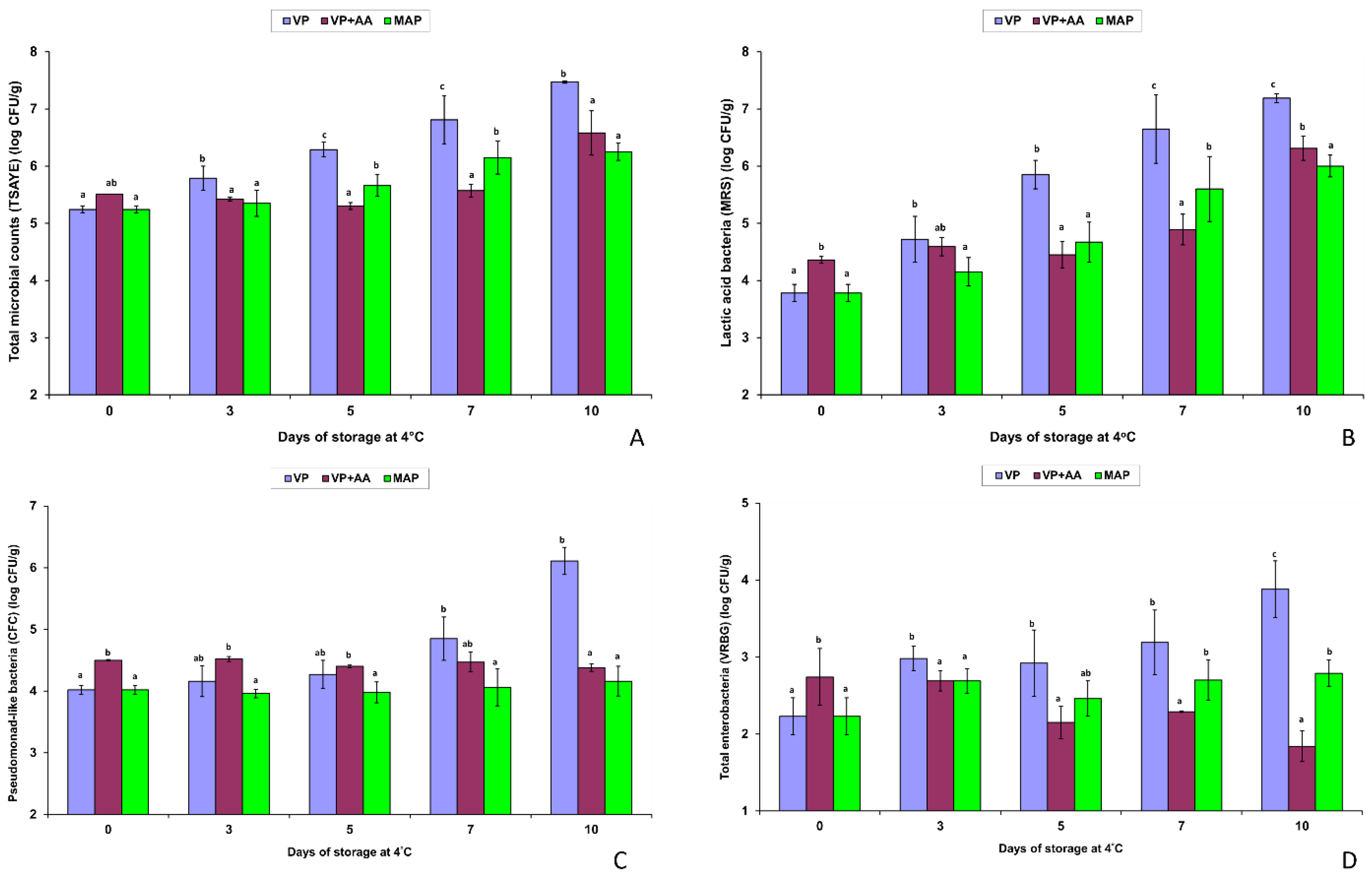
3.5. Major Variations in the Spoilage Pattern of the Fresh VP Chicken Meat Minces Compared to the Resultant MAP Products and the High-Acetate-Containing VP + AA Mince during Storage at 4 °C

To compare the changes in L-lactate, D-lactate, total lactate, and acetate concentrations (Figure 1) in association with the evolution of the TVCs (Figure 2A), spoilage LAB (Figure 2B), pseudomonad-like bacteria (Figure 2C), and enterobacteria (Figure 2D) in the VP3 and VP4 batches of this study with the corresponding changes that occurred in the resultant MAP products studied by Tsafrakidou et al. [32], the above two datasets were plotted together. The VP + AA batch data were also inserted in the plots for comparison (Figure 1 and Figure 2). Additionally, the corresponding pH values of the above three sets of minced fresh chicken samples were compared statistically because the changes in pH are related to the organic acid (Figure 1) and microbial (Figure 2) changes in meat during storage.
The mean initial (day 0) concentration (mg/100 g of meat) of L-lactate was 832 mg/100 g for the VP and their resultant MAP batch samples, whereas it was as high as 1257 mg/100 g in the VP + AA (Figure 1) due to the addition of ‘external’ L-lactate. On day 7, L-lactate decreased by at least 3-fold to 6-fold in the VP (253 mg/100 g) and VP + AA (200 mg/100 g) batches, respectively, whereas it remained high (792 mg/100 g) in the MAP batches. Changes in total lactate showed a similar pattern with L-lactate naturally found in meat, because the initial (day 0) D-lactate content was very low (16–18 mg/100 g) in all batches. On day 7, D-lactate was minimized, except for the VP samples (114 mg/100 g of meat) (Figure 1). The mean initial (day 0) acetate was only 19 mg/100 g in the VP and their counterpart MAP samples. Conversely, the VP + AA batch contained 243 mg/100 g of ‘external’ acetate on day 0, which increased to 278 mg/100 g on day 7 of storage. Acetate increased in the VP (44.3 mg/100 g) and MAP (92 mg/100 g) samples, too (Figure 1).
In accordance with the data in Figure 1, on day 0, the VP samples (pH 6.06 ± 0.06) and their resultant MAP samples (pH 6.01 ± 0.06) had similar pH values, which were higher (p < 0.05) than the pH 5.77 ± 0.03 of the acid-pretreated VP + AA samples. After 5 days, however, the pH drop (ca. 5.8) in the VP/MAP samples caused by the TVC/LAB growth below (viz. Figure 2A,B) reached a value similar to the initial low pH of the VP + AA samples that remained unchanged during storage. On day 7, the pH reversed from 5.8 to 6.0 in the VP samples that developed putrid spoilage; this reversal was ca. 0.1 pH unit smaller in the resultant MAP products that developed ‘blown-pack’ sulfur-type spoilage [32]. In contrast, no significant pH reversal increases occurred in the VP + AA samples during storage. Eventually, in the terminally spoiled samples, the mean pH was higher in the order of MAP (pH 5.96) > VP (pH 5.91) > VP + AA (pH 5.73).
Microbiologically, although the fresh mince of the VP + AA batch was found to be more contaminated on day 0 than the remaining batches (Figure 2), TVCs increased in the order of VP > MAP > VP + AA from day 3 to day 7 (p < 0.05). However, a delayed 10-fold TVC increase in the VP + AA samples from day 7 to day 10 resulted in similar TVCs to the MAP samples by the end (day 10) of storage at 4 °C (Figure 2A). Unsurprisingly, MAP retarded growth of LAB (Figure 2B) and pseudomonads (Figure 2C) by 1–2 log units, at final levels below 6.5 and 4.5 log cfu/g, respectively, compared to VP, whereas the VP + AA batch behaved similarly to the MAP batches throughout storage at 4 °C for 10 days (Figure 2B,C). Thus, the growth-retarding effects of VP combined with the lactate/acetate antimicrobial effects on the spoilage microbiota were comparable to the effects of MAP alone (Figure 2A–C). Significant growth of enterobacteria occurred in the VP samples only, whereas VP + AA was more inhibitory than MAP for enterobacteria (Figure 2D). Growth of staphylococci, enterococci, and yeasts was suppressed in all batches due to refrigeration rather than due to the VP, MAP, or VP + AA treatments.
Finally, comparing the percentage isolation frequency of the LAB species and B. thermosphacta from the VP3 and VP4 batches (Table 4) and the resultant MAP1 and MAP2 products (Table 6 in Ref. [32]), it can be suggested that the packaging method did not exert major selective effects on the composition of the terminal spoilage microbiota of minced, free-range chicken meat at 4 °C. The spoilage community was dominated by Lb. sakei, either under VP (57.5%) or MAP (67.5%), followed by Lc. carnosum (10.0% vs. 12.5%), B. thermosphacta (10.0% vs. 7.5%), C. divergens (5.0% vs. 5.0%), and Lb. fuchuensis (5.0% vs. 2.5%); the rest of the LAB species were sporadic isolates (0–2.5%). Compared to MAP, VP likely reduced the prevalence of Lb. sakei by increasing the isolation frequency of Lb. curvatus and Lb. fuchuensis (Table 4), but additional studies are required to conclude this.
Being less effective than MAP in suppressing growth of Gram-negative bacteria (Figure 2C,D), VP may also affect and thus modulate differently the composition of the spoilage community by selecting for certain gram-negative bacteria species. Under MAP, the subdominant Gram-negative microbiota that survived without growth consisted mainly of members of the genera Pseudomonas, Moraxella, and Alcaligenes, while Serratia liquefaciens was the most abundant member of psychrotrophic Enterobacteriaceae [32]. The biochemical characterization of the Gram-negative spoilage bacteria isolated from the VP samples during this study still is in progress.

4. Discussion

Alternative (organic, free-range, low-input) poultry systems represent only a small portion (about 5%) of the poultry production in the EU [42]. Despite the fact that their popularity is increasing worldwide, microbiological shelf-life, spoilage ecology, and pathogen control studies on alternative poultry products are still limited [5,29,32]. In contrast, since 2002, numerous EU and US survey studies have shown the superior quality of commercially available broiler carcass meat from alternative compared to conventional chickens in terms of bird race, production properties, sensorial attributes, surface color, texture of muscle, and chemical indicators [43,44,45,46,47]. Relative to the methods of this study, only the pH and moisture (i.e., both relating to the water holding capacity; WHC) data of the above studies are offered for comparison with the present results. Of note, the organic, free-range chicken meat samples contained higher levels of polyunsaturated fatty acids (PUFAs) compared to conventional chicken meat samples, resulting in higher Thio Barbituric Acid Reactive Substance (TBA-RS) values and thereby enhanced lipid oxidation, despite the fact that they also contained higher amounts of antioxidants (i.e., tocopherols) transferred from the pasture [43,44,46,48]. This negative aspect is probably associated with the selection of an anoxic MAP atmosphere (30:70 CO2/N2) by ‘PINDOS’ for packaging all retail, free-range chicken meat products, as suggested by Abdullach et al. [48]. Conversely, packaging of fresh (poultry) meat products in high-O2 MAP (i.e., 20:80 or 30:70 CO2/O2) [9,11,13,18,22] has been a frequent industrial trend since the mid-1990s to prevent the bright red color of the meat during storage [49].
Regarding the pH and moisture, Castellini et al. [43] first reported that the organic chicken muscles had lower ultimate pH (pHu) and WHC values than the control chicken muscles, depending on the muscle type and the bird age at slaughter. For instance, at 81 days of age, the pHu of the organic breast and thigh muscles was 5.8 and 6.1, whereas the control muscles had pHu of 6.0 and 6.3, respectively. The moisture content of the organic breast (75.8%) and thigh (76.9%) was higher than that of the control breast (74.8%) and thigh (75.4%) muscles [43]. Similar trends were later reported by others. Specifically, a US study by Husak et al. [45] discriminated between retail broilers from free-range (fed outside), organic-fed inside, and conventional chickens, and amongst them, the former breast and thigh products had the lowest pH of 5.72 and 6.07, respectively. Higher mean pH values for the organic skinless chicken breast (pH 5.95) and thigh (pH 6.45) muscle, and pH of 6.3–6.4 for the skin of both, were reported in a recent Czech study by Hulankova et al. [5]. Of note, the initial pH of 6.0–6.1 of the mixed (ca. 50:50 breast/thigh) VP/MAP batch samples coincided with the above literature pH data, whereas it was lower than the initial pH of conventional fresh raw chicken thigh (pH 6.2–6.5) or breast (pH 5.8–6.4) meat usually reported in the literature [50,51,52], including conventional chicken breast fillets (initial pH 6.4) from the same plant previously analyzed by others [25,27]. Thus, a lowered pH may be a constant intrinsic (abiotic) characteristic of free-range chicken meat. The mean moisture (73.3%) content of the VP/MAP batches was lower compared to the aforementioned moisture values [43], probably because mincing increased water loss from the fresh samples of this study. The VP + AA batch (pH 5.77) had a higher (74.8%) moisture content compared to the rest VP batches probably due to the presence in the blend of sodium or potassium lactate, which are known to increase WHC in meat formulations [7].
Consistent with their fairly low initial pH, the fairly high initial L-lactate content of the VP/MAP samples (Table 1; Figure 1) could also be an intrinsic characteristic of each mince rather than the consequence of pretreatment with lactic acid (for decontamination) or salts (for active packaging) during processing [7,53,54,55]. Castellini et al. [43] suggested that the lower pHu of organic chickens could be due to the better welfare conditions that reduced the stress pre-slaughter and thus glycogen consumption, but neither of the above authors measured L-lactate in organic versus control chicken samples, nor relevant data found in the literature, to justify this. Nevertheless, raw poultry meat has an approximate 0.2–0.4-unit higher pH than raw pork and mainly beef, while it is well-established that the chicken breast and thigh represent the normal (white) and high-pH DFD (red) meat, respectively [56]. In general, the pH of the animal carcass meat, categorized as Dark Firm Dry (DFD; pH 6.4–6.8), normal (ca. pH 6.2), or Pale Soft Exudative (PSE; <6.2, even down to 5.6), reflects the concentration of L-lactate produced via the anaerobic breakdown of the glycogen in the meat muscles after slaughter [55]. When the animal is exposed to long-term stress before slaughter, glycogen is converted by respiration, resulting in deficient L-lactate production post-mortem and thus in high-pH DFD meat, which is more prone to rapid bacterial deterioration than normal meat [55]. Regardless of stress, the chicken thigh contains less L-lactate than the breast because it absorbs the kinetic activity of the live birds. For instance, Kakouri and Nychas [56] reported that the initial (day 0) L-lactate content of conventional chicken thigh meat or skinless breast fillets stored in VP or MAP (100% CO2 and 100% N2) was 244 and 679 mg/100 g of meat, respectively. In this study, the initial L-lactate of the VP batches (mean 748 mg/100 g; VP4 had ca. 900 mg/100 g) was even higher than that of the breast fillets in that previous study [56]. Thus, despite the fact that all fresh minces contained ca. 50% breast and thigh, L-lactate remained high probably because the free-range birds were exposed to reduced stress before slaughter [43].
Along with the physicochemical (abiotic) parameters of the muscle, the initial microbial contamination is the primary biotic factor affecting the rate and type of meat spoilage [1,7,57]. In principle, the higher the TVC in meat before storage, the faster the onset of microbial spoilage under a constant set of packaging and storage conditions [7]. Hulankova et al. [5] reported that both the muscle and skin of organic chicken breast and thigh samples showed good initial microbiological quality, with TVCs being of less than 3 and 5 log cfu/g, respectively. Skinless breast fillets from conventional chickens also had initial TVCs below 3 log cfu/g [19], including fillets produced in Epirus [58]. However, other previous studies reported remarkably higher mean initial (day 0) TVCs, which, for instance, were 4.45 log cfu/g in freshly packaged Korean chicken breast fillets [2] or up to 6.74 log cfu/g in skinless breast fillets obtained from a small Greek processing plant [56]. Overall, initial contamination levels of fresh poultry products may vary greatly depending on the plant hygiene and the time passed after portioning or mincing for retail distribution and storage [1,50]. Usually, thigh is more heavily contaminated than breast, and although mincing further increases the TVCs by ca. 10-fold [7], fresh US ground turkey meat patties still had TVCs slightly above 3 log units/g on day 0 [21]. In this study, all free-range chicken meat samples potentially minced with the skin had initial TVCs of 5.1–5.5 log cfu/g (Table 2). Although, to the authors’ knowledge, no official regulations or industrial standards exist [32], an approximate 5-log TVC level in fresh minced meats, including poultry, indicates an overall good microbiological quality, according to previous conventional microbiological data [1,23] and recent non-invasive assessment studies using hyperspectral scattering techniques as, for example, in chilled pork [59].
However, apart from the TVC levels [2,18], the type of naturally contaminating spoilage bacteria and their growth potential during storage, depending on the packaging method (air, VP, high-O2 or low-O2 MAP; passive or active) and the storage temperature, are critical factors in determining progressive and terminal population changes and species successions in the microbial community, the biochemistry of raw meat spoilage, and eventually the product shelf-life [1,7,24,57,60]. Regarding growth, the complete failure of total pseudomonad-like bacteria to increase above 4.5 log cfu/g of minced meat in the MAP and VP + AA samples (Figure 2C), and the low initial Enterobacteriaceae counts followed by restricted or no growth in all minced samples at 4 °C (Figure 2D), indicated that psychrotrophic Gram-negative bacteria were unlikely to have had a drastic in situ contribution to spoilage of free-range chicken meat. Only in the VP samples did pseudomonad-like bacteria increase to ca. 6 log cfu/g, but with major delay compared to TVC/LAB (Table 2). According to Nychas et al. [57], 7–8 log cfu/g for Pseudomonas spp. is the determinant of raw meat spoilage, while several studies consider the upper limit of microbiological acceptability for chicken meat to be 7-log cfu/g [1,2,11,19,26]. In some studies, Pseudomonas grew close to the 7-log threshold in high-pH conventional fresh chicken products stored in VP or high-O2 MAP [19,23,56], thereby contributing to the volatilome associated with their spoilage [24,56]. In contrast, numerous studies have noted strong growth inhibition of Gram-negative bacteria along with major growth retardation of TVC and LAB, in conventional low-O2 MAP poultry compared to high-O2 MAP, VP, or air, resulting in significant product shelf-life extensions [11,21,25,27,28,56,57], as noted in this study, too (Figure 2). Likewise, Hulankova et al. [5] found restricted or no growth of Pseudomonas and Enterobacteriaceae during storage of organic breast and thigh muscle meat in MAP1 (20:80 CO2/O2) and mainly in MAP2 (30:70 CO2/N2), i.e., the retail MAP condition applied by ‘PINDOS’, too [32]. Pseudomonas grew above 5.0 log cfu/g only in the MAP2 skin after 10–14 days at 2 ± 2 °C, whereas major growth (>6.0–9.0 log units) occurred only in muscle and mainly skin samples stored in air [5]. To sum up, based on populations (Table 2; Figure 2), Gram-negative bacteria were minor spoilers of the minced VP free-range chicken meat samples, unless we accept that the spoilage potential of aerobic pseudomonad-like bacteria was high at subdominant levels well below 5 and 6 log cfu/g by the middle (day 5) and end (days 7 and 10) of storage at 4 °C, respectively. Major spoilers were the LAB species (Table 2, Table 3 and Table 4) retrieved from the VP batch samples by the end of storage, once along with B. thermosphacta, in general agreement with the literature [1,4,10,13,24,26,60].
In terms of microbial ecology, the typical, facultative heterofermentative species of the genus Latilactobacillus, Lb. sakei [17,37], was the primary specific spoilage organism (SSO), i.e., the single species being responsible for the minced free-range chicken meat spoilage [61], in both packaging conditions, i.e., according to its high isolation frequency among the dominant microbiota (TSAYE and MRS) of the VP1 (65%), VP + AA (70%), VP3 + VP4 (57.5%), and the resultant MAP products (67.5%) [32], at the end of storage. However, apart from Lb. sakei, Lb. fuchuensis, C. divergens, Lc. carnosum, and B. thermosphacta were major subdominant members of the ‘metabiotic spoilage association’, a term introduced to describe situations where two or more microbial species contribute to food (meat) spoilage through the exchange of metabolites or nutrients [24,62]. According to the literature, all four species above are amongst the most frequently isolated psychrotrophic bacteria from raw, fresh or marinated, meat and poultry products stored in VP or MAP at 0–4 °C; in most previous cases, they were the predominant spoilers, along with Carnobacterium maltaromaticum and Leuconostoc gelidum subsp. gelidum or Lc. gelidum subsp. gasicomitatum [24,50,60,63,64,65,66]. In this study, all C. divergens and B. thermosphacta and most Lc. carnosum isolates were recovered from the most diversified TSAYE plates of the VP batches bearing TVCs well above the 7-log threshold on day 10 (Table 2). This finding suggests that at least the above three meat-specific species interacted with Lb. sakei and contributed to spoilage, which was earlier and more offensive in vacuum as regards consumer (our laboratory personnel) perception. Contribution to spoilage was seemingly higher for C. divergens in VP1, B. thermosphacta in VP3, and Lc. carnosum in VP4. In contrast, neither TVCs nor LAB or any non-LAB populations reached the 7-log level in the anoxic MAP [32] or VP + AA (Table 2) batches; even Lb. sakei was at ca. 10-fold lower levels on day 10. Therefore, despite the fact that spoilage community was similar in terms of species composition, sharing the SSO Lb. sakei, the sensorial manifestation of spoilage (i.e., based on visual and orthonasal impression) was different among samples, described as ‘putrid without blowing’ in the VP1 and VP3 + VP4 batches, ‘blown-pack/sulphur’ in both resultant MAP batches, and acidic-vinegar/‘malty/cheesy’ in the VP + AA.
Tsafrakidou et al. [32] discussed certain in vitro metabolic capabilities of the predominant SSO Lb. sakei, other meat LAB, and B. thermosphacta to explain ‘blown-pack’ sulfur spoilage manifested in all retail MAP samples after 7 to 10 days. Most of those discussions are extended to explain why spoilage in the VP samples was sensed as more putrefactive than in the resultant MAP samples. So, although its natural concentration in raw meat is limited (ca. 0.1%) and is even ca. 10-fold lower in fresh poultry than beef and pork, glucose is the first (key) substrate preferentially utilized by most bacterial contaminants developing in meat during cold storage in any packaging condition [24,67,68]. In anoxic (i.e., 30:70 CO2/N2) MAP trays or ‘firm’ VP bags limiting residual oxygen within the product and the packaging film interface, glucose is fermented by LAB (mainly), B. thermosphacta, and less likely by Enterobacteriacae, whereas Pseudomonas have a major disadvantage in glucose uptake, i.e., otherwise they rapidly transform glucose to gluconate to initiate growth in air-stored meat [7,57,67]. Because most psychrotrophic meat LAB species are facultative or obligatory heterofermentative, they produce two moles of lactate, or one mole of lactate, acetate, and CO2 from glucose (hexoses), respectively. Under glucose limitation, the former LAB species, including Lb. sakei, can ferment ribose (pentoses), also limited in meat, to form lactate and acetate, by shifting their sugar metabolism from homofermentative to heterofermenative without gas [7,17,24].
In this study, the slight progressive reduction in the fresh chicken meat pH by ca. 0.2–0.3 pH units from day 0 to day 5 in all VP and MAP samples reflected sugar (glucose) fermentation. Whereas Lb. sakei has the ability to highly express L-lactate dehydrogenase and form much more (or only) L-lactate than D-lactate in the racemase [37,69], and because most other LAB species (Table 3) and B. thermosphacta are L-lactate producers and none, except for Lc. carnosum, are sole D-lactate producers [37,38,39,40,41], the low-to-negligible D-lactate content in all samples on day 7 is justified. Indeed, according to the literature, lactate (i.e., the major L-lactate fraction contained in the chicken minces post-slaughter plus the minor L, DL, or D-lactate amounts above) is the third substrate, in order, catabolized by LAB and other spoilage bacteria after the depletion of glucose/glucose-6-phosphate in fresh meat ecosystems, mainly by oxidation in air-packed, high-O2 MAP and VP products [24]. However, the capability of Lb. sakei to catabolize both lactate isomers and form acetate by shifting its metabolism to heterolactic under glucose limitation in vitro [70] and during anaerobic cold storage of fresh meat [68] increased its competitive dominant growth after the first 3 to 5 days herein (Table 2). Particularly in the VP samples, acetate from glucose and lactate could be produced by all subdominant meat LAB because C. divergens and Lc. carnosum are obligatory heterofermenters [39,40] and Lb. fuchuensis and Lb. curvatus are close relatives of Lb. sakei sharing meat as their main habitat and glycolytic pathways. B. thermosphacta is a homofermentative bacterium under low-oxygen- and glucose-availability conditions, while it shifts to heterofermentative under high-oxygen and/or glucose-limiting conditions, producing high amounts of acetate during meat spoilage [24].
Based on the preceding discussion, the major decrease in L-lactate in the VP samples compared to its minor decrease in the resultant oxygen-free MAP (30:70 CO2/N2) samples (which was the most prominent difference between the two packaging methods after 7 days of storage at 4 °C (Figure 1)) could be explained: lactate catabolism via oxidation was much reduced, almost prevented, in the anoxic MAP of free-range chicken meat minces because of the antioxidant and the antimicrobial growth retardation effects of the solubilized 30% CO2 against all bacteria (LAB) spoilers, including the dominant Lb. sakei. In contrast, in the VP samples, the antimicrobial effects of CO2 were minimal, i.e., practically absent, because typical CO2 producers, like Lc. carnosum, were subdominant and C. divergens possesses a unique heterolactic metabolism that leads to low, even undetectable, CO2 formation in vitro [71]. Hence, apart from the fact that growth of Lb. sakei, the other LAB spoilers, and B. thermosphacta was more pronounced in VP than MAP, the oxygen remaining in the former samples after sealing, also because vacuum bags had a higher oxygen transmission rate (OTR) (175 cm3/m2, 24 h at 23 °C; 0% RH) than the MAP trays [32], enhanced lactate oxidation to acetate during storage. Similar differences were noted by Kakouri and Nychas [56] between VP and MAP (100% CO2) fresh chicken meat muscles stored at 3 °C for up to 13 days. Specifically, the initial L-lactate in breast and thigh remained constant in the 100% CO2 MAP samples, whereas it decreased more in the high-O2 MAP (80:20 O2/CO2) than in the 100% N2 MAP and VP thigh samples during storage at 3 °C. In all cases, the decrease in L-lactate was accompanied by an increase in acetate which was greater, in the order of 80:20 O2/CO2 > 100% N2 > VP > 100% CO2 [56]. Several later studies confirmed the above trends and provided advanced data on the in situ formation of volatile glycolytic end products affecting (poultry) meat spoilage, such as acetate, acetoin, diacetyl, ethanol, other alcohols, aldehydes, ketones, butyric acid, and esters (such as butyl acetate and ethyl acetate) [22,24,72]. These volatile compounds (VOCs) can be formed in situ either by Lb. sakei and Lb. fuchuensis or by C. divergens/C. maltaromaticum and Lc. gelidum/Lc. gasicomitatum, Lc. carnosum/Lc. mesenteroides,or B. thermosphacta [24,72,73]; three of the latter meat spoilers were frequent in the VP samples (Table 4). Depending on the strain, all Lb. sakei and C. divergens isolates from VP and VP + AA were strong to moderate acetoin producers in vitro, followed by Lb. fuchuensis, as the previous MAP isolates were, too [32]. The greatest decrease in L-lactate occurred in the VP + AA samples, providing clear evidence that Lb. sakei and to a lesser extent Lb. fuchuensis and Lb. curvatus were responsible for L-lactate utilization under vacuum, regardless of the high external acetate in this batch.
With progressive storage, after glucose depletion and in parallel to utilizing the glycolytic intermediates (glucose-6-phosphate, gluconate, lactate, pyruvate, formate, ethanol, acetate), the best-adapted meat spoilage bacteria start attacking amino acids, water-soluble meat proteins, and lipids to cause different types of spoilage, depending on the SSOs and the oxygen availability in the package [7,23,24]. In this aspect, the preferential use by Lb. sakei of the arginine present in meat as a principal substrate to produce ATP when glucose is close to depletion [17] further supported its prevalence and increased its spoilage potential. Ammonia readily produced in vitro from arginine hydrolysis by Lb. sakei and other arginine-positive LAB, mainly Carnobacterium spp. (viz. Table 3), contributes greatly to the volatilome associated with (poultry) meat spoilage in air, and significantly more in VP compared to low-O2 MAP [17,24,74]; thus, it can serve as a primary chemical indicator of ‘putrid’ spoilage in fresh poultry [56]. Indeed, ammonia increased more in spoiled VP than MAP/100% CO2 breast and thigh samples during storage, while, unsurprisingly, increases in ammonia were the highest in thigh samples stored at 80:20 O2/CO2 at 3 °C for 13 days [56]. Likewise, ammonia increased by ca. 2-fold more in air-stored organic chicken breast and thigh, but it remained constant in MAP2 (30:70 CO2/N2) at 2 ± 2 °C for 14 days [48], as it probably did in the retail MAP samples which released a ‘spoiled-egg’ sulfite smell upon opening [32] rather than putrid off odors, like the VP samples. Apparently, MAP triggered H2S production by Lb. sakei previously associated with offensive (sulfurous offodor) spoilage of VP or low-O2 MAP (5:95 CO2/N2) beef [68,74,75,76], whereas VP further allowed more oxidative/putrefactive (ammonia-releasing) spoilage reactions associated with decarboxylation of arginine, other amino acids (methionine, cystine, cysteine), peptides, and water-soluble proteins by mainly Lb. sakei [74] and C. divergens and/or B. thermosphacta in the VP samples.
The VP + AA batch spoiled less rapidly and offensively and was highly selective for Lb. sakei, as well as for Lb. fuchuensis and Lb. curvatus due to the protective residual (in case of decontamination) or additive (in case of supplementation) [1,53,54] antimicrobial effects of the external lactate and acetate throughout storage [7,35]. Similar results regarding a high selective predominance of Latilactobacillus spp., namely Lb. graminis or Lb. curvatus/graminis along with Leuconostoc spp., during refrigerated or chill storage of fresh pork model or retail French pork sausages pretreated with a commercial lactate/diacetate blend or potassium lactate and sodium acetate were reported by Benson et al. [77] and Boujou-Albert et al. [78], respectively. The removal of both organic salt preservatives resulted in control sausages which were more diversified throughout storage [77,78], as also were the three VP batch samples without (at least) external acetate.
Before 2016, lactates and diacetates were prohibited in both processing and final packaging of organic poultry due to additional restrictions that limit the use of antimicrobials in alternative production systems, despite concerns regarding a potentially increased pathogen persistence and growth potential in these products, including L. monocytogenes [29]. However, in 2016, the US National Organic Standards Board approved the use of lactate as an antimicrobial agent and pH regulator in organic processing, whereas diacetate was determined not to be ancillary and therefore not approved for use yet [29]. For this reason, at first glance, we suspected the routine use of solely sodium or potassium lactate in all fresh industrial batches, which would increase L-lactate and the WHC of the free-range chicken meat mince without much affecting the pH before storage [7]. Later, during the challenge experiments, the failure of L. monocytogenes to grow in all VP batches supported our suspicion. However, previous challenge studies on conventional poultry products have also noted the absence or limited growth of L. monocytogenes in (control) VP samples within the short time (<10 days) of the product’s shelf-life under refrigerated storage [29,52,79]. In one of those studies, Argyri et al. [52] found that B. thermosphacta, rather than a psychrotrophic meat LAB species, was the predominant SSO in both the control and HPP-treated chicken fillets. Overall, according to the literature, the natural spoilage microbiota in fresh meat and poultry often exerts growth-inhibitory effects against coexisting pathogens, including L. monocytogenes [7]. In particular, the main psychrotrophic meat LAB species of Latilactobacillus, Leuconostoc, and Carnobacterium play a controversial role; most strains contribute to fresh meat spoilage through the generation of offensive metabolites discussed above, but many competitive strains of low spoilage potential serve as bioprotective (antilisterial) agents by the in situ production of natural antimicrobials, mainly bacteriocins [64,66]. Therefore, LAB bacteriocins and/or protective cultures are amongst the primary control measures against L. monocytogenes in conventional or alternative poultry products [29,51]. In this study, the L. monocytogenes inocula failed to grow because bacteriocin-producing strains of the predominant Lb. sakei (mainly), C. divergens, or Lc. carnosum might have been numerous within the spoilage LAB biota growing competitively in the VP samples; moreover, the combined LAB-inhibitory effects were enhanced by refrigeration, considering that the growth potential of L. monocytogenes increases under temperature abuse (i.e., 10–12 °C) storage conditions [52] that should be carefully avoided in fresh meat distribution anyway [7,28,57]. The absence of detectable listerial growth in the uninoculated VP batches stored at 4 °C was another positive finding because Listeria spp. prevalence in poultry meat is noticeable, with L. innocua as the dominant species followed by L. monocytogenes [1]. Indeed, one of our early studies showed that turkey necks and breasts and mechanically deboned meat (MDM) were the principal sources of Listeria spp. contamination in a Greek meat processing plant, with L. monocytogenes isolations being primarily distributed in turkey necks (30.1%), MDM (20.5%), and turkey breasts (19.2%), respectively [31]. Twenty years later, a meta-analysis study reported that L. monocytogenes was frequently found in chicken meat, at a mean prevalence of about 21%, at the industry or retail level and for both packed and unpacked products [80]. Although natural Listeria spp. contamination in fresh (poultry) meat products would be killed during cooking and thus the incidence of L. monocytogenes in chicken meat is not required to be reported to the European Food Safety Authority (EFSA) [52,80], further systematic surveillance and challenge data on the prevalence and control of this stress-resistant pathogen [30] in alternative poultry processing and retail are required [29]. Similar research data are required for the free-range chicken rearing systems in Epirus or other Greek areas because, as mentioned, this meat type is currently used in the production of alternate chicken sausages or other ready-to-eat (RTE) deli meats and thus it may serve as an incoming raw material source of L. monocytogenes in meat processing plants.
The high predominance of Lb. sakei in all VP (Table 4) or MAP [32] samples suggests that free-range chicken meat may be more selective for Lb. sakei growth during storage due to its lowered pH and higher lactate and possibly glucose content, or other genetic or nutritional factors compared to conventional chicken meat. On this basis, the spoilage microbial (LAB) community of free-range chicken MAP meat may constantly share a higher similarity with that of fresh minced MAP beef dominated by Lb. sakei [81] rather than by Carnobacterium spp. and B. thermosphacta or by Lc. gelidum/gasicomitatum, being the dominant spoilers in retail high-O2 MAP, fresh [1,4,9,10,13,26] or marinated [63,66], chicken meat products, respectively. Further research is required to validate the above hypothesis. Also, based on the preceding discussion, the in vitro and in situ antilisterial potential of the isolated Lb. sakei (mainly), C. divergens, and Lc. carnosum strain biotypes/genotypes from the VP or MAP free-range chicken meat minces would be useful to evaluate in future screening studies to potentially detect novel bacteriocinogenic strains suitable for use as bioprotective cultures in the poultry meat industry.

5. Conclusions

This is the first report on the microbial spoilage of minced Greek free-range chicken meat stored in VP compared to low-O2 MAP (30%/70% CO2/N2) at 4 °C for up to 10 days. Comparisons were not based only on TVCs and selective microbial enumerations across storage, but they were extended to identifying the SSOs in the VP meat batches of this study vs. the resultant MAP products studied previously [32]. Results indicated that the free-range chicken minces spoiled more rapidly and offensively in VP than in anoxic MAP conditions. LAB and B. thermosphacta were major spoilers, whereas Pseudomonas and Enterobacteriacae likely were minor spoilers. Detected use of an acetate-containing antimicrobial in one of the industrial batches (VP + AA) before mincing retarded TVC and LAB growth comparably to low-O2 MAP and altered the sensory spoilage to the mildest acidic offodor type at 4 °C. Latilactobacillus, primarily Lb. sakei, were favored to dominate in MAP and VP + AA, whereas C. divergens, Lc. carnosum, and/or B. thermosphacta were more frequent in the spoiled VPs, without erasing the predominance of Lb. sakei. L. monocytogenes failed to grow, but survived without death, in all free-range VP chicken meat minces at 4 °C for 10 days, regardless of whether the mince contained ‘external’ lactate/acetate. Although culture-dependent studies based on a higher number of isolates during storage might detect additional LAB or non-LAB species, the composition of the terminal spoilage community based on conventional methods would remain valid with regard to the SSOs identified. However, additional metagenomic analyses combined with advanced biochemical and sensory quality studies are required to provide in-depth descriptions of the diverse microbial ecology and the volatilome associated with the spoilage of free-range compared to conventional chicken meat products.

Author Contributions

Conceptualization, J.S.; methodology, P.T., N.S. and J.S.; formal analysis, P.T., N.S., A.K. and J.S.; investigation, L.B.; data curation, P.T. and J.S.; writing—original draft preparation, P.T. and J.S.; writing—review and editing, P.T., L.B. and J.S.; supervision, J.S.; project administration, L.B. All authors have read and agreed to the published version of the manuscript.

Funding

This research is part of the research program entitled ‘Production of processed meats by applying novel biotechnological methods for increasing their shelf life, microbiological safety and nutritional value’ (MIS number: 5033164), supported by the action ‘Strengthening of small and medium-sized enterprises for research programs in the fields of agro-nutrition, health and biotechnology’, co-financed by the European Union (European Regional Development Fund) and Greece, under the ‘Operational Program Epirus 2014–2020’ of the National Strategic Reference Framework.

Data Availability Statement

Data are contained within the article.

Acknowledgments

We thank the company PINDOS for providing the chicken meat samples.

Conflicts of Interest

The authors declare no conflict of interest.

References

  1. Rouger, A.; Tresse, O.; Zagorec, M. Bacterial contaminants of poultry meat: Sources, species, and dynamics. Microorganisms 2017, 5, 50. [Google Scholar] [CrossRef] [PubMed]
  2. Yimenu, S.M.; Koo, J.; Kim, B.S.; Kim, J.H.; Kim, J.Y. Freshness-based real-time shelf-life estimation of packaged chicken meat under dynamic storage conditions. Poult. Sci. 2019, 98, 6921–6930. [Google Scholar] [CrossRef] [PubMed]
  3. Lytou, A.E.; Renieri, C.T.; Doulgeraki, A.I.; Nychas, G.J.E.; Panagou, E.Z. Assessment of the microbiological quality and safety of marinated chicken products from Greek retail outlets. Int. J. Food Microbiol. 2020, 320, 108506. [Google Scholar] [CrossRef] [PubMed]
  4. Samapundo, S.; de Baenst, I.; Aerts, M.; Cnockaert, M.; Devlieghere, F.; Van Damme, P. Tracking the sources of psychrotrophic bacteria contaminating chicken cuts during processing. Food Microbiol. 2019, 81, 40–50. [Google Scholar] [CrossRef] [PubMed]
  5. Hulankova, R.; Borilova, G.; Abdullah, F.A.A.; Buchtova, H. Microbiological quality of organic chicken meat during refrigerated storage in air and modified atmospheres. Brit. Poult. Sci. 2018, 59, 506–513. [Google Scholar] [CrossRef] [PubMed]
  6. Jin, S.; Yang, L.; Zang, H.; Xu, Y.; Chen, X.; Chen, X.; Liu, P.; Geng, Z. Influence of free-range days on growth performance, carcass traits, meat quality, lymphoid organ indices, and blood biochemistry of Wannanyellow chickens. Poult. Sci. 2019, 98, 6602–6610. [Google Scholar] [CrossRef] [PubMed]
  7. Samelis, J. Managing microbial spoilage in the meat industry. In Food Spoilage Microorganisms; Blackburn, C.W., Ed.; Woodhead Publishing Limited: Cambridge, UK, 2006; pp. 213–286. [Google Scholar]
  8. Odeyemi, O.A.; Alegbeleye, O.O.; Strateva, M.; Stratev, D. Understanding spoilage microbial community and spoilage mechanisms in foods of animal origin. Comp. Rev. Food Sci. Food Saf. 2020, 19, 311–331. [Google Scholar] [CrossRef]
  9. Lauritsen, C.V.; Kjeldgaard, J.; Ingmer, H.; Bisgaard, M.; Christensen, H. Microbiota encompassing putative spoilage bacteria in retail packaged broiler meat and commercial broiler abattoir. Int. J. Food Microbiol. 2019, 300, 14–21. [Google Scholar] [CrossRef]
  10. Rouger, A.; Moriceau, N.; Prévost, H.; Remenant, B.; Zagorec, M. Diversity of bacterial communities in French chicken cuts stored under modified atmosphere packaging. Food Microbiol. 2018, 70, 7–16. [Google Scholar] [CrossRef]
  11. Franke, C.; Höll, L.; Langowski, H.C.; Petermeier, H.; Vogel, R.F. Sensory evaluation of chicken breast packed in two different modified atmospheres. Food Packag. Shelf Life 2017, 13, 66–75. [Google Scholar] [CrossRef]
  12. Moutiq, R.; Misra, N.; Mendonça, A.; Keener, K. In-package decontamination of chicken breast using cold plasma technology: Microbial, quality and storage studies. Meat Sci. 2020, 159, 107942. [Google Scholar] [CrossRef] [PubMed]
  13. Rossaint, S.; Klausmann, S.; Kreyenschmidt, J. Effect of high-oxygen and oxygen-free modified atmosphere packaging on the spoilage process of poultry breast fillets. Poult. Sci. 2015, 94, 96–103. [Google Scholar] [CrossRef] [PubMed]
  14. Morales, P.A.; Aguirre, J.S.; Troncoso, M.R.; Figueroa, G.O. Phenotypic and genotypic characterization of Pseudomonas spp. present in spoiled poultry fillets sold in retail settings. LWT Food Sci. Technol. 2016, 73, 609–614. [Google Scholar] [CrossRef]
  15. Mellor, G.E.; Bentley, J.A.; Dykes, G.A. Evidence for a role of biosurfactants produced by Pseudomonas fluorescens in the spoilage of fresh aerobically stored chicken meat. Food Microbiol. 2011, 28, 1101–1104. [Google Scholar] [CrossRef] [PubMed]
  16. Wang, G.Y.; Wang, H.H.; Han, Y.W.; Xing, T.; Ye, K.P.; Xu, X.L.; Zhou, G.H. Evaluation of the spoilage potential of bacteria isolated from chilled chicken in vitro and in situ. Food Microbiol. 2017, 63, 139–146. [Google Scholar] [CrossRef]
  17. Labadie, J. Consequences of packaging on bacterial growth. Meat is an ecological niche. Meat Sci. 1999, 52, 299–305. [Google Scholar] [CrossRef]
  18. Herbert, U.; Albrecht, A.; Kreyenschmidt, J. Definition of predictor variables for MAP poultry fillets stored under different temperature conditions. Poult. Sci. 2015, 94, 424–432. [Google Scholar] [CrossRef]
  19. Chmiel, M.; Roszko, M.; Hać-Szymańczuk, E.; Adamczak, L.; Florowski, T.; Pietrzak, D.; Cegiełka, A.; Bryła, M. Time evolution of microbiological quality and content of volatile compounds in chicken fillets packed using various techniques and stored under different conditions. Poult. Sci. 2020, 99, 1107–1116. [Google Scholar] [CrossRef]
  20. Nieminen, T.T.; Nummela, M.; Björkroth, J. Packaging gas selects lactic acid bacterial communities on raw pork. J. Appl. Microbiol. 2015, 119, 1310–1316. [Google Scholar] [CrossRef]
  21. Dhananjayan, R.; Han, I.Y.; Acton, J.C.; Dawson, P.L. Growth depth effects of bacteria in ground turkey meat patties subjected to high carbon dioxide or high oxygen atmospheres. Poult. Sci. 2006, 85, 1821–1828. [Google Scholar] [CrossRef]
  22. Klein, D.; Maurer, S.; Herbert, U.; Kreyenschmidt, J.; Kaul, P. Detection of volatile organic compounds arising from chicken breast filets under modified atmosphere packaging using TD-GC/MS. Food Anal. Methods 2018, 11, 88–98. [Google Scholar] [CrossRef]
  23. Dainty, R.H.; Mackey, B.M. The relationship between the phenotypic properties of bacteria from chill-stored meat and spoilage processes. J. Appl. Microbiol. 1992, 73, 103S–114S. [Google Scholar] [CrossRef] [PubMed]
  24. Casaburi, A.; Piombino, P.; Nychas, G.J.; Villani, F.; Ercolini, D. Bacterial populations and the volatilome associated to meat spoilage. Food Microbiol. 2015, 45, 83–102. [Google Scholar] [CrossRef] [PubMed]
  25. Patsias, A.; Badeka, A.V.; Savvaidis, I.N.; Kontominas, M.G. Combined effect of freeze chilling and MAP on quality parameters of raw chicken fillets. Food Microbiol. 2008, 25, 575–581. [Google Scholar] [CrossRef] [PubMed]
  26. Höll, L.; Behr, J.; Vogel, R.F. Identification and growth dynamics of meat spoilage microorganisms in modified atmosphere packaged poultry meat by MALDI-TOF MS. Food Microbiol. 2016, 60, 84–91. [Google Scholar] [CrossRef] [PubMed]
  27. Chouliara, E.; Karatapanis, A.; Savvaidis, I.N.; Kontominas, M.G. Combined effect of oregano essential oil and modified atmosphere packaging on shelf-life extension of fresh chicken breast meat, stored at 4 °C. Food Microbiol. 2007, 24, 607–617. [Google Scholar] [CrossRef]
  28. Luong, N.D.M.; Coroller, L.; Zagorec, M.; Membré, J.M.; Guillou, S. Spoilage of chilled fresh meat products during storage: A quantitative analysis of literature data. Microorganisms 2020, 8, 1198. [Google Scholar] [CrossRef]
  29. Rothrock, J.M.J.; Micciche, A.C.; Bodie, A.R.; Ricke, S.C. Listeria occurrence and potential control strategies in alternative and conventional poultry processing and retail. Front. Sustain. Food Syst. 2019, 3, 33. [Google Scholar] [CrossRef]
  30. Roberts, B.N.; Chakravarty, D.; Gardner, J.C., III; Ricke, S.C.; Donaldson, J.R. Listeria monocytogenes response to anaerobic environments. Pathogens 2020, 9, 210. [Google Scholar] [CrossRef]
  31. Samelis, J.; Metaxopoulos, J. Incidence and principal sources of Listeria spp. and Listeria monocytogenes contamination in processed meats and a meat processing plant. Food Microbiol. 1999, 16, 465–477. [Google Scholar] [CrossRef]
  32. Tsafrakidou, P.; Sameli, N.; Bosnea, L.; Chorianopoulos, N.; Samelis, J. Assessment of the spoilage microbiota in minced free range chicken meat during storage at 4 °C in retail modified atmosphere packages. Food Microbiol. 2021, 99, 103822. [Google Scholar] [CrossRef] [PubMed]
  33. Vandera, E.; Lianou, A.; Kakouri, A.; Feng, J.; Koukkou, A.I.; Samelis, J. Enhanced control of Listeria monocytogenes by Enterococcus faecium KE82, a multiple enterocin-producing strain, in different milk environments. J. Food Prot. 2017, 80, 74–85. [Google Scholar] [CrossRef] [PubMed]
  34. Samelis, J.; Kakouri, A.; Rementzis, J. The spoilage microflora of cured, cooked turkey breasts prepared commercially with or without smoking. Int. J. Food Microbiol. 2000, 56, 133–143. [Google Scholar] [CrossRef] [PubMed]
  35. Samelis, J.; Kakouri, A. Growth inhibitory and selective pressure effects of sodium diacetate on the spoilage microbiota of frankfurters stored at 4 °C and 12 °C in vacuum. Foods 2021, 10, 74. [Google Scholar] [CrossRef] [PubMed]
  36. Association of the Official Chemists (AOAC) International. Official Methods of Analysis; Cunniff, P., Ed.; AOAC International: Rockville, MD, USA, 1998. [Google Scholar]
  37. Zheng, J.; Wittouck, S.; Salvetti, E.; Franz, C.M.A.P.; Harris, H.M.B.; Mattarelli, P.; O’Toole, P.W.; Pot, B.; Vandamme, P.; Walter, J.; et al. A taxonomic note on the genus Lactobacillus: Description of 23 novel genera, emended description of the genus Lactobacillus Beijerinck 1901, and union of Lactobacillaceae and Leuconostocaceae. Int. J. Syst. Evol. Microbiol. 2020, 70, 2782–2858. [Google Scholar] [CrossRef]
  38. Sakala, R.M.; Kato, Y.; Hayashidani, H.; Murakami, M.; Kaneuchi, C.; Ogawa, M. Lactobacillus fuchuensis sp. nov., isolated from vacuum-packaged refrigerated beef. Int. J. Syst. Evol. Microbiol. 2002, 52, 1151–1154. [Google Scholar]
  39. Shaw, B.G.; Harding, C.D. Leuconostocgelidum sp. nov. and Leuconostoccarnosum sp. nov. from chill-stored meats. Int. J. Syst. Bacteriol. 1989, 39, 217–223. [Google Scholar] [CrossRef]
  40. Collins, M.D.; Farrow, J.A.E.; Phillips, B.A.; Ferusu, S.; Jones, D. Classification of Lactobacillus divergens, Lactobacillus piscicola, and some catalase-negative, asporogenous, rod-shaped bacteria from poultry in a new genus, Carnobacterium. Int. J. Syst. Bacteriol. 1987, 37, 310–316. [Google Scholar] [CrossRef]
  41. Lee, J.S.; Lee, K.C.; Ahn, J.S.; Mheen, T.I.; Pyun, Y.R.; Park, Y.H. Weissellakoreensis sp. nov., isolated from kimchi. Int. J. Syst. Evol. Microbiol. 2002, 52, 1257–1261. [Google Scholar]
  42. Dal Bosco, A.; Mattioli, S.; Cartoni Mancinell, A.; Cotozzolo, E.; Castellini, C. Extensive rearing systems in poultry production: The right chicken for the right farming system. A review of twenty years of scientific research in Perugia University, Italy. Animals 2021, 11, 1281. [Google Scholar] [CrossRef]
  43. Castellini, C.; Mugnai, C.; Dal Bosco, A. Effect of organic production system on broiler carcass and meat quality. Meat Sci. 2002, 60, 219–225. [Google Scholar] [CrossRef] [PubMed]
  44. Fanatico, A.C.; Pillai, P.B.; Emmert, J.L.; Owens, C.M. Meat quality of slow- and fast-growing chicken genotypes fed low-nutrient or standard diets and raised indoors or with outdoor access. Poult. Sci. 2007, 86, 2245–2255. [Google Scholar] [CrossRef] [PubMed]
  45. Husak, R.L.; Sebranek, J.G.; Bregendahl, K. A survey of commercially available broilers marketed as organic, free-range, and conventional broilers for cooked meat yields, meat composition, and relative value. Poult. Sci. 2008, 87, 2367–2376. [Google Scholar] [CrossRef] [PubMed]
  46. Dal Bosco, A.; Mugnai, C.; Mattioli, S.; Rosati, A.; Ruggeri, S.; Ranucci, D.; Castellini, C. Transfer of bioactive compounds from pasture to meat in organic free-range chickens. Poult. Sci. 2016, 95, 2464–2471. [Google Scholar] [CrossRef]
  47. Abdullah, F.A.A.; Buchtová, H. Quantity and quality properties of breast and thigh of chicken broilers from organic and conventional production systems. J. Food Saf. Food Quality—Arch. Für Leb. 2017, 68, 56–62. [Google Scholar]
  48. Abdullah, F.A.A.; Buchtová, H.; Turek, P. Influence of modified atmosphere packaging on freshness parameters of organic chicken meat—Short communication. Czech J. Food Sci. 2017, 35, 466–468. [Google Scholar] [CrossRef]
  49. Borch, E.; Kant-Muermans, M.L.; Blixt, Y. Bacterial spoilage of meat and cured meat products. Int. J. Food Microbiol. 1996, 33, 103–120. [Google Scholar] [CrossRef]
  50. Vihavainen, E.; Lundström, H.S.; Susiluoto, T.; Koort, J.; Paulin, L.; Auvinen, P.; Björkroth, K.J. Role of broiler carcasses and processing plant air in contamination of modified-atmosphere-packaged broiler products with psychrotrophic lactic acid bacteria. Appl. Environ. Microbiol. 2007, 73, 1136–1145. [Google Scholar] [CrossRef]
  51. Melero, B.; Vinuesa, R.; Diez, A.M.; Jaime, I.; Rovira, J. Application of protective cultures against Listeria monocytogenes and Campylobacter jejuni in chicken products packaged under modified atmosphere. Poult. Sci. 2013, 92, 1108–1116. [Google Scholar] [CrossRef]
  52. Argyri, A.A.; Papadopoulou, O.S.; Sourri, P.; Chorianopoulos, N.; Tassou, C.C. Quality and safety of fresh chicken fillets after high pressure processing: Survival of indigenous Brochothrixthermosphacta and inoculated Listeria monocytogenes. Microorganisms 2019, 7, 520. [Google Scholar] [CrossRef]
  53. Zeitoun, A.A.M.; Debevere, J.M. Decontamination with lactic acid/sodium lactate buffer in combination with modified atmosphere packaging effects on the shelf life of fresh poultry. Int. J. Food Microbiol. 1992, 16, 89–98. [Google Scholar] [CrossRef] [PubMed]
  54. Jiménez, S.M.; Salsi, M.S.; Tiburzi, M.C.; Rafaghelli, R.C.; Pirovani, M.E. Combined use of acetic acid treatment and modified atmosphere packaging for extending the shelf-life of chilled chicken breast portions. J. Appl. Microbiol. 1999, 87, 339–344. [Google Scholar] [CrossRef] [PubMed]
  55. Dave, D.; Ghaly, A.E. Meat spoilage mechanisms and preservation techniques: A critical review. Am. J Agric. Biol. Sci. 2011, 6, 486–510. [Google Scholar]
  56. Kakouri, A.; Nychas, G.J.E. Storage of poultry meat under modified atmospheres or vacuum packs: Possible role of microbial metabolites as indicator of spoilage. J. Appl. Bacteriol. 1994, 76, 163–172. [Google Scholar] [CrossRef] [PubMed]
  57. Nychas, G.J.E.; Skandamis, P.N.; Tassou, C.C.; Koutsoumanis, K.P. Meat spoilage during distribution. Meat Sci. 2008, 78, 77–89. [Google Scholar] [CrossRef]
  58. Latou, E.; Mexis, S.F.; Badeka, A.V.; Kontakos, S.; Kontominas, M.G. Combined effect of chitosan and modified atmosphere packaging for shelf-life extension of chicken breast fillets. LWT Food Sci. Technol. 2014, 55, 263–268. [Google Scholar] [CrossRef]
  59. Zhang, L.; Peng, Y. Non-invasive qualitative and quantitative assessment of spoilage attributes of chilled pork using hyperspectral scattering technique. Appl. Spectrosc. 2016, 70, 1309–1320. [Google Scholar] [CrossRef]
  60. Doulgeraki, A.I.; Ercolini, D.; Villani, F.; Nychas, G.J.E. Spoilage microbiota associated to the storage of raw meat in different conditions. Int. J. Food Microbiol. 2012, 157, 130–141. [Google Scholar] [CrossRef]
  61. Dalgaard, P.; Gram, L.; Huss, H.H. Spoilage and shelf life of cod fillets packed in vacuum or modified atmosphere. Int. J. Food Microbiol. 1993, 19, 283–294. [Google Scholar] [CrossRef]
  62. Gram, L.; Ravn, L.; Rasch, M.; Bruhn, J.B.; Christensen, A.B.; Givskov, M. Food spoilage—Interactions between food spoilage bacteria. Int. J. Food Microbiol. 2002, 78, 79–97. [Google Scholar] [CrossRef]
  63. Susiluoto, T.; Korkeala, H.; Björkroth, K.J. Leuconostocgasicomitatum is the dominating lactic acid bacterium in retail modified-atmosphere-packaged marinated broiler meat strips on sell-by-day. Int. J. Food Microbiol. 2003, 80, 89–97. [Google Scholar] [CrossRef] [PubMed]
  64. Laursen, B.G.; Bay, L.; Cleenwerck, I.; Vancanneyt, M.; Swings, J.; Dalgaard, P.; Leisner, J.J. Carnobacteriumdivergens and Carnobacteriummaltaromaticum as spoilers or protective cultures in meat and seafood: Phenotypic and genotypic characterization. Syst. Appl. Microbiol. 2005, 28, 151–164. [Google Scholar] [CrossRef] [PubMed]
  65. Pothakos, V.; Snauwaert, C.; De Vos, P.; Huys, G.; Devlieghere, F. Psychrotrophic members of Leuconostocgasicomitatum, Leuconostocgelidum and Lactococcus piscium dominate at the end of shelf-life in packaged and chilled-stored food products in Belgium. Food Microbiol. 2014, 39, 61–67. [Google Scholar] [CrossRef] [PubMed]
  66. Pothakos, V.; Devlieghere, F.; Villani, F.; Björkroth, J.; Ercolini, D. Lactic acid bacteria and their controversial role in fresh meat spoilage. Meat Sci. 2015, 109, 66–74. [Google Scholar] [CrossRef] [PubMed]
  67. Nychas, G.; Dillon, V.; Board, R. Glucose, the key substrate in the microbiological changes occurring in meat and certain meat products. Biotechnol. Appl. Biochem. 1988, 10, 203–231. [Google Scholar] [CrossRef] [PubMed]
  68. Borch, E.; Agerhem, H. Chemical, microbial and sensory changes during the anaerobic cold storage of beef inoculated with a homofermentative Lactobacillus sp. or a Leuconostoc sp. Int. J. Food Microbiol. 1992, 15, 99–108. [Google Scholar] [CrossRef]
  69. Kim, K.H.; Chun, B.H.; Baek, J.H.; Roh, S.W.; Lee, S.H.; Jeon, C.O. Genomic and metabolic features of Lactobacillus sakei as revealed by its pan-genome and the metatranscriptome of kimchi fermentation. Food Microbiol. 2020, 86, 103341. [Google Scholar] [CrossRef]
  70. Borch, E.; Berg, H.; Holst, O. Heterolactic fermentation by a homofermentative Lactobacillus sp. during glucose limitation in anaerobic continuous culture with complete cell recycle. J. Appl. Bacteriol. 1991, 71, 265–269. [Google Scholar] [CrossRef]
  71. DeBruyn, I.N.; Holzapfel, W.H.; Visser, L.; Louw, A.I. Glucose-metabolism by Lactobacillus divergens. Microbiology 1988, 134, 2103–2109. [Google Scholar] [CrossRef]
  72. Papadopoulou, O.S.; Iliopoulos, V.; Mallouchos, A.; Panagou, E.Z.; Chorianopoulos, N.; Tassou, C.C.; Nychas, G.J.E. Spoilage potential of Pseudomonas (P. fragi, P. putida) and LAB (Leuconostocmesenteroides, Lactobacillus sakei) strains and their volatilome profile during storage of sterile pork meat using GC/MS and data analysis. Foods 2020, 9, 633. [Google Scholar] [CrossRef]
  73. Chen, S.; Liu, S.; Ma, J.; Xu, X.; Wang, H. Evaluation of the spoilage heterogeneity of meat-borne Leuconostocmesenteroides by metabonomics and in-situ analysis. Food Res. Int. 2022, 156, 111365. [Google Scholar] [CrossRef] [PubMed]
  74. Barbieri, F.; Laghi, L.; Gardini, F.; Montanari, C.; Tabanelli, G. Metabolism of Lactobacillus sakei Chr82 in the presence of different amounts of fermentable sugars. Foods 2020, 9, 720. [Google Scholar] [CrossRef] [PubMed]
  75. Egan, A.F.; Shay, B.J.; Rogers, P.J. Factors affecting the production of hydrogen sulphide by Lactobacillus sakei L13 growing on vacuum-packaged beef. J. Appl. Bacteriol. 1989, 67, 255–262. [Google Scholar] [CrossRef]
  76. Leisner, J.J.; Greer, G.G.; Stiles, M.E. Control of beef spoilage by a sulfide-producing Lactobacillus sake strain with bacteriocinogenic Leuconostocgelidum UAL187 during anaerobic storage at 2 °C. Appl. Environ. Microbiol. 1996, 62, 2610–2614. [Google Scholar] [CrossRef] [PubMed]
  77. Benson, A.K.; David, J.R.D.; Gilbreth, S.E.; Smith, G.; Nietfeldt, J.; Legge, R.; Kim, J.; Sinha, R.; Duncan, C.E.; Ma, J.; et al. Microbial successions are associated with changes in chemical profiles of a model refrigerated fresh pork sausage during an 80-day shelf-life study. Appl. Environ. Microbiol. 2014, 80, 5178–5194. [Google Scholar] [CrossRef] [PubMed]
  78. Bouju-Albert, A.; Pilet, M.F.; Guillou, S. Influence of lactate and acetate removal on the microbiota of French fresh pork sausages. Food Microbiol. 2018, 76, 328–336. [Google Scholar] [CrossRef]
  79. Irkin, R.; Kizilirmak Esmer, O. Control of Listeria monocytogenes in ground chicken breast meat under aerobic, vacuum and modified atmosphere packaging conditions with or without the presence of bay essential oil at 4 °C. Food Sci. Technol. Res. 2010, 16, 285–290. [Google Scholar] [CrossRef]
  80. Gonçalves-Tenório, A.; Nunes Silva, B.; Rodrigues, V.; Cadavez, V.; Gonzales-Barron, U. Prevalence of pathogens in poultry meat: A meta-analysis of European published studies. Foods 2018, 7, 69. [Google Scholar] [CrossRef]
  81. Doulgeraki, A.I.; Paramithiotis, S.; Kagkli, D.M.; Nychas, G.J.E. Lactic acid bacteria population dynamics during minced beef storage under aerobic or modified atmosphere packaging conditions. Food Microbiol. 2010, 27, 1028–1034. [Google Scholar] [CrossRef]
Figure 1. Concentrations (mg/100 g of meat) of L-lactate, D-lactate, total lactate, and acetate of vacuum-packaged ‘normal’ (VP), acetate-pretreated (VP + AA), and packaged-in-modified-atmosphere (MAP) minced, free-range chicken meat, on day 0 and day 7 of refrigerated storage. Within each sampling day, bars with different letters are significantly different (p < 0.05). The MAP bars refer to the previous tabular data adapted from Tsafrakidou et al. [32].
Applmicrobiol 03 00088 g001
Figure 2. Evolution (log cfu/g) of total microbial counts (A), lactic acid bacteria (B), pseudomonad-like bacteria (C), and enterobacteria (D) in vacuum-packaged ‘normal’ (VP), acetate-pretreated (VP + AA), and packaged-in-modified-atmosphere (MAP) minced, free-range chicken meat samples during a 10-day refrigerated storage period. Whiskers for each bar indicate standard deviations. Within each sampling day, mean bars with different letters are significantly different (p < 0.05). The MAP bars refer to the previous tabular data adapted from Tsafrakidou et al. [32].
Applmicrobiol 03 00088 g002
Table 1. pH values and concentrations (mg/100 g of meat) of the main organic acids in vacuum-packaged samples of ‘normal’ (VP) or acetate-pretreated (VP + AA) free-range minced chicken meat during refrigerated storage 1.
Biochemical
Parameter
TreatmentStorage at 4 °C (Days)
035710
Chicken meat pHVP6.04 ± 0.05 Bb5.93 ± 0.08 Bab5.88 ± 0.13 Ba5.96 ± 0.08 Bb5.91 ± 0.01 Bab
VP + AA5.77 ± 0.03 Aa5.80 ± 0.05 Aab5.76 ± 0.11 Aa5.86 ± 0.01 Ab5.73 ± 0.07 Aa
Organic acid 2
L-lactic acidVP748.3 ± 251.5 AbNTNT217.9 ± 92.4 AaNT
VP + AA1256.6 ± 590.3 BbNTNT199.8 ± 92.9 AaNT
D-lactic acidVP17.1 ± 3.7 AaNTNT80.3 ± 100.7 BbNT
VP + AA25.6 ± 0.0 AbNTNTn.d. AaNT
Total lactic acidVP765.4 ± 255.2 AbNTNT298.2 ± 193.1 AaNT
VP + AA1282.2 ± 590.3 BbNTNT199.8 ± 92.9 AaNT
Acetic acidVP17.4 ± 23.3 AaNTNT37.5 ± 15.2 AaNT
VP + AA242.9 ± 17.0 BaNTNT278.2 ± 37.2 BaNT
1 Values for the VP treatment are the means ± standard deviation of three independent chicken meat batches with two individual samples analyzed for each batch (n = 6). Values for the VP + AA treatment are the means ± standard deviation of two individual samples of the independent chicken meat batch (VP2) found to contain a high acetate content before storage (n = 2). Means lacking a common lowercase (a,b) letter within a row are significantly different (p < 0.05). For each parameter, means lacking a common uppercase (A,B) letter within a column are significantly different (p < 0.05). 2 The concentrations of organic acids were determined on day 0 (freshly minced VP product) and day 7 (spoiled minced VP product); n.d., not detected (i.e., organic acid level equals zero in calculations); NT, not tested.
Table 2. Populations (log cfu/g) of the main spoilage microbial groups and the inoculated (3-strain cocktail) Listeria monocytogenes enumerated in vacuum-packaged samples of ‘normal’ (VP) or acetate-pretreated (VP + AA) free-range minced chicken meat during refrigerated storage 1.
Microbial GroupTreatmentStorage at 4°C (Days)
035710
Total mesophilic microbiotaVP5.33 ± 0.14 Aa5.75 ± 0.17 Aab6.30 ± 0.10 Bb6.93 ± 0.38 Bc7.47 ± 0.02 Bd
VP + AA5.51 ± 0.00 Aa5.42 ± 0.03 Aa5.30 ± 0.06 Aa5.57 ± 0.11 Aa6.58 ± 0.39 Ab
Lactic acid bacteriaVP4.15 ± 0.59 Aa4.81 ± 0.34 Aa5.69 ± 0.32 Bb6.53 ± 0.51 Bc7.19 ± 0.08 Bd
VP + AA4.36 ± 0.06 Aa4.59 ± 0.16 Aa4.45 ± 0.23 Aa4.89 ± 0.27 Aa6.31 ± 0.21 Ab
Pseudomonad-like bacteriaVP4.17 ± 0.26 Aa4.30 ± 0.30 Aa4.59 ± 0.52 Aa5.14 ± 0.53 Bab6.11 ± 0.22 Bb
VP + AA4.50 ± 0.01 Aa4.52 ± 0.04 Aa4.40 ± 0.02 Aa4.47 ± 0.16 Aa4.38 ± 0.06 Aa
EnterobacteriaceaeVP2.25 ± 0.19 Aa2.95 ± 0.23 Ab3.22 ± 0.58 Bab3.50 ± 0.58 Bb3.88 ± 0.37 Bb
VP + AA2.74 ± 0.37 Ac2.69 ± 0.13 Ac2.15 ± 0.21 Aab2.29 ± 0.01 Ab1.84 ± 0.20 Aa
EnterococciVP3.14 ± 0.56 Aa3.06 ± 0.18 Aa3.18 ± 0.30 Aa3.18 ± 0.33 Ba2.69 ± 0.13 Aa
VP + AA3.91 ± 0.29 Ab3.00 ± 0.00 Aa2.63 ± 0.21 Aa2.50 ± 0.28 Aa2.93 ± 0.04 Aa
StaphylococciVP4.05 ± 0.29 Aa4.00 ± 0.17 Aa3.95 ± 0.28 Aa3.78 ± 0.56 Aa3.96 ± 0.14 Aa
VP + AA4.07 ± 0.01 Aa4.13 ± 0.09 Aa4.04 ± 0.08 Aa4.01 ± 0.06 Aa3.71 ± 0.18 Aa
YeastsVP3.61 ± 0.37 Aa3.28 ± 0.84 Aa3.60 ± 0.43 Aa3.67 ± 0.42 Aa3.54 ± 0.08 Aa
VP + AA4.04 ± 0.02 Aa3.89 ± 0.00 Aa4.01 ± 0.08 Aa4.14 ± 0.20 Aa3.83 ± 0.18 Aa
Listeria monocytogenesVP3.24 ± 0.07 Aa3.09 ± 0.09 Aa3.31 ± 0.13 Aa3.12 ± 0.28 Aa3.19 ± 0.08 Aa
VP + AA3.28 ± 0.03 Aa3.48 ± 0.00 Βa3.30 ± 0.31 Aa2.89 ± 0.58 Aa3.14 ± 0.08 Aa
1 Values for the VP treatment are the means ± standard deviation of three independent chicken meat batches with two individual samples analyzed for each batch (n = 6). Values for the VP + AA treatment are the means ± standard deviation of two individual samples of the independent chicken meat batch (VP2) found to contain a high acetate content before storage (n = 2). Means lacking a common lowercase (a–d) letter within a row are significantly different (p < 0.05). For each separate microbial group, the two treatment means lacking a common uppercase (A,B) letter within a column are significantly different (p < 0.05).
Table 3. Biochemical species identification of 76 LAB isolates from the terminally spoiled, vacuum-packaged samples of four batches (VP; VP + AA in Table 1 and Table 2) of minced free-range chicken meat stored at 4 °C.
LAB SpeciesBasic Differentiating Phenotypic CharacteristicsBasic Differentiating Sugar
Fermentation Reactions
Total Isolates
MACO2NH337 °C45 °C6.5%8%LAraGalMalMelSucTrehXyl
Latilactobacillus sakei (typical isolates)R+++++++/+d++48
Latilactobacillus sakei (atypical isolates)R+++−/+d+++2
Latilactobacillus
curvatus
CR+++++2/44
Latilactobacillus
fuchuensis
SR++++7
Leuconosto ccarnosumCB++3/5+++5
Carnobacterium
divergens
SR(+)d+++d2/5+++5
Weissella koreensisR(+)+++2
Enterococcus faecalisC++++++++++2
Abiotrophia/FacklamiaC(+)((+))(+)(+)1
76
ΜA, microscopic appearance as rods (R), curved rods (CR), slender rods (SR), coccobacilli (CB) or cocci (C); CO2, gas production from glucose; ΝH3, ammonia production from arginine; 37 °C/45 °C, growth at 37 °C or 45 °C; 6.5%/8.0%, growth in 6.5% or 8.0% sodium chloride; LAra, L-arabinose; Gal, D-galactose; Mal, D-maltose; Mel, D-melibiose; Suc, D-surcose; Treh, D-trehalose; Xyl, D-xylose. +, positive reaction; −, negative reaction; (+), weak positive reaction; ((+)) very weak reaction; +d, delayed (>3 days) growth or delayed (>48 h) positive fermentation reaction; 3/5, three of the five isolates were positive.
Table 4. Numerical distribution of the isolates of the identified LAB species within each individual batch of terminally spoiled, vacuum-packaged ‘normal’ (VP1, VP3, VP4) or acetate-pretreated (VP +AA) minced, free-range chicken meat after 7 or 10 days of storage at 4 °C.
Biochemically Identified LAB SpeciesNumber of IsolatesCorresponding Group/Biotype in Retail MAP Products 1Representative 16S rRNA Identified MAP Isolates 2Number of Isolates from Each Chicken Meat Batch
and Isolation Agar Medium
Batch 1 (VP1)Batch 2
(VP + AA)
Batch 3 (VP3)Batch 4 (VP4)
TSAYEMRSTSAYEMRSTSAYEMRSTSAYEMRS
Latilactobacillus
sakei(typical)
48A (A1 + A2)MCM3, MCM10, MCM34, MCM46310682937
Latilactobacillus
sakei (atypical)
2A (A3)MCM44------11
Latilactobacillus
curvatus
4n.d. in MAP 3-2-1--1--
Latilactobacillus
fuchuensis
7EMCM401-31--2-
Leuconostoc
carnosum
5BMCM43---1--22
Carnobacterium
divergens
5CMCM9, MCM313---2---
Weissella
koreensis
2DMCM501---1---
Enterococcus faecalis2n.d. in MAP 3-------2-
Abiotrophia/
Facklamia
1FMCM37----1---
Total isolates76 101010106101010
1 The LAB species identification and grouping of the present 76 isolates from VP samples in Table 3 correspond biochemically to groups A-F of the 37 LAB isolates (coded with the prefix MCM) retrieved from the resultant retail MAP products analyzed by Tsafrakidou et al. [32]. 2 The biochemical LAB species identification of the MAP and their corresponding VP isolates within each phenotypic group was confirmed by 16S rRNA identification of the representative MCM isolates included in this column. 3 No isolates of the species Latilactobacillus curvatus or Enterococcus faecalis were previously detected in the respective retail MAP products analyzed by Tsafrakidou et al. [32]. TSAYE, Tryptic Soy Agar with 0.6% Yeast Extract; MRS, de Man Rogosa Sharpe.
Disclaimer/Publisher’s Note: The statements, opinions and data contained in all publications are solely those of the individual author(s) and contributor(s) and not of MDPI and/or the editor(s). MDPI and/or the editor(s) disclaim responsibility for any injury to people or property resulting from any ideas, methods, instructions or products referred to in the content.

