Article

Therapeutic Effects of a Dry Powder Prepared from the Green Microalga Coccomyxa sp. KJ in Mice Infected with Influenza A Virus

1
College of Life and Health Sciences, Chubu University, Kasugai 487-8501, Japan
2
DENSO Corporation, Kariya 448-8661, Japan
3
Graduate School of Medicine and Pharmaceutical Sciences, University of Toyama, Toyama 930-0194, Japan
4
Graduate School of Engineering, Chubu University, Kasugai 487-8501, Japan
*
Author to whom correspondence should be addressed.
Appl. Microbiol. 2022, 2(3), 481-491; https://doi.org/10.3390/applmicrobiol2030037
Submission received: 6 June 2022 / Revised: 5 July 2022 / Accepted: 12 July 2022 / Published: 15 July 2022

Abstract

:
Influenza virus is a seasonal respiratory pathogen that produces global pandemics by genome reassortments. This rapid evolution creates difficulty in producing vaccines. Although several anti-influenza drugs have been developed, acquisition of rapid drug resistance by viruses is common. Therefore, it is important to develop novel therapeutic and prophylactic strategies. In this study, we evaluated the antiviral effects of a microalgae Coccomyxa sp. KJ (IPOD FERM BP-22254) extract in a BALB/c mouse model of influenza. Oral administration of dry algal powder (5 mg/day or 20 mg/day) before infection with influenza A virus (IFV) suppressed viral proliferation in the lungs and bronchoalveolar lavage fluid (BALF). It also exhibited stimulatory effects on systemic and local production of neutralizing antibodies. These results suggest that this powder is a promising candidate for the therapeutic and prophylactic management of influenza.

1. Introduction

Influenza viruses cause contagious acute respiratory disease with seasonal variations. Although healthy individuals recover within two weeks, immunocompromised individuals, such as the elderly, are at higher risk of developing serious complications including death [1,2]. To date, vaccination is the most effective strategy for preventing influenza, with a good safety profile and acceptable tolerability. However, vaccines sometimes fail to provide cross-protection against variant strains and can exhibit unsatisfactory efficacy in high-risk individuals. Alternatively, the disease may be treated with antiviral agents, including neuraminidase inhibitors (oseltamivir and zanamivir) [3,4] and cap-dependent endonuclease inhibitors (baloxavir marboxil) [5]. However, influenza viruses have been found to evolve rapidly to acquire rapid drug resistance [6,7]. Given the unsatisfactory efficacy of available vaccines and the emergence of drug-resistant strains, there remains an unmet need for the management of this pathogen.
Coccomyxa sp. KJ (Pseudochoricystis ellipsoidea) is a green microalga isolated in Japan. It has been reported to accumulate lipids to concentrations greater than 30% of its dry weight and is therefore considered to be of industrial importance for biofuel production [8,9]. Recently, we investigated its antiviral component(s) using a bioactivity-guided assay. Among these components, monogalactosyl diacylglyceride (MGDG) exerted virucidal effects against herpes simplex virus type 2, severe acute respiratory syndrome coronavirus 2 (SARS-CoV-2), and norovirus [10,11,12]. In vivo studies of norovirus have shown that MGDG suppresses viral replication and induces the production of virus-specific antibodies [10]. However, because MGDG constitutes only 4–5% of microalgal dry weight, it is expensive to use.
The aim of this study was to evaluate the potential of a more crude and dry Coccomyxa sp. KJ powder in a mouse model of influenza. Our findings may offer a new alternative for influenza management.

2. Materials and Methods

2.1. Algal Material

Coccomyxa sp. KJ was isolated from a hot spring in Japan and cultivated in the inorganic medium of a culture pond, as described elsewhere [8]. Dry algae were prepared by freeze-drying, followed by autoclaving at 121 °C for 20 min, and crushing in a hammer mill (Fuji Paudal Co., Ltd., Osaka, Japan). MGDG has been obtained from Coccomyxa sp., and its chemical structure was determined as previously described [12]. The neuraminidase inhibitor oseltamivir phosphate ((-)-ethyl(3R, 4R, 5S)-4-acetamide-5-amino-3-(1-ethylpropoxy)cyclohex-1-ene-1-carboxylate monophosphate) was purchased from Hoffman-La Roche, Ltd. (Basel, Switzerland) and stored at 4 °C.

2.2. Cell Line and Virus

Madin–Darby canine kidney (MDCK) cells obtained from Denka Seiken Co., Ltd. (Tokyo, Japan) were grown in Eagle’s minimum essential medium (MEM) supplemented with 5% fetal bovine serum (FBS) and antibiotics (100 units/mL penicillin and 100 μg/mL streptomycin; Nacalai Tesque, Inc., Kyoto, Japan). Influenza A virus (A/NWS/33, H1N1 subtype, Denka Seiken Co., Ltd.) (IFV) was propagated in MDCK cells. IFV stocks were titrated by plaque assays. MDCK cell monolayers in 35-mm dishes were incubated for 1 h at room temperature with virus that had been 10-fold serially diluted with phosphate-buffered saline (PBS). Cell monolayers were washed with PBS, overlaid with MEM supplemented with 0.5% agarose, and incubated at 37 °C for 2 days. After fixation with 10% formaldehyde solution, the cell monolayers were stained with crystal violet solution and subjected to plaque counting under a microscope (Olympus Corporation, Tokyo, Japan) [13].

2.3. Mice

Female BALB/c mice (5–6 weeks old) were obtained from Japan SLC (Shizuoka, Japan). All animal experiments were conducted in accordance with the guidelines of Chubu University and were approved by its Animal Care Committee (permission number 202110047). Mice were euthanized by anesthesia within 24 h (experimental endpoint) when their body weight increased by more than 20% per day.

2.4. Murine Influenza Infection

In our initial experiments, we evaluated the effects of dry algal powder on in vivo viral replication and virus-specific antibody production. BALB/c mice (five per treatment) were intranasally infected with 2 × 10⁴ PFU of IFV in 50 μL PBS on day 0. Dry algal powder (5 mg/day or 10 mg/day) suspended and oseltamivir phosphate (0.2 mg/day) dissolved in sterile water were orally administered twice daily for 14 days (7 days before and 7 days after infection). Mice in the control group received intranasal vehicle (sterile water) alone. Lung samples and bronchoalveolar lavage fluids (BALFs) were collected on day 3, and blood and BALF samples were collected 14 days post infection. Lung samples were sonicated for 10 s after adding 10 μL PBS per 1 mg of tissue and centrifuged at 10,000 rpm for 30 min to separate the supernatants, which were stored at −80 °C. BALF was collected after four washes with 0.8 mL ice-cold PBS using a tracheal cannula and centrifuged at 1500 rpm for 10 min to obtain supernatants that were stored at −80 °C. Blood samples were centrifuged at 3000 rpm for 10 min, then sera were harvested and stored at −20 °C. In a second experiment, we determined the effects of dry algal powder on virus-specific antibody production two and four weeks after infection. Mice (five per treatment) were intranasally infected with 2 × 10⁴ PFU in 50 μL PBS. Dry algal powder (5 mg/day or 20 mg/day) was orally administered twice daily for 14 or 28 days 1 h post infection, and sterile water was administered to the control group. Blood samples were collected at 14 and 28 days after infection to obtain sera. To compare the therapeutic effect of purified MGDG with that of dry algal powder, mice were infected with 2 × 10⁴ PFU as described above. MGDG (1 mg/day) suspended in sterile water was orally administered twice a day for 14 days 1 h after infection. BALF samples were collected on days 3 and 14.

2.5. Neutralizing Antibody Assay

Anti-influenza virus titers in sera and BALFs were determined using a 50% plaque reduction assay. Approximately 200 PFU of virus (0.1 mL) was mixed with 20- to 50,000-fold serial dilutions (0.1 mL) of sera or 1- to 500-fold serial dilutions (0.1 mL) of BALFs, and incubated at 37 °C for 1 h. Serum and BALFs were diluted in PBS. MDCK cell monolayers in 35-mm dishes were incubated with 0.1 mL of each mixture at room temperature for 1 h, and overlaid with MEM including 0.5% agarose. Dishes were fixed, stained, and quantified as described above. Titers are expressed as the highest dilution of the serum or BALF sample that reduced the plaque number by 50% relative to the PBS control.

2.6. Statistics

Comparisons between two groups were performed using Student’s t-test. Statistical significance was set at p < 0.05.

3. Results

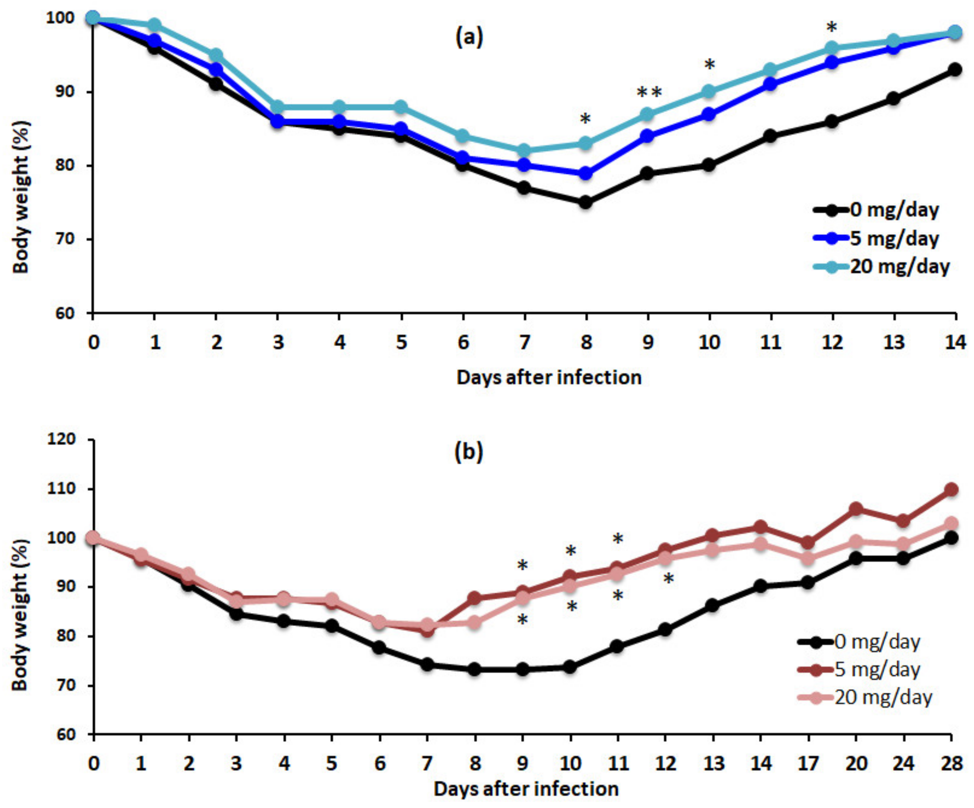
3.1. Algal Powder Attenuates Body Weight Decrease after IFV Infection

No observable side effects, such as diarrhea, were observed during the experimental period. None of the mice showed a decrease in body weight greater than 20% within a day throughout the experimental period. In the control group (water only), mice showed an approximately 30% reduction in weight seven days after infection (Figure 1). Mice treated with 5 and 10 mg/day showed approximately 30% and 20% reduction in body weight 7 days after infection, respectively. Dry algal powder administration caused no prolonged weight loss compared to that in control mice, followed by rapid weight recovery. The body weight of oseltamivir-treated mice did not decrease markedly (by less than 6%) throughout the 14-day period following infection.

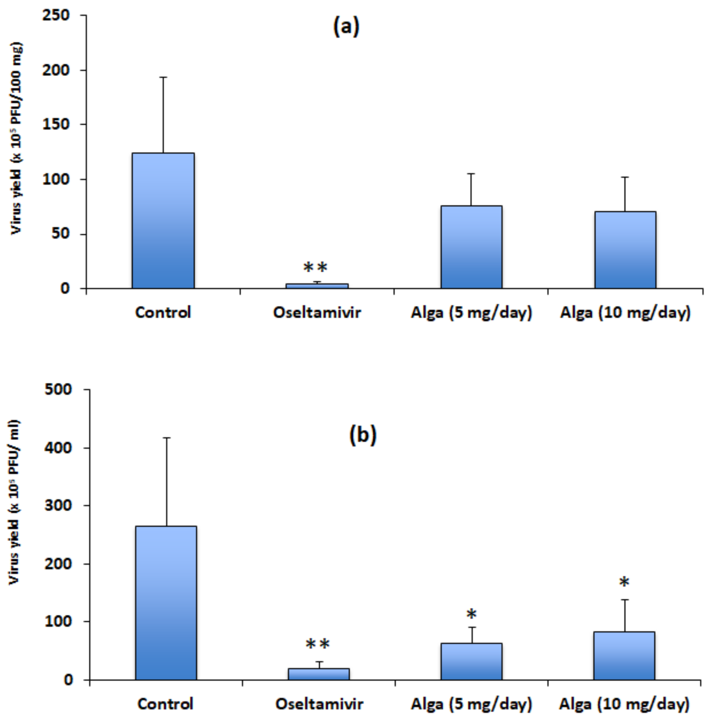
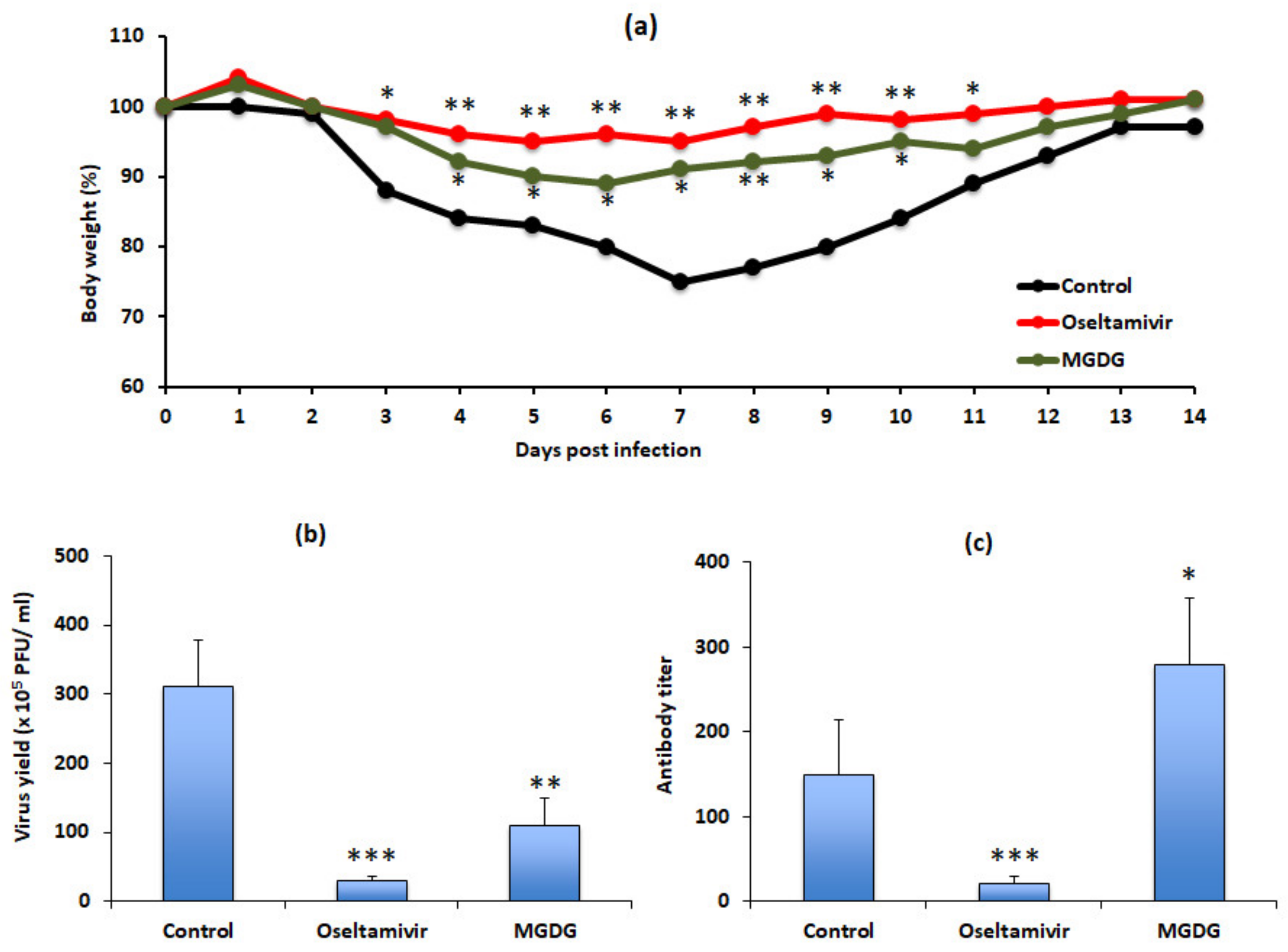
3.2. Algal Powder Inhibits Viral Replication

When BALB/c mice were intranasally infected with IFV, virus yields in both lung and BALF peaked at 3–4 days and then decreased gradually until day 7 post infection (data not shown). To evaluate the effects of 5 and 10 mg/day dry algal powder on IFV proliferation, we titered lung tissue and BALF three days after infection. The powder showed a tendency to reduce titers in the lungs (Figure 2a) and significantly reduced titers in the BALFs (p < 0.05) (Figure 2b). Titers did not differ significantly between mice treated with 5 and 10 mg/day of dry algal powder. The positive control oseltamivir significantly suppressed viral replication (p < 0.01).

3.3. Algal Powder Increases Neutralizing Antibody Responses

Resistance to influenza virus infection is partially mediated by the local and systemic secretion of neutralizing antibodies [14,15]. Therefore, we titered neutralizing antibody in sera and BALF samples collected 14 and 28 days post infection. The neutralizing antibody titers in both serum (Figure 3a) and BALF (Figure 3b) (p < 0.01) samples were significantly higher in mice treated with (5 mg/day or 10 mg/day) dry algal powder than in control mice. The antibody titers did not differ significantly between mice treated with 5 or 10 mg/day. In contrast, antibody titers in both serum and BALF samples were significantly lower in mice from the oseltamivir-treated group (p < 0.001) than in controls (Figure 3).

3.4. Algal Powder Increases Weight and Antibody Production at 20 mg/day

In our second in vivo experiment, the effects of dry algal powder on body weight and antibody production were compared between groups treated with 5 or 20 mg/day powder for two or four weeks. Weight loss was attenuated by algal powder for two (Figure 4a) and four (Figure 4b) weeks. During the recovery period, that is, at 7 days post infection, we failed to detect virus in the lungs and bronchia and observed a significant increase in weight (p < 0.05) in the alga-treated group. The time required to return to original body weight was 24–28, 13–14, and 13–14 days for the control, 5 mg/day, and 20 mg/day alga-treated groups, respectively. Compared to control mice, treatment with 5 mg/day or 20 mg/day dry algal powder increased serum neutralizing antibody titers at 14 and 28 days post infection (Figure 5). Antibody titers at 4 weeks post infection were higher than those at 2 weeks post infection.

3.5. MGDG Accounts for Algal Powder’s Therapeutic Effects

To address the likely active component of the powder, MGDG, we treated mice with it in the same manner. No side effects were observed following MGDG administration. Mice treated with 1 mg/day MGDG showed no prolonged weight loss compared to control mice treated with water (Figure 6a). MGDG significantly suppressed IFV replication (p < 0.01) in BALFs collected 3 d post infection (Figure 6b). The neutralizing antibody titers in BALF samples collected 14 days post infection were significantly higher (p < 0.05) in mice treated with MGDG than in controls (Figure 6c).

4. Discussion

Influenza virus infection is initiated in the upper respiratory tract and encounters pulmonary phagocytic cells, such as alveolar macrophages, which constitute the first line of defense. Macrophages respond rapidly and create an environment that supports the induction of the immune response [16]. The production of neutralizing antibodies is an important host defense mechanism that provides protection against reinfection by primary or closely related strains [17,18]. However, there have been reports of reinfection with pandemic strains of H1N1 influenza A virus after successful oseltamivir treatment [19]. In the present study, significantly lower titers of neutralizing antibodies were observed in oseltamivir-treated mice (Figure 3). This decrease is likely to increase susceptibility to reinfection. In contrast, administration of Coccomyxa sp. KJ powder enhanced the production of neutralizing antibodies after primary infection (Figure 3 and Figure 5). Consequently, it is plausible that this alga could help to prevent reinfection.
We examined viral loads in the lungs and BALFs of mice three days post infection and observed that viral titers in Coccomyxa sp. KJ KJ-treated mice were lower than those of controls (Figure 2). These protective effects were confirmed by determining virus-specific antibody production (Figure 3 and Figure 5). These results suggest that dry algal powder may stimulate host immune responses. We recently reported that MGDG, a component of Coccomyxa sp. KJ, exerts virucidal effects against herpes simplex virus type 2, SARS-CoV-2, murine norovirus, and feline calicivirus [10,11,12]. As shown in Figure 6, 1 mg/day of MGDG exerted therapeutic effects in IFV-infected mice. Because the dry algal powder used in the other experiments contained approximately 4% MGDG, 20 mg of powder contains approximately 0.8 mg MGDG. Therefore, it is likely that the decrease in viral replication by powder can be attributed to a direct interaction between MGDG and virus particles. Recently, Ohshima et al. reported that a water-soluble extract of Coccomyxa sp. KJ affected T-cell fates and helped ameliorate superantigen-induced T cell hyperactivation and immune suppression [20]. Alternatively, it is plausible that the therapeutic effects we observed may have been mediated, at least in part, by other algal components. Suppression of virus replication and stimulation of virus-specific antibody production by the alga likely contributed to the more rapid recovery of body weight observed in the alga-treated group (Figure 4 and Figure 5).
Several types of algae are known to possess sulfated polysaccharides, including fucoidan and rhamnan sulfate, which have antiviral effects [21,22,23,24,25,26,27,28,29]. These components are transported to Peyer’s patches in the intestinal epithelium by microfold cells (M cells) [21,30,31]. M cells capture bacteria [32] and deliver them to antigen-presenting cells. Thus, these sulfated polysaccharides may stimulate the immune response. However, because the molecular weight of MGDG is low, it is unlikely to be transported to Peyer’s patches by M cells.

5. Conclusions

Here, we evaluated the therapeutic and prophylactic effects of orally administered Coccomyxa spp. KJ dry powder in virus-infected mice by measuring the body weight, virus replication, and virus-specific antibody titers. We report that oseltamivir, a currently approved anti-influenza drug, prevents a decrease in body weight and suppresses viral proliferation and antibody production. In contrast, oral dry algal powder attenuated weight loss, suppressed virus proliferation, and stimulated antibody production. Although MGDG is thought to be one of the active components in this algal powder, further studies are needed to characterize its active component(s).

Author Contributions

Conceptualization, K.H. and T.K.; methodology, K.H. and S.K.; chemical analysis, J.-B.L.; investigation, K.H., S.K. and T.K.; resources, S.K. and H.K.; data curation, T.K.; writing—original draft, K.H.; writing—review and editing, J.-B.L., H.K. and T.K.; visualization: J.-B.L.; supervision, H.K. and T.K.; funding acquisition, H.K. and T.K. All authors have read and agreed to the published version of the manuscript.

Funding

This research was funded by the JST Adaptable and Seamless Technology Transfer Program through Target-driven R&D (A-STEP) grant JPMJTR204H. Part of this work was supported by the JST-Mirai grant JPMJMI19D4 and by Grant-in-Aid for Scientific Research (C) 22K04902 under the Ministry of Education, Culture, Sports, Science, and Technology (MEXT) of Japan. The Cooperative Research Program of the “Network Joint Research Center for Materials and Devices” also provided support.

Institutional Review Board Statement

The animal study protocol was approved by the Institutional Review Board of Chubu University (protocol code 2710056, date of approval 16 April 2015; protocol code 3010057, date of approval 20 April 2020).

Informed Consent Statement

Not applicable.

Data Availability Statement

Not applicable.

Acknowledgments

We thank Kinya Atsumi for preparing the materials used for experiments.

Conflicts of Interest

The authors declare no conflict of interest.

References

  1. Taubenberger, J.K.; Morens, D.M. The pathology of influenza virus infections. Ann. Rev. Pathol. 2008, 3, 499–522. [Google Scholar] [CrossRef] [PubMed]
  2. Grounder, A.P.; Boon, A.C.M. Influenza pathogenesis: The effect of host factors on severity of disease. J. Immunol. 2019, 202, 341–350. [Google Scholar] [CrossRef] [Green Version]
  3. Hurt, A.C.; Kelly, H. Debate regarding oseltamivir use for seasonal and pandemic influenza. Emerg. Infect. Dis. 2016, 22, 949–955. [Google Scholar] [CrossRef]
  4. Heneghan, C.J.; Onakpoya, I.; Thompson, M.; Spencer, E.A.; Jones, M.; Jefferson, T. Zanamivir for influenza in adults and children: Systematic review of clinical study reports and summary of regulatory comments. BMJ 2014, 384, g2547. [Google Scholar] [CrossRef] [PubMed] [Green Version]
  5. Hayden, F.G.; Sugaya, N.; Hirotsu, N.; Lee, N.; de Jong, M.D.; Hurt, A.C.; Ishida, T.; Sekino, H.; Yamada, K.; Portsmouth, S.; et al. Baloxavir marboxil for uncomplicated influenza in adults and adolescents. N. Engl. J. Med. 2018, 379, 913–923. [Google Scholar] [CrossRef] [PubMed]
  6. Hussain, M.; Galvin, H.D.; Haw, T.Y.; Nutsford, A.N.; Husain, M. Drug resistance in influenza A virus: The epidemiology and management. Infect. Drug Resist. 2017, 10, 121–134. [Google Scholar] [CrossRef] [Green Version]
  7. Takashita, E.; Kawakami, C.; Morita, H.; Ogawa, R.; Fujisaki, S.; Shirakura, M.; Miura, H.; Nakamura, K.; Kishida, N.; Kuwahara, T.; et al. Detection of influenza A (H3N2) viruses exhibiting reduced susceptibility to the novel cap-dependent endonuclease inhibitor baloxavir in Japan, December 2018. Eurosurveillance 2019, 24, 1800698. [Google Scholar] [CrossRef] [Green Version]
  8. Satoh, A.; Kato, M.; Yamamoto, K.; Ishibashi, M.; Sekiguchi, H.; Kurano, N.; Miyachi, S. Characterization of the Lipid Accumulation in a New Microalgal Species, Pseudochoricystis ellipsoidea (Trebouxiophyceae). J. Jpn. Inst. Energy 2010, 89, 909–913. [Google Scholar] [CrossRef] [Green Version]
  9. Kasai, Y.; Oshima, K.; Ikeda, F.; Abe, J.; Yoshimitsu, Y.; Harayama, S. Construction of a self-cloning system in the unicellular green alga Pseudochoricystis ellipsoidea. Biotechnol. Biofuels 2015, 8, 94. [Google Scholar] [CrossRef] [Green Version]
  10. Hayashi, K.; Komatsu, S.; Kuno, H.; Asai, S.; Matsuura, I.; Kudkyal, V.R.; Kawahara, T. Virucidal and immunostimulating activities of monogalactosyl diacyglyceride from Coccomyxa sp. KJ, a green microalga, against murine norovirus and feline calicivirus. Mar. Drugs 2022, 20, 131. [Google Scholar] [CrossRef]
  11. Hayashi, K.; Asai, S.; Umezawa, K.; Kakizoe, H.; Miyachi, H.; Morita, M.; Akaike, T.; Kuno, H.; Komatsu, S.; Watanabe, T.; et al. Virucidal effect of monogalactosyl diacyglyceride from a green microalga, Coccomyxa sp. KJ, against clinical isolates of SARS-CoV-2 as assessed by a plaque assay. J. Clin. Lab. Anal. 2022, 36, e24146. [Google Scholar] [CrossRef]
  12. Hayashi, K.; Lee, J.-B.; Atsumi, K.; Kanazashi, M.; Shibayama, T.; Okamoto, K.; Kawahara, T.; Hayashi, T. In vitro and in vivo anti-herpes simplex virus activity of monogalactosyl diacylglyceride from Coccomyxa sp. KJ (IPOD FERM BP-22254), a green microalga. PLoS ONE 2019, 14, e0219305. [Google Scholar] [CrossRef] [PubMed] [Green Version]
  13. Ito, T.; Hayashi, K.; Nishiguchi, M.; Hayashi, T.; Iinuma, M. Resveratrol oligomer C-glucosides and antiviral resveratrol tetramers isolated from the stem bark of Shorea uliginosa. Phytochem. Lett. 2018, 28, 1–7. [Google Scholar] [CrossRef]
  14. Clements, M.L.; Betts, R.F.; Tierney, E.L.; Murphy, B.R. Serum and nasal wash antibodies associated with resistance to experimental challenge with influenza A wild virus. J. Clin. Microbiol. 1986, 24, 157–160. [Google Scholar] [CrossRef] [PubMed] [Green Version]
  15. Tophan, D.J.; Tripp, R.A.; Doherty, P.C. CD8+ T cells clear influenza virus by perforin or Fas-dependent process. J. Immunol. 1997, 159, 5197–5200. [Google Scholar]
  16. Burleson, G.R.; Burleson, F.G. Influenza virus host resistance model. Methods 2007, 41, 31–37. [Google Scholar] [CrossRef]
  17. Lee, C.-C.; Yang, C.-Y.; Lin, L.-L.; Ko, T.-P.; Chang, A.H.-L.; Chang, S.S.-C.; Wang, A.H.-J. An effective neutralizing antibody against influenza virus H1N1 from human B cells. Sci. Rep. 2019, 9, 4546. [Google Scholar] [CrossRef]
  18. Padilla-Quirarte, H.O.; Lopez-Guerrero, D.V.; Gutierrez-Xicotencatl, L.; Esquivel-Guandarrama, F. Protective antibodies against influenza proteins. Front. Immunol. 2019, 10, 1677. [Google Scholar] [CrossRef] [Green Version]
  19. Perez, C.M.; Ferres, M.; Labarca, J.A. Pandemic (H1N1) 2009 reinfection, Chile. Emerg. Infect. Dis. 2010, 16, 156–157. [Google Scholar] [CrossRef]
  20. Ohshima, S.; Komatsu, S.; Kashiwagi, H.; Goto, Y.; Ohno, Y.; Yamada, S.; Kanno, A.; Shimizu, T.; Seki, T.; Yasuda, A.; et al. Coccomyxa sp. KJ extract affects the fate of T cells stimulated by toxic shock syndrome toxin-1, a superantigen secreted by Staphylococcus aureus. Microbiol. Immunol. 2022, 12982. [Google Scholar] [CrossRef]
  21. Terasawa, M.; Hayashi, K.; Lee, J.-B.; Nishiura, K.; Matsuda, K.; Hayashi, T.; Kawahara, T. Anti-influenza A virus activity of rhamnan sulfate from green algae Monostroma nitidum in mice with normal and compromised immunity. Mar. Drugs 2020, 18, 254. [Google Scholar] [CrossRef] [PubMed]
  22. Lee, J.-B.; Hayashi, K.; Hashimoto, M.; Nakano, T.; Hayashi, T. Novel antiviral fucoidan from sporophyll of Undaria pinnatifida (Mekabu). Chem. Pharm. Bull. 2004, 52, 1091–1094. [Google Scholar] [CrossRef] [PubMed] [Green Version]
  23. Synytsya, A.; Bleha, R.; Synytsya, A.; Pohl, R.; Hayashi, K.; Yoshinaga, K.; Nakano, T.; Hayashi, T. Mekabu fucoidan: Structural complexity and defensive effects against avian influenza A Viruses. Carbohydr. Polym. 2014, 111, 633–644. [Google Scholar] [CrossRef] [PubMed]
  24. Negishi, H.; Mori, M.; Mori, H.; Yamori, Y. Supplementation of elderly Japanese men and women with fucoidan from seaweed increases immune responses to seasonal influenza vaccination. J. Nutr. 2013, 143, 1794–1798. [Google Scholar] [CrossRef]
  25. Hayashi, K.; Lee, J.-B.; Nakano, T.; Hayashi, T. Anti-influenza A virus characteristics of a fucoidan from sporophyll of Undaria pinnatifida in mice with normal and compromised immunity. Microbes Infect. 2013, 15, 302–309. [Google Scholar] [CrossRef]
  26. Lee, J.-B.; Koizumi, S.; Hayashi, K.; Hayashi, T. Structure of rhamnan sulfate from the green alga Monostroma nitidum and its anti-herpetic effect. Carbohydr. Polym. 2010, 81, 572–577. [Google Scholar] [CrossRef]
  27. Wang, S.; Wang, W.; Hao, C.; Yunjia, Y.; Qin, L.; He, M.; Mao, W. Antiviral activity against enterovirus 71 of sulfated rhamnan isolated from the green alga Monostroma latissimum. Carbohydr. Polym. 2018, 200, 43–53. [Google Scholar] [CrossRef]
  28. Lee, J.-B.; Hayashi, K.; Hayashi, T.; Sankawa, U.; Maeda, M. Antiviral activities against HSV-1, HCMV, and HIV-1 of rhamnan sulfate from Monostroma latissimum. Planta Med. 1999, 65, 439–441. [Google Scholar] [CrossRef]
  29. Lee, J.-B.; Hayashi, K.; Maeda, M.; Hayashi, T. Antiherpetic activities of sulfated polysaccharides from green algae. Planta Med. 2004, 70, 813–817. [Google Scholar] [CrossRef]
  30. Hong, F.; Yan, J.; Baran, J.T.; Allendorf, D.J.; Hansen, R.D.; Ostroff, G.R.; Xing, P.X.; Cheung, N.K.; Ross, G.D. Mechanism by which orally administered beta-1,3-glucans enhance the tumoricidal activity of antitumor monoclonal antibodies in murine tumor models. J. Immunol. 2004, 173, 797–806. [Google Scholar] [CrossRef] [Green Version]
  31. Kawashima, T.; Murakami, K.; Nishimura, I.; Nakano, T.; Obata, A. A sulfated polysaccharide, fucoidan, enhances the immunomodulatory effects of lactic acid bacteria. Int. J. Mol. Med. 2012, 29, 447–453. [Google Scholar] [CrossRef] [PubMed] [Green Version]
  32. Ohno, H. Intestinal M cells. J. Biochem. 2016, 159, 151–160. [Google Scholar] [CrossRef] [PubMed] [Green Version]
Figure 1. Dry Coccomyxa sp. KJ powder and oseltamivir inhibit weight loss. Influenza A virus (IFV)-infected mice (n = 5 per group) received oral water (control), 0.2 mg oseltamivir, or 5 or 10 mg/day alga for 14 days (7 days before and 7 days after infection). Body weight on the day of infection was taken as 100%. * p < 0.05 and *** p < 0.001 versus the control group.
Applmicrobiol 02 00037 g001
Figure 2. Dry Coccomyxa sp. KJ powder inhibits IFV replication. IFV-infected mice (n = 5 per group) were orally administered water (control), 0.2 mg oseltamivir, or 5 mg/day or 10 mg/day dry algal powder for 14 days (7 days before and 7 days post infection). Virus yields in the lung (a) and BALF (b) are shown. Each value represents the mean ± standard deviation (SD) (n = 5). * p < 0.05 and ** p < 0.01 versus the control group.
Applmicrobiol 02 00037 g002
Figure 3. Dry Coccomyxa sp. KJ powder increases IFV neutralizing antibody production. IFV-infected mice (n = 5 per group) were orally administered water (control), 0.2 mg oseltamivir, and 5 or 10 mg/day dry algal powder for 14 days (7 days before and 7 days post infection). The titers in the serum (a) and BALF (b) are shown. Each value represents the mean ± SD (n = 5). ** p < 0.01 and *** p < 0.001 versus control.
Applmicrobiol 02 00037 g003
Figure 4. Dry Coccomyxa sp. KJ powder attenuates weight loss. IFV-infected mice (n = 5 per group) were orally administered water (control), or 5 mg/day or 20 mg/day dry algal powder 1 h post infection for 14 days (a) or 24 days after infection (b). Body weight on the day of infection was taken as 100%. * p < 0.05 and ** p < 0.01 versus the control group.
Applmicrobiol 02 00037 g004
Figure 5. Effects of dry Coccomyxa sp. KJ powder on the production of serum IFV-neutralizing antibodies. IFV-infected mice (n = 5 per group) were orally administered water (control), or 5 mg/day or 20 mg/day algal powder 1 h post infection for 14 days (a) or 28 days (b) after inoculation. Each value represents the mean ± SD (n = 5). * p < 0.05 and *** p < 0.001 versus the control group at 14 days post infection; # p < 0.05 and ### p < 0.001 versus the control at 28 days post infection.
Applmicrobiol 02 00037 g005
Figure 6. Effects of MGDG isolated from Coccomyxa sp. KJ and oseltamivir in IFV-infected mice. The mice (n = 5 per group) were orally administered water (control), 0.2 mg oseltamivir, or 1 mg/day MGDG for 14 days after virus inoculation. Body weight (a) on the day of virus inoculation (day 0) was taken as 100%. Virus yields in BALF samples (b) were evaluated after three days of IFV inoculation. Neutralizing antibody titers in BALF samples (c) were evaluated after 14 days of IFV inoculation. * p < 0.05, ** p < 0.01, and *** p < 0.001 versus the control group.
Applmicrobiol 02 00037 g006
Publisher’s Note: MDPI stays neutral with regard to jurisdictional claims in published maps and institutional affiliations.

