Perspective

A Pragmatic Perspective of the Initial Stages of the Contact Killing of Bacteria on Copper-Containing Surfaces

Département de Génie Physique, Polytechnique Montréal, CP 6079, Succursale C-V, Montréal, QC H3C 3A7, Canada
Appl. Microbiol. 2022, 2(3), 449-452; https://doi.org/10.3390/applmicrobiol2030033
Submission received: 16 May 2022 / Revised: 20 June 2022 / Accepted: 22 June 2022 / Published: 23 June 2022

Abstract

:
A consideration of the outer structures of Gram-positive and Gram-negative bacteria, and of the surface contaminants present on Cu-containing substrates, has led to the identification of Cu2O as a potent antimicrobial. In the presence of adsorbed water, the hydrated form ionizes to CuI-O, which is capable of degrading the protective polysaccharide layer of the outer lipopolysaccharide membranes of Gram-negative bacteria; it is equally capable of attacking the peptidoglycan lattices present in both Gram-positive and Gram-negative bacteria. This Perspective underlines the importance of CuI-O in the early stages of contact killing, and points to information, still lacking, that would optimize contact killing and lead to broader applications in the therapeutic management of bacterial infections.

Graphical Abstract

1. Introduction

In a recent Comment [1], the present writer presented a short view of the initial stages of the contact killing of Gram-positive Bacillus subtilis on copper-containing substrates. On further reflection, it became obvious that the view presented was not limited to the single instance of Gram-positive bacteria. This Perspective is offered as a demonstration of the generality of that view and as a guide to identifying further information required, so as to obtain a better understanding of the processes involved in contact killing.
To begin, a review of the outer-surface bacterial cell structures is needed. Two broad categories exist: Gram-positive and Gram-negative, depending on whether they are stained with Gram dye.

2. Cell Structure

The outer layer of a Gram-positive bacterium is composed of a peptidoglycan lattice, which is comprised of glycan (i.e., polysaccharide) strands crosslinked by short peptides of D-amino acids [2,3]; the lattice also contains some teichoic acids, which are phosphate–carbohydrate copolymers, linked by phosphodiester bonds. Beneath the lattice lies the cytoplasmic membrane, composed of a glycerophospholipid bilayer, which surrounds and protects the cell from the external environment. Gram-positive bacteria found in a nosocomial setting, for reasons that will shortly become obvious, include Staphylococcus aureus, Enterococcus and Streptococcus.
In the case of a Gram-negative bacterium, the peptidoglycan lattice is much thinner, and is surrounded by another lipid bilayer, whose outer leaf contains lipopolysaccharides [3]. These include polysaccharide chains extending outward from the surface of the bilayer. It is generally accepted that their presence blocks the approach of drugs to the membrane, and is the reason why Gram-negative illnesses are more difficult to treat. Gram-negative bacteria found in a nosocomial setting include Escherichia coli, Pseudomonas aeruginosa, Klebsiella pneumoniae and Proteus vulgaris.

3. A Classic Contact Killing Study and Its Significance

A recent study, which occurred in a nosocomial setting [4], has a specific bearing on this Perspective. It was summarized in our recent paper on the physicochemical characterization of the Cu nanoparticle surface [5], and is cited here verbatim:
A toilet seat, a set of tap handles and a ward entrance door push plate, all containing Cu, were installed six months before the study began; halfway through the ten-week study, they were interchanged with similar items that did not contain Cu, and their use was continued at other sites. At whichever locations they were placed, none of the ten Cu-containing sites failed the benchmark antimicrobial test of a total aerobic count of less than 5 colony-forming units/cm2; that is, even contaminated Cu surfaces were antimicrobial. These results were confirmed by several similar studies listed in [6].
The reader will note that no mention is made of whether the results apply uniquely to either Gram-positive or Gram-negative bacteria. That is, they apply to both.
Further, based on our Time-of-Flight Secondary Ion Mass Spectrometry (TOF-SIMS) results on the Cu nanoparticle surface [5], which showed the almost complete absence of zerovalent Cu (based on the almost complete absence of Cun fragments having n > 1), it would appear that it is the Cu surface contaminants that are uniquely responsible for its antimicrobial efficacy. In our study [5], a Selected-Area Electron Diffraction reconstruction indicated that these contaminants included Cu2O, CuCO3, malachite (Cu2CO3(OH)2) and azurite (Cu3(CO3)2(OH)2). As noted there [5], Cu2O does not oxidize directly to CuO at room temperature, nor is it found in nature, except at the sites of cataclysmic events; rather, Cu2O oxidizes by combining with water vapor and carbon dioxide to form malachite and azurite, which decompose at elevated temperatures to form CuO.
Thus, one or more of these Cu compounds is capable of killing both Gram-positive and Gram-negative bacteria. At this point in this Perspective, it is unclear which of the contaminants is involved or whether the same compounds are involved in both cases.
Further, while dry contact is implied, this is not the case, since both the Cu substrate and the bacterium are capable of adsorbing water.

4. Water Adsorption onto Solids

Adsorption is a spontaneous thermodynamic process that lowers the surface energy of the solid [7]. This is the action behind corrosion and the deposition of adventitious carbon, as well as the adsorption of surface water from the background humidity. The simultaneous adsorption of several contaminants, as in the present case, depends on the initial interaction of the components with the substrate, their subsequent interaction with what was already deposited, and their interactions among themselves. It is common to obtain different TOF-SIMS responses from different locations on the same sample, indicating that adsorption is not uniform.
The adsorption of water onto solid surfaces has long been of interest [8,9]. Initially, experiments were limited to only detecting the presence of moisture, eventually followed by attempts to quantify the layer thickness. A feature of all these attempts is the assumption of layer uniformity.
The present writer is aware of only one study that actually observed the adsorbed water directly [10], showing exactly how and where it appeared. Using a specially designed Scanning Tunneling Microscope, those authors found that as the humidity increased, droplets of water, 30–50 nm in diameter, were formed on a freshly sputtered Au surface. Droplets were formed because the water did not wet the substrate and, as a result, retracted.
These droplets grew in height and diameter as the humidity increased and, at 75% humidity, finally contacted each other to give a uniform film. Judging from our Cu nanoparticle results [5], the oxygen-containing organic fragments that were found using X-ray photoelectron spectroscopy (XPS) suggest that some of the water may have reacted with depositing adventitious carbon, although the majority was exuded to the continually growing surface, where it retracted to form droplets.
Those authors also carried out similar experiments on Ti and HOPG [10]; being more wettable, those substrates formed continuous water films at lower humidities. While the water layer heights determined by the authors have been criticized [11], the criticism is based on assumptions that may not be correct. In any case, surface water is present.
The present writer is unaware of similar studies on bacterial cells. However, the outer structures of both Gram-positive and Gram-negative bacterial cells suggest the presence of at least hydrogen-bonded water. Thus, contact between the Cu-containing substrate and the bacterium appears to always occur through an aqueous interphase.

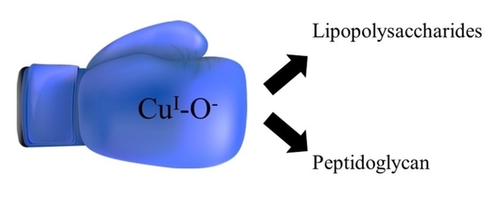
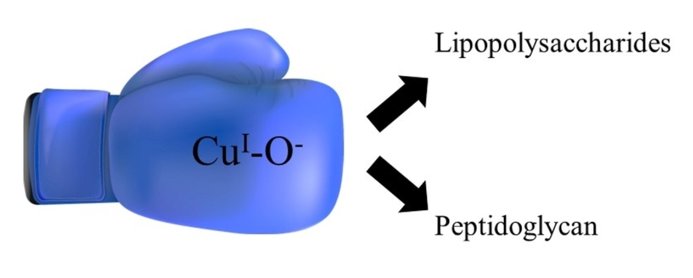
5. Attack on Bacteria

As previously noted [1], in the presence of water, the M-O-M structure of surface oxides is hydrolyzed to M-OH, which ionizes according to its isoelectric point (i.e., M-O or M-OH2+). Of the Cu-containing contaminants listed earlier (Cu2O, CuCO3, malachite and azurite), only the isoelectric point of Cu2O is available [12]; it is at a pH of ~5, which means that, at the pH of water (i.e., 7), it exists mainly as CuI-O. We tentatively assume that both malachite and azurite, which contain cupric hydroxide structures, have isoelectric points not far from that of CuO, which is at a pH of ~9 [12]; this means that, at the pH of water, they would exist as CuII-OH2+. While it is clear that both CuI-O and CuII-OH2+ are capable of attacking the lipid and peptidoglycan structures found in Gram-positive and Gram-negative bacteria [13,14,15,16,17], studies of the chemistries involved in these attacks are lacking. Such studies are needed for the optimization of contact killing.
The reader is reminded that the outer surfaces of Gram-negative bacteria are composed of lipopolysaccharides, whose outer leaves have polysaccharide chains extending outward from the surface of the bilayer. These form a protective layer over the membrane, and must be degraded before the membrane can be attacked. While polysaccharide degradation is usually cited as being accomplished enzymatically, it has recently come to light that Cu2O is also capable of achieving this [18,19]; other studies have also confirmed the potency of Cu2O under similar, although not identical, circumstances, with Cu2O substantially outperforming CuO [20,21].

6. Initial Stages of Contact Killing

Thus, it appears that, at the initial stages of the contact killing of Gram-negative bacteria, the structure that has antimicrobial activity at the Cu surface is Cu2O in its hydrated, ionized form: CuI-O. Once the polysaccharide structure is degraded, the CuI-O can continue to degrade the peptidoglycan lattice below, at the peptide crosslinks, as previously suggested [1], and/or along the glycan (polysaccharide) strands [18,19], as noted above. Whether the malachite and azurite participate, and to what extent, is presently unknown.
In the case of Gram-positive bacteria, there is no lipopolysaccharide barrier to overcome, but the peptidoglycan lattice is much thicker. Here, too, CuI-O is capable of initiating the attack, with malachite and azurite, again, participating to unknown extents.

7. Summary and Conclusions

The literature data quoted here strongly suggest that, of the contaminants formed on a Cu-containing substrate, Cu2O, hydrated and ionized (CuI-O), is a potent antimicrobial against both Gram-positive and Gram-negative bacteria: it attacks and degrades the lipopolysaccharide membrane of Gram-negative bacteria, as well as the peptidoglycan lattices of both Gram-positive and Gram-negative bacteria. Unfortunately, how these feats are accomplished is presently unknown. The lack of such chemical information puts an unfortunate cap on the optimization of contact killing, its medical management of bacterial infections, and its applicability to other situations that might benefit from its use.

Funding

This research received no external funding.

Institutional Review Board Statement

Not applicable.

Informed Consent Statement

Not applicable.

Data Availability Statement

Not applicable.

Conflicts of Interest

The author declares no conflict of interest.

References

  1. Sacher, E. Comment on “High-Resolution Microscopical Studies of Contact Killing Mechanisms on Copper-Based Surfaces”. ACS Appl. Mater. Interfaces 2022, 14, 16959–16960. [Google Scholar] [CrossRef] [PubMed]
  2. Vollmer, D.; de Pedro, M.A. Peptidoglycan Structure and Architecture. FEMS Microbiol. Rev. 2008, 32, 149–167. [Google Scholar] [CrossRef] [PubMed] [Green Version]
  3. Epand, R.M.; Walker, C.; Epand, R.F.; Magarvey, N.A. Molecular Mechanisms of Membrane Targeting Antibiotics. Biochem. Biophys. Acta 2016, 1858, 970–987. [Google Scholar] [CrossRef] [PubMed]
  4. Casey, A.L.; Adams, T.J.; Karpanen, P.A.; Cookson, B.D.; Nightingale, P.; Miruszenko, L.; Shillam, R.; Christian, P.; Slliot, T.S.J. Role of Cu in Reducing Hospital Environment Contamination. J. Hosp. Infect. 2010, 74, 72–77. [Google Scholar] [CrossRef]
  5. Loran, S.; Cheng, S.; Botton, G.A.; Yahia, L.H.; Yelon, A.; Sacher, E. The Physicochemical Characterization of the Cu Nanoparticle Surface, and of its Evolution on Atmospheric Exposure: Application to Antimicrobial Bandages for Wound Dressings. Appl. Surf. Sci. 2019, 473, 25–30. [Google Scholar] [CrossRef]
  6. Grass, G.; Rensing, C.; Solioz, M. Metallic Copper as an Antimicrobial Surface. Environ. Microbiol. 2011, 77, 1541–1547. [Google Scholar] [CrossRef] [Green Version]
  7. Bavand, R.; Chen, L.; França, R.; Loran, S.; Yang, D.-Q.; Yelon, A.; Zhang, G.-X.; Sacher, E. Comment on “Intensity modulation of the Shirley backgroundof the Cr3p spectra with photon energies around the Cr2p edge”. Surf. Interface Anal. 2018, 50, 683–685. [Google Scholar] [CrossRef]
  8. Vogler, E.A. Structure and Reactivity of Water at Biomaterial Surfaces. Adv. Coll. Interface Sci. 1998, 74, 69–117. [Google Scholar] [CrossRef]
  9. Xiao, C.; Shi, P.; Yan, W.; Chen, L.; Qian, L.; Kim, S.H. Thickness and Structure of Adsorbed Water Layer and Effects on Adhesion and Friction at Nanoasperity Contact. Colloids Interfaces 2019, 3, 55. [Google Scholar] [CrossRef] [Green Version]
  10. Freund, J.; Halbritter, J.; Hörber, J.K.H. How Dry are Dried Samples? Water Adsorption Measured by STM. Microsc. Res. Tech. 1999, 44, 327–338. [Google Scholar] [CrossRef]
  11. Opitz, A.; Scherge, M.; Ahmed, S.I.-U.; Schaefer, J.A. A Comparative Investigation of Thickness Measurements of Ultra-Thin Water Films by Scanning Probe Techniques. J. Appl. Phys. 2007, 101, 064310. [Google Scholar] [CrossRef]
  12. Kosmulski, M. Isoelectric Points and Points of Zero Charge of Metal (Hydro)Oxides: 50 Years After Parks’s Review. Adv. Colloid Interface Sci. 2016, 238, 1–61. [Google Scholar] [CrossRef]
  13. Limo, M.J.; Sola-Rabada, A.; Boix, E.; Thota, V.; Westcott, Z.; Puddu, V.; Perry, C.C. Interactions Between Metal Oxides and Biomolecules: From Fundamental Understanding to Applications. Chem. Rev. 2018, 118, 11118–11193. [Google Scholar] [CrossRef] [Green Version]
  14. Whitaker, J.R.; Feeney, R.E.; Sternberg, M.M. Chemical and Physical Modification of Proteins by the Hydroxyl Ion. CRC Crit. Rev. Food Sci. Nutr. 1983, 19, 173–212. [Google Scholar] [CrossRef]
  15. Schubert, J.; Radeke, C.; Fery, A.; Chanana, M. The Role of pH, Metal Ions and Their Hydroxides in Charge Reversal of Protein-coated Nanoparticles. Phys. Chem. Chem. Phys. 2019, 21, 11011–11018. [Google Scholar] [CrossRef] [Green Version]
  16. Brockerhoff, H. Breakdown of Phospholipids in Mild Alkaline Hydrolysis. J. Lipid Res. 1983, 4, 96–99. [Google Scholar] [CrossRef]
  17. Wang, L.; Hu, C.; Shao, L. The Antimicrobial Activity of Nanoparticles: Present Situation and Prospects for the Future. Int. J. Nanomed. 2017, 12, 1227–1249. [Google Scholar] [CrossRef] [Green Version]
  18. Gopalakrishnan, K.; Ramesh, C.; Ragunathan, V.; Thamilselvan, M. Antibacterial Activity of Cu2O Nanoparticles on E. coli Synthesized from Tridax Procumbrens Leaf Extract and Surface Coating with Polyaniline. Digest J. Nanomater. Biostruct. 2012, 7, 833–839. [Google Scholar]
  19. Bezza, F.A.; Tichapondwa, S.M.; Chrwa, E.M.N. Fabrication of Monodispersed Copper Oxide Nanoparticles with Potential Application as Antimicrobial Agents. Sci. Rep. 2020, 10, 16680. [Google Scholar] [CrossRef]
  20. Hans, M.; Erbe, A.; Mathews, S.; Chen, Y.; Solioz, M.; Mücklich, F. Role of Copper Oxides in Contact Killing of Bacteria. Langmuir 2013, 29, 16160–16166. [Google Scholar] [CrossRef]
  21. Abicht, H.K.; Gonskikh, Y.; Gerber, S.D.; Solioz, M. Non-Enzymic Copper Reduction by Menaquinone Enhances Copper Toxicity in Lactococcus lactis IL1403. Microbiology 2013, 159, 1190–1197. [Google Scholar] [CrossRef] [Green Version]
Publisher’s Note: MDPI stays neutral with regard to jurisdictional claims in published maps and institutional affiliations.

