Article

Binge-like Alcohol Administration Alters Decision Making in an Adolescent Rat Model: Role of N-Methyl-D-Aspartate Receptor Signaling

1
Laboratorio de Función y Patología Neuronal, Departamento de Biología Celular y Molecular, Facultad de Ciencias Biológicas, Pontificia Universidad Católica de Chile, Av. Libertador Bernardo O’Higgins 340, Santiago 8331150, Chile
2
Centro de investigación y Estudio del Consumo de Alcohol en Adolescentes (CIAAA), Santiago 8331150, Chile
3
Centro de Excelencia en Biomedicina de Magallanes (CEBIMA), Universidad de Magallanes, Punta Arenas 6213515, Chile
*
Author to whom correspondence should be addressed.
Stresses 2024, 4(1), 1-13; https://doi.org/10.3390/stresses4010001
Submission received: 8 November 2023 / Revised: 15 December 2023 / Accepted: 19 December 2023 / Published: 22 December 2023
(This article belongs to the Section Animal and Human Stresses)

Abstract

:
Alcohol is one of the most used legal drugs abused worldwide, and its consumption is associated with high mortality and morbidity rates. There is an increasing concern about the starting age of consumption of this drug since it has become evident that it is at younger ages. The so-called “pattern of consumption by binge” corresponds to ingesting large amounts of alcohol in a short period and is the most popular among young people. Previous studies show that alcohol causes damage in different areas, such as the hippocampus, hypothalamus, and prefrontal cortex, and adolescents are more susceptible to alcohol toxicity. Alcohol inhibits the membrane glutamate receptor, NMDA-type glutamate receptors (NMDAR). Using a binge-like alcohol administration protocol in adolescent rats (PND25), we investigate decision making through the attentional set-shifting test (ASST) and alterations in the NMDAR signaling in related areas. We observe an impairment in executive function without alterations in NMDAR abundance. However, binge alcohol changes NMDAR signaling and decreases quantity in the synapse, mainly in the hippocampus and hypothalamus. We suggest that prefrontal cortex impairment could arise from damaged connections with the hippocampus and hypothalamus, affecting the survival pathway and memory and learning process.

1. Introduction

Alcohol is the most common licit substance, and its excessive consumption is the third cause of death worldwide [1] through injuries (including road traffic injuries), cardiovascular diseases, and cancer. Also, alcohol causes mental health conditions like depression and anxiety disorders [2,3]. The annual consumption during 2016 was equal to 6.4 L of pure alcohol per person aged 15 years or older worldwide, which implies using 13.9 g of pure alcohol per day [2]. Young people between 15 and 24 years present mainly a troubling pattern of alcohol consumption known as “binge drinking” [2,4]. According to the National Institute on Alcohol Abuse and Alcoholism [5], “binge drinking” refers to alcohol consumption that brings the blood alcohol concentration (BAC) to 0.08 g/dL. This value is reached by consuming five or more drinks in males or four or more in females in a time window of 2 h [6,7]. That type of alcohol drinking has short and long-term problems for the adolescent, like intoxication, depression [8], social rejection [9], and other neuronal alterations, like reduction in the volume of the grey matter and decrease in white matter integrity [10]. During childhood and adolescence, the brain undergoes several maturation processes that require neurotransmission changes and synaptic plasticity related to structural modifications in different brain regions [11,12,13,14]. Some studies reported smaller hippocampal volume, lower neuronal activity (measured as the blood-oxygen-level-dependent (BOLD) activation in MRI) at baseline in frontal, temporal, and parietal cortices [15], and alterations in connectivity between frontal and limbic regions [10]. Also, the neuroendocrine function during puberty is altered by alcohol [16].
Among brain areas affected by alcohol are found in the hypothalamus, the hippocampus, and the prefrontal cortex [17]. Briefly, The hypothalamus (Ht) is located in the forebrain [18], and one of its primary functions is the regulation of pituitary hormones, fluid balance, stress control, and others [19,20]. Some studies show that alcohol produces a similar response to stress in the hypothalami–pituitary–adrenal (HPA) axis, providing a behavioral and emotional response [21]. The hippocampus (Hc) is located in the medial temporal lobe [22], and it is crucial for long-term declarative memory, spatial memory, recognition memory, and short-term memory [22,23,24], and alcohol induces cognitive impairment in spatial processing tasks [25,26]. The medial prefrontal cortex (mPFC) has four divisions: two dorsal regions that play a role in motor behavior, and two ventral areas that are related to emotional, cognitive, and mnemonic processes [27], and it is related to alcohol-seeking behavior [28]. Evidence shows that those areas are connected through neuronal circuits and fiber projections. Indeed, there are established connections between Hc and PFC, Ht and PFC, and Ht and Hc [27,29,30].
mPFC is a zone of the brain where all inputs are processed to generate a complete evaluation of them. It is responsible for executive functions necessary for appropriately controlling the behavior. Those executive functions include attention control, inhibition of control, working memory, and all those functions that help to concentrate on a specific task or process, generating a final output [31,32]. Additionally, the mPFC is involved in a process called decision making. Decision making requires the evaluation of costs and benefits to see which option is better long-term [33]. Decision making is heavily affected by alcohol abuse. It impacts behavioral learning and its associated cellular signaling, producing a weak ventromedial PFC activity [34].
The brain circuits that control these processes communicate within the circuit (for example, CA3-CA1 synapses in the hippocampus) and between different nuclei. This communication is carried out through various neurotransmitters, among which glutamate appears prominently. Glutamatergic transmission is controlled, among others, by ionotropic receptors, within which communication through N-methyl-D-aspartate receptor (NMDAR)-type mediates critical processes of synaptic plasticity and long-term memory. [35,36]. The NMDAR is located in the postsynaptic density (PSD) and extrasynaptic domains, where its localization is regulated by phosphorylation. One mark of PSD positioning is the phosphorylation of tyrosine 1472 in the GluN2B subunit [37], produced by Fyn kinase. The dephosphorylation of this residue triggers NMDAR endocytosis and is driven by Striatal-enriched tyrosine phosphatase (STEP) [38]. STEP protein displays several isoforms, and the best characterized is STEP61, a membrane-associated protein located in the postsynaptic density of neurons. As a result of excitotoxic stimuli and the elevated levels of calcium, STEP61 is cleaved by calpain to STEP33, inactivating it [39]. The signaling downstream of synaptic NMDAR involves multiple proteins with a consequent pro-survival and anti-apoptotic gene expression. This response includes the phosphorylation and, consequently activation of ERK1/2 and the transcription factor CREB, and other signaling proteins [40,41]. But beyond the particular molecular mechanisms, glutamate receptors containing the GluN2B subunit have been implicated in a series of complex processes at the level of synaptic connectivity [42], with a particularly relevant role in memory and learning processes [43].
We have previously shown that our binge-like alcohol administration model affects the hippocampal synaptic transmission and recognition memory but not spatial memory [25]. Recognition memory requires a subject to remember that an item was associated with a particular place, memory judgments, and decision making. So, the recognition memory was related to the hippocampus and medial prefrontal cortex functions [44]. As we know that alcohol mimics the effects of stress in hypothalamic function and stress-impaired decision making [45,46], we believe that mPFC function and decision making could be impaired by binge-like alcohol administration through the impaired interplay between Hp, Ht, and mPFC. To tackle this idea, we evaluate decision making and mPFC function through attention tests and evaluate NMDAR signaling, a known player in neuron survival, in mPFC, Hp, and Ht.

2. Results

We have previously shown that adolescent binge-like alcohol treatment impaired recognition memory in novel object recognition test (NOR) and social interaction test, but spatial memory remains unchanged compared to sham animals [25]. So, we decided to evaluate the decision making involving prefrontal cortex function. The ASST consists of seeking and learning where a hidden reward is. To perform this task, two bowls containing different mediums and odors are presented to the rats, who are trained to look for the reward hidden inside the bowl. Rats must learn where the reward is depending on the medium and/or odors present. The ASST consists of different stages where the medium and odors are changed to measure different learning and memory skills such as memory flexibility and shift attention (Figure 1A) [47]. Before starting the ASST, the rats completed the habituation phase that included digging establishment and exploratory digging in the testing apparatus. The test was performed for digging establishment until rats correctly dug and found the reward for 12 non-consecutive trials. Thus, we quantified the number of trials needed to reach the criterion. The results are shown in Figure 1B, and there was no difference between sham and BEP animals. Then, rats were submitted to different stages of the ASST test. In this case, the criterion was six consecutive successful trials, where the number of trials to reach the criterion (Figure 1C) and the number of errors (Figure 1D) were measured. The effect of binge-like alcohol administration on adolescent rats is that BEP animals need more trials to reach the criterion than sham rats. Still, it only showed a difference in the CD reverse stage (t-test, t(9) = 2.361, p = 0.0094, Figure 1C). There was a difference in the number of errors when rats had to choose the bowl to receive the reward; the BEP group had more erroneous trials than the sham group (t-test, t(57) = 2.961, p = 0.0045, Figure 1D). The final graph shows that rats needed more trials to reach the criterion in the extradimensional discrimination (ED) stage than the trials they needed to finish the intra-dimensional discrimination (ID) stage. Also, the BEP group needed more trials than the sham group to finish ED (Figure 1E). These results indicate mild cognitive impairment in prefrontal cortex function after binge-like alcohol administration.
We analyze the NMDAR signaling pathway to evaluate the neuron function involved in synaptic plasticity and neuronal survival [48]. It is well established that NMDAR is one of the molecular targets of ethanol [49,50,51]. Thus, we evaluated NMDAR signaling and abundance in the mPFC, Hp, and Ht synapses. We have previously shown that our binge-like administration protocol impaired synaptic transmission in the hippocampus [25], and we believe that NMDAR impairment could be related to ASST performance and involved in the decision-making process.
Immunoblotting from the prefrontal cortex, hippocampi, and hypothalamic tissues was performed on crucial signaling proteins related to NMDAR function and localization. In the prefrontal cortex, most of the proteins analyzed remain unchanged one week after binge-like alcohol administration, with the only exception of p-CREB (t(4) = 2.605; p = 0.0299) (Figure 2A,B), suggesting that pro-survival signaling from the NMDARs is reduced following alcohol binge-drinking in adolescent rats in the main brain area related in the ASST performance. In the hippocampus, STEP61 showed a significant decrease (t(4) = 2.044; p = 0.05) while STEP33 remained unchanged (t(4) = 0.1709; p = 0.4363) (Figure 2C,D). The Fyn kinase also showed a decreasing trend (t(4) = 1.652; p = 0.087) (Figure 2C,D), indicating the possibility that NMDA receptors have been taken away from the synapse. Concerning signaling proteins downstream of synaptic NMDAR activation, p-CREB was reduced (t(4) = 4.598; p = 0.005), while ERK proteins remain unchanged (t(4) = 0.6951; p = 0.2626) (Figure 2C,D). Finally, in the hypothalamus, the levels of the cleavage product of the phosphatase STEP (STEP33) increased one week after binge-like protocol (t(4) = 2.402; p = 0.0371), although the levels of the active isoform 61 (STEP61) remains unaltered (t(4) = 0.6703; p = 0.2697) (Figure 2E,F). In the same line, the kinase Fyn showed decreased levels (t(4) = 3.199; p = 0.0165) (Figure 2E,F), suggesting that the NMDA receptor could be removed from the synapse. Furthermore, we analyzed two proteins related to NMDAR signaling the phosphorylated form of ERK (ERK1 Thr202/Tyr204 and ERK2 Thr185/Tyr187) and CREB (Ser133) and found that p-ERK (t(4) = 5.631; p = 0.0024) and p-CREB (t(4) = 2.332; p = 0.04) both decreased compared to control animals (Figure 2E,F). These results may indicate a reduced function of synaptic NMDA receptors one week after binge-like alcohol administration in adolescent rats in brain areas that could affect mPFC function and ASST performance.
Then, we wanted to assess if, effectively, there are fewer NMDA receptors in the synapses of the animals treated with binge-like alcohol administration. To test this, we performed synaptosomal isolation from brain tissue: hypothalamus, hippocampus, and prefrontal cortex. The synaptosomal fractions were treated with Triton buffer to isolate PSD fractions, and we analyzed the presence of the GluN2B subunit of the NMDA receptor in these PSD fractions. As Fyn kinase and STEP phosphatase act over tyrosine 1472 of the GluN2B subunit, we expected a decrease in the amount of this receptor subunit in the PSD fractions, which was corroborated by immunoblots (Figure 3). GluN2B was less abundant in the PSD of the hypothalamus (t(4) = 2.020; p = 0.049) and hippocampus (t(4) = 2.508; p = 0.0331) from BEP animals (Figure 3B–E) and remain unchanged in the prefrontal cortex (t(3) = 0.3192; p = 0.3853) (Figure 3A,B), as we expected, given the same levels of the kinase and phosphatase in lysates. All this evidence suggests that one week after binge-like alcohol administration in adolescent rats decreases the number of NMDA receptors in the synapses of the hypothalamus and hippocampus, and this phenomenon brings a decrease in pro-survival signaling to neurons. We suggest that alcohol-induced dysfunction of the hippocampus and hypothalamus could alter mPFC function, altering decision making.

3. Discussion

In the present study, we have shown that binge-like alcohol exposure produces changes in ASST, a well-established cognitive task to evaluate fronto-cortical function and neuropathology through assessing executive functions as cognitive flexibility [52]. We have observed decreased NMDAR signaling and abundance in the hypothalamus and hippocampus, compromising neuronal survival [53], with a slight effect on the mPFC.
These results are directly connected with the fact that alcohol produces damage in the brain, and one of the targets is the glutamatergic receptor type NMDA, producing inhibitory action or increasing the function depending on the type of consumption [50,54,55]. After chronic ethanol exposure, for example, the clustering of the receptor increases, and shortly after ethanol withdrawal, GluN2B colocalization with PSD-95 is decreased, indicating the movement of receptors away from the PSD [56]. This background makes us think that the effect of alcohol on glutamatergic transmission is related to the dynamics of the GluN2B subunit of the receptor, but we certainly cannot rule out that at least part of the effect has its origin in a change in composition where the subunit GluN2A is involved. Moreover, recent studies have shown that binge alcohol exposure produces a series of adaptations in the neuroendocrine circuit, specifically producing a tolerance response to stress [21].
The mPFC plays many roles in complex processing, like decision making, adaptive responses, long-term memory, and memory of recent events (a few days ago) [57]. However, the function of mPFC is related to other brain structures, and these connections could alter its function. For example, the CA1 region of the hippocampus projects to the mPFC with axon collaterals [58], so alterations in the NMDAR abundance and signaling in the hippocampus may affect the function and performance of the cortex in the ASST procedure. Indeed, the information about objects and events is processed in cortical areas which project to the hippocampus. Here, the dorsal and ventral hippocampus play different roles in this information processing and finally project to the mPFC [52], and even a dissociation exists between both the dorsal and ventral hippocampus in decision making [59]. It has been described that reward-based learning lies in the correct function of the mPFC and the dorsal hippocampus [60]. As ASST depends on the learning to dig for a reward, the processing step in the hippocampus could be impaired in alcohol-treated rats altering the performance in the mPFC.
Regarding this issue, we have used whole hippocampal lysates in immunoblot experiments, and the evidence showed that the hippocampus plays different roles across different circuits in the longitudinal axis [61]. For example, place cells, which decode spatial processing, are more distributed and have lower place fields in the dorsal hippocampus, contrary to the ventral [62]. Previous experiments suggest that the dorsal hippocampus (a quarter of total hippocampal volume) plays a role in spatial processing [63]. Thus, we think that further experiments could address the question of whether there are differences in NMDAR signaling across the longitudinal axis after alcohol binge-like administration and how it could impact mPFC function.
Recent studies have shown that binge alcohol exposure produces a series of adaptations in the neuroendocrine circuit, specifically producing a tolerance response to stress. Alcohol plays a similar response to stress in the brain [21]. Interestingly, stress affects the function of mPFC and hippocampus [64,65,66], and we have shown that the hypothalamus is the most altered brain area in our binge-like alcohol model. Thus, we suggest that the hypothalamic stress-like response and/or hypothalamic dysfunction could alter the performance of mPFC in the ASST task.
It is worth noting that studies in humans showed that binge drinking impairs decision making in young students, including increasing unplanned sexual encounters, suggesting reduced impulse control [67]. Affective decision making is also impaired in adolescent humans considered binge drinkers, along with hyperreactivity of neural circuits revealed by functional magnetic resonance [68]. Animal models have shown that binge drinking in mice alters the excitability of pyramidal neurons of the PFC, impairing its functions, such as working memory and decision making [69]. Binge drinking induces changes in the physiology of PFC, such as alterations in the expression of corticotropin-releasing factor [70] and neuropeptide Y [71]. Expression profiles in PFC and ventral hippocampus are also altered in alcohol-preferring rats undergoing binge drinking treatment [72].
Therefore, mPFC function and other cognitive impairments due to binge drinking could be explained based on changes in the electrophysiological properties of neurons and alterations in downstream signaling pathways that modulate gene expression. Although the signaling pathways we analyzed are relevant to the downstream signaling of NMDARs, these effectors, including STEP 33 and 61, pERK1/2 and pCREB, are not specific to this particular pathway. However, they are markers used for this purpose, i.e., the generation of crosstalk with other signaling pathways may provide greater complexity to the response system to alcohol exposure.
In this investigation, we showed that NMDAR signaling, one of the principal molecular effectors in the learning process and memory, was impaired by binge-like alcohol administration along with mild dysfunction in the executive function of mPFC. Binge-like administration also damages other brain areas, such as the hypothalamus and the hippocampus. In subsequent research, we intend to delve into the particular mechanism of the participation of NMDARs with interventions in the receptor by genetic and pharmacological tools. Our findings are consistent with our previous studies, which showed impairment in recognition memory. Finally, adolescence is a critical period in brain maturation, and we have reported brain impairment produced by alcohol. These findings could contribute to the reduction in alcohol consumption in teenagers and avoid its detrimental effects, for example, in the decision-making process.

4. Materials and Methods

4.1. Animals

Adolescent male Sprague Dawley rats, postnatal day 25 (PND25), were housed in groups of 3–4 rats per cage and maintained at 22 °C on a 12:12 h light–dark cycle, with water ad libitum. Food was restricted according to the ASST procedure. Animals were obtained from CIBEM-UC (Center for Innovation in Biomedical Experimental Models from the Pontificia Universidad Católica de Chile). The animals were handled according to the National Institutes of Health guidelines (NIH, Baltimore, MD, USA). The Bioethical Committee of the Faculty of Biological Sciences of the Pontificia Universidad Católica de Chile approved the experimental procedures (CBB-186/2014). The number of animals used per experimental group depends on the experimental approach and is indicated in the legend of each figure with a total of 22 animals.

4.2. Reagents and Antibodies

Ethanol was obtained from Merck Millipore (catalog number 107017). The primary antibodies used here were mouse anti-STEP (23E5 Novus Biologicals), rabbit anti-pCREB (Ser133) (NB100-92512, Novus Biologicals, Centennial, CO, USA), mouse anti-Fyn (15) (sc-434; Sta. Cruz Biotechnology, Inc., Dallas, TX, USA), rabbit anti-pERK1/2 (T204/Y202) (14-9109-82; Invitrogen, Waltham, MA, USA), mouse anti-ERK1/2 (3F8B3; Invitrogen), mouse anti-Tubulin (NB500-333, Novus Biologicals), rabbit anti-GluN2B subunit of NMDAR (A6474, Invitrogen and mouse anti-PSD95 (7E3) (sc-32290; Sta. Cruz Biotechnology Inc., Dallas, TX, USA).

4.3. Binge-like Ethanol Protocol in Rats

Doses of ethanol (3.0 g/kg, 25% v/v mixed in isotonic saline, BEP, and binge ethanol pretreatment) or saline solution (sham) were administrated through intraperitoneal (i.p) injections beginning on PND25 as previously described [25,73]. A second dose was given on PND26, followed by 2 consecutive days with gaps of 2 days without injections for two weeks (PND25, 26, 29, 30, 33, 34, 37, and 38). The injected i.p volumes were dependent on the weight of each animal, varying from 1 to 3 mL for both sham and BEP groups. From a methodological perspective, our ethanol exposure model has a good level of control regarding the consistency of the observed effects, but one of its limitations is that the animals are not exposed to ethanol voluntarily. Other models could offer a better approximation, with the drinking in the dark (DID) model [74], a model that we have used for other works, we consider that the approach used in this manuscript is adequate to analyze the parameters we propose. The maximum blood ethanol concentrations (BEC) reach 210 ± 11 mg⁄dL at 30 min post-injection.

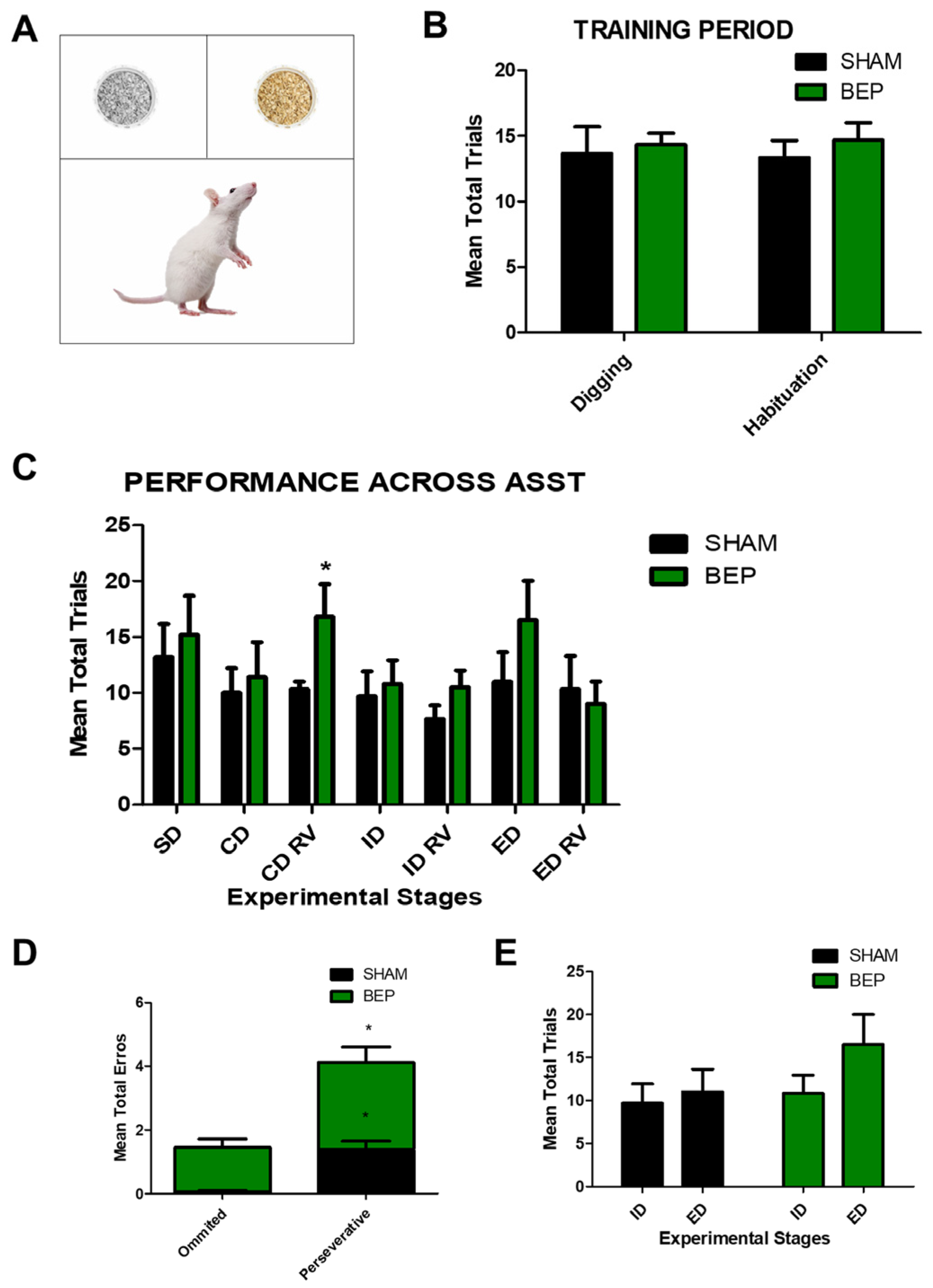
4.4. Attentional Set-Shifting Task (ASST)

The test followed the protocol described by Lynley [75] with modifications. Animals were placed on a food-restricted diet for one week before the beginning of the test and housed individually to maintain the diet. The habituation to the bowl started on the third day of the diet, and a piece of food was gradually hidden since the fourth day to train the rat to seek food in the bowl. The digging training was performed on the seventh day of the diet in the home cage. After 24 h, and one day before the test, the rats were trained on the apparatus with simple discrimination. In this stage, rats were submitted to different trials until they accomplished the criterion of 12 non-successive successful trials. A trial was considered successful when the rat chose the correct bowl, dug in the medium, and obtained the reward.
After the training process, the rats were tested across seven stages of the ASST. In this stage, rats were submitted to the trials necessary to achieve the criterion of 6 consecutive successful trials. Both the number of trials to reach the criterion and the number of errors were quantified. During the first stage (1), simple discrimination (SD), the rat had to choose the texture along one dimension. In the following stages, (2) compound discrimination (CD), (3) reversal learning (RL) of CD discrimination (CD RV), (4) intradimensional shift (ID), RL of ID (ID RV), and (5) extradimensional shift (ED) and RL of ED (ED RV), a second dimension was added—the odor. This second dimension consisted of using essential oils (Table 1) which were put in the bowl following the instructions established by Popik [47]. Briefly, the odor was introduced in the CD stage, but the reward still depended on the medium. In the CD RV stage, odors should have been ignored, and the reward was in the opposite medium. In the ID stage, the medium and odors were changed but the cue was still the medium. In the ID RV stage odors should have been ignored and the reward was changed to the opposite medium. In the ED stage, the odor was the cue to obtain the reward, not the medium. Finally, in the ED RV stage, the opposite odor was the cue to obtain the reward [47]. To avoid the rats learning the bowl’s position, the correct bowl’s location was randomly assigned in every trial.

4.5. Western Blot

The hippocampi, medial prefrontal cortex, and hypothalamus of treated or control animals were dissected on ice and immediately processed. Briefly, the hippocampal tissue was homogenized in RIPA buffer (25 mM Tris-Cl, pH 7.6, 150 mM NaCl, 1% NP-40, 1% sodium deoxycholate, and 0,1% SDS) supplemented with a protease inhibitor mixture and phosphatase inhibitors (25 mM NaF, 100 mM Na3VO4, and 30 μM Na2P2O7). The protein samples were centrifuged at 13,500 rpm for 15 min at 4 °C. The protein concentrations were determined using the BCA Protein Assay Kit (Pierce). The samples were resolved by SDS-PAGE, followed by immunoblotting on PVDF membranes. The membranes were incubated with the primary antibodies and anti-mouse, anti-goat, or anti-rabbit IgG peroxidase-conjugated antibodies (Jackson Immunoresearch, Inc., West Grove, PA, USA) and developed using an ECL kit (Westar Sun, Cyanagen, Bologna, Italia, or Westar Supernova, Cyanagen. Bologna, Italia).

4.6. PSD Isolation

Tissues were homogenized in buffer A containing 5 mM Hepes, pH 7.4, and 320 mM sucrose supplemented with protease and phosphatase inhibitor mixture. Cell debris was removed by centrifugation for 10 min at 1000× g (P1). The supernatant (S1) was centrifuged for 20 min at 20,000× g, obtaining S2 (cytosol and microsomes) and P2 fractions. P2 pellet was resuspended in Triton buffer (20 mM HEPES, 100 mM NaCl, 0.5% Triton X-100, pH 7.2) and rotated slowly for 30 min at 4 °C. Once incubated, samples were centrifugated at 12,000× g for 20 min to obtain PSD (pellet) and extrasynaptic (supernatant) fractions. PSD fraction was resuspended in Triton buffer containing protease and phosphatase inhibitor mixture. All manipulations were carried out on the ice or at 4 °C; samples were stored at −80 °C until use. Protein concentration was quantified and analyzed via Western blot.

4.7. Analysis

The images were loaded into ImageJ software. 1,5 (NIH) for densitometry analysis via Western blot. The results are presented as graphs depicting the mean ± standard error. According to the experiment, statistical significance was determined using one-way ANOVA with Tukey’s post-test, Student’s t-test, or two-way ANOVA with Bonferroni’s post-test. p values > 0.05 and ≤0.05 were regarded, respectively, as not statistically significant (n.s) and statistically significant (*). All statistical analyses were performed using Prism software, version 5.01 (GraphPad Software, Inc., Boston, MA, USA).

Author Contributions

C.A. and W.C. conceived the project and designed the experiments. C.A., R.G.M. and M.L. performed the experiments, analyzed the data, and wrote the manuscript. W.C. supervised the study. R.G.M., M.L. and W.C. edited the final version. All authors have read and agreed to the published version of the manuscript.

Funding

This work has been supported by grants: Research Team Project in Science and Technology (ACT1411), Basal Center for Excellence in Science and Technology (AFB 170005, PFB 12/2007), and Agencia Nacional de investigación y Desarrollo (ANID) FONDECYT grant: 1190620 (to W.C.).

Data Availability Statement

The datasets generated during the current study are available from the corresponding author on request.

Acknowledgments

We thank researchers from the Department of Cellular and Molecular Biology of the Faculty of Biological Sciences of the Pontificia Universidad Católica de Chile for their willingness to access spaces and equipment under their administration.

Conflicts of Interest

The authors declare no conflicts of interest. The funders had no role in the study’s design, in the collection, analyses, or interpretation of data, in the writing of the manuscript, or in the decision to publish the results.

References

  1. Koob, G.F.; Le Moal, M. Plasticity of reward neurocircuitry and the ‘dark side’ of drug addiction. Nat. Neurosci. 2005, 8, 1442–1444. [Google Scholar] [CrossRef] [PubMed]
  2. WHO. Global Status Report on Alcohol and Health 2018; World Health Organization: Geneva, Switzerland, 2018. [Google Scholar]
  3. Hendriks, H.F.J. Alcohol and Human Health: What Is the Evidence? Ann. Rev. Food Sci. Technol. 2020, 11, 1–21. [Google Scholar] [CrossRef] [PubMed]
  4. SAMHSA. Substance Abuse and Mental Health Services Administration. National Survey on Drug Use and Health: National Findings; U.S. Department of Health and Human Services: Rockville, MA, USA, 2007.
  5. NIAAA. National Institute on Alcohol Abuse and Alcoholism. Strategic Plan 2017–2021; U.S. Department of Health and Human Services: Rockville, MA, USA, 2017.
  6. Chung, T.; Creswell, K.G.; Bachrach, R.; Clark, D.B.; Martin, C.S. Adolescent Binge Drinking. Alcohol Res. Curr. Rev. 2018, 39, 5–15. [Google Scholar]
  7. Merrill, J.E.; Carey, K.B. Drinking Over the Lifespan: Focus on College Ages. Alcohol Res. Curr. Rev. 2016, 38, 103–114. [Google Scholar]
  8. Briones, T.L.; Woods, J. Chronic binge-like alcohol consumption in adolescence causes depression-like symptoms possibly mediated by the effects of BDNF on neurogenesis. Neuroscience 2013, 254, 324–334. [Google Scholar] [CrossRef] [PubMed]
  9. McBride, O.; Cheng, H.G. Exploring the emergence of alcohol use disorder symptoms in the two years after onset of drinking: Findings from the National Surveys on Drug Use and Health. Addiction 2011, 106, 555–563. [Google Scholar] [CrossRef]
  10. Spear, L.P. Effects of adolescent alcohol consumption on the brain and behaviour. Nat. Rev. Neurosci. 2018, 19, 197–214. [Google Scholar] [CrossRef]
  11. Fuhrmann, D.; Knoll, L.J.; Blakemore, S.J. Adolescence as a Sensitive Period of Brain Development. Trends Cogn. Sci. 2015, 19, 558–566. [Google Scholar] [CrossRef]
  12. Dahl, R.E. Adolescent brain development: A period of vulnerabilities and opportunities. Keynote address. Ann. N. Y. Acad. Sci. 2004, 1021, 1–22. [Google Scholar] [CrossRef]
  13. Crone, E.A.; Dahl, R.E. Understanding adolescence as a period of social-affective engagement and goal flexibility. Nat. Rev. Neurosci. 2012, 13, 636–650. [Google Scholar] [CrossRef]
  14. Lauharatanahirun, N.; Maciejewski, D.F.; Kim-Spoon, J.; King-Casas, B. Risk-related brain activation is linked to longitudinal changes in adolescent health risk behaviors. Dev. Cogn. Neurosci. 2023, 63, 101291. [Google Scholar] [CrossRef] [PubMed]
  15. Squeglia, L.M.; Jacobus, J.; Tapert, S.F. The effect of alcohol use on human adolescent brain structures and systems. Handb. Clin. Neurol. 2014, 125, 501–510. [Google Scholar] [CrossRef] [PubMed]
  16. Dees, W.L.; Hiney, J.K.; Srivastava, V.K. Alcohol and Puberty. Alcohol Res. Curr. Rev. 2017, 38, 277–282. [Google Scholar]
  17. Seemiller, L.R.; Gould, T.J. The effects of adolescent alcohol exposure on learning and related neurobiology in humans and rodents. Neurobiol. Learn. Mem. 2020, 172, 107234. [Google Scholar] [CrossRef] [PubMed]
  18. Xie, Y.; Dorsky, R.I. Development of the hypothalamus: Conservation, modification and innovation. Development 2017, 144, 1588–1599. [Google Scholar] [CrossRef] [PubMed]
  19. Bear, M.H.; Bhimji, S.S. Neuroanatomy, Hypothalamus; StatPearls: Treasure Island, FL, USA, 2018. [Google Scholar]
  20. Flament-Durand, J. The hypothalamus: Anatomy and functions. Acta Psychiatr. Belg. 1980, 80, 364–375. [Google Scholar] [PubMed]
  21. Blaine, S.K.; Sinha, R. Alcohol, stress, and glucocorticoids: From risk to dependence and relapse in alcohol use disorders. Neuropharmacology 2017, 122, 136–147. [Google Scholar] [CrossRef] [PubMed]
  22. Bird, C.M.; Burgess, N. The hippocampus and memory: Insights from spatial processing. Nat. Rev. Neurosci. 2008, 9, 182–194. [Google Scholar] [CrossRef]
  23. Eichenbaum, H. A cortical-hippocampal system for declarative memory. Nat. Rev. Neurosci. 2000, 1, 41–50. [Google Scholar] [CrossRef]
  24. Eichenbaum, H. Hippocampus: Cognitive processes and neural representations that underlie declarative memory. Neuron 2004, 44, 109–120. [Google Scholar] [CrossRef]
  25. Tapia-Rojas, C.; Carvajal, F.J.; Mira, R.G.; Arce, C.; Lerma-Cabrera, J.M.; Orellana, J.A.; Cerpa, W.; Quintanilla, R.A. Adolescent Binge Alcohol Exposure Affects the Brain Function Through Mitochondrial Impairment. Mol. Neurobiol. 2018, 55, 4473–4491. [Google Scholar] [CrossRef] [PubMed]
  26. Mira, R.G.; Lira, M.; Tapia-Rojas, C.; Rebolledo, D.L.; Quintanilla, R.A.; Cerpa, W. Effect of Alcohol on Hippocampal-Dependent Plasticity and Behavior: Role of Glutamatergic Synaptic Transmission. Front. Behav. Neurosci. 2019, 13, 288. [Google Scholar] [CrossRef]
  27. Vertes, R.P. Interactions among the medial prefrontal cortex, hippocampus and midline thalamus in emotional and cognitive processing in the rat. Neuroscience 2006, 142, 1–20. [Google Scholar] [CrossRef] [PubMed]
  28. Klenowski, P.M. Emerging role for the medial prefrontal cortex in alcohol-seeking behaviors. Addict. Behav. 2018, 77, 102–106. [Google Scholar] [CrossRef] [PubMed]
  29. Arnsten, A.F. Stress signalling pathways that impair prefrontal cortex structure and function. Nat. Rev. Neurosci. 2009, 10, 410–422. [Google Scholar] [CrossRef]
  30. Itoga, C.A.; Chen, Y.; Fateri, C.; Echeverry, P.A.; Lai, J.M.; Delgado, J.; Badhon, S.; Short, A.; Baram, T.Z.; Xu, X. New viral-genetic mapping uncovers an enrichment of corticotropin-releasing hormone-expressing neuronal inputs to the nucleus accumbens from stress-related brain regions. J. Comp. Neurol. 2019, 527, 2474–2487. [Google Scholar] [CrossRef] [PubMed]
  31. Girotti, M.; Adler, S.M.; Bulin, S.E.; Fucich, E.A.; Paredes, D.; Morilak, D.A. Prefrontal cortex executive processes affected by stress in health and disease. Prog. Neuro-Psychopharmacol. Biol. Psychiatry 2018, 85, 161–179. [Google Scholar] [CrossRef] [PubMed]
  32. Logue, S.F.; Gould, T.J. The neural and genetic basis of executive function: Attention, cognitive flexibility, and response inhibition. Pharmacol. Biochem. Behav. 2014, 123, 45–54. [Google Scholar] [CrossRef]
  33. Orsini, C.A.; Heshmati, S.C.; Garman, T.S.; Wall, S.C.; Bizon, J.L.; Setlow, B. Contributions of medial prefrontal cortex to decision making involving risk of punishment. Neuropharmacology 2018, 139, 205–216. [Google Scholar] [CrossRef]
  34. Galandra, C.; Basso, G.; Cappa, S.; Canessa, N. The alcoholic brain: Neural bases of impaired reward-based decision-making in alcohol use disorders. Neurol. Sci. 2018, 39, 423–435. [Google Scholar] [CrossRef]
  35. Kumar, A. NMDA Receptor Function During Senescence: Implication on Cognitive Performance. Front. Neurosci. 2015, 9, 473. [Google Scholar] [CrossRef] [PubMed]
  36. Burgdorf, J.; Colechio, E.M.; Stanton, P.; Panksepp, J. Positive Emotional Learning Induces Resilience to Depression: A Role for NMDA Receptor-mediated Synaptic Plasticity. Curr. Neuropharmacol. 2017, 15, 3–10. [Google Scholar] [CrossRef] [PubMed]
  37. Goebel-Goody, S.M.; Davies, K.D.; Alvestad Linger, R.M.; Freund, R.K.; Browning, M.D. Phospho-regulation of synaptic and extrasynaptic N-methyl-d-aspartate receptors in adult hippocampal slices. Neuroscience 2009, 158, 1446–1459. [Google Scholar] [CrossRef] [PubMed]
  38. Lau, C.G.; Zukin, R.S. NMDA receptor trafficking in synaptic plasticity and neuropsychiatric disorders. Nat. Rev. Neurosci. 2007, 8, 413–426. [Google Scholar] [CrossRef] [PubMed]
  39. Nguyen, T.H.; Paul, S.; Xu, Y.; Gurd, J.W.; Lombroso, P.J. Calcium-dependent cleavage of striatal enriched tyrosine phosphatase (STEP). J. Neurochem. 1999, 73, 1995–2001. [Google Scholar] [PubMed]
  40. Carvajal, F.J.; Mattison, H.A.; Cerpa, W. Role of NMDA Receptor-Mediated Glutamatergic Signaling in Chronic and Acute Neuropathologies. Neural Plast. 2016, 2016, 2701526. [Google Scholar] [CrossRef] [PubMed]
  41. Hardingham, G.E.; Bading, H. Synaptic versus extrasynaptic NMDA receptor signalling: Implications for neurodegenerative disorders. Nat. Rev. Neurosci. 2010, 11, 682–696. [Google Scholar] [CrossRef]
  42. Ge, Y.; Wang, Y.T. GluN2B-containing NMDARs in the mammalian brain: Pharmacology, physiology, and pathology. Front. Mol. Neurosci. 2023, 16, 1190324. [Google Scholar] [CrossRef]
  43. Sun, Y.Y.; Cai, W.; Yu, J.; Liu, S.S.; Zhuo, M.; Li, B.M.; Zhang, X.H. Surface expression of hippocampal NMDA GluN2B receptors regulated by fear conditioning determines its contribution to memory consolidation in adult rats. Sci. Rep. 2016, 6, 30743. [Google Scholar] [CrossRef]
  44. Warburton, E.C.; Brown, M.W. Neural circuitry for rat recognition memory. Behav. Brain Res. 2015, 285, 131–139. [Google Scholar] [CrossRef]
  45. Clay, J.M.; Parker, M.O. The role of stress-reactivity, stress-recovery and risky decision-making in psychosocial stress-induced alcohol consumption in social drinkers. Psychopharmacology 2018, 235, 3243–3257. [Google Scholar] [CrossRef] [PubMed]
  46. Dias-Ferreira, E.; Sousa, J.C.; Melo, I.; Morgado, P.; Mesquita, A.R.; Cerqueira, J.J.; Costa, R.M.; Sousa, N. Chronic stress causes frontostriatal reorganization and affects decision-making. Science 2009, 325, 621–625. [Google Scholar] [CrossRef] [PubMed]
  47. Popik, P.; Nikiforuk, A. Attentional Set-Shifting Paradigm in the Rat. Curr. Protoc. Neurosci. 2015, 72, 9.51.1–9.51.13. [Google Scholar] [CrossRef] [PubMed]
  48. Bartlett, T.E.; Wang, Y.T. The intersections of NMDAR-dependent synaptic plasticity and cell survival. Neuropharmacology 2013, 74, 59–68. [Google Scholar] [CrossRef] [PubMed]
  49. Lovinger, D.M.; White, G.; Weight, F.F. Ethanol inhibits NMDA-activated ion current in hippocampal neurons. Science 1989, 243, 1721–1724. [Google Scholar] [CrossRef] [PubMed]
  50. Lovinger, D.M.; White, G.; Weight, F.F. NMDA receptor-mediated synaptic excitation selectively inhibited by ethanol in hippocampal slice from adult rat. J. Neurosci. 1990, 10, 1372–1379. [Google Scholar] [CrossRef] [PubMed]
  51. Mira, R.G.; Tapia-Rojas, C.; Perez, M.J.; Jara, C.; Vergara, E.H.; Quintanilla, R.A.; Cerpa, W. Alcohol impairs hippocampal function: From NMDA receptor synaptic transmission to mitochondrial function. Drug Alcohol Depend. 2019, 205, 107628. [Google Scholar] [CrossRef]
  52. Tait, D.S.; Bowman, E.M.; Neuwirth, L.S.; Brown, V.J. Assessment of intradimensional/extradimensional attentional set-shifting in rats. Neurosci. Biobehav. Rev. 2018, 89, 72–84. [Google Scholar] [CrossRef]
  53. Wang, R.; Reddy, P.H. Role of Glutamate and NMDA Receptors in Alzheimer’s Disease. J. Alzheimer’s Dis. JAD 2017, 57, 1041–1048. [Google Scholar] [CrossRef]
  54. Lu, S.M.; Yeh, H.H. Ethanol modulates AMPA-induced current responses of primary somatosensory cortical neurons. Neurochem. Int. 1999, 35, 175–183. [Google Scholar] [CrossRef]
  55. Roberto, M.; Varodayan, F.P. Synaptic targets: Chronic alcohol actions. Neuropharmacology 2017, 122, 85–99. [Google Scholar] [CrossRef] [PubMed]
  56. Clapp, P.; Gibson, E.S.; Dell’acqua, M.L.; Hoffman, P.L. Phosphorylation regulates removal of synaptic N-methyl-D-aspartate receptors after withdrawal from chronic ethanol exposure. J. Pharmacol. Exp. Ther. 2010, 332, 720–729. [Google Scholar] [CrossRef] [PubMed]
  57. Euston, D.R.; Gruber, A.J.; McNaughton, B.L. The role of medial prefrontal cortex in memory and decision making. Neuron 2012, 76, 1057–1070. [Google Scholar] [CrossRef] [PubMed]
  58. Chiba, T. Collateral projection from the amygdalo--hippocampal transition area and CA1 to the hypothalamus and medial prefrontal cortex in the rat. Neurosci. Res. 2000, 38, 373–383. [Google Scholar] [CrossRef] [PubMed]
  59. Schmidt, B.; Hinman, J.R.; Jacobson, T.K.; Szkudlarek, E.; Argraves, M.; Escabi, M.A.; Markus, E.J. Dissociation between dorsal and ventral hippocampal theta oscillations during decision-making. J. Neurosci. 2013, 33, 6212–6224. [Google Scholar] [CrossRef] [PubMed]
  60. Le Merre, P.; Esmaeili, V.; Charriere, E.; Galan, K.; Salin, P.A.; Petersen, C.C.H.; Crochet, S. Reward-Based Learning Drives Rapid Sensory Signals in Medial Prefrontal Cortex and Dorsal Hippocampus Necessary for Goal-Directed Behavior. Neuron 2018, 97, 83–91.E5. [Google Scholar] [CrossRef] [PubMed]
  61. Strange, B.A.; Witter, M.P.; Lein, E.S.; Moser, E.I. Functional organization of the hippocampal longitudinal axis. Nat. Rev. Neurosci. 2014, 15, 655–669. [Google Scholar] [CrossRef] [PubMed]
  62. Kjelstrup, K.B.; Solstad, T.; Brun, V.H.; Hafting, T.; Leutgeb, S.; Witter, M.P.; Moser, E.I.; Moser, M.B. Finite scale of spatial representation in the hippocampus. Science 2008, 321, 140–143. [Google Scholar] [CrossRef]
  63. Moser, M.B.; Moser, E.I.; Forrest, E.; Andersen, P.; Morris, R.G. Spatial learning with a minislab in the dorsal hippocampus. Proc. Natl. Acad. Sci. USA 1995, 92, 9697–9701. [Google Scholar] [CrossRef]
  64. McEwen, B.S. Stress and hippocampal plasticity. Annu. Rev. Neurosci. 1999, 22, 105–122. [Google Scholar] [CrossRef]
  65. Cook, S.C.; Wellman, C.L. Chronic stress alters dendritic morphology in rat medial prefrontal cortex. J. Neurobiol. 2004, 60, 236–248. [Google Scholar] [CrossRef]
  66. Liston, C.; McEwen, B.S.; Casey, B.J. Psychosocial stress reversibly disrupts prefrontal processing and attentional control. Proc. Natl. Acad. Sci. USA 2009, 106, 912–917. [Google Scholar] [CrossRef] [PubMed]
  67. Townshend, J.M.; Kambouropoulos, N.; Griffin, A.; Hunt, F.J.; Milani, R.M. Binge drinking, reflection impulsivity, and unplanned sexual behavior: Impaired decision-making in young social drinkers. Alcohol. Clin. Exp. Res. 2014, 38, 1143–1150. [Google Scholar] [CrossRef] [PubMed]
  68. Xiao, L.; Bechara, A.; Gong, Q.; Huang, X.; Li, X.; Xue, G.; Wong, S.; Lu, Z.L.; Palmer, P.; Wei, Y.; et al. Abnormal affective decision making revealed in adolescent binge drinkers using a functional magnetic resonance imaging study. Psychol. Addict. Behav. J. Soc. Psychol. Addict. Behav. 2013, 27, 443–454. [Google Scholar] [CrossRef] [PubMed]
  69. Salling, M.C.; Skelly, M.J.; Avegno, E.; Regan, S.; Zeric, T.; Nichols, E.; Harrison, N.L. Alcohol Consumption during Adolescence in a Mouse Model of Binge Drinking Alters the Intrinsic Excitability and Function of the Prefrontal Cortex through a Reduction in the Hyperpolarization-Activated Cation Current. J. Neurosci. 2018, 38, 6207–6222. [Google Scholar] [CrossRef]
  70. Ketchesin, K.D.; Stinnett, G.S.; Seasholtz, A.F. Binge Drinking Decreases Corticotropin-Releasing Factor-Binding Protein Expression in the Medial Prefrontal Cortex of Mice. Alcohol. Clin. Exp. Res. 2016, 40, 1641–1650. [Google Scholar] [CrossRef]
  71. Robinson, S.L.; Marrero, I.M.; Perez-Heydrich, C.A.; Sepulveda-Orengo, M.T.; Reissner, K.J.; Thiele, T.E. Medial prefrontal cortex neuropeptide Y modulates binge-like ethanol consumption in C57BL/6J mice. Neuropsychopharmacology 2019, 44, 1132–1140. [Google Scholar] [CrossRef]
  72. McClintick, J.N.; McBride, W.J.; Bell, R.L.; Ding, Z.M.; Liu, Y.; Xuei, X.; Edenberg, H.J. Gene expression changes in the ventral hippocampus and medial prefrontal cortex of adolescent alcohol-preferring (P) rats following binge-like alcohol drinking. Alcohol 2018, 68, 37–47. [Google Scholar] [CrossRef]
  73. Tapia-Rojas, C.; Torres, A.K.; Quintanilla, R.A. Adolescence Binge Alcohol Consumption Induces Hippocampal Mitochondrial Impairment that Persists during the Adulthood. Neuroscience 2019, 406, 356–368. [Google Scholar] [CrossRef]
  74. Mira, R.G.; Lira, M.; Quintanilla, R.A.; Cerpa, W. Alcohol consumption during adolescence alters the hippocampal response to traumatic brain injury. Biochem. Biophys. Res. Commun. 2020, 528, 514–519. [Google Scholar] [CrossRef]
  75. Linley, S.B.; Gallo, M.M.; Vertes, R.P. Lesions of the ventral midline thalamus produce deficits in reversal learning and attention on an odor texture set shifting task. Brain Res. 2016, 1649, 110–122. [Google Scholar] [CrossRef] [PubMed]
Figure 1. Adolescent alcohol effects on attentional set-shifting task. (A) Representative drawing of ASST. (B) Mean trials to reach the criteria on the training period. (C) The number of mean total trials to reach the criterion of association learning. Abbreviations: SD, simple discrimination; CD, compound discrimination; CD RV, compound discrimination reversal; ID, intradimensional shift; ID REV, intradimensional shift reversal; ED, extradimensional shift; ED REV: extradimensional shift reversal. (D) Mean of total errors before reaching the criterion. (E) Comparison between mean total trials to reach the ID and ED stages criterion. All tests were performed one week after ethanol treatment. * p < 0.05, two-way ANOVA, t-test. N = 5.
Stresses 04 00001 g001
Figure 2. Adolescent alcohol exposure decreases NMDAR pro-survival signaling. (A,C,E) Immunoblot images from whole lysates from the hypothalamus (A), hippocampus (C), and Prefrontal Cortex (E). Main regulators of GluN2B-Y1472 phosphorylation, Fyn kinase, and STEP phosphatase were analyzed along with pERK1/2 and pCREB (Ser133) downstream effectors of synaptic NMDAR signaling proteins. (B,D,F) Immunoblots quantification. Every protein was normalized with its respective loading control and analyzed independently by t-test. * p < 0.05, ** p < 0.01. N = 3 rats.
Stresses 04 00001 g002
Figure 3. Adolescent alcohol exposure decreases the abundance of synaptic NMDARs. (A,C,E) Immunoblot images from isolated PSD fractions from the hypothalamus (A), hippocampus (C), and Prefrontal Cortex (E). The GluN2B subunit of NMDA receptors was analyzed. (B,D,F) Immunoblots quantification. The protein densitometry was normalized with its respective loading control and analyzed independently by t-test. * p < 0.05. N = 3 rats.
Stresses 04 00001 g003
Table 1. Stimuli in the attentional set-shifting task. Odors and mediums are used for different stages and their combination. Reward stimuli are shown in bold.
STAGESTIMULI COMBINATION
TRAINING
Digging trainingCorn cob bedding
TASKODORMEDIUM(+)(−)
SD M1 corn cob beddingM2M1
M2 paper bedding
CDO1 gingerM1 corn cob beddingO1/M1O2/M2
O2 cinnamonM2 paper beddingO2/M1O1/M2
CD revO1 gingerM1 corn cob beddingO2/M1O1/M2
O2 cinnamonM2 paper beddingO2/M2O1/M1
IDO3 lemonM3 cellulose beddingO3/M3O4/M4
O4 fennelM4 rubber beddingO4/M3O3/M4
ID revO3 lemonM3 cellulose beddingO3/M4O4/M3
O4 fennelM4 rubber beddingO4/M4O3/M3
EDO5 citronellaM5 gravel beddingM5/O5M6/O6
O6 cloveM6 plush beddingM6/O5M5/O6
ED revO5 citronellaM5 gravel beddingM5/O6M6/O5
O6 cloveM6 plush beddingM6/O6M5/O5
Disclaimer/Publisher’s Note: The statements, opinions and data contained in all publications are solely those of the individual author(s) and contributor(s) and not of MDPI and/or the editor(s). MDPI and/or the editor(s) disclaim responsibility for any injury to people or property resulting from any ideas, methods, instructions or products referred to in the content.

