Review

Insulin Resistance Develops Due to an Imbalance in the Synthesis of Cyclic AMP and the Natural Cyclic AMP Antagonist Prostaglandylinositol Cyclic Phosphate (Cyclic PIP)

by
Heinrich K. Wasner
BioReg Biopharm, Technology Innovation Laboratory, University of Illinois at Chicago, Chicago, IL 60612, USA
Stresses 2023, 3(4), 762-772; https://doi.org/10.3390/stresses3040052
Submission received: 21 September 2023 / Revised: 10 November 2023 / Accepted: 14 November 2023 / Published: 17 November 2023
(This article belongs to the Collection Feature Papers in Human and Animal Stresses)

Abstract

:
The reasons initiating insulin resistance are not identified. Various metabolic derailments have been characterized. These are the outcome and not the initiation of insulin resistance. In animal models of type 2 diabetes and hypertension, a decreased hormonal stimulation of the synthesis of the cyclic AMP antagonist prostaglandylinositol cyclic phosphate (cyclic PIP) was determined. The resultant imbalance of the action of cyclic AMP and cyclic PIP shifts metabolic regulation to the dominance of catabolism and a decrease in imperative anabolism. This dominance develops gradually since the more cyclic AMP dominates, the more the synthesis of cyclic PIP will be inhibited. Vanishing actions of cyclic PIP are its 10-fold activation of glucose uptake in adipocytes, its inhibition of insulin release from pancreatic β-cells, its inhibition of PKA and its 7-fold activation of protein ser/thr phosphatase. Reduced synthesis of cyclic PIP results from (a) decreased substrate availability, (b) long-time elevated cyclic AMP levels resulting from stress overloads and (c) aging and the gradual decrease in the synthesis of hormones which likely maintain mechanisms that stimulate cyclic PIP synthesis. The need is to discover which hormones, such as growth hormone, insulin-like growth factor-1, dehydroepiandrosterone, and testosterone, are involved in maintaining the stimulation of cyclic PIP synthesis.

1. Introduction

Type 2 diabetes and hypertension are acute health problems of today’s world. The percentage of people with type 2 diabetes is continuously increasing, and there are at least 422 million people with diabetes and 1.28 billion adults with hypertension worldwide according to the WHO. Many scientists are engaged in identifying the primary causes that initiate the development of insulin resistance, a starting condition of type 2 diabetes. Using various inbred mouse strains, Nelson et al. determined the effects of diet on the development of insulin resistance primarily in muscle, liver and white adipose tissue [1], and Mejhert and Ryden commented on this report [2]. Petersen and Shulman published a comprehensive and detailed review on the “mechanism of insulin action and insulin resistance” and came to the conclusion that the “sheer complexity of biological systems means that any effort to understand insulin resistance with a unified, succinct and straightforward model may be a fool’s errand” [3]. Galicia-Garcia et al. [4] proposed that type 2 diabetes is caused by a combination of two primary factors, namely a defective insulin secretion of the pancreatic β-cells and the inability of insulin-sensitive tissues to respond appropriately to insulin, and they concluded that there is still a long way to go to fully understand insulin resistance.
The mechanism of insulin action, the understanding of which is a prerequisite for determining defects in insulin’s action, appears to be nearly solved: Insulin binds to its receptor, whose intrinsic tyrosine kinase is in this way activated; this kinase auto-phosphorylates itself and then phosphorylates primarily the insulin receptor substrates IRS1-4 and several Shc proteins [5,6]. The IRS proteins are suggested to regulate metabolism, whereas Shc proteins appear to be involved in the regulation of proliferation by insulin. The IRS proteins are considered to be docking proteins to which various enzymes bind via Src-homology-2 (SH-2) domains and send signals further into a cell via phosphorylation pathways. Additionally, the IRS proteins are inactivated by serine/threonine phosphorylation by several different protein ser/thr kinases [5].
Earl Sutherland articulated many years ago that a decrease in the strength of the insulin signal will increase the strength of the counterregulatory signal, leading to the dominance of regulations triggered by cyclic AMP [7]. This view has not been adequately pursued. A characteristic of insulin resistance is that it progresses slowly and gradually for reasons not fully understood at the present time. Inhibition of single proteins/enzymes such as the IRS proteins cannot explain this slow progression, though they play a crucial role in signal transduction. Current thinking is that protein phosphorylation pathways are of primary importance [5,6], but fails to consider that insulin antagonizes glucagon action. Insulin rapidly reverses cyclic AMP-triggered protein ser/thr phosphorylation. The key player in this scenario is the natural cyclic AMP antagonist, prostaglandylinositol cyclic phosphate (cyclic PIP) [8,9]. The synthesis of cyclic PIP is stimulated for instance by insulin and also noradrenaline. Cyclic PIP synthesis is decreased in type 2 diabetic and also hypertensive rodents [8]. The effect of decreasing cyclic PIP synthesis and consequently increasing cyclic AMP synthesis on the development of insulin resistance is the topic of this report.

2. Cyclic AMP

After its discovery by Earl Sutherland, cyclic AMP was for many years viewed as the unique intracellular regulator that activates the cyclic AMP-dependent protein kinase (PKA). By protein ser/thr phosphorylation, catabolic enzymes are predominantly activated and anabolic enzymes are predominantly inactivated. Further ways of action of cyclic AMP are (a) its activation of guanine exchange factors, (b) its binding to cyclic AMP binding proteins and (c) its regulation of cyclic nucleotide-gated ion channels [10]. Cyclic AMP synthesis is stimulated for instance by glucagon and adrenaline via its β-receptors. Adenylate cyclase is activated by the Gαs subunit of the stimulatory, heterotrimeric Gs protein [11]. The reversal of this activation is controlled by the GTPase activity of the Gαs protein which converts the bound GTP to GDP inactivating adenylate cyclase. Cyclic PIP inhibits adenylate cyclase dose-dependently up to 100% [8]. In contrast, the Gi protein inhibits adenylate cyclase by approximately 30% at maximal hormone concentration [12]. This effect is declared to be an adrenergic α2-receptor action (see below).

3. The Natural Cyclic AMP Antagonist Cyclic PIP

The search for an antagonist to cyclic AMP started in the laboratory of the late Earl Sutherland. Cyclic PIP was primarily isolated from rat livers, which were extra-corporally perfused with buffer and stimulated with noradrenaline or insulin, homogenized within 1 to 3 min, and then put under denaturing conditions to minimize the enzymatic degradation of cyclic PIP. From 2 kg of rat livers, approximately 60 g of water-soluble, low-molecular-weight compounds were obtained, which also contained cyclic PIP. A rough calculation indicated that 500,000 to 1,000,000 molecules of this extract contain 1 molecule of cyclic PIP. But more challenging was the finding that cyclic PIP is one of the most labile molecules of this extract. It decays by 80% within 30 min held at an ionic strength comparable to a 0.5 molar sodium chloride solution, approximately a 3-fold concentrated physiological saline solution. The labilities of cyclic PIP are connected to the tension of the 5-ring phosphodiester, which is the most labile bond of cyclic PIP, as well as the allyl-ether bond combining two secondary alcohols and the beta-keto-hydroxy structure of the C5-ring of prostaglandin E (PGE). Chemically it is O-(prostaglandyl-E)-(15-4′)-(myo-inositol 1′:2′-cyclic phosphate) [8]. It is biosynthesized from PGE and activated inositol phosphate by cyclic PIP synthase [8], which is active in a tyrosine-phosphorylated form [13]. The synthesis of cyclic PIP is stimulated also by adrenergic α1- and α2-receptors [14]. This result contradicts the present view of adrenergic α1- and α2-receptor action, as discussed in [14,15]. The adrenergic receptors are transmembrane, G protein-coupled receptors; thus, cyclic PIP synthase should also be activated by a G protein [13]. It is not known whether there are two different variants of cyclic PIP synthase, but it is known that the biosynthesis of the substrates for cyclic PIP synthesis involves phospholipase A2 and phospholipase C, both of which are activated by these two modes of activation as summarized in [15]. The synthesis of cyclic AMP from ATP is a one-step reaction, and ATP is generally always present in a high enough amount in all cells to warrant maximal synthesis of cyclic AMP. In contrast, the biosynthesis of cyclic PIP needs at least five reaction steps (Figure 1), and both substrates are obtained from membrane-bound lipids [15].
The primary action of cyclic AMP is to activate PKA, whereas cyclic PIP inhibits PKA. PKA is activated by increasing concentrations of cyclic AMP (10−8 to 10−6 molar) at least 10-fold. Increasing concentrations of cyclic PIP led to increasing, up to 100%, inhibition of basal and cyclic AMP-activated PKA. For an equal percentage of inhibition of basal and cyclic AMP-activated PKA, approximately 4 times more cyclic PIP is needed in the case of the cyclic AMP-activated kinase [14,16]. It was assumed that protein ser/thr phosphatase, the counter-regulatory enzyme to PKA, would also be regulated by these two compounds. Phosphatase is a rather labile enzyme. In order to cope with this problem, scientists decided to isolate and characterize the catalytic subunits of this class of enzymes [17]. For this reason, the holoenzyme of protein ser/thr phosphatase was partly purified [9]. The obtained holoenzyme has a slightly higher molecular weight than PKA and is 7-fold activated by cyclic PIP and completely inhibited by cyclic AMP in the physiological action range [8]. The effects of cyclic PIP in viable cells are as follows: (1) 10-fold activation of glucose uptake into adipocytes; (2) shut-off of insulin release from pancreatic β-cells; (3) inhibition of glucagon-stimulated autophagy and proteolysis; (4) 2,7-fold positive inotropic effect on the papillary muscle of the heart, which is, different from the action of cyclic AMP, connected with an elongation of the contraction time [8]. As was the case with cyclic AMP [10], further modes of action of cyclic PIP are likely to be found in the coming years as the chemical synthesis of cyclic PIP allows additional studies.

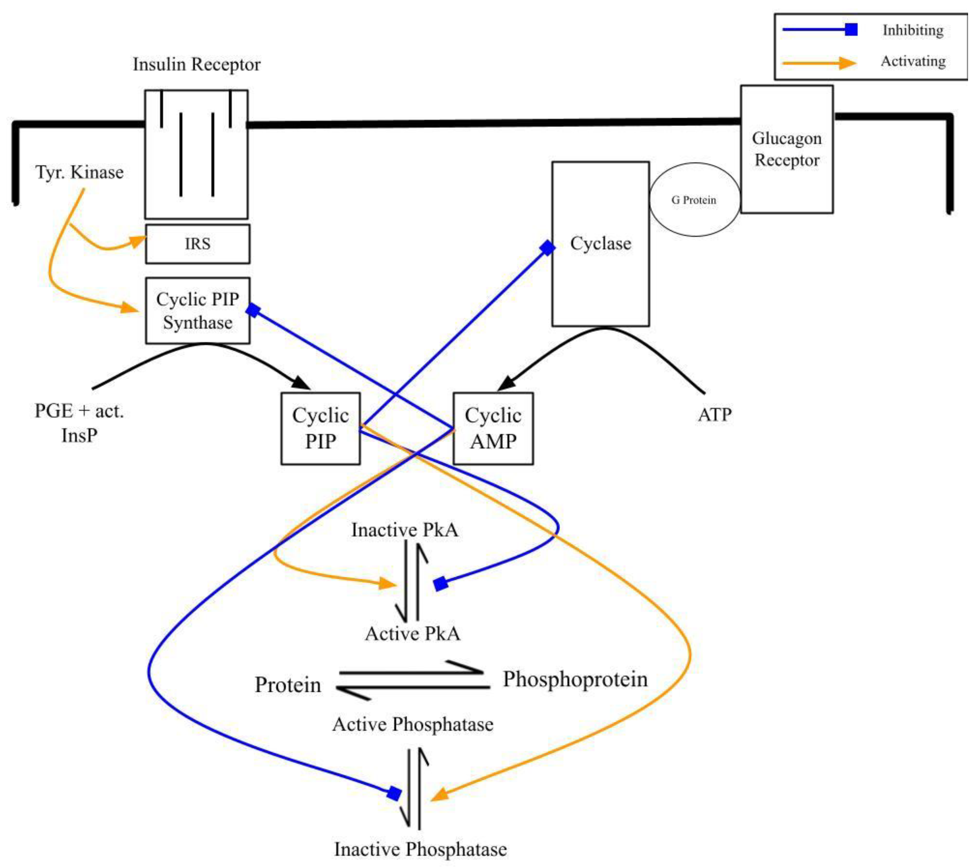
4. The Interplay of Cyclic AMP and Cyclic PIP

4.1. At the Level of Their Biosynthesis: Cyclic AMP Inhibits Cyclic PIP Synthase and Cyclic PIP Inhibits Adenylate Cyclase

Cyclic PIP synthase is activated by tyrosine phosphorylation [9,13], which correlates with the signal transduction and the tyrosine kinase activity of the insulin receptor [5,6]. Apart from the activation and inhibition by tyrosine phosphorylation and dephosphorylation, respectively, cyclic PIP synthase is inhibited by cyclic AMP in the presence of ATP by a protein ser/thr kinase [18]. This inhibition may ensure that no cyclic PIP is synthesized at a time when cyclic AMP is required to respond to appropriate signals, since it would be counterproductive if cyclic AMP and cyclic PIP were synthesized at the same time [9]. With respect to selectivity, it is assumed that this phosphorylation is carried out by a specific kinase and not by a half dozen enzymes as discussed for the IRS proteins [5,6]. The protein ser/thr phosphatase, which reverses the protein ser/thr phosphorylation of cyclic PIP synthase, has yet to be found.
Cyclic PIP was found by its inhibition of adenylate cyclase [8] indicating that cyclic PIP antagonizes the action of cyclic AMP comparably to the inhibition of cyclic PIP synthase by cyclic AMP. This inhibition is most likely the result of a phosphorylation reaction [19]. However, this kinase has not been characterized further, and it is not known if it phosphorylates tyrosine or serine/threonine residues of adenylate cyclase or its regulatory structures involved in the activation of adenylate cyclase. The group of Pradipta Ghosh reported recently that receptor tyrosine kinases, after activation by growth factors such as EGF, PDGF and insulin, phosphorylate tyrosine residues of the Gαi protein, which then efficiently inhibits adenylate cyclase. This inhibition was demonstrated in intact cells, but difficult to show in “in vitro” assays. They explained that in the “in vitro” assay, a part of this complex assay mixture may be missing or not be present in the needed amount [20]. This missing component could be cyclic PIP since under the experimental conditions used, the synthesis of cyclic PIP is stimulated very well in intact cells but not in disrupted cells [21]. One may question if these three different inhibitory effects of adenylate cyclase, namely the partial inhibition by Gi protein [12], the complete inhibition by cyclic PIP [8] and the complete inhibition by the tyrosine-phosphorylated Gαi protein [20], could be parts of one complex reaction sequence leading to inhibition of adenylate cyclase. It would be bewildering if adenylate cyclase is inhibited in various ways that are triggered by the same hormones. That means insulin stimulates the synthesis of cyclic PIP and also the tyrosine phosphorylation of Gαi protein, and noradrenaline stimulates the synthesis of cyclic PIP and triggers the inhibition via the Gi protein, which was determined in cell homogenates by Sabol and Nirenberg. They most likely could determine only a partial inhibition of adenylate cyclase [12], since cyclic PIP is not synthesized in cell homogenates [21].
The point of this section is that cyclic AMP inhibits cyclic PIP synthase [18] and cyclic PIP inhibits adenylate cyclase [8]. The following two experiments visualize the effect of cyclic PIP on the hormone-stimulated adenylate cyclase of hepatocytes of untreated and diabetic rats. (A) Glucagon stimulates cyclic AMP synthesis about 30-fold in isolated rat hepatocytes, whereas adrenaline triggers only a 2- to 3-fold increase. However, when hepatocytes were first stimulated with glucagon and 40 min later with adrenaline, adrenaline stimulated cyclic AMP synthesis 6- to 9-fold [8,22]. The explanation is that on the first stimulation with adrenaline, cyclic PIP synthesis is also increased, which inhibits adenylate cyclase, limiting the increase in cyclic AMP. Glucagon stimulates a much greater synthesis of cyclic AMP, which will inhibit cyclic PIP synthase. Hence, after a first stimulation with glucagon, on a second stimulation with adrenaline, the synthesis of cyclic PIP is decreased, leading to an increased cyclic AMP synthesis. For a perfect intracellular control of the activity of adenylate cyclase and also cyclic PIP synthase, one would expect that both enzymes are not only inhibited by their antagonistic regulator but that there must be mechanisms by which either cyclic PIP synthase or adenylate cyclase is specifically reactivated. (B) The time course of cyclic AMP synthesis was determined in rat hepatocytes of untreated and diabetic rats, obtained by low-dose streptozotocin treatment. Both cell suspensions were stimulated with adrenaline at 0 and at 30 min. The basal cyclic AMP level in the hepatocytes of the diabetic rats was approximately 3-fold higher than that in the cells of non-diabetic control rats. The adrenaline-stimulated cyclic AMP synthesis of the controls was set to 100%, and in the hepatocytes of the diabetic rats, cyclic AMP synthesis was then increased to 155%. The second stimulation with adrenaline at 30 min triggered an approximately 90% decreased synthesis of cyclic AMP in the controls, because of the stimulation of cyclic PIP synthesis by adrenaline at 0 min. However, in the hepatocytes of the diabetic rats, the synthesis of cyclic AMP following a second stimulation with adrenaline was nearly as great as it was in the first stimulation. The reduced cyclic PIP synthesis, which was observed in the livers of diabetic rodents, is the obvious explanation [8,18]. This result is in accordance with Sutherland’s observation that cyclic AMP synthesis increases in cells with a reduced insulin response [7]. This finding is not unique to rats treated with streptozotocin. Cyclic PIP synthase activity in the livers of Ksj db/db mice is 36% lower than that in Ksj control mice, and cyclic PIP synthase activity in the livers of spontaneously hypertensive rats (SHR) at three months of age is decreased by 27% when compared with the activity of Wistar–Kyoto control rats [18].
In summary, decreasing stimulation of cyclic PIP synthesis leads to increasing cyclic AMP synthesis. This appears to play an essential role in the development of insulin resistance because elevated cyclic AMP levels will further inhibit the stimulation of cyclic PIP synthesis and in this way continuously increase cyclic AMP synthesis and action with time, leading to predominantly serine/threonine-phosphorylated proteins (see below). The resulting downward spiral of the stimulation of cyclic PIP synthesis can explain the nearly absent hormonal stimulation of cyclic PIP synthesis in type 2 diabetic rats (Table 1). In other words, the mutual inhibition of the synthesis stimulation of the two regulators, cyclic AMP and cyclic PIP, is certainly necessary because it enables the regulator, whose synthesis is stimulated by a hormone, to perform its task unimpaired. However, in the event of a weakening of the stimulation of one of these two regulators, this positive effect of mutual inhibition enforces the imbalance in the synthesis of these two regulators and consequently in the regulation of metabolism. In the case of the development of insulin resistance, the stimulation of cyclic PIP synthesis becomes weaker for various reasons (see below).

4.2. At the Level of the Regulation of Metabolism: Increasing Cyclic AMP and Decreasing Cyclic PIP Synthesis Lead to Dominance of Catabolism over Anabolism

The activity of PKA and of protein ser/thr phosphatase is regulated by cyclic AMP and cyclic PIP [8,9], creating an equilibrium between the dephospho- and phospho-forms of interconvertible enzymes. For instance, insulin stimulates the synthesis of cyclic PIP, which shuts off cyclic AMP synthesis, activates protein ser/thr phosphatase, inhibits PKA [8,9] and shifts the equilibrium between the dephospho- and phospho-forms of interconvertible enzymes to the left side, the dephosphorylated form (Figure 2). However, a decreasing stimulation of cyclic PIP synthesis will not adequately inhibit adenylate cyclase and will also not appropriately shift the equilibrium to the left side. The result is that with time, the equilibrium between the dephospho- and phospho-forms of interconvertible enzymes will be more and more shifted to the phospho-form, and the metabolism will be more and more stuck in the catabolic state.

5. Illnesses Connected with Decreased Synthesis of Cyclic PIP

The stimulation of cyclic PIP synthesis decreases in low-dose streptozotocin diabetic rats. That means within 35 days after streptozotocin application, the stimulation of cyclic PIP synthesis gradually nearly vanished, but the basal levels of cyclic PIP synthesis appear to be somewhat increased, which is, presently, unexplained [18,23]. Additionally, arachidonic acid feeding improves streptozotocin-induced type 2 diabetes in rats [24]. The obvious explanation is that arachidonic acid is the substrate for prostaglandin and cyclic PIP synthesis. Furthermore, inhibition of cyclic PIP synthesis by non-steroidal anti-inflammatory drugs (NSAIDs) leads to a metabolic state of the treated rats, which is characteristic of insulin resistance [21]. Glucose tolerance tests showed that in these treated rats, the serum insulin levels increased at least 3 times higher compared to the untreated control rats. Cyclic PIP inhibits the release of insulin in accordance with insulin’s action [8], and a reduced synthesis of cyclic PIP leads to a decreasing shut-off of insulin’s secretion from pancreatic β-cells and thus to increased serum insulin levels [21]. Additionally, the blood glucose levels of these animals were 50% more increased and declined slower in comparison with the values from untreated control rats. Accordingly, the liver and muscle of the treated rats had highly reduced glycogen contents. Insulin and also cyclic PIP stimulated a 10-fold increase in glucose uptake in adipocytes of untreated rats. But in adipocytes of the treated rats, the stimulation of glucose uptake by insulin was decreased by 70%, but added cyclic PIP triggered an unchanged glucose uptake [21], indicating that the defect in the signal transduction of the developing insulin resistance should be found between the insulin receptor and cyclic PIP synthesis [9].
In the brain, type 2 diabetes leads to increased phosphorylation (hyper-phosphorylation) of the tau protein, a characteristic of Alzheimer’s disease. This diabetic situation changes the activity of glycogen synthase kinase-3β (GSK3β), which is a suggested reason for the enhanced phosphorylation of the tau protein. The authors discussed that PKB/Akt, after its activation by PI3-kinase, inactivates GSK3β by phosphorylation. Hyper-phosphorylation of the tau protein results from a decreased phosphorylation of GSK3β in type 2 diabetic patients [25]. Cyclic PIP synthase is found to act upstream of PI3-kinase [26], which means this interpretation has to be reconsidered. Furthermore, the tau hyper-phosphorylation correlates with the inhibited protein phosphatase-2A (PP-2A), the main tau phosphatase [27]. Cyclic AMP inhibits protein ser/thr phosphatase [8,9], and type 2 diabetes leads to increased cyclic AMP levels (see above). This could explain the inhibition of PP-2A in type 2 diabetics. An international group of scientists led by Ron Kahn characterized the metabolism of the cell-culture line iMyos, which is derived from muscle cells of type 2 diabetic patients [28]. They determined over-phosphorylation of various proteins. Increased serine/threonine phosphorylation of IRS1 and IRS2 is discussed to be the result of various over-active serine/threonine kinases. Additionally, they found over-phosphorylation of many proteins/enzymes downstream of different regulatory pathways. They concluded that there must be a factor that has a regulatory effect on several kinases or phosphatases and that this factor must be urgently identified [28]. It is very likely that they will rediscover cyclic AMP and cyclic PIP. Presently, there is still a gap between the increasing cyclic AMP and decreasing cyclic PIP levels and the over-phosphorylated proteins/enzymes. That means it remains to be determined (a) to what extent cyclic AMP is involved in this over-phosphorylation and (b) by which biochemical pathways the elevated cyclic AMP levels cause this over-phosphorylation of some of these proteins and enzymes, depending on the progression of insulin resistance. It is unlikely that the decreasing effectiveness of insulin could be the cause for over-phosphorylation, because (a) the regulations of insulin, which are triggered via phosphorylation pathways, should lead to decreasing phosphorylation in the case of developing insulin resistance and (b) the pathways of insulin action, which trigger dephosphorylation, would stay in a phosphorylated state, depending on the degree of the developing insulin resistance. But the evolving increase in cyclic AMP levels could explain the over-phosphorylation. Enzymes/proteins remain phosphorylated because of insulin’s weaker regulation and, for instance, a successive glucagon stimulation or maybe the increased basal cyclic AMP levels will further phosphorylate these enzymes. Ron Kahn reported that “over-phosphorylation occurs both inside and outside the classical insulin-signaling pathway” [28]. The gradually increasing cyclic AMP levels, a result of developing insulin resistance, could be a possible explanation for the over-phosphorylation outside of insulin’s signaling pathways.
In the introduction, some present views on insulin resistance were mentioned. These reports describe metabolic conditions, which show the extent of the derailment of metabolism in the course of the development of insulin resistance. However, the events that initiated the derailment have not been identified and are mostly not discussed. In particular, the action of cyclic AMP and the existence and action of cyclic PIP are rarely considered. But it has been recognized that many different pathways are derailed in the case of the development of insulin resistance [1,3,4,29]. However, since insulin triggers a multitude of regulations in all the different organs of a body, one could realize that a single, essential derailment of insulin’s signal transduction, such as the gradual loss of stimulation of the synthesis of cyclic PIP, which triggers many of insulin’s actions [8], will result in a gradual decrease in all these regulations stimulated by insulin and, importantly, can lead to increasing dominance of cyclic AMP and its catabolic regulations.
As mentioned in the introduction, Galicia-Garcia et al. concluded that type 2 diabetes is caused by defective insulin secretion and the inability of insulin-responsive tissues to respond to insulin [4]. It could have been recognized that these defects are related to the decreasing action of cyclic PIP. Firstly, cyclic PIP inhibits the release of insulin from pancreatic β-cells [8], and in the case of a vanishing stimulation of cyclic PIP synthesis, the shut-off of insulin’s secretion seizes, and the resulting continuous excretion of insulin will gradually lead to an exhaustion of the pancreatic β-cells [30]. Secondly, all organs/cells of a body and not just skeletal muscles, liver and white adipose cells are responsive to insulin, and so far as cyclic PIP is the intracellular executor of many regulations of insulin [8], these functions will vanish in the case of a decreasing cyclic PIP synthesis.
What causes lead to the decreasing stimulation of cyclic PIP synthesis? (1) One possibility is that needed substrates can be limiting. This problem should play a minor role in the synthesis of activated inositol phosphate since generally there should be no deficit in myo-inositol intake by food. However, an increasing number of reports show that polycystic ovary syndrome, gestational diabetes mellitus and metabolic syndrome can be improved by increased intake of inositol [31,32]. Myo-inositol is a structural component of cyclic PIP, and one may ask if inositol feeding could lead to an improved synthesis of a decreased cyclic PIP synthesis. However, the defect causing inositol deficiency is not yet identified, and thus, this question cannot be answered presently. (2) Substrate deficiency will more likely play a role in the case of the synthesis of prostaglandins. The supply of lipids containing polyunsaturated fatty acids such as omega-6 fatty acids can be limiting, especially since humans cannot synthesize these acids themselves and are dependent on food as their source. Along these lines, pain relievers of the NSAID family inhibit the biosynthesis of prostaglandins, whose decrease impairs the synthesis of cyclic PIP [21]. More recently, it was reported that streptozotocin-induced type 2 diabetes can be improved by support with arachidonic acid [24]. (3) Too many humans have high stress levels that result from problems of daily life such as too-long exposures to too much noise and to challenges at work [33], causing elevated cyclic AMP levels [34,35]. (4) Another consideration is that regulatory mechanisms decline with aging. Though not finally proven, key hormones such as growth hormone, dehydroepiandrosterone, insulin-like growth factor-1 and testosterone appear to be involved in aging and in insulin action [36]. Presently, the mechanisms of action of these hormones are still under investigation. Pataky, Young and Nair recommend in their review on aging that caution be taken in treatment with these hormones since too many negative side effects of these hormonal replacements could outweigh the positive effects. They suggest that, presently, it is better to maximally apply physical exercise and calorie restrictions since this way of lifestyle has positive effects on aging and on insulin sensitivity and has no negative side effects, though the mechanisms of action of lifestyle changes are still largely unknown. These hormones start to decrease at around the age of 30 by around 1 to 2% annually [36]. This is most likely a further reason affecting the slow development of type 2 diabetes over many years. Unfortunately, not enough is known about the biochemical mechanism of action of these hormones, how their decrease might lead to type 2 diabetes and hypertension and which one of them has an effect on the stimulatory mechanism of cyclic PIP synthesis.
In summary, the imbalance in the mutual inhibition of the synthesis of cyclic PIP and cyclic AMP, as outlined above (Figure 2), leads to the derailment of the regulation of metabolism. There appear to be compensatory mechanisms, which may slow down this derailment of the metabolism. Matching with cyclic PIP action, insulin inhibits skeletal muscle PKA as shown, for instance, in healthy Rhesus monkeys [37]. Diabetes, apart from the disrupted insulin signaling, moreover leads to defective signaling of adrenergic β-receptors. In the focus stays the not adequately responsive PKA [38,39,40]. The decreasing cyclic PIP synthesis leads initially to increasing cyclic AMP levels, whose actions appear to be subsequently opposed by an unresponsive PKA in a, presently, not finally discovered way. If such a down-regulation can improve diabetic conditions or if an unresponsive PKA makes the treatment of diabetes more complicated has to be determined.
There are reports that type 2 diabetes is curable through lifestyle changes in diet and exercise. Certainly, there are environmental conditions that should be altered to improve the metabolic situation. However, other factors such as the aging process need to be better understood. Of particular note here are the four hormones discussed to have effects on aging and insulin action. Such studies would lead to substantial progress in understanding insulin resistance. This report discusses the effects of decreasing stimulation of cyclic PIP synthesis with respect to the regulation of the equilibrium between the dephospho- and phospho-forms of interconvertible enzymes. When further regulatory properties of cyclic PIP are better characterized in the coming years, further aspects of the development of insulin resistance may be found.

Funding

This research received no external funding.

Institutional Review Board Statement

Not applicable.

Informed Consent Statement

Not applicable.

Data Availability Statement

Not applicable.

Acknowledgments

The author sincerely thanks Kathy Rundell for critical suggestions and for improving the language of the manuscript and Samantha Meister for skillful help in getting the figures and tables in shape and proofreading the manuscript.

Conflicts of Interest

The author declares no conflict of interest.

References

  1. Nelson, M.E.; Madsen, S.; Cooke, K.C.; Fritzen, A.M.; Thorius, I.H.; Masson, S.W.C.; Carroll, L.; Weiss, F.C.; Seldin, M.M.; Potter, M.; et al. Systems-level analysis of insulin action in mouse strains provides insight into tissue- and pathway-specific interaction that drive insulin resistance. Cell Metab. 2022, 34, 227–239. [Google Scholar] [CrossRef]
  2. Mejhert, N.; Ryden, M. Understanding the complexity of insulin resistance. Nat. Rev. Endocrinol. 2022, 18, 269–270. [Google Scholar] [CrossRef]
  3. Petersen, M.C.; Shulman, G.I. Mechanisms of insulin action and insulin resistance. Physiol. Rev. 2018, 98, 2133–2223. [Google Scholar] [CrossRef]
  4. Galicia-Garcia, U.; Benito-Vicente, A.; Jebari, S.; Larrea-Sebal, A.; Siddiqi, H.; Uribe, K.B.; Ostolaza, H.; Martin, C. Pathophysiology of type 2 diabetes mellitus. Int. J. Mol. Sci. 2020, 21, 6275. [Google Scholar] [CrossRef]
  5. White, M.F.; Kahn, C.R. Insulin action at a molecular level—100 years of progress. Mol. Metab. 2021, 52, 101304. [Google Scholar] [CrossRef] [PubMed]
  6. Nagao, H.; Cai, W.; Brandao, B.B.; Wever Albrechtsen, N.J.; Steger, M.; Gattu, A.K.; Pan, H.; Dreyfuss, J.M.; Wunderlich, T.; Mann, M.; et al. Leucine-973 is a crucial residue differentiating insulin and IGF-1 receptor signaling. J. Clin. Invest. 2023, 133, E161472. [Google Scholar] [CrossRef]
  7. Robison, G.A.; Butcher, R.W.; Sutherland, E.W. Cyclic AMP; Academic Press: New York, NY, USA, 1971. [Google Scholar]
  8. Wasner, H.K. Prostaglandylinositol cyclic phosphate, the natural antagonist of cyclic AMP. IUBMB Life 2020, 72, 2282–2289. [Google Scholar] [CrossRef] [PubMed]
  9. Wasner, H.K. Metformin’s mechanism of action is stimulation of the biosynthesis of the natural cyclic AMP antagonist prostaglandylinositol cyclic phosphate (cyclic PIP). Int. J. Mol. Sci. 2022, 23, 2200. [Google Scholar] [CrossRef] [PubMed]
  10. Yarwood, S.J. Special issue on “new advances in cyclic AMP signaling”—An editorial overview. Cells 2020, 9, 2274. [Google Scholar] [CrossRef] [PubMed]
  11. von Keulen, S.C.; Rothlisberger, U. Exploring the inhibition mechanism of adenylyl cyclase type 5 by n-terminal myristoylated Gai1. PLoS. Comput. Biol. 2017, 13, e1005673. [Google Scholar] [CrossRef]
  12. Sabol, S.L.; Nirenberg, M. Regulation of adenylate cyclase of neuroblastoma x glioma hybrid cells by a-adrenergic receptors. J. Biol. Chem. 1979, 254, 1913–1920. [Google Scholar] [CrossRef] [PubMed]
  13. Wasner, H.K.; Gebel, M.; Hucken, S.; Schaefer, M.; Kincses, M. Two different mechanisms for activation of cyclic PIP synthase: By a G protein or by protein tyrosine phosphorylation. Biol. Chem. 2000, 382, 145–153. [Google Scholar] [CrossRef] [PubMed]
  14. Wasner, H.K.; Salge, U.; Gebel, M. The endogenous cyclic AMP antagonist, cyclic PIP: Its ubiquity, hormone-stimulated synthesis and identification as prostaglandylinositol cyclic phosphate. Acta Diabetol. 1993, 30, 220–232. [Google Scholar] [CrossRef] [PubMed]
  15. Wasner, H.K.; Lessmann, M.; Conrad, M.; Amini, H.; Psarakis, E.; Mir-Mohammad-Sadegh, A. Biosynthesis of the endogenous cyclic adenosine monophosphate (AMP) antagonist, prostaglandylinositol cyclic phosphate (cyclic PIP), from prostaglandin E and activated inositol polyphosphate in rat liver plasma membranes. Acta Diabetol. 1996, 33, 126–138. [Google Scholar] [CrossRef]
  16. Wasner, H.K.; Salge, U. Prostaglandylinositol cyclic phosphate, a second messenger for insulin. Excerpta Medica Int. Congr. Ser. 1987, 726, 226–231. [Google Scholar]
  17. Brautigan, D.L. Protein ser/thr phosphatases—The ugly ducklings of cell signaling. FEBS J. 2013, 280, 324–345. [Google Scholar] [CrossRef]
  18. Wasner, H.K.; Salge, U.; Psarakis, E.; Niktopoulos, A. Insulin resistance, a result of reduced synthesis of prostaglandylinositol cyclic phosphate, a mediator of insulin action? Regulation of cyclic PIP synthase activity by oral antidiabetic and antihypertensive drugs. Acta Diabetol. 1997, 34, 257–264. [Google Scholar] [CrossRef] [PubMed]
  19. Weber, S.; Lemoine, H.; Wasner, H.K. Prostaglandin deficiency promotes sensitization of adenylyl cyclase. Biol. Chem. 2000, 381, 525–529. [Google Scholar] [CrossRef]
  20. Kalogriopoulos, N.A.; Lopez-Sanchez, I.; Lin, C.; Ngo, T.; Midde, K.K.; Roy, S.; Aznar, N.; Murray, F.; Garcia-Marcos, M.; Kufareva, I.; et al. Receptor tyrosine kinases activate heterotrimeric G proteins via phosphorylation within the interdomain cleft of Gai. Proc. Natl. Acad. Sci. USA 2020, 117, 28763–28774. [Google Scholar] [CrossRef]
  21. Wasner, H.K.; Weber, S.; Partke, H.J.; Amini-Hadi-Kiashar, H. Indomethacin treatment causes loss of insulin action in rats: Involvement of prostaglandins in the mechanism of insulin action. Acta Diabetol. 1994, 31, 175–182. [Google Scholar] [CrossRef]
  22. Wasner, H.; Lemoine, H.; Junger, E.; Lessmann, M.; Kaufmann, R. Prostaglandyl-inositol cyclic phosphate, a new second messenger. In Prostaglandins, Leukotrienes, Lipoxins and PAF; Bailey, J.M., Ed.; Plenum Press: New York, NY, USA, 1991; pp. 153–168. [Google Scholar]
  23. Salge, U. Untersuchungen zur Biologischen Synthese des Cyclo AMP-Antagonisten. Ph.D. Thesis, Heinrich-Heine-University Duesseldorf, Duesseldorf, Germany, 1987. [Google Scholar]
  24. Gundula, N.K.V.; Naidu, V.G.M.; Das, U.N. Amelioration of streptozotocin-induced type 2 diabetes mellitus in Wistar rats by arachidonic acid. Biochem. Biophys. Res. Commun. 2018, 496, 105–113. [Google Scholar] [CrossRef] [PubMed]
  25. Hobday, A.L.; Parmar, M.S. The link between diabetes mellitus and tau hyperphosphorylation: Implications for risk of Alzheimer’s disease. Cureus 2021, 13, e18362. [Google Scholar] [CrossRef]
  26. Gypakis, A.; Wasner, H.K. Phosphatidyl 3-kinase and prostaglandylinositol cyclic phosphate (cyclic PIP), a mediator of insulin action, in the signal transduction of insulin. Biol. Chem. 2000, 381, 1139–1141. [Google Scholar] [CrossRef] [PubMed]
  27. Gratuze, M.; Julien, J.; Petry, F.R.; Morin, F.; Planel, E. Insulin deprivation induces PP2A inhibition and tau hyperphosphorylation in htau mice, a model of Alzheimer’s disease-like tau pathology. Sci. Rep. 2017, 7, 46359. [Google Scholar] [CrossRef]
  28. Batista, T.M.; Jayavelu, A.K.; Wewer Albrechtsen, N.J.; Iovino, S.; Lebastchi, J.; Pan, H.; Dreyfuss, J.M.; Krook, A.; Zierath, J.R.; Mann, M.; et al. A cell-autonomous signature of dysregulated protein phosphorylation underlies muscle insulin resistance in type 2 diabetes. Cell Metab. 2020, 32, 844–859. [Google Scholar] [CrossRef]
  29. James, D.E.; Stöckli, J.; Birnbaum, M.J. The aetiology and molecular landscape of insulin resistance. Nat. Rev. Mol. Cell Biol. 2018, 22, 751–771. [Google Scholar] [CrossRef]
  30. Suiso, A.; Glaser, B.; Dor, Y. Metabolic stress and compromised identity of pancreatic beta cells. Front. Genet. 2017, 8, 21. [Google Scholar] [CrossRef]
  31. Michell, R.B. Do inositol supplements enhance phosphatidylinositol supply and thus support endoplasmic reticulum function? Br. J. Nutr. 2018, 120, 301–316. [Google Scholar] [CrossRef] [PubMed]
  32. Lepore, E.; Lauretta, R.; Bianchini, M.; Mormando, M.; Di Lorenzo, C.; Unfer, V. Inositols depletion and resistance: Principal mechanisms and therapeutic strategies. Int. J. Mol. Sci. 2021, 22, 6796. [Google Scholar] [CrossRef] [PubMed]
  33. Wong, H.; Singh, J.; Go, R.M.; Ahluwalia, N.; Guerrero-Go, M.A. The effect of mental stress on non-insulin-dependent diabetes: Determining the relationship between catecholamine and adrenergic signals from stress, anxiety, and depression on the physiological changes in the pancreatic hormone secretion. Cureus 2019, 11, e5474. [Google Scholar] [CrossRef]
  34. Reich, K.M.; Gay, C.V.; Frangos, J.A. Fluid shear stress as a mediator of osteoblast cyclic adenosine monophosphate production. J. Cell. Physiol. 1990, 143, 100–104. [Google Scholar] [CrossRef]
  35. Stone, E.A.; John, S.M. Stress-induced increase of extracellular levels of cyclic AMP in rat cortex. Brain Res. 1992, 597, 144–147. [Google Scholar] [CrossRef]
  36. Pataky, M.W.; Young, W.F.; Nair, K.S. Hormonal and metabolic changes of aging and the influence of lifestyle modifications. Mayo Clin. Proc. 2021, 96, 788–814. [Google Scholar] [CrossRef]
  37. Ortmeyer, H.K. Insulin decreases skeletal muscle cAMP-dependent protein kinase (PKA) activity in normal monkeys and increases PKA activity in insulin-resistant Rhesus monkeys. J. Basic Clin. Physiol. Pharmacol. 1997, 8, 223–236. [Google Scholar] [CrossRef]
  38. Eyster, C.A.; Matsuzaki, S.; Newhardt, M.F.; Giorgione, J.R.; Humphries, K.M. Diabetes induced decreases in PKA signaling in cardiomyocytes: The role of insulin. PLoS ONE 2020, 15, e0231806. [Google Scholar] [CrossRef] [PubMed]
  39. Bockus, L.B.; Humphries, K.M. cAMP-dependent protein kinase (PKA) signaling is impaired in diabetic heart. J. Biol. Chem. 2015, 290, 29250–29258. [Google Scholar] [CrossRef] [PubMed]
  40. London, E.; Bloyd, M.; Stratakis, C.A. PKA functions in metabolism and resistance to obesity: Lessons from mouse and human studies. J. Endocrinol. 2020, 246, R51–R64. [Google Scholar] [CrossRef] [PubMed]
Figure 1. Cyclic PIP biosynthesis. After hormonal stimulation, phospholipase A2 liberates unsaturated fatty acids from membrane-bound phospholipids, and these unsaturated fatty acids are converted to prostaglandins; phospholipase C liberates various inositol phosphates, of which one is converted to activated inositol phosphate [8]. Cyclic PIP synthase combines the inositol 1:2-cyclic phosphate part of activated inositol phosphate and PGE to cyclic PIP. The ATP is solely needed to activate cyclic PIP synthase by tyrosine phosphorylation [13]. Apart from the insulin receptor tyrosine kinase, other protein kinases and protein phosphatases discussed in the text are not shown.
Stresses 03 00052 g001
Figure 2. Scheme illustrating the biosynthesis of cyclic AMP and cyclic PIP, their inhibition of their counterparts and one prominent way of their regulatory properties that is the regulation of the equilibrium between dephospho- and phospho-forms of interconvertible enzymes (the blue arrow indicates inhibition; the yellow one, activation; the black arrows indicate the synthesis of either cyclic AMP from ATP or of cyclic PIP from PGE and activated inositol phosphate (act. InsP)).
Stresses 03 00052 g002
Table 1. Insulin-stimulated cyclic PIP synthesis in organs of control rats and type 2 diabetic rats.
Cyclic PIP Synthesis (Units/g Wet Weight)
Control RatsDiabetic Rats
OrgansBasalInsulin stim.BasalInsulin stim.
Brain1.79.33.65
Heart4.715.13.24.9
Intestine5.215.7n.d.n.d.
Kidney4.612.87.79
Liver4.18.775.5
Lung4.675.76.7
Musclen.d.n.d.78.2
Spleen1.519.756.2
(n.d. = not determined; stim. = stimulated; 1 unit of cyclic PIP is the amount that inhibits PKA by 50% in a 0.1 mL assay and equals approximately 5 pmol; the values are adapted from [18,23].)
Disclaimer/Publisher’s Note: The statements, opinions and data contained in all publications are solely those of the individual author(s) and contributor(s) and not of MDPI and/or the editor(s). MDPI and/or the editor(s) disclaim responsibility for any injury to people or property resulting from any ideas, methods, instructions or products referred to in the content.

