Article

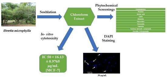
Phytochemical and In Vitro Cytotoxic Screening of Chloroform Extract of Ehretia microphylla Lamk

1
Department of Pharmaceutical Sciences and Drug Research, Punjabi University, Patiala 147002, Punjab, India
2
Khalsa College of Pharmacy, Amritsar 143001, Punjab, India
*
Authors to whom correspondence should be addressed.
Stresses 2022, 2(4), 384-394; https://doi.org/10.3390/stresses2040027
Submission received: 19 August 2022 / Revised: 23 September 2022 / Accepted: 27 September 2022 / Published: 3 October 2022
(This article belongs to the Special Issue Cancer and Stresses)

Abstract

:
Ehretia microphylla of the Boraginaceae family has been extensively used as a folklore remedy for the treatment of a wide range of ailments such as cough, cancer, allergies, and gastrointestinal and venereal disorders. Extensive literature review reports have revealed these findings due to the presence of numerous phytomolecules. To validate traditional claims for cytotoxic activity of E. microphylla, the present study was undertaken. Dried leaves of the plant were powdered and defatted with petroleum ether followed by hot continuous extraction with chloroform. The chloroform extract was subjected to in vitro cytotoxic screening against a panel of human cancer cell lines such as HCT-116 (colon), MCF-7 (breast), PC-3 (prostate), A-549 (lung), HL-60 (leukemia) and MiaPaCa-2 (pancreatic) at 50 µM using SRB assay. The extract exhibited noteworthy cytotoxicity activity against breast and lung cancer. It exhibited 85.55% and 77.93% inhibition against MCF-7 and A-549 cancer cell lines, respectively. The mechanism behind cell death was determined using the DAPI staining method, which induces alteration in nuclear morphology in MCF-7 cell lines evidenced through DAPI staining. Phytochemical screening of E. microphylla extract showed the presence of saponins, steroids, lipids, tannins and triterpenoids. The chemoprofile of the chloroform extract of E. microphylla leaves was established using an n-hexane:ethyl acetate solvent system in a ratio of 6:4. The developed chromatogram showed five spots both in visible and UV light at 254 nm. The information provided in the present study will enable further studies on the isolation and characterization of bioactive compounds/fractions by following bioactivity-guided fractionation, and thus, the plant has the potential to reduce proliferation and may induce cell death via apoptosis in breast cancer cells.

Graphical Abstract

1. Introduction

Cancer is a critical non-communicable public health menace. Cancer is a general term for a large assemblage of diseases that can affect any portion of the body and is characterized by uncontrolled proliferation of abnormal cells. It is the second leading cause of mortality after cardiovascular disorders around the world, with an incidence rate of more than 9.6 million cases per year [1]. All over the world, people suffer from different types of cancers; among these, lung cancer is reported as the most prevalent in males followed by breast cancer in females [2,3,4,5]. Generally, normal body cells grow, proliferate and die in a programmed channel, whereas cancer cells do not die, but they continue to grow and proliferate in a disorderly manner. Research indicates that cancers are caused by a dysfunction of many genes coding for proteins such as anti-apoptotic proteins, transcription factors, growth factor receptors, tumor suppressor genes and cancer-promoting enzymes [6,7]. Cancer management has largely benefited from the discovery of medicinal plants. Phytomolecules and their derivatives are being explored for the prevention or cure of cancer via diverse mechanisms [8,9]. Researchers are not only tirelessly trying to isolate phytochemicals from medicinal plants to conquer this growing threat of cancer but are also exploring the mechanism behind cell death [10,11]. A thorough survey of literature reveals that a large number of phytomolecules have been evaluated for in vitro anticancer activity in various cancer cell lines [12].
Ehretia microphylla Lamk (Boraginaceae), also known as Scorpion bush, is a medium-sized dicot shrub with a height around 1–4 m [13]. The plant is locally known as Wild Tea and Tsaang Gubat by tribal communities of Asia [14,15]. E. microphylla has been documented in the Siddha Materia Medica [16]. It is widely distributed in the Guangdong, Hainan, and Taiwan provinces of South China, as well as in subtropical areas of southern and southeastern Asia. The plant is widely used for its ample range of activities such as antibacterial, anti-viral, and anti-allergic as well as its wound-healing actions. [17,18,19]. Numerous reports documented on E. microphylla reveals this plant as a potential candidate for the treatment of eczema, scabies and pruritus [20], tooth decay [21] cachexia, and food poisoning [22]. It is used in the management of diarrhea, and it is antispasmodic and a disinfectant. [23]. Traditionally, the decoction of the dried leaves has been used for stomach and cough problems; moreover, the fresh leaves are used as a substitute for tea [24]. The plant is also used in the treatment of diabetes, allergies and hyperacidity [25].
Plant secondary metabolites and their derivatives such as vinca alkaloids, taxanes, podophyllotoxins and camptothecin have been clinically employed in combating cancer [3]. The outcome of these studies not only signifies the use of phytomolecules but has also attracted the attention of scientists/researchers to explore the anticancer potential of other traditionally used and medicinally promising plants [26,27].
Keeping in view its efficacy and relative safety, it seems to be a viable approach to investigate this traditional plant to search for new anticancer molecules. Several researchers have established that E. microphylla contains numerous bioactive metabolites such as triterpenes, alkaloids, glycosides, tannins, α-amyrin and β-amyrin, saponins, flavonoids and rosmarinic acid [28,29].

2. Results

2.1. Plant Extract

The yield of E. microphylla extract as well as color and physical appearance are presented in Table 1.

2.2. Phytochemical Screening of Chloroform Extract of E. microphylla

Crude extract of chloroform of E. microphylla was subjected to phytochemical screening to reveal the presence of phytomolecules, viz., carbohydrates, proteins, amino acids, lipids, alkaloids, glycosides, flavonoids, saponins, tannins, steroids, triterpenoids and phenolic compounds, which are depicted in Table 2.

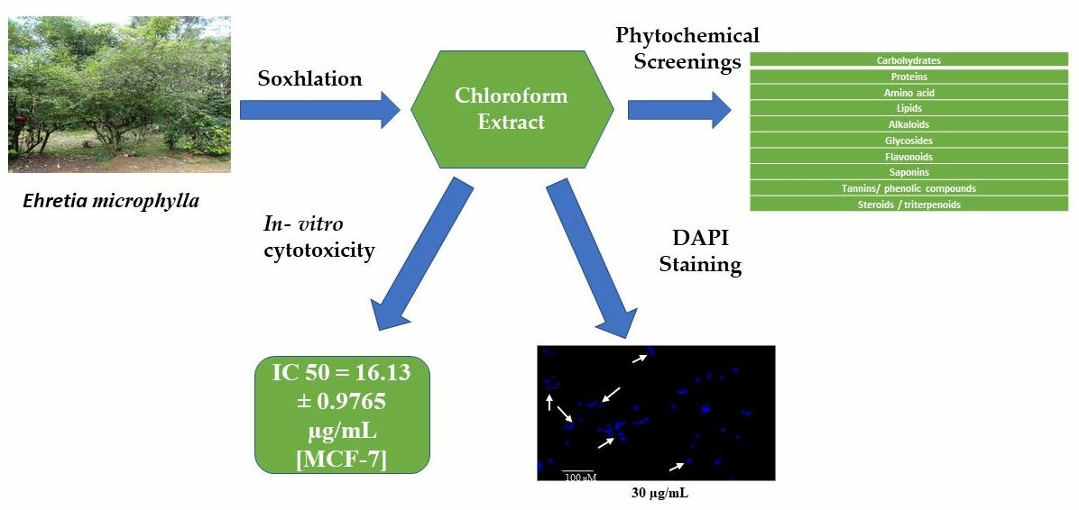
2.3. Thin-Layer Chromatography (TLC) Finger Printing

Thin-layer chromatography is a technique for examining mixtures and determining the number of components present in them. It is also used for identification and to check the purity of a compound. The chemoprofile of chloroform extract of E. microphylla was developed using a solvent system (n-hexane:ethyl acetate: 6:4), and the chromatogram showed five spots. The developed chromatogram depicted in Figure 1 and retention factor (Rf) for each spot was calculated and presented in Table 3.

2.4. Biological Evaluation

2.4.1. In Vitro Cytotoxicity

The dried chloroform extract of E. microphylla (PSEM) was tested for in vitro cytotoxicity against human cancer cell lines HCT-116 (colon), MiaPaCa-2 (pancreatic), PC-3 (prostate) A-549 (lung), and HL-60 (promyelocytic leukemia) malignant cell lines using sulforhodamine B. The results of the preliminary screening of SRB assay are presented in Table 4. Careful observation of the results reveals that the cell lines HCT-116, PC-3, A-549 and MCF-7 were the most sensitive, while HL-60 and MiaPaCa-2 cell lines were almost resistant to the chloroform extract at 50 µg/mL. The chloroform extract produced significant effects against PC-3, HL-60, HCT-116 and MCF-7 cell lines. The chloroform extract elicited 75.45%, 74.50%, 77.93% and 85.55% inhibition against HCT-116, PC-3, A-549 and MCF-7, respectively, which are presented in Table 4.
The chloroform extract displaying more than 70% inhibition was only evaluated against those cell lines at different concentrations (10, 20, 30, 40 and 50 µg/mL), and the IC50 values (Table 5) were calculated. The chloroform extract of E. microphylla was also assessed for selective killing against “FR2” normal human breast cells. The extract did not attain IC50 values until >97 µM, as depicted in Table 5.
Careful observations of Table 5 reveal that the IC50 values of PSEM were found to be 16.13 ± 0.9765 and 26.40 ± 1.2445 µg/mL against MCF-7 and HCT-116 human cancer cell lines, respectively.

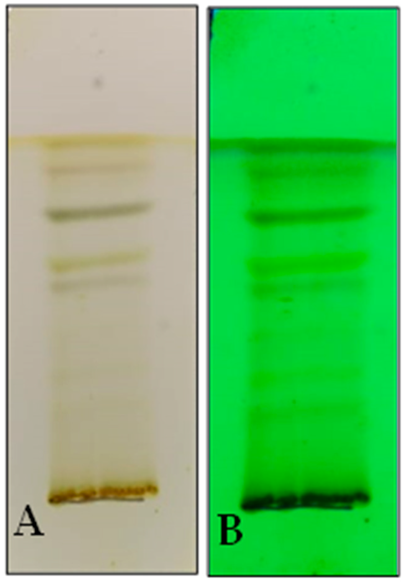
2.4.2. Effect of Chloroform Extract on Nuclear Morphology

The apoptotic effect of chloroform extract of Ehretia microphylla was observed using 4’,6-Diamidino-2-phenylindole (DAPI) staining. Noticeable changes were observed in the nuclear morphology of the MCF cell lines. The formation of apoptotic bodies and condensed nuclei was observed in a concentration-dependent manner, which is indicated in white colored arrows in Figure 2, whereas the nuclei of untreated MCF- 7 cells exhibited normal intact morphology.

3. Discussions

Cancer is one of the foremost threats to mankind worldwide after cardiovascular disorders. Numerous chemotherapeutic agents are clinically available and are approved by the FDA for the mitigation of cancer. Cancer therapy in the form of surgery or radiotherapy is effective when the disease is detected early, but many cancers are still diagnosed after the cells from a primary tumor have metastasized to other parts of the body, and chemotherapy is the main form of treatment at this point [30]. Chemotherapy involves delivering drugs systemically so that they can reach and kill tumor cells, but most of these drugs have severe side effects in patients and must therefore be used at low doses. Researchers claim that developing new drugs that are more selective for cancer cells and have fewer side effects is a major challenge [31].
Integrative medicine, which combines traditional western medicine with alternative or complementary treatments such as herbal medicine, acupuncture, massages, biofeedback, yoga, and stress management techniques, is being used to supplement orthodox medicines and treatment approaches in the management of cancer patients. Despite the availability of chemotherapeutic agents, the scientific community is still facing challenges to seek out novel entities for the treatment and management of different types of cancer because of the agents’ harmful side effects.
Researchers are currently inclined toward the search for natural products because of the presence of abundant therapeutically accepted phytomolecules such as flavonoids, tannins, amino acids, alkaloids, sugars, tannins and polyphenolic compounds, despite their ethno-pharmacological significance in the mitigation of jaundice, inflammation, asthma, cancer, diarrhea, wound healing and skin diseases by tribes of Africa, Australia and Asia [15,32].
Kudera et al. established in vitro antiproliferative and antibacterial actions of ethanolic extract of E. microphylla against intestinal human cancer cell lines such as Caco-2 and HT-29 with IC50 values of 37.89 ± 2.68 and 130.89 ± 13.99 μg/mL, respectively, of which this plant was used earlier as folk medicine in the mitigation of diarrhea, dysentery and abdominal pain. [33,34]. Similarly, Shukla et al. reported antioxidant, antibacterial and antiallergic effects of ethanolic leave extract of E. microphylla [35]. Using another approach, Simpol et al. documented that the leaves of the plant Scorpion Bush were traditionally used for numerous medicinal purposes, viz., as an antispasmodic, body cleanser and mouth wash, which are attributed due to the presence of phenolic acids, benzoquinone, fatty acids and flavonoids [36]. Henry et al. also reported some bioactive metabolites from E. mircophylla such as ursolic acid, baurneol, microphyllone, hydroxymicrophyllone, and dehydromicrophyllone and recognized them for the cure of scabies, itching, stomach ache, psoriasis and skin allergies [37].
Charisse et al. documented an ample range of pharmacological actions from the different parts of E. microphylla, including the aerial part, leaves, root, stem and fruits. The plant is recognized to contain a variety of phytoconstituents such as rosmarinic acid, bauerenol, α-amyrin, β-amyrin, kaempferol-3-O-rutinoside and kaempferol-3-O-glucoside from methanol extract of the leaves of E. microphylla. Methanol extract of the plant contains ehretianone, stigmasterol, β-sitosterol and α-spinasterol. E. microphylla exhibited numerous activities, viz., anticancer, antioxidant, anti-inflammatory, wound healing, antimicrobial, anti-allergy and anti-angiogenesis. Methanolic extract of leaves of the plant induces apoptosis and DNA fragmentation [38,39]. With this background, several studies of this plant motivated us to explore the anticancer potential for chloroform extract of the leaves of E. microphylla, as this plant has not yet been explored as an anticancer agent, keeping in view that E. microphylla is documented to have numerous phytomolecules that are responsible for its use in the mitigation of cancer. In the present investigation, the phytochemical screening and cytotoxic evaluation of chloroform extract of E. microphylla have been established. The chloroform extract of E. microphylla exhibits significant anticancer activity and is attributed to the presence of triterpenoid, saponins and phenolic compounds. The available literature also reveals that triterpenoids and phenolic compounds are the most bioactive compounds that are responsible for anticancer potential of most traditionally used plants such as α-amyrins and β-amyrins and ursolic acid, which belong to the triterpenoid class, whereas bergenin and gallic acid are phenolic compounds that exhibit their significant anticancer activities against several human cancer cell lines [14,40,41,42,43,44,45].
Observations of the present study are in agreement with aforementioned reports, as phenolic compounds and triterpenoids have been observed as the main compounds in the phytochemical screening of chloroform extract of E. microphylla leaves. The thin-layer chromatographic pattern of chloroform extract of E. microphylla leaves reveals the presence of five spots that have established the chemoprofile of chloroform extract of E. microphylla. Identification of separated compounds can be achieved on the basis of their retention factor (Rf) values [46].
The cytotoxicity results reveal that HCT-116, PC-3, A-549 and MCF-7 were the most sensitive, while HL-60 and MiaPaCa-2 cell lines were almost resistant to chloroform extract at 50 µg/mL. The chloroform extract displaying more than 70% inhibition was only evaluated against those cell lines at different concentrations and to determine the IC50 values. The chloroform extract of E. microphylla exhibits selective cytotoxicity toward human breast cancer cell lines and has been found to be non-toxic against “FR2” normal human breast epithelial cells. Furthermore, the IC50 value of the extract was found to be high in normal human breast epithelial cells (FR2), which validates its effectiveness as a potential candidate for the mitigation of cancer. Careful observation of Table 5 reveals that the IC50 value of chloroform extract of E. microphylla is 16.25 µg/mL against MCF-7 cell lines.
Apoptosis is the hallmark of cancer and plays a critical role in the amelioration of breast cancer. The mechanism behind cell death was determined using the DAPI staining method. DAPI is a fluorescent stain that binds to DNA at furrows of the A-T region. The results revealed that the chloroform extract of E. microphylla induces apoptosis in a concentration-dependent manner. Thus, these classes of phytomolecules may be responsible for anticancer activity. However, further work is required to elucidate the mechanism behind cell death in MCF-7 cell lines, and thus, E. microphylla can be a potential lead and therapeutic candidate for next level of evaluation for the mitigation of cancer.

4. Materials and Methods

4.1. Plant Materials

The dried leaves of E. microphylla were procured from the Department of Botany, Sri Venkateshwara University, Tirupati. The authentication of E. microphylla (0850) was confirmed by Assistant Professor Dr. K. Madhava Chetty from the department of Botany of S.V. University, Tirupati, Andhra Pradesh. Specimen voucher 0850 was submitted to the herbarium of the same department.

4.2. Chemicals and Reagents

The chemicals, solvents, and reagents such as chloroform, hexane, ethanol and ethylacetate were purchased from Loba chem., CDH, and Spectro chem India. RPMI-1640, fetal calf serum, minimum essential medium (MEM), trypan blue, trypsin, streptomycin, gentamycin, penicillin, dimethyl sulfoxide, (DMSO), mitomycin-C, sulforhodamine, and adriamycin were from Sigma Aldrich, Chemical Co., Burlington, MA, USA. Distilled water, Tris buffer, Tris-EDTA buffer, (Hi-Media) acetic acid, sodium bicarbonate, hydrochloric acid were from spectrochem. All other chemicals used in this study were of analytical grade and were purchased from local dealers.

4.3. Preparation of the Extract

The shade-dried leaves of E. microphylla were ground using mixer to form coarse powder. The dried coarsely powdered plant (1 Kg) was defatted by Soxhlation apparatus using petroleum ether 60–80 °C (5 L). The defatted material was then extracted using chloroform (5 L) by soxhlation process. The exhaustive extraction was carried out at a temperature of 80°C. The extraction was continued until few drops of solvent collected during siphoning did not leave any residue after evaporation on watch glass. Lastly, chloroform extract was dried under vacuum [32]. The dried extract was further subjected to phytochemical screening and cytotoxicity studies.

4.4. Phytochemical Screening of Extract of E. microphylla

The chloroform extract was subjected to phytochemical screening to detect the presence of phytomolecules such as carbohydrates, proteins, amino acids, alkaloids, glycosides, flavonoids, saponins, tannins, steroids, triterpenoids and phenolic compounds as per the standard protocol mentioned in the literature [47].

4.5. Thin-Layer Chromatography (TLC) of Extracts

The chloroform extract of E. microphylla leaves was subjected to thin-layer chromatography to develop a chromatographic pattern to analyze the number of constituents present in it. The chemoprofile of chloroform extract of E. microphylla was developed on pre-coated silica gel plates (Merck) using solvent system n-hexane:ethyl acetate: 6:4, with detection by visible light and UV light at 254 nm [46].

4.6. Cell Culture, Growth Conditions, and Treatment

HCT-116 (colon), MiaPaCa-2 (pancreatic), PC-3 (prostate) A-549 (lung), HL-60 (leukemia), MCF-7 (breast) human cancer cells and fR2 (normal human breast epithelial) lines used in this study were obtained from the National Cancer Institute (NCI), Frederick, Burlington, MA, USA. All human cancer cells were grown and maintained in RPMI1640/MEM/DMEM medium supplemented with 10% heat-inactivated fetal bovine serum (FBS), L-glutamine (0.3 mg/mL), penicillin (100 units/mL), streptomycin (100 µg/mL), 0.37% NaHCO3 and pyruvic acid (0.11 mg/mL). Cells were grown in CO2 incubator (Thermocon Electron Corporation, Waltham, MA) at 37° C temperature, 90% humidity and 5% CO2 gas environment, and subjected to treatment with chloroform extract of E. microphylla dissolved in DMSO, while untreated control received only DMSO (<0.2%) as a vehicle.

4.7. Biological Evaluation

4.7.1. In Vitro Cytotoxic Assay

In vitro cytotoxicity of chloroform extract of E. microphylla was determined using semi-automated sulforhodamine-B (SRB) assay against a panel of human cancer cell lines, viz., HCT-116 (colon), MiaPaCa-2 (pancreatic), PC-3 (prostate), A-549 (lung), HL-60 (leukemia), MCF-7 (breast) and FR-2 (normal human breast epithelial) malignant cells (Monks et al., 1991). Cancer cells/mL of suspension were counted and seeded in 96-well plates at a density of 10,000 cells per well/100 µL. Seeded plates were incubated at 37 °C in an environment of 90% relative humidity and 5% CO2 for 24 h. Stock solutions of chloroform extract (20 mg/mL) were prepared in DMSO, and 50 µL concentrations were prepared using serial dilution method. Then, 50 µL of test material in 100 µL of complete growth medium was added and allowed to incubate for 48 h at 37 °C temperature, 90% relative humidity and 5% CO2 environment. After that, 50 µL of 50% chilled TCA (trichloroacetic acid) was added to each well and kept for 1 h to immobilize the cells at 4 °C. After that, plates were washed five to six times with water, air dried and stained with sulforhodamine-B dye (100 µL, 0.4% w/v in 1% acetic acid) for 30 min. Plates were again washed with 1% (v/v) acetic acid to remove the unbound dye. Plates were air dried, and absorbed dye was dissolved by adding 100 µL of 10 mM Tris buffer (pH 10.5). Thereafter, plates were placed on plate shaker for 10 min. Wells with no treatments were considered as control wells. The optical density (OD) was measured using an ELISA reader at 540 nm. Cell growth was obtained by subtracting the mean OD (optical density) values of blanks and mean OD values of the treatments. Percent growth in the presence of extracts was calculated considering the growth devoid of any test substance (chloroform extract) as 100%, and in turn, % age growth inhibition in the presence of test substance was determined. Extracts showing greater than 70% inhibition were selected to determine the dose–response curve (IC50) [48,49,50,51,52,53,54].

4.7.2. Effect of Chloroform Extract of E. microphylla on Nuclear Morphology

Human breast cancer cells (MCF-7) were seeded at a density of 1 × 10⁴ cells/well in a 12-well plate. The cells were treated with or without different concentrations of chloroform extract of E. microphylla for 24 h and incubated at 37 °C temperature, 95% air and 5% CO2 atmosphere for 6 h. The treated and untreated cells were collected and washed with ice-cold PBS and fixed with methanol: acetic acid (3:1) for 30 min at room temperature. The cells were stained using DAPI (1 µg/mL) for 20 min and analyzed under fluorescent microscope. (Nuclear morphology was examined using DAPI staining as described earlier.) The presented images were magnified at ×20 and scale bars, 100 µm [53].

5. Conclusions

The results of the present investigation revealed that chloroform extract of E. microphylla holds great potential. The induction of apoptosis leading to cell death was determined using DAPI staining. The results of the DAPI staining revealed that chloroform extract of E. microphylla exhibited apoptotic body formation in a dose-dependent manner. The IC50 values of PSEM were determined against selected human cancer cell lines. It is inferred from the results that chloroform extract of E. microphylla is highly effective against MCF-7 and HCT-116 cell lines, as evident from their lower IC50 values against MCF-7 and HCT-116 human cancer cell lines versus other cell lines. Moreover, it was found to be non-toxic against normal human breast epithelial cells, and it could be developed as a source of a novel anticancer molecule. The anticancer activity of the plant may be attributed to alkaloids, triterpenoids and phenolic compounds, as apparent from preliminary phytochemical screening. The research work is in progress to isolate and characterize bioactive chemical constituent(s)/bioactive fraction.

Author Contributions

Conceptualization, S.K.; methodology, P.S.; software, data curation, P.S. and S.K.; writing—original draft preparation, P.S.; writing—review and editing, P.S. and S.K.; visualization, R.S. and S.K.; supervision, S.K. and R.S.; project administration, S.K. and P.S. All authors have read and agreed to the published version of the manuscript.

Funding

This research received no external funding.

Data Availability Statement

All the data have been presented in the manuscript.

Acknowledgments

The authors are thankful to the Director of the Indian Institute of Integrative Medicine, Canal Road, Jammu, for providing the necessary research facilities. The authors are also grateful to the Vice-Chancellor of Punjabi University, Patiala, India, for his encouragement and to the Head of Department, Department of Pharmaceutical Sciences and Drug Research, Punjabi University, Patiala, for the providing necessary research facilities.

Conflicts of Interest

The authors declare no conflict of interest.

References

  1. Bray, F.; Ferlay, J.; Soerjomataram, I.; Siegel, R.L.; Torre, L.A.; Jemal, A. Global Cancer Statistics 2018: GLOBOCAN estimates of incidence and mortality worldwide for 36 cancers in 185 countries. CA Cancer J. Clin. 2018, 68, 394–424. [Google Scholar] [CrossRef] [Green Version]
  2. Horn, L.; Pao, W.; Johnson, D.H. Neoplasms of the lung, Chapter 89. In Harrisons Principles of Internal Medicine, 18th ed.; Longo, D.L., Kasper, D.L., Jamson, J.L., Fauci, A.S., Hauser, S.L., Loscalzo, J., Eds.; MacGraw-Hill: New York, NY, USA, 2012; ISBN 978-0-07-174889-6. [Google Scholar]
  3. Kumar, D.; Sharma, P.; Singh, H.; Nepali, K.; Gupta, G.K.; Jain, S.K.; Ntie-Kang, F. The value of pyrans as anticancer scaffolds in medicinal chemistry. RSC Adv. 2017, 7, 36977. [Google Scholar] [CrossRef] [Green Version]
  4. Zhou, Z.; Tang, M.; Liu, Y.; Zhang, Z.; Lu, R.; Lu, J. Apigenin inhibits cell proliferation, migration, and invasion by targeting Akt (Protein kinase B) in the A549 human lung cancer cell line. Anticancer Drugs 2017, 28, 446–456. [Google Scholar] [CrossRef] [PubMed]
  5. Kumar, D.; Jain, S.K. A Comprehensive Review of N-Heterocycles as Cytotoxic Agents. Curr. Med. Chem. 2016, 23, 4338–4394. [Google Scholar] [CrossRef]
  6. Singh, S.; Jarial, R.; Kanwar, S.S. Therapeutic effect of herbal medicines on obesity, herbal pancreatic lipase inhibitors. Wudpecker J. Med. Plants. 2013, 2, 53–65. [Google Scholar]
  7. Kumar, D.; Singh, O.; Nepali, K.; Bedi, P.; Qayum, A.; Singh, S.; Jain, S.K. Naphthoflavones as Antiproliferative Agents: Design, Synthesis and Biological Evaluation. Anticancer Agents Med. Chem. 2016, 16, 881–890. [Google Scholar] [CrossRef]
  8. Sharma, P.; Sharma, R.; Rao, H.S.; Kumar, D. Phytochemistry and Medicinal Attributes of A. Scholaris: A Review. Int. J. Pharm. Sci. Res. 2015, 6, 505–513. [Google Scholar]
  9. Kumar, D.; Malik, F.; Bedi, P.M.S.; Jain, S. 2,4-diarylpyrano[3,2-c]chromen-5(4H)-ones as coumarin-chalcone conjugates: Design, synthesis and biological evaluation as apoptosis inducing agents. Chem. Pharm. Bull. 2016, 64, 399–409. [Google Scholar] [CrossRef] [PubMed] [Green Version]
  10. Kiselova, Y.; Ivanova, D.; Chervenkov, T.; Gerova, D.; Galunska, B.; Yankova, T. Correlation between the in vitro antioxidant activity and polyphenol content of aqueous extracts from Bulgarian herbs. Phytother. Res. 2006, 20, 961–965. [Google Scholar] [CrossRef]
  11. Kumar, D.; Nepali, K.; Bedi, P.M.S.; Kumar, S.; Malik, F.; Jain, S. 4,6-diaryl Pyrimidones as Constrained Chalcone Analogues: Design, Synthesis and Evaluation as Antiproliferative Agents. Anticancer Agents Med. Chem. 2015, 15, 793–803. [Google Scholar] [CrossRef] [PubMed]
  12. Singla, R.K.; Sharma, P.; Dubey, A.K.; Gundamaraju, R.; Kumar, D.; Kumar, S.; Madaan, R.; Shri, R.; Tsagkaris, C.; Parisi, S.; et al. Natural Product-Based Studies for the Management of Castration-Resistant Prostate Cancer: Computational to Clinical Studies. Front. Pharmacol. 2021, 12, 732266. [Google Scholar] [CrossRef] [PubMed]
  13. Cagampan, D.M.; Lacuata, K.A.; Ples, M.B.; Vitor, R.J.S. Effect of Ehretia microphylla on the blood cholesterol and weight of ICR mice (Mus musculus). Natl. J. Physiol. Pharm. Pharmacol. 2018, 8, 983–987. [Google Scholar] [CrossRef]
  14. Sharma, P.; Shri, R.; Ntie-Kang, F.; Kumar, S. Phytochemical and Ethnopharmacological Perspectives of Ehretia laevis. Molecules 2021, 26, 3489. [Google Scholar] [CrossRef]
  15. Demetrio, L.V.J.; Jeannie, I.A.; Juliana, J.M.P.; Esperanza, C.C.; Windell, L.R. Antibacterial activities of ethanol extracts of Philippine medicinal plants against multidrug-resistant bacteria. Asian Pac. J. Trop. Biomed. 2015, 5, 532–540. [Google Scholar]
  16. Murugesa, M.K.S. Gunapadam. Part-I; Tamil Nadu Siddha Medical Board: Chennai, India, 1956; p. 274. [Google Scholar]
  17. Kaur, R.; Sharma, P.; Gupta, G.K.; Ntie-Kang, F.; Kumar, D. Structure Activity Relationship and Mechanistic Insights for Anti-HIV Natural Products. Molecules 2020, 25, 2070. [Google Scholar] [CrossRef]
  18. Kumar, D.; Sharma, P.; Kaur, R.; Lobe, M.M.; Gupta, G.K.; Ntie-Kang, F. In search of therapeutic candidates for HIV/AIDS: Rational approaches, design strategies, structure–activity relationship and mechanistic insights. RSC Adv. 2021, 11, 17936–17964. [Google Scholar] [CrossRef]
  19. Roeder, E.; Wiedenfeld, H. Plants containing pyrrolizidine alkaloids used in the traditional Indian medicine—Including ayurveda. Pharmazie 2013, 68, 83–92. [Google Scholar]
  20. Vista, F.E.S.; Dalmacio, L.M.M.; Corales, L.G.M.; Salem, G.M.; Galula, J.U.; Chao, D.Y. Antiviral Effect of Crude Aqueous Extracts from Ten Philippine Medicinal Plants against Zika virus. Acta Med. Phillipina 2022, 54, 195–202. [Google Scholar] [CrossRef]
  21. Bhatia, P.; Sharma, A.; George, A.J.; Anvitha, D.; Kumar, P.; Dwivedi, V.P.; Chandra, N.S. Antibacterial activity of medicinal plants against ESKAPE: An update. Heliyon 2021, 7, e06310. [Google Scholar] [CrossRef]
  22. Zheng, X.L.; Wei, J.H.; Sun, W.; Li, R.T.; Liu, S.B.; Dai, H.F. Ethnobotanical study on medicinal plants around Limu Mountains of Hainan Island, China. J. Ethnopharmacol. 2013, 148, 964–974. [Google Scholar] [CrossRef]
  23. Tsaang Gubat or Wild Tea (Ehretia microphylla Lam.). Available online: http://www.philippineherbalmedicine.org/tsaang_gubat.htm (accessed on 14 August 2022).
  24. Anonymous. The Wealth of India; CSIR: New Delhi, India, 1952; Volume 3, p. 129. [Google Scholar]
  25. Dapar, M.L.G.; Meve, U.; Liede-Schumann, S.; Alejandro, G.J.D. Ethnomedicinal appraisal and conservation status of medicinal plants among the Manobo tribe of Bayugan City, Philippines. Biodiversitas 2020, 21, 3843–3855. [Google Scholar] [CrossRef]
  26. Wall, M.E. Camptothecin and taxol: Discovery to clinic. Med. Res. Rev. 1998, 18, 299–314. [Google Scholar] [CrossRef]
  27. Kaur, T.; Sharma, P.; Gupta, G.; Ntie-Kang, F.; Kumar, D. Treatment of tuberculosis by natural drugs: A review. Plant Arch. 2019, 19, 2168–2176. [Google Scholar]
  28. Rimando, A.M.; Inoshiri, S.; Otsuka, H.; Kohda, H.; Yamasaki, K.; Padolina, W.G.; Torres, L.; Quintana, E.G.; Cantoria, M.C. Screening for mast cell histamine release inhibitory activity of Philippine medicinal plants. Active constituent of Ehretia microphylla. Jpn. J. Pharmacog. 1987, 41, 242–247. [Google Scholar]
  29. Cordero, M.; Leon-Gonzalez, A.J.; Montano, J.M.C.; Moron, E.B.; Lazaro, L.M. Pro-oxidant natural products as anticancer agents. Curr. Drug Targets. 2012, 13, 1006–1028. [Google Scholar] [CrossRef]
  30. Yokoyama, C.; Sueyoshi, Y.; Ema, M.; Mori, Y.; Takaishi, K.; Hisatomi, H. Induction of oxidative stress by anticancer drugs in the presence and absence of cells. Oncol. Lett. 2017, 14, 6066–6070. [Google Scholar] [CrossRef]
  31. Chopra, R.N.; Nayar, S.L.; Chopra, I.C. Glossary of Indian Medicinal Plants; Council of Scientific and Industrial Research: New Delhi, India, 1986; p. 104. [Google Scholar]
  32. Safe, S.H.; Prather, P.L.; Brents, L.K.; Chadalapaka, G.; Jutooru, I. Unifying mechanisms of action of the anticancer activities of triterpenoids and synthetic analogs. Anticancer Agents Med. Chem. 2012, 12, 1211–1220. [Google Scholar] [CrossRef]
  33. Kudera, T.; Fiserova, B.; Korytakova, M.; Doskocil, I.; Salmonova, H.; Tulin, E.E.; Nguon, S.; Bande, M.M.; Kokoska, L. In vitro Selective Antibacterial and Antiproliferative Effects of Ethanolic Extracts from Cambodian and Philippine Plants Used in Folk Medicine for Diarrhea Treatment. Front. Pharmacol. 2021, 12, 746808. [Google Scholar] [CrossRef]
  34. Stuart, G.U. List of Philippine Herbal Medicinal Plants. Philippine Medicinal Plants. 2017. StuartXchange-SX. Available online: http://www.stuartxchange.org/CompleteList.html (accessed on 14 August 2022).
  35. Shukla, A.; Kaur, A. A systematic review of traditional uses bioactive phytoconstituents of genus Ehretia. Asian J. Pharmaceut. Clin. Res. 2018, 11, 6–17. [Google Scholar] [CrossRef]
  36. Simpol, L.R.; Otsuka, H.; Ohtani, K. Nitrile glucosides and rosmarinic acid, the histamine inhibitor from Ehretia philippinensis. Phytochemistry 1994, 36, 91–95. [Google Scholar] [CrossRef]
  37. Henry, I.A.B.; Alfred, J.H.R.; Kimbberly, A.S.; Allister, M.T.T.; Alicia, I.Y.; Tooba, M.; Jitbanjong, T.; Veeranoot, N. Recommended Medicinal Plants as Source of Natural Products: A Review. Digital Chin. Med. 2018, 1, 131–142. [Google Scholar]
  38. Legaspi, C.L.B.; Bagaoisan, D.M.A. Ehretia microphylla Tablet Formulation for Biliary and Gastrointestinal Colic: A Review of its Phytochemical Constituents, Pharmacologic Activities and Clinical Researches. Acta Med. Phillipina 2020, 54, 80–85. [Google Scholar] [CrossRef]
  39. Nayak, J.; Basak, U.C. Antioxidant potential of some lesser known wild edible fruits of Odisha. Eur. J. Exp. Biol. 2015, 5, 60–70. [Google Scholar]
  40. Ghante, M.H.; Jamkhande, P.G. Role of Pentacyclic Triterpenoids in Chemoprevention and Anticancer Treatment: An Overview on Targets and Underling Mechanisms. J. Pharmacopunct. 2019, 22, 55–67. [Google Scholar] [CrossRef] [PubMed]
  41. Abdal Dayem, A.; Choi, H.Y.; Yang, G.M.; Kim, K.; Saha, S.K.; Cho, S.G. The Anti-Cancer Effect of Polyphenols against Breast Cancer and Cancer Stem Cells: Molecular Mechanisms. Nutrients 2016, 8, 581. [Google Scholar] [CrossRef]
  42. Niedzwiecki, A.; Roomi, M.W.; Kalinovsky, T.; Rath, M. Anticancer Efficacy of Polyphenols and Their Combinations. Nutrients 2016, 8, 552. [Google Scholar] [CrossRef] [Green Version]
  43. Mukharjee, P.K. Quality Control of Herbal Drug, 1st ed.; Business Horizon Publication: New Delhi, India, 2002; pp. 186–193. [Google Scholar]
  44. Sharma, P.; Kumar, D.; Shri, R.; Kumar, S. Mechanistic insights and docking studies of phytomolecules as potential candidates in the management of cancer. Curr. Pharma. Design. 2022; advance online publication. [Google Scholar] [CrossRef]
  45. Madaan, R.; Kumar, D.; Singla, R.K.; Kumar, S.; Sharma, P.; Dubey, A.K.; Shen, B. Mechanistic Insight of Pharmacological and nanotechnological perspectives of Bergenin. Curr. Top. Med. Chem. 2022, 22, 132–149. [Google Scholar] [CrossRef]
  46. WHO. Quality Control Methods for Medicinal Plant Materials; World Health Organization: Geneva, Switzerland, 2010.
  47. Khandelwal, K.R. Preliminary Phytochemical Screening in Practical Pharmacognosy; Nirali Parkashan: Pune, India, 2004; pp. 1156–1449. [Google Scholar]
  48. Keepers, Y.P. Comparison of the Sulforhodamine B Protein and Tetrazolium (MTT) Assays for in vitro Chemosensitivity Testing. Eur. J. Cancer 1991, 27, 897–900. [Google Scholar] [CrossRef] [Green Version]
  49. Skehan, P.; Storeng, R. New Colorimetric Cytotoxicity Assay for Anticancer-Drug Screening. J. Nat. Cancer Instit. 1990, 82, 1107–1112. [Google Scholar] [CrossRef] [PubMed]
  50. Akindele, A.J.; Wani, Z.A.; Sharma, S.; Mahajan, G.; Satti, N.K.; Adeyemi, O.O.; Mondhe, D.M.; Saxena, A.K. In vitro and In vivo Anticancer Activity of Root Extracts of Sansevieria liberica Gerome and Labroy (Agavaceae). Evid. Based Complement Alternat. Med. 2015, 15, 560404. [Google Scholar]
  51. Kumar, A.; Qayum, A.; Sharma, P.R.; Singh, S.K.; Shah, B.A. Synthesis of β-boswellic acid derivatives as cytotoxic and apoptotic agents. Bioorg. Med. Chem. Lett. 2016, 26, 76–81. [Google Scholar] [CrossRef] [PubMed]
  52. Kumar, D.; Sharma, P.; Nepali, K.; Mahajan, G.; Mintoo, M.J.; Singh, A.; Singh, G.; Mondhe, D.M.; Singh, G.; Jain, S.K.; et al. Antitumour, acute toxicity and molecular modeling studies of 4-(pyridin-4-yl)-6-(thiophen-2-yl)pyrimidin-2(1H)-one against Ehrlich ascites carcinoma and sarcoma-180. Heliyon 2018, 4, e00661. [Google Scholar] [CrossRef]
  53. Kumar, D.; Singh, G.; Sharma, P.; Qayum, A.; Mahajan, G.; Mintoo, M.J.; Singh, S.K.; Mondhe, D.M.; Bedi, P.; Jain, S.K.; et al. 4-aryl/heteroaryl-4H-fused Pyrans as Anti-proliferative Agents: Design, Synthesis and Biological Evaluation. Anticancer Agents Med. Chem. 2018, 18, 57–73. [Google Scholar] [CrossRef]
  54. Mihaljevic, A.L.; Michalski, C.W.; Friess, H.; Kleeff, J. Molecular mechanism of pancreatic cancer-Understanding proliferation, invasion, and metastasis. Langenbeck’s Arch. Surg. 2010, 395, 295–308. [Google Scholar] [CrossRef]
Figure 1. Thin-layer chromatogram of chloroform extract of Ehretia microphylla leaves. (A) TLC chromatogram observed in visible light; (B) TLC chromatogram observed in UV light at 254 nm in UV chamber.
Stresses 02 00027 g001
Figure 2. DAPI staining in MCF-7 cell lines. Variation in nuclear morphology induced with PSEM in a concentration-dependent manner, observed under fluorescence microscope. PSEM induced the formation of apoptotic bodies, as shown by arrows. The photographs depicted here are magnified at ×20, and scale bars, 100 µm.
Stresses 02 00027 g002
Table 1. Yield of extracts of E. microphylla.
Sr. No.Extract (g/Kg)Percentage YieldColorPhysical Appearance
1Chloroform (54.347)5.43GreenSticky mass
Table 2. Results of phytochemical screening of chloroform extract of E. microphylla.
Sr. No.Class of PhytoconstituentsTestsChloroform
1CarbohydratesMolisch’s test
2ProteinsBiuret test
3Amino acidNinhydrin test
4LipidsSolubility test+
5AlkaloidsMayer’s test
6GlycosidesLegal test
7FlavonoidsShinoda test
8SaponinsFroth test+
9Tannins and phenolic compoundsFerric chloride test+
10Steroids and triterpenoidsLiebermann–Burchard test+
(+) present; (−) absent.
Table 3. Results of TLC fingerprinting of chloroform extract of E. microphylla leaves.
S. No.ExtractSolvent SystemNo. of SpotsRf Value
1Chloroform extractHexane: ethyl acetate50.57, 0.65, 0.78, 0.87, 0.92
Table 4. Percentage inhibition of cancer cells by different extracts of E. microphylla at 50 µg/mL.
EntryHCT-116 (Colon Cancer)MiaPaCa-2
(Pancreatic Cancer)
PC-3
(Prostate Cancer)
A-549
(Lung Cancer)
HL-60
(Leukemia Cancer)
MCF-7
(Breast Cancer)
PSEM75.45 ± 0.039919.36 ± 0.042274.50 ± 0.047377.93 ± 0.045134.67 ± 0.041085.55 ± 0.0389
Results are represented as mean ± SD (n = 3).
Table 5. IC50 values of chloroform extract against different human cancer and normal cells.
EntryHCT-116 (Colon Cancer)MiaPaCa-2
(Pancreatic Cancer)
PC-3
(Prostate Cancer)
A-549
(Lung Cancer)
HL-60
(Leukemia Cancer)
MCF-7
(Breast Cancer)
FR2
(Normal Human Breast Epithelial)
PSEM26.40 ± 1.2445ND21.56 ± 1.547329.47 ± 1.6532ND16.13 ± 0.9765˃97
Adriamycin------1.75 ± 0.143------------------1.10 ± 0.437ND
Results are represented as mean ± SD (n = 3); ND, not determined.
Publisher’s Note: MDPI stays neutral with regard to jurisdictional claims in published maps and institutional affiliations.

