Review

Stem Cell Scaffolds for the Treatment of Spinal Cord Injury—A Review

1
College of Medicine, University of Florida, Gainesville, FL 32611, USA
2
Department of Neurosurgery, University of Florida, Gainesville, FL 32611, USA
*
Author to whom correspondence should be addressed.
Biomechanics 2023, 3(3), 322-342; https://doi.org/10.3390/biomechanics3030028
Submission received: 11 May 2023 / Revised: 6 July 2023 / Accepted: 14 July 2023 / Published: 1 August 2023
(This article belongs to the Section Neuromechanics)

Abstract

:
Spinal cord injury (SCI) is a profoundly debilitating yet common central nervous system condition resulting in significant morbidity and mortality rates. Major causes of SCI encompass traumatic incidences such as motor vehicle accidents, falls, and sports injuries. Present treatment strategies for SCI aim to improve and enhance neurologic functionality. The ability for neural stem cells (NSCs) to differentiate into diverse neural and glial cell precursors has stimulated the investigation of stem cell scaffolds as potential therapeutics for SCI. Various scaffolding modalities including composite materials, natural polymers, synthetic polymers, and hydrogels have been explored. However, most trials remain largely in the preclinical stage, emphasizing the need to further develop and refine these treatment strategies before clinical implementation. In this review, we delve into the physiological processes that underpin NSC differentiation, including substrates and signaling pathways required for axonal regrowth post-injury, and provide an overview of current and emerging stem cell scaffolding platforms for SCI.

1. Introduction

Spinal cord injury (SCI) is an incredibly devastating central nervous system condition resulting in significant morbidity and mortality rates worldwide. Recent estimates of traumatic SCI include about 26.5 cases per 1,000,000 citizens with greater predominance in males [1,2]. Approximately 50% of SCIs are cervical and result in greater mortality rates, especially in older adults [1]. Common causes of SCI in the United States include motor vehicle accidents, sports injuries, and traumatic falls [2]. SCI pathogenesis is hypothesized to occur in two stages—primary and secondary [3]. Primary SCI refers to the initial mechanical injury while secondary SCI involves acute and chronic cascades of increased immune activation, neuroinflammation, and excitotoxicity [3,4]. Specific mechanisms of secondary SCI include lipid peroxidation, axon degeneration and demyelination, increased calcium influx, free radical formation, and pathological remodeling of the surrounding extracellular matrix [3]. Secondary SCI is presumed to predict and influence overall SCI severity, highlighting its potential role as a target for intervention [4]. However, continued efforts are needed to characterize the role of inflammation in SCI and whether certain cell types and molecules are beneficial or detrimental to recovery [4].
Neural regeneration following axonal injury is a complex process involving multiple proteins, signaling molecules, and genes [5]. Axonal regeneration begins with rapid sealing of the plasma membrane, followed by axonal growth cone formation and stabilization [6,7]. Regrowth is further mediated by several neurotrophic factors such as brain-derived neurotrophic factor (BDNF), neurotrophin-3 (NT3), and nerve growth factor (NGF), all of which act on tyrosine kinase receptors [8]. Intraoperative electrical stimulation (ES) additionally has the ability to promote axonal regrowth, though the biological mechanisms remain relatively unknown.
Despite the fact that neurons in the central nervous system regenerate poorly in response to trauma, a major goal in the treatment of SCI is restoration of neurologic functioning. The current standard of care treatment for acute SCI includes pharmacologic agents such as paracetamol, weak opioids, or non-steroidal anti-inflammatory drugs [9]. In some cases, surgical intervention such as a spinal decompression may be necessary [9]. However, there are contradictory findings in the literature regarding the effects of surgical timing on SCI outcomes. A meta-analysis by Hsieh et al. demonstrated that early decompression within 8–12 h of SCI was associated with improvement of at least one AIS grade, regardless of spinal location or completeness of injury [10]. Similarly, another study involving 1548 patients showed that surgery within 24 h was associated with improved recovery [11]. However, findings by Aarabi et al. revealed the timing of surgery does not influence 6 month post-injury AIS conversion in cervical SCI patients [12]. As such, clinical management of SCI differs with respect to individual patient differences and medical center protocols.
To improve patient outcomes in a highly individualized manner, novel treatment strategies for SCI involving use of stem cell scaffolds have been explored [9]. Stem cell scaffolding involves the creation of a three-dimensional structure designed to imitate the extracellular matrix surrounding cells in the spinal cord [13,14]. By providing stem cells with a highly biocompatible environment suitable for stem cell differentiation, adhesion, and proliferation, stem cell scaffolds can theoretically be used to treat SCI [15]. A variety of scaffolding modalities have been explored including hydrogels, natural polymers, synthetic polymers, and composites [14,16]. However, many stem cell scaffolding models for SCI remain largely in the preclinical stage, highlighting the need to investigate this potential therapeutic further. As such, this review aims to examine current SCI stem cell scaffolding models and their potential applications for clinical adoption in SCI patients.

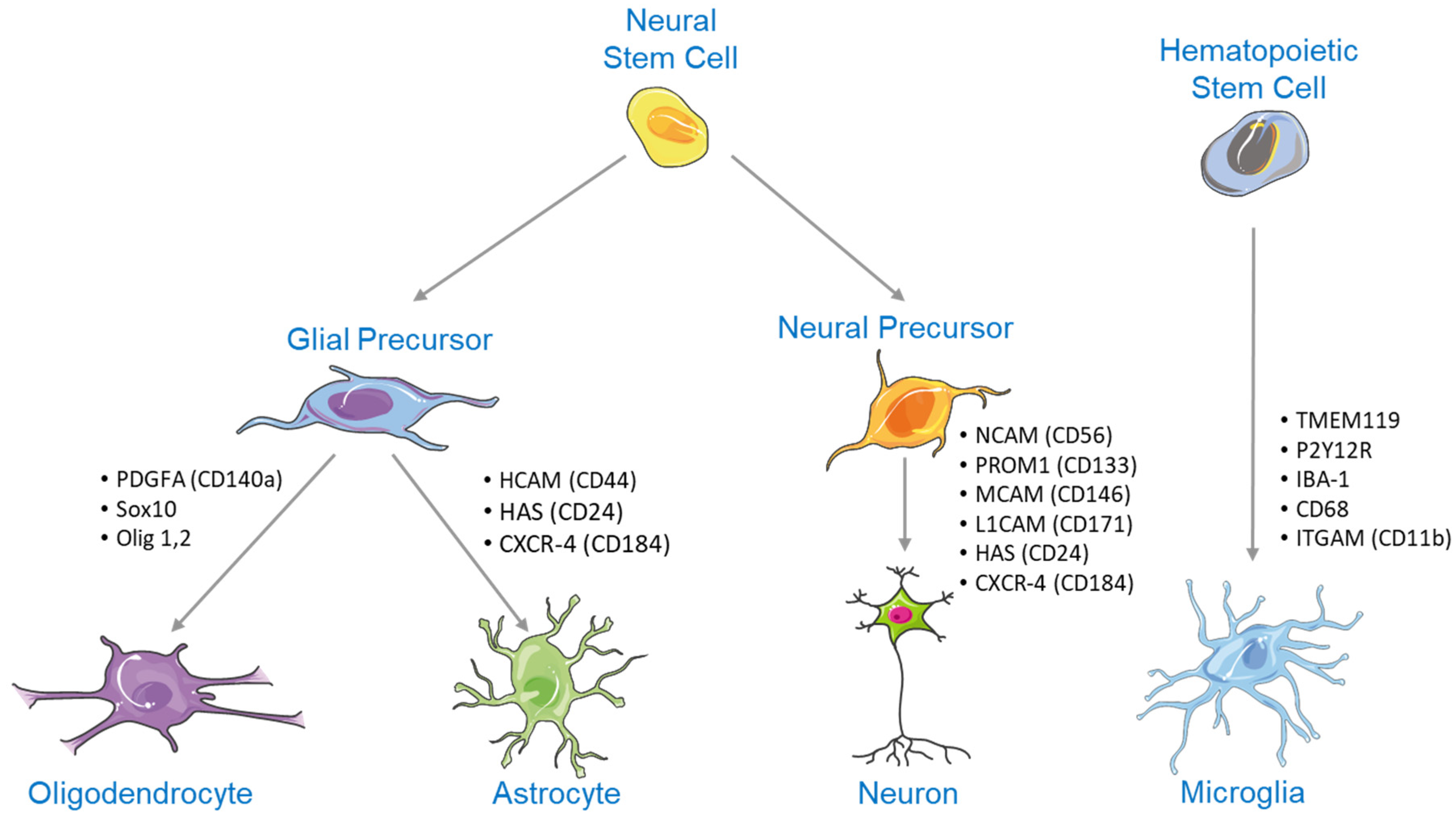
2. NSC Differentiation

NSC differentiation begins when NSCs differentiate into neural or glial progenitor cells. Neural progenitors can then differentiate into a wide range of mature neurons, while glial progenitor cells ultimately differentiate into oligodendrocytes and astrocytes (Figure 1) [17,18]. Contrastingly, microglia are of hematopoietic origin [19]. Surface markers are seen at each stage of differentiation, and as such, can be used to determine neural cell differentiation progress [20].
NSC differentiation is regulated by a variety of signaling pathways and transcription factors that influence both embryological development and adult neural neogenesis [21]. Specific signaling pathways highly implicated in NSC differentiation include the Notch, Wnt/β-catenin, sonic hedgehog (Shh), and bone morphogenetic protein (BMP) signaling pathways [21].

2.1. Notch Signaling in NSC Differentiation

The Notch signaling pathway plays a crucial role in the inhibition of NSC differentiation through direct interactions of Jagged1, Jagged2, delta-like 1, -3, and -4 ligands with the four Notch receptors (NOTCH1, NOTCH2, NOTCH3, and NOTCH4) [22]. Binding of these ligands to Notch receptors on the surface of NSCs induces proteolytic cleavage of the receptor and subsequent release of the Notch intracellular domain (NICD) into the cytoplasm of the cell [23]. The released NICD translocases to the nucleus of the NSC where it complexes with the DNA-binding protein RBPj, forming the NICD-RBPj complex [24,25]. The NICD-RBPj complex promotes downstream expression of basic helix-loop-helix transcriptional repressors such as hairy and enhancer of split which inhibit the expression of Ng2 and Mash1, transcription factors responsible for activating NSC differentiation [22,26]. Downregulation of these transcription factors ultimately results in the inability to initiate NSC differentiation [25].
Following SCI, Notch signaling is activated which may be responsible for the failure of NSCs to mature into functional neurons at the site of the lesion [27]. As such, therapeutics that inhibit Notch signaling have the potential to restore neurologic functioning and reduce symptomatology associated with SCI. Several studies have investigated Notch inhibition as a potential therapeutic for SCI through the use of electroacupuncture [28], oligodendrocyte precursor cell transplantation at the SCI lesion [29], and bone marrow mesenchymal stem cell transplantation at the SCI lesion [30]. Inhibition of the Notch signaling pathway has been shown to promote proliferation and differentiation of neurons at the SCI lesion [29,30], restore altered levels of protein expression following SCI [28], and suppress activation of neurotoxic astrocytes following SCI [31]. Thus, therapeutics designed to inhibit Notch signaling may serve as an effective treatment option for SCI.

2.2. Wnt/β-Catenin Signaling in NSC Differentiation

The highly conserved canonical Wnt/β-catenin signaling pathway is believed to play a significant role in the promotion of embryological and adult NSC differentiation [32]. Wnt ligands are secreted by various autocrine and paracrine pathways, where they then interact with the surface G-protein coupled Frizzled receptor and co-receptor of the low-density lipoprotein-related protein receptor 5/6 to form a ternary cell surface complex [21]. The formation of this ternary complex ultimately leads to the inactivation of glycogen-synthase-kinase-3β (GSK-3β), a serine/threonine kinase that plays a multifunctional role in signaling pathways responsible for cell growth, inflammation, glucose metabolism, and embryologic development [21,32,33]. In the absence of Wnt signaling, GSK-3β activation promotes the phosphorylation, ubiquitination, and degradation of β-catenin by proteosomes [34]. By inhibiting GSK-3β, Wnt signaling allows for stabilized β-catenin to enter the cell nucleus and interact with T-cell factor/lymphoid enhancer-binding factor transcription factors, promoting the transcription of numerous genes implicated in NSC differentiation [33,35].
It has been demonstrated throughout the literature that Wnt signaling increases at SCI lesions following the initial injury [36,37,38]. Because the Wnt/β-catenin pathway plays a key role in stem cell differentiation, therapeutics that utilize and activate this pathway have the potential to treat SCI [39]. For example, overactivation of miR-124, a regulatory coding gene of NSC differentiation and proliferation, promotes functional recovery in mice with SCIs by activation of the Wnt/β-catenin pathway [40]. Similarly, administration of salvianolic acid B [38], sirtuin-1 [41], and rapamycin [42] have been shown to improve neurologic functioning, NSC differentiation, and NSC proliferation following SCI through increased Wnt/β-catenin signaling. Taken together, these results highlight the potential of Wnt/β-catenin activation as a therapeutic for SCI.

2.3. Shh Signaling in NSC Differentiation

Shh signaling has been shown to play a significant role in limb bud development, early central nervous system (CNS) development, and adult NSC differentiation [43]. In adult mammals, Shh signaling regulates NSC differentiation and migration in the subventricular zone of the lateral ventricle [21,44]. This process begins when Shh ligands bind to the Patched receptor resulting in the activation and expression of G protein-coupled receptor-like protein Smoothened [45,46]. Smoothened then activates several transcription factors belonging to the Gli family, which are responsible for promoting NSC differentiation and proliferation [43]. Although there is a lack of evidence in the literature showing a connection between Shh signaling and SCI, several recent studies have suggested that increased Shh signaling may improve patient outcomes following traumatic brain injury and ischemia [47]. Therefore, it is possible that increased Shh signaling may serve as a therapeutic for SCI; however, more research is required before this idea can be established as fact.

2.4. BMP Signaling in NSC Differentiation

BMPs are polyfunctional cytokines that regulate a variety of cellular functions including cell proliferation, differentiation, and death [48]. As such, BMP signaling plays a crucial role in regulating the process of NSC development and differentiation in the embryologic and adult CNS [49]. Specifically, BMP ligands bind to a tetrameric receptor complex containing two type I and two type II transmembrane serine/threonine kinases on the surface of NSCs [50]. The formation of this complex results in the upregulation of Smad1, Smad5, and Smad8 transcription factors [50]. Once phosphorylated, these transcription factors bind and form an activate complex with Smad4, which translocates to the cell nucleus to activate genes responsible for inhibiting NSC differentiation [51]. Additionally, BMP signaling plays a crucial role in regulating glial scar formation which further prevents NSC differentiation and axonal growth following SCI [52].
BMP signaling has been shown to pathologically increase at the site of SCI lesions resulting in diminished functional recovery, lipid peroxidation, increased cell death, disruption of the extracellular matrix, and reduced axon regeneration [48,50]. As such, inhibition of BMP signaling has the potential to serve as a therapeutic for SCI. This idea has been investigated primarily in murine models of SCI through administration of noggin, an endogenous antagonist of BMP ligands [53,54]. For example, Matsuura et al. [55] discovered that noggin-induced inhibition of BMP resulted in enhanced locomotor activity and significant corticospinal tract regrowth following spinal cord contusion. A later study by Hart et al. [54]. confirmed these findings by demonstrating that acute blockage of BMP with noggin promoted remyelination, oligodendrogenesis, and acute functional recovery in rats with SCI [54]. It is important to note that administration of noggin failed to provide long-term functional recovery, highlighting how BMP inhibition with noggin may only be effective for the treatment of acute SCI [54]. Nonetheless, these results ultimately demonstrate how BMP antagonists have the potential to restore neurologic functioning and promote axonal regrowth following SCI.

3. Substrates Indicated for Axonal Regrowth Post-Injury

Three-dimensional (3D) axonal regrowth is a complex process that occurs after an injury involving multiple genes, signaling molecules, proteins, and extracellular environment components [5]. For axonal regrowth to occur, the primary neural cell body must physically disconnect from its distal target [55]. When this occurs, the remaining portion of the axon connected proximally to the neural cell body will undergo a regenerative phase, which entails the creation of the axonal growth cone. This growth cone can be described as a mobile structure that uses various growth factors and signaling molecules in the surrounding environment to guide and elongate the axon [5].

3.1. Plasma Membrane Sealants

An initial injury to an axon first necessitates a rapid sealing of the damaged plasma membrane, as the expected rupture would cause spillage of intracellular contents out of the cell or extracellular contents, such as Ca2+ into the cell [6]. This process naturally occurs in vivo, through physical phenomena involving line tension created via hydrophobic interactions between free lipid edges and membrane tension induced by cytoskeletal organization and physical membrane curvature [6]. However, studies have demonstrated that this process can be facilitated via the exogenous delivery of hydrophilic polymers (e.g., polyethylene glycol) or surfactants such as poloxamers [56,57].
Polyethylene glycol specifically has been shown to artificially seal transected axons and bypass calcium signaling to artificially fuse adjacent cells [58,59]. This process is enhanced with the addition of methylene blue to the polyethylene glycol, highlighting how methylene blue can be used to assess the efficacy and safety of polyethylene glycol-based scaffolds in human clinical trials [60]. Furthermore, polyethylene glycol-mediated axonal fusion has additionally been shown to improve behavioral functioning and strengthen neuromuscular structures in a rat model of SCI [61]. Taken together, these results demonstrate the efficacy and therapeutic value of polyethylene glycol for plasma membrane sealing following axonal injury. It is important to note that the influence of other stem cell scaffolding platforms on plasma membrane sealing post-injury remains largely unstudied. Future research should be conducted to better understand how stem cell scaffolds composed of natural polymers, synthetic polymers, or composite materials influence axonal sealing to improve our understanding of stem cell scaffold safety, efficacy, and functionality.

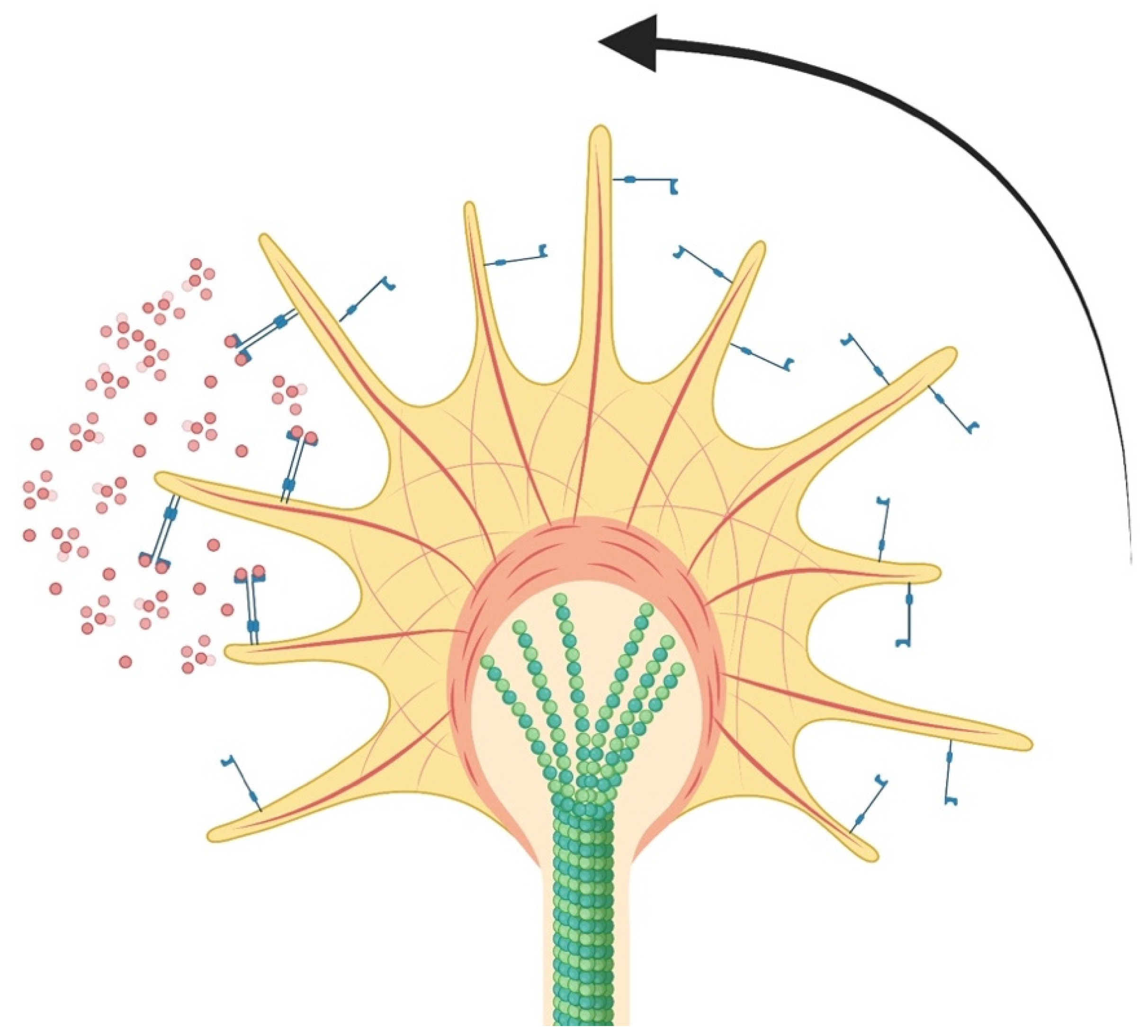
3.2. Growth Cone Formation and Stability

Once the plasma membrane has been sealed, axonal regrowth continues with the formation of the axonal growth cone, a highly specialized motile structure reminiscent of the growth cone formed during neurogenesis. The axonal growth cone takes on fan-like morphology and has the ability to respond to extracellular cues that guide axonal regrowth through complex interactions with actin filaments and microtubules [62]. Specifically, growth cones are organized with a central substructure made of microtubules and a peripheral substructure made of actin filaments [7]. Microtubules within the axonal growth cone serve as a driving force of axonal growth and are primarily responsible for guiding a growing axon toward extracellular growth factors and signaling molecules in a process known as axonal turning [62]. Similarly, the retrograde flow of the actin cytoskeleton against microtubules creates traction that allows for more precise and controlled axonal growth [62]. In addition to axonal growth, this symbiotic relationship allows for stability of the axonal growth cone. Axonal growth cone stability is incredibly important in the process of axonal regrowth because axons or growth cones with unstable microtubule polymers often retract back onto themselves, forming what is known as retraction bulbs [7,63,64], which terminates axonal regrowth.
Providing exogenous influence over microtubule stability potentially can facilitate growth cone formation and navigation during axonal regrowth. For example, a commonly used chemotherapy drug, paclitaxel (taxol), is a known microtubule stabilizer and thus is a good candidate for improving stability for growth cones. One study by Hur et al. [7] investigated the effects of locally administered taxol on both neurons derived from the central and peripheral nervous systems. This study determined that low doses of taxol administration increase the growth capacity of both types of neurons in myelin media that resembles the environment of an injured spinal cord, while also minimizing the prevalence of retraction bulbs that prematurely abort the axon regeneration process [7]. As such, incorporation of molecules designed to promote microtubule stability into stem cell scaffolds allows for enhanced growth cone formation and stability. Collagen-based NSC scaffolds enhanced with taxon have been shown to promote neural regeneration, demonstrating direct influence of NSC scaffolds on successful axonal growth cone formation and stability [65].

3.3. Neurotrophic Factors and Guidance

The formation and stabilization of a growth cone is necessary for axonal regrowth, but not entirely sufficient by itself. An equally important aspect of this complicated process is the mediation of axon guidance during regeneration (Figure 2). Of particular note, neurotrophic factors including BDNF, NT3, and NGF as are known to exert positive effects on neural outgrowth and survival, as well as promote axonal guidance during regeneration [8]. These factors bind preferentially to tyrosine kinase receptors, known as trkA, trkB, and trkC, which respectively bind and activate several major downstream signaling pathways including the mitogen activated protein kinase (MAPK) pathway and phospholipase C-γ pathway, which in turn encourage axonal regrowth [8,66,67]. For example, NGF binds specifically to trkA [68]. This interaction results in the dimerization and activation of trkA through the phospholipase C-γ, MAPK, and phosphatidylinositol 3-kinase (PI3K) signaling pathways [69]. When the activated NGF/TrkA complex is internalized into the axon by clathrin-mediated endocytosis or pincher-mediated micropinocytosis, retrograde transport occurs, and the complex is able to exert its positive effects on axonal growth and regeneration [69]. Similarly, BDNF binds with trkB, resulting in the translation of actin mRNA and consequent turning of axonal growth cones during chemoattraction [8,70]. The exogenous application of BDNF to damaged axons has additionally been shown to increase the transportation of actin proteins and promote the formation and forward expansion of growth cones [8,71,72,73]. Furthermore, NT3 interacts with trkC to activate transcription and translation of various genes that promote NSC survival and differentiation [74]. It is important to note that although NT3 primarily interacts with trkC, NT3 can also interact with trkA and trkB with less efficiency [68].
Taken together, neurotrophic factors are an obvious preliminary choice when considering potential therapeutics for facilitating axonal regrowth due to their demonstrated in vitro and in vivo capacity to promote neural survival and guidance during regeneration. However, it must be noted that the application of these growth factors must be judiciously monitored, as they could potentially induce inappropriate levels of neural growth that could paradoxically have a negative functional impact [75]. Future studies should investigate the relationship between stem cell scaffolds and neurotrophic factor abundance and physiological functioning to improve the safety of current and emerging stem cell scaffolds for SCI.

3.4. Matrix Vehicles for Axonal Regeneration

Despite the effectiveness of neurotrophic factors in facilitating axonal regeneration alone, studies have shown that this process is significantly enhanced when neurotrophic factors are contained within a matrix vehicle [76]. The principle of these matrices is relatively simple; the matrix provides a structured environment that allows the growth cone of a regenerating neuron to have access to growth factors while limiting the spread of growth factors to prevent aberrant neural growth elsewhere [76]. For example, collagen type I filaments derived from the extracellular matrix of regenerating axons have been shown to facilitate axonal reconnection over a distance of 30 mm [77,78,79]. It is believed that collagen provides both a foundation for blood vessel development and physical guide for axons to follow during the process of axonal regrowth [77,78,79].
Modern matrix vehicles for axonal regeneration including polyethylene glycol [80], fibrin [81], and peptide hydrogels [82] aim to deliver critical growth factors and stem cells directly to the SCI lesion in a highly biocompatible, patient-specific manner. For example, cross-linkage of polyethylene glycol produces hydrogels that have the ability to deliver bone marrow mononuclear cells and growth factors at the SCI lesion [80]. Hydrogel-based matrix vehicles can be enhanced further through the addition of proteins and NSCs that aim to promote axonal regrowth in a way that is unique to the biochemical profile of the patient. A study by Wiseman et al. [82] investigated this idea using a biomimetic self-assembling peptide hydrogel enhanced with NSCs known as Fmoc-DIKVAV. This hydrogel was found to provide a microenvironment suitable for axonal regrowth while also improving behavioral functioning in rodents [82]. Furthermore, hydrogels enhanced with fibrin, a non-globular protein derived from fibrinogen, have the ability to promote axonal regrowth and improve behavioral functioning in rodents following SCI [81]. It is important to note that studies regarding hydrogel-based matrix vehicles for axonal regeneration remain largely in the preclinic phase, highlighting the need for additional research before implementation into clinical practice.

3.5. Electrical Stimulation for Axonal Growth

Electrical stimulation (ES) has the ability to promote axonal regrowth. However, the mechanisms responsible for this phenomenon remain not completely understood. It has been proposed that ES imitates retrograde propagation of intracellular calcium waves traveling toward the neural cell body and as such, plays a key role in action potential propagation, allowing for axonal regrowth [83]. Furthermore, ES in the neural cell body upregulates expression of recombination activating genes (RAGs) necessary for axonal regeneration and elongation by increasing BDNF expression [84]. Upregulation of BDNF in a calcium-dependent manner results in concurrent upregulation of Tα1 tubulin and growth associated protein-43 as well as Rho inhibition, all of which enhance cytoskeletal assembly [83]. Other pathways such as a p38 mitogen-activated protein kinase pathway may play a role in promoting neurite outgrowth by activation of cAMP response element-binding protein [85].
The role of ES on axonal regrowth has been explored in several murine models. One study by Geremia et al. [86] found that 1 h of intraoperative ES (20 Hz) significantly increased dorsal root ganglia neuron regeneration and branching in mice with complete femoral nerve transections. Additionally, ES was correlated with a significant increase in growth-associated protein 43 mRNA expression 2 days after neural repair [86]. A later study by Keane et al. [87] demonstrated increased axonal density, improved nerve regeneration, and increased macrophage recruitment in rats receiving 16 Hz of ES for 1 h. In mice with complete tibial nerve resections, as little as 10 min of intraoperative ES at 16 Hz increased axon regeneration and facilitated functional recovery [88]. Similarly, Koh et al. [89] found significant improvements in motor functioning following 10 min of intraoperative ES in rats with isografts to treat sciatic nerve transections. Several ongoing clinical trials are currently investigating the potential role of intraoperative ES on axonal regrowth in humans [90].
Given the potential for stem cell scaffolds to significantly improve SCI recovery, it is possible that combination therapy involving ES followed by stem cell scaffold injection may greatly enhance neuroregeneration and improve neurologic functioning after SCI. However, there are currently no reports in the literature detailing this potential relationship. Future research endeavors should aim to determine the optimal timing, duration, and intensity of ES, as well as the selection of appropriate stem cell types and scaffold materials before experimentation and clinical use in humans.

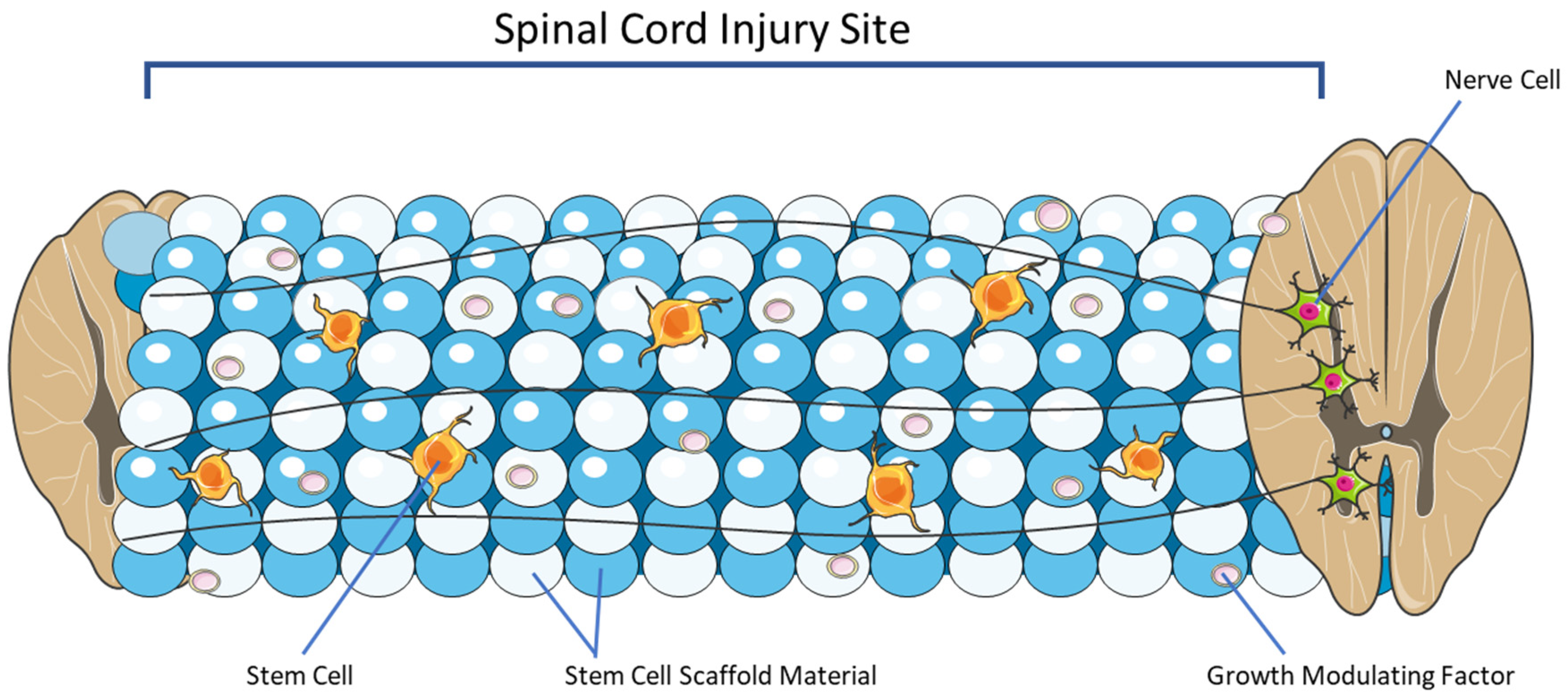
4. Overview of Stem Cell Scaffolding

The stem cell scaffold is a lab-created, three-dimensional structure designed to imitate and modulate many of the key characteristics of the naturally occurring ECM (Figure 3) [13,14]. The scaffold is meant to provide an environment for stem cell differentiation, attachment, and growth [15]. There are several broad categories of scaffolds including hydrogels, natural polymers, synthetic polymers, and composites [14,16]. A key feature of stem cell scaffolds is their ability to be modified for specific applications [91]. Routes of modification that will be reviewed here include the incorporation of growth-modulating molecules, surface adhesion molecules, and electrical stimulation.
One of the key purposes of a scaffold is to provide a mechanical support network for stem cells to reside within. This framework protects growing cells and axons from applied forces post-SCI [92,93]. The porosity or the percentage of pores per area of scaffold is a vital feature. It defines the surface area for cell attachment within the scaffold environment [94]. Higher porosity precents have been associated with increased stem cell growth and differentiation [95,96]. While to our knowledge, no study has examined the ideal scaffold pore diameter for SCI repair, it is notable that spinal corticospinal tract axons are believed to range from 1 to 22 μm in diameter, with the majority of axons falling in a range of 1–4 μm. This implies that while scaffold pore diameters as low as 4 μm are large enough for the passage of most axons, the lower end of the pore diameter limit must be no less than 22 μm to accommodate all corticospinal tract axons. In addition, even more space may be required for the ideal passage of waste, nutrients, and other important axons with larger diameters such as pyramidal tract axons, purkinje cell axons, Aα sensory fibers, and Aβ sensory fibers. Conversely, increasing porosity and pore diameter has the negative effect of reducing compressive strength and elastic modulus [97,98,99]. Another crucial property of a scaffold is biodegradability with minimal cytotoxic byproducts [100]. Biodegradability allows the scaffold to be fully absorbed with time and replaced by ECM. This reduces lasting inflammation, permits further axonal and cell proliferation, and allows for the release of infused growth-modulating factors [14,100].

4.1. Natural Polymer Scaffolds

Natural polymer scaffolds are constructed from molecules that exist in nature. Consequently, natural polymer scaffolds generally have increased cell adhesion and biocompatibility as compared to their synthetic counterparts [15]. Furthermore, given the large natural abundance of materials used to create natural polymer scaffolds, this scaffolding modality offers a more cost-effective therapeutic with more efficient synthesis [15]. With respect to SCI, natural polymer scaffolds have the ability to restore motor function and promote axonal regrowth following injury [101]. Materials commonly utilized in natural polymer scaffolds include collagen, chitosan, gelatin, fibrin, and alginate [14,102,103]. However, a Bayesian network meta-analysis conducted by Zhang et al. [101]. determined collagen, fibrin, and gelatin to be the most ideal compounds used to create natural polymer stem cell scaffolds in a rat model of SCI.
Collagen, the most abundant protein in the ECM, is composed of a-chains synthesized into triple helical fiber unites [104]. Collagen has a number of desirable characteristics including high biocompatibility and full biodegradability [102,104]. One key feature of collagen is its versatility as it can be fabricated through a number of means including electrospinning 3D printing and hydrogels [105,106]. Collagen can be denatured to form gelatin, which expresses similar biodegradability and biocompatibility properties [104]. Because gelatin possesses no triple helix structure, the randomly distributed a-chains are able to polymerize into a gel structure [104]. However, gelatin is unique in the sense that it has heightened hydrophilic properties and a diverse range gel densities as compared to collagen [107]. A recent study by Ke et al. [107] demonstrated that gelatin could be 3D printed at low cost and high consistency into microsphere scaffolds that could be easily implanted into SCI lesion sites. As stem cell scaffolds for SCI repair near wide medical application, many of these properties of reliability and lower production costs may become increasingly desirable. Furthermore, fibrin forms naturally during blood clotting as the product of thrombin and fibrinogen [108,109]. As such, fibrin is highly biocompatible, bio-degradable, and promotes cell adhesion [94]. A systemic review by Ortiz et al. [94]. found that fibrin scaffolds with mesenchymal stem cells promoted axonal renewal, remyelination, and recovery of motor function, indicating that fibrin may be an exceptional scaffold for recovery post-SCI.

4.2. Synthetic Polymer Scaffolds

There are a number of synthetic materials that can be fabricated into stem cell scaffolds including poly(lactic-co-glycolic acid), polycaprolactone, polyethylene glycol, poly(glycolic acid), poly(2-hydroxyethyl methacrylate), poly(N-(2-hydroxypropyl) methacrylamide), and polyacrylonitrile/polyvinyl chloride [102]. Generally speaking, some of the advantages of synthetic polymers include their wider range of mechanical traits including increased durability and strength [102]. Synthetic scaffolds also have the potential to be synthesized in mass through a number of innovative techniques including 3D printing by inkjet systems, micro-extrusion, stereolithography, and fused deposition modeling [102]. With respect to SCI, synthetic polymer scaffolds have the ability to hold highly complex biomaterials such as superparamagnetic iron oxide nanoparticles [110]. The complexity of synthetic polymer scaffolds allows for enhanced axonal regeneration and restoration of motor functioning following SCI, which may offer an additional advantage over natural polymer scaffolds [102]. However, these polymers can be synthesized from materials that may damage, degrade, or reduce the overall efficacy of the therapeutic encapsulated within. As such, it is important to determine the optimal polymer formulation before conducting in vivo experiments.

4.3. Hydrogel Scaffolds

Hydrogels are hydrophilic polymers that can be either synthetic or natural and offer a number of advantages as stem cell scaffolds [111]. They are generally highly permeable, biocompatible, and biodegradable [111]. One advantage of hydrogels is their potential to transition from a liquid to gel upon injection into the lesion site, a modality known as injectable hydrogels [112]. Injectable hydrogels ultimately provide a more convenient therapeutic delivery method which is often favorable in research. Furthermore, hydrogels often provide an environment that promotes cellular survival and proliferation, which is critical when creating a NSC scaffold for SCI [113]. However, a major barrier to achieving high in vivo compatibility with hydrogel-based scaffolds is the presence of toxic moieties and chemicals used in hydrogel synthesis [113]. Examples of toxic chemicals commonly used in hydrogel synthesis include stabilizers, initiators, organic solvents, and emulsifiers, all of which can damage or destroy normal cells surrounding the lesion [113].

4.4. Hybrid or Composite Scaffolds

Recent research has focused on combining two or more biomaterials into hybrid or composite scaffolds that can be further fine-tuned by blending advantages of multiple materials [114,115]. For example, gelatin-based hydrogel layered with the synthetic polymer polycaprolactone has been shown to add support to the scaffold and provide a directed path for axon regrowth [115]. Similarly, introduction of a conductive polymer into a photocrosslinkable gelatin/polyethylene glycol matrix creates a more stable scaffold able to deliver more NSCs to SCI lesions [116]. Due to the high degree of variability and heterogenous nature of composite scaffolds, the overall synthesis, biocompatibility, and degradability varies between composite scaffold modalities due to their unique composition. However, the practice of combining multiple biomaterials to create a single scaffold gives composite scaffolds an advantage over traditional single-material natural or synthetic polymer scaffolds because composite scaffolds can be designed to hold a more diverse group of biomaterials designed to promote axonal regeneration allowing for more robust, patient-specific care for SCI. Furthermore, composite scaffolds have the potential to meet both mechanical and physiological requirements necessary for efficacy, safety, and viability in vivo. However, because composite scaffolds are often complex, they can be more expensive and less efficient to produce as compared to single-material scaffolds. Furthermore, it may take researchers more time to perfect composite scaffold formulations before translation to in vivo experiments due to their high degree of complexity.

4.5. Growth Modulating Factors

Growth-modulating factors that can be integrated into stem cell scaffolds offer a significant method for promoting neural, axonal, and vascular growth and integration [117,118,119,120]. The incorporation of growth-modulating factors into the scaffold offers several key benefits. Firstly, as the scaffold degrades, it can potentially release a steady dose of growth factors to the lesion site [121,122]. This is particularly effective as growth factors often degrade quickly within tissue [123]. In addition, there are theoretically fewer off target effects as the total dose of growth factor is substantially less than a systemic treatment [123].
BDNF is an example of a growth modulating factor known to promote NSC survival and differentiation [103]. In addition, glial cell line-derived neurotrophic factor (GDNF), IGF-1, NGF, and vascular endothelial growth factor (VEGF) promote angiogenesis and healing following injury [117,118,119,120,124,125]. More recently, neurotropin has been investigated as a growth modulating factor implicated in SCI recovery as it has been shown to modulate cytokines and inhibit apoptosis [126]. Neuregulin-1 is another growth factor that has been shown to facilitate recovery following SCI mediated by interactions with the Nrg-1/ErbB network that facilitates NSC differentiation, neural migration, and myelination [127]. Given the extensive role growth modulating factors play in axonal regeneration, incorporation of growth modulating factors into stem cell scaffolds has the potential to improve various scaffolding modalities.

5. Emerging Pre-Clinical Studies and Their Applications for Clinical Adoption

Given the significant therapeutic potential of scaffolding for SCI, a variety of preclinical studies have investigated a diverse range of scaffolding constructs for their therapeutic potential in the treatment of SCI. Collagen is the most abundant protein in mammals primarily used to synthesize connective tissues such as skin, bone, muscles, tendons, and cartilage. Its high biocompatibility, hydrophilic nature, abundance in somatic tissues, and high degree of cellular adhesion allows it to be synthesized into scaffolds [106]. Consequently, collagen-derived stem cell scaffolds for SCI have been studied extensively throughout the literature (Table 1) [128,129,130,131,132,133,134,135]. Specifically, collagen-based stem cell scaffolds recruit and protect embryonic neural stem progenitor cells (NSPCs) at SCI lesions, promote neural stem cell (NSC) adhesion, proliferation, and differentiation, and improve locomotor behaviors in murine models [128,129]. When seeded with NSPCs, the efficacy of collagen scaffolding for SCI is enhanced through improved axonal elongation, neural regeneration at SCI lesions, enhanced NSPC differentiation, and functional integration of the regenerated cells into the preexisting neural network [129]. This strategy has been shown to improve hindlimb motor function, nerve regeneration, and neural cell extension in rat models of complete spinal cord transection [131].
Collagen scaffolds for SCI can be further modified through the addition of patient-specific bone marrow mononuclear cells or mesenchymal stem cells (MSCs) [130,134]. In murine and canine models of complete spinal cord transection, administration of collagen scaffolds seeded with MSCs derived from neonatal umbilical cord tissue resulted in increased motor scores and reduced injury area [130]. Consequently, a phase I clinical trial (NCT 02510365) was conducted by Deng et al. [130] to investigate the efficacy of this scaffold in 40 patients with acute complete cervical injuries. Twelve months after transplantation of the human umbilical cord MSC collagen scaffold at the site of SCI, novel nerve fiber growth emerged and electrophysiological activity of the adjacent neurons improved [130]. Increased daily life scores, American Spinal Injury Association scores, and bowel and urinary functioning were additionally observed [130]. The results of this clinical trial are significant because there are few reports in the literature detailing results from humans subjects involved in clinical trials investigating the role of stem cell scaffolds as a treatment option for SCI. Furthermore, a later study by Tang et al. [117] investigating the longitudinal effects (2–5 years) of collagen scaffolds loaded with patient-specific bone marrow mononuclear cells or human umbilical cord MSCs for SCI demonstrated similar results with improvements in bowel and bladder sensation, voluntary walking ability, and enhanced finger activity [130,134].
The addition of proteins expressed in MSCs can enhance the efficacy of collagen scaffolds for SCI [128]. One study by Liu et al. [128] investigating the effects of a linearly ordered collagen scaffold modified with N-cadherin, a protein expressed in mesenchymal cells, found that adhesion of NSPCs onto the collagen scaffold improved with the introduction of N-cadherin. When transplanted into rats with complete spinal cord transections, the N-cadherin-modified collagen scaffold recruited more NSPCs to the lesion site and consequently, LOCS-Ncad rats demonstrated significantly improved locomotor function as compared to controls [128]. Similarly, collagen scaffolds seeded with MSCs can be further enhanced by the addition of silk fibroin or heparan sulfate [132,133,135]. Silk fibroin is a natural fibrous protein found in silk and spider webs. Like collagen, silk fibroin demonstrates high biocompatibility and mechanical strength allowing for its use in adipose tissue, bone, and skin regeneration [136]. When compared to collagen/silk scaffolds lacking MSC seeds, collagen/silk scaffolds seeded with MSCs can facilitate nerve fiber regeneration, enhance remyelination, and accelerate the establishment of synaptic connections at the injury site [135]. As such, human umbilical cord MSCs embedded on collagen/silk fibroin scaffolds have been shown to induce functional recovery and improve locomotor behaviors in rats with complete SCIs [133,135]. Similarly, heparan sulfate is a polysaccharide involved in a number of biological processes including cell proliferation, inflammation, and angiogenesis [137]. Collagen scaffolds enhanced with heparan sulfate and MSCs demonstrate significant improvements in locomotor activity, motor evoked potential, urodynamic parameters, and modulation of inflammatory cytokines in canines with complete spinal cord transections [132].
In addition to collagen, stem cell scaffolds constructed with hydrogel have been explored as a therapeutic for SCI (Table 2) [138,139,140,141,142,143]. Matrigel, a solubilized basement membrane protein composed of laminin, collagen IV, heparan sulfate proteoglycans, entactin/nidogen, and growth factors, can be employed for treatment of SCI [144]. Matrigel has been shown to support neural stem cell survival in vitro and in vivo [144]. When implanted into a murine model of SCI, administration of Matrigel shows slight functional repair and neural recovery, though nonsignificant [144]. Hyaluronic acid hydrogel dotted with manganese dioxide nanoparticles has the ability to bridge nerve tissue and enhance adhesive growth of MSCs [142]. When implanted into rats with SCIs, MSC differentiation is enhanced and motor function is significantly restored [142]. NSCs obtained from epileptic human brain specimens seeded in PuraMatrix peptide hydrogel enhance cell survival and differentiation, reduce SCI lesion volume, and improve neurological functioning in rats with SCIs [140]. Hydrogel enhanced with agarose, gelatin, and polypyrrole additionally improves NSPC differentiation, reduces SCI lesion volume, and provides a biocompatible microenvironment suited for tissue repair in vivo [143]. Similarly, gelatin methacryloyl (GelMA) hydrogel constructed with decellularized spinal cord extracellular matrix-gel (DSCG) provides a robust microenvironment in vitro favoring menstrual blood-derived mesenchymal stem cells (MenSCs) adhesion, proliferation, and differentiation [138]. DSCG/GelMA scaffolded with MenSCs improved motor function, reduces neural inflammation, and promotes neural differentiation in rats with complete spinal cord transections [138]. The same effects are seen in murine models of SCI utilizing grooved GelMA-MXene hydrogel loaded with NSCs [139]. Because SCI creates a pathologically inflamed microenvironment characterized by immune activation damage-associated molecular patterns and activation of immune cells that impair neurologic recovery, more modern hydrogel scaffolds aim to restore the pathological SCI microenvironment [141]. For example, hydrogel scaffolds constructed to release interleukin-10, an anti-inflammatory cytokine, have the ability to enhance NSC differentiation, neural regeneration, and axonal growth in mice with SCIs [141].
The development of 3D bioprinting technology has allowed for the refinement of stem cell scaffolds for SCI [145]. Specifically, 3D bioprinting technology precisely enhances neural regeneration by creating biomimetic scaffolds tailored to the dimensions of the subject or patient in a time-sensitive manner [146]. Bioprinting of a sodium alginate/gelatin scaffold loaded with NSPCs and oligodendrocytes demonstrates improved hindlimb motor function and nerve regeneration in a rodent model of SCI [145]. Similarly, 3D bioprinted scaffolds loaded with induced pluripotent stem cells derived from urine cells have the ability to improve SCI in mice [147]. The precision of 3D bioprinted scaffolds can additionally be enhanced with the addition of small molecules. By loading 3D bioprinted scaffolds with OSMI-4, a small molecule O-GlcNAc transferase inhibitor, differentiation of NSCs can be induced and specifically guided to the SCI lesion for more efficient SCI repair [148]. Consequently, the OSMI-4-refined bioscaffold promoted neural regeneration and axonal growth, leading to significant motor recovery in rats with SCIs [148]. As such, construction of stem cell scaffolds for SCI can be refined by means of 3D bioprinting.

6. Conclusions and Future Research Directions

Trauma to the central nervous system is incredibly difficult to treat as neurons of the central nervous system regenerate poorly in response to injury. However, stem cell scaffolds have the potential to restore neurologic function, effectively allowing for repair of axons in the central nervous system. Different stem cell scaffolding platforms utilize unique stem cell populations highlighting the highly modifiable and patient-specific nature of stem cell scaffolding for SCI. This property of stem cell scaffolds provides patients with a variety of platforms that can be tailored to the type, location, and severity of SCI. Although stem cell scaffolding serves as a potential therapeutic for the treatment and clinical management of SCI, trials remain largely in the preclinical phase. Future research is needed to better understand the underlying biological mechanisms of stem cell scaffold success in murine models to determine their potential safety and efficacy in humans. Specific properties of stem cell scaffolds that must be investigated include the optimal cell type for different SCIs as well as optimal transplantation dosages, timings, and administration methods. For example, intravenous administration of stem cell scaffolds is less invasive and far more convenient for patients and providers as compared to infusion directly at the SCI lesion, especially if multiple infusions are necessary. These key formulation and dosage properties may create challenges to providers looking to translate these preclinical findings into clinical practice. It is possible that future research endeavors could explore the use of stem cell scaffolds in combination with other novel therapeutics including biomaterials and other modifiers of the SCI ECM. Furthermore, stem cell scaffolds created from highly complex polymers may provide a barrier to accessing care due to the high associated therapeutic cost. As such, it is important to consider the cost patients may have to pay for stem cell scaffolding therapeutics to ensure stem cell scaffolds are as accessible as possible.
As aforementioned, NSCs have the potential to differentiate into a variety of mature cells via several critical signaling pathways. Because biomaterials used to create stem cell scaffolds are designed to deliver key growth factors indicated for axonal regrowth to the site of injury for an extended period of time, it is possible that stem cell scaffolds have the potential to modulate key signaling pathways involved in NSC differentiation. Future studies should investigate how novel stem cell scaffolding platforms influence NSC differentiation pathways as well as how to monitor NSC differentiation patterns within stem cell scaffolds. Nonetheless, although significant validation is required before implementation into the clinic, current results and ongoing trials highlight the potential for stem cell scaffolding to improve and revolutionize the treatment of SCI.

Author Contributions

All authors contributed to original draft preparation, reviewing, and editing. All authors have read and agreed to the published version of the manuscript.

Funding

This research received no external funding.

Institutional Review Board Statement

Not applicable.

Informed Consent Statement

Not applicable.

Data Availability Statement

Not applicable.

Conflicts of Interest

The authors declare no conflict of interest.

Abbreviations

3DThree dimensional
AISAmerican Spinal Injury Association Impairment Scale
BDNFBrain-derived neurotrophic factor
BMPBone morphogenetic protein
CNSCentral nervous system
CSFCerebrospinal fluid
ESElectrical stimulation
GDNFGlial cell line-derived neurotrophic factor
GFAPGlial fibrillary acidic protein
GSK-3βGlycogen-synthase-kinase-3β
GelMAGelatin methacryloyl
IMLLIntramedullary lesion length
MAPKMitogenactivated protein kinase
MPMethylprednisolone
MRIMagnetic resonance imaging
MSCMesenchymal stem cell
MenSCsMenstrual blood-derived mesenchymal stem cells
NCIDNotch intracellular signaling domain
NF-LNeurofilament light
NGFNerve growth factor
NSCNeural stem cell
NSPCsNeural stem progenitor cells
NT3Neurotrophin-3 (NT3)
PI3KPhosphatidylinositol 3-kinase
RAGRecombination activating gene
SCISpinal cord injury
ShhSonic hedgehog
VEGFVascular endothelial growth factor

References

  1. Barbiellini Amidei, C.; Salmaso, L.; Bellio, S.; Saia, M. Epidemiology of traumatic spinal cord injury: A large population-based study. Spinal Cord 2022, 60, 812–819. [Google Scholar] [CrossRef]
  2. Aarabi, B.; Albrecht, J.S.; Simard, J.M.; Chryssikos, T.; Schwartzbauer, G.; Sansur, C.A.; Crandall, K.; Gertner, M.; Howie, B.; Wessell, A.; et al. Trends in Demographics and Markers of Injury Severity in Traumatic Cervical Spinal Cord Injury. J. Neurotrauma 2021, 38, 756–764. [Google Scholar] [CrossRef] [PubMed]
  3. Alizadeh, A.; Dyck, S.M.; Karimi-Abdolrezaee, S. Traumatic Spinal Cord Injury: An Overview of Pathophysiology, Models and Acute Injury Mechanisms. Front. Neurol. 2019, 10, 282. [Google Scholar] [CrossRef] [PubMed] [Green Version]
  4. Sterner, R.C.; Sterner, R.M. Immune response following traumatic spinal cord injury: Pathophysiology and therapies. Front. Immunol. 2022, 13, 1084101. [Google Scholar] [CrossRef]
  5. Gumy, L.F.; Tan, C.L.; Fawcett, J.W. The role of local protein synthesis and degradation in axon regeneration. Exp. Neurol. 2010, 223, 28–37. [Google Scholar] [CrossRef] [PubMed] [Green Version]
  6. Hendricks, B.K.; Shi, R. Mechanisms of neuronal membrane sealing following mechanical trauma. Neurosci. Bull. 2014, 30, 627–644. [Google Scholar] [CrossRef] [Green Version]
  7. Hur, E.M.; Saijilafu; Zhou, F.Q. Growing the growth cone: Remodeling the cytoskeleton to promote axon regeneration. Trends Neurosci. 2012, 35, 164–174. [Google Scholar] [CrossRef] [Green Version]
  8. Guthrie, S. Neurotrophic factors: Are they axon guidance molecules? Adv. Exp. Med. Biol. 2007, 621, 81–94. [Google Scholar] [CrossRef]
  9. Fiani, B.; Arshad, M.A.; Shaikh, E.S.; Baig, A.; Farooqui, M.; Ayub, M.A.; Zafar, A.; Quadri, S.A. Current updates on various treatment approaches in the early management of acute spinal cord injury. Rev. Neurosci. 2021, 32, 513–530. [Google Scholar] [CrossRef]
  10. Hsieh, Y.L.; Tay, J.; Hsu, S.H.; Chen, W.T.; Fang, Y.D.; Liew, C.Q.; Chou, E.H.; Wolfshohl, J.; d’Etienne, J.; Wang, C.H.; et al. Early versus Late Surgical Decompression for Traumatic Spinal Cord Injury on Neurological Recovery: A Systematic Review and Meta-Analysis. J. Neurotrauma 2021, 38, 2927–2936. [Google Scholar] [CrossRef]
  11. Badhiwala, J.H.; Wilson, J.R.; Witiw, C.D.; Harrop, J.S.; Vaccaro, A.R.; Aarabi, B.; Grossman, R.G.; Geisler, F.H.; Fehlings, M.G. The influence of timing of surgical decompression for acute spinal cord injury: A pooled analysis of individual patient data. Lancet Neurol. 2021, 20, 117–126. [Google Scholar] [CrossRef]
  12. Aarabi, B.; Akhtar-Danesh, N.; Chryssikos, T.; Shanmuganathan, K.; Schwartzbauer, G.T.; Simard, J.M.; Olexa, J.; Sansur, C.A.; Crandall, K.M.; Mushlin, H.; et al. Efficacy of Ultra-Early (<12 h), Early (12–24 h), and Late (>24–138.5 h) Surgery with Magnetic Resonance Imaging-Confirmed Decompression in American Spinal Injury Association Impairment Scale Grades A, B, and C Cervical Spinal Cord Injury. J. Neurotrauma 2020, 37, 448–457. [Google Scholar] [CrossRef] [Green Version]
  13. Zarepour, A.; Hooshmand, S.; Gokmen, A.; Zarrabi, A.; Mostafavi, E. Spinal Cord Injury Management through the Combination of Stem Cells and Implantable 3D Bioprinted Platforms. Cells 2021, 10, 3189. [Google Scholar] [CrossRef] [PubMed]
  14. Blando, S.; Anchesi, I.; Mazzon, E.; Gugliandolo, A. Can a Scaffold Enriched with Mesenchymal Stem Cells Be a Good Treatment for Spinal Cord Injury? Int. J. Mol. Sci. 2022, 23, 7545. [Google Scholar] [CrossRef] [PubMed]
  15. Dorazco-Valdes, J. Estudio comparativo entre los aspectos clinicos, electroencefalograficos y la prueba del dibujo de la figura humana en el nino epileptico [Comparative study between the clinical aspects, the encephalographic and the draw-a-person test in epileptic children]. J. Neurol. Sci. 1968, 6, 373–380. [Google Scholar] [CrossRef] [PubMed]
  16. Valdoz, J.C.; Johnson, B.C.; Jacobs, D.J.; Franks, N.A.; Dodson, E.L.; Sanders, C.; Cribbs, C.G.; Van Ry, P.M. The ECM: To Scaffold, or Not to Scaffold, That Is the Question. Int. J. Mol. Sci. 2021, 22, 12690. [Google Scholar] [CrossRef]
  17. Tang, Y.; Yu, P.; Cheng, L. Current progress in the derivation and therapeutic application of neural stem cells. Cell Death Dis. 2017, 8, e3108. [Google Scholar] [CrossRef] [Green Version]
  18. Kaminska, A.; Radoszkiewicz, K.; Rybkowska, P.; Wedzinska, A.; Sarnowska, A. Interaction of Neural Stem Cells (NSCs) and Mesenchymal Stem Cells (MSCs) as a Promising Approach in Brain Study and Nerve Regeneration. Cells 2022, 11, 1464. [Google Scholar] [CrossRef]
  19. de Vasconcelos, P.; Lacerda, J.F. Hematopoietic Stem Cell Transplantation for Neurological Disorders: A Focus on Inborn Errors of Metabolism. Front. Cell. Neurosci. 2022, 16, 895511. [Google Scholar] [CrossRef]
  20. De Gioia, R.; Biella, F.; Citterio, G.; Rizzo, F.; Abati, E.; Nizzardo, M.; Bresolin, N.; Comi, G.P.; Corti, S. Neural Stem Cell Transplantation for Neurodegenerative Diseases. Int. J. Mol. Sci. 2020, 21, 3103. [Google Scholar] [CrossRef]
  21. Navarro Quiroz, E.; Navarro Quiroz, R.; Ahmad, M.; Gomez Escorcia, L.; Villarreal, J.L.; Fernandez Ponce, C.; Aroca Martinez, G. Cell Signaling in Neuronal Stem Cells. Cells 2018, 7, 75. [Google Scholar] [CrossRef] [PubMed] [Green Version]
  22. Penton, A.L.; Leonard, L.D.; Spinner, N.B. Notch signaling in human development and disease. Semin. Cell Dev. Biol. 2012, 23, 450–457. [Google Scholar] [CrossRef] [PubMed] [Green Version]
  23. Manzari-Tavakoli, A.; Babajani, A.; Farjoo, M.H.; Hajinasrollah, M.; Bahrami, S.; Niknejad, H. The Cross-Talks Among Bone Morphogenetic Protein (BMP) Signaling and Other Prominent Pathways Involved in Neural Differentiation. Front. Mol. Neurosci. 2022, 15, 827275. [Google Scholar] [CrossRef] [PubMed]
  24. Teodorczyk, M.; Schmidt, M.H.H. Notching on Cancer’s Door: Notch Signaling in Brain Tumors. Front. Oncol. 2014, 4, 341. [Google Scholar] [CrossRef] [Green Version]
  25. Imayoshi, I.; Kageyama, R. The role of Notch signaling in adult neurogenesis. Mol. Neurobiol. 2011, 44, 7–12. [Google Scholar] [CrossRef]
  26. Nian, F.S.; Hou, P.S. Evolving Roles of Notch Signaling in Cortical Development. Front. Neurosci. 2022, 16, 844410. [Google Scholar] [CrossRef]
  27. Yamamoto, S.; Nagao, M.; Sugimori, M.; Kosako, H.; Nakatomi, H.; Yamamoto, N.; Takebayashi, H.; Nabeshima, Y.; Kitamura, T.; Weinmaster, G.; et al. Transcription factor expression and Notch-dependent regulation of neural progenitors in the adult rat spinal cord. J. Neurosci. 2001, 21, 9814–9823. [Google Scholar] [CrossRef] [Green Version]
  28. Wang, X.; Wang, Q.; Tian, H.; Lv, W.; Song, L.; Li, Z.; Yao, H.; Shi, S. Electroacupuncture in promoting neural repair after spinal cord injury: Inhibiting the Notch signaling pathway and regulating downstream proteins expression. Anat. Rec. 2021, 304, 2494–2505. [Google Scholar] [CrossRef]
  29. Yuan, H.; Fang, C.L.; Deng, Y.P.; Huang, J.; Niu, R.Z.; Chen, J.L.; Chen, T.B.; Zhu, Z.Q.; Chen, L.; Xiong, L.L.; et al. A2B5-positive oligodendrocyte precursor cell transplantation improves neurological deficits in rats following spinal cord contusion associated with changes in expression of factors involved in the Notch signaling pathway. Neurochirurgie 2022, 68, 188–195. [Google Scholar] [CrossRef]
  30. Chen, Y.; Lian, X.H.; Liao, L.Y.; Liu, Y.T.; Liu, S.L.; Gao, Q. Transplantation of bone marrow mesenchymal stem cells alleviates spinal cord injury via inhibiting Notch signaling. Eur. Rev. Med. Pharmacol. Sci. 2019, 23, 31–38. [Google Scholar] [CrossRef]
  31. Qian, D.; Li, L.; Rong, Y.; Liu, W.; Wang, Q.; Zhou, Z.; Gu, C.; Huang, Y.; Zhao, X.; Chen, J.; et al. Blocking Notch signal pathway suppresses the activation of neurotoxic A1 astrocytes after spinal cord injury. Cell Cycle 2019, 18, 3010–3029. [Google Scholar] [CrossRef] [PubMed]
  32. Toledo, E.M.; Colombres, M.; Inestrosa, N.C. Wnt signaling in neuroprotection and stem cell differentiation. Prog. Neurobiol. 2008, 86, 281–296. [Google Scholar] [CrossRef] [PubMed]
  33. Logan, C.Y.; Nusse, R. The Wnt signaling pathway in development and disease. Annu. Rev. Cell Dev. Biol. 2004, 20, 781–810. [Google Scholar] [CrossRef] [Green Version]
  34. Nusse, R.; Clevers, H. Wnt/beta-Catenin Signaling, Disease, and Emerging Therapeutic Modalities. Cell 2017, 169, 985–999. [Google Scholar] [CrossRef] [PubMed] [Green Version]
  35. Tamai, K.; Semenov, M.; Kato, Y.; Spokony, R.; Liu, C.; Katsuyama, Y.; Hess, F.; Saint-Jeannet, J.P.; He, X. LDL-receptor-related proteins in Wnt signal transduction. Nature 2000, 407, 530–535. [Google Scholar] [CrossRef] [PubMed]
  36. Rodriguez, J.P.; Coulter, M.; Miotke, J.; Meyer, R.L.; Takemaru, K.; Levine, J.M. Abrogation of beta-catenin signaling in oligodendrocyte precursor cells reduces glial scarring and promotes axon regeneration after CNS injury. J. Neurosci. 2014, 34, 10285–10297. [Google Scholar] [CrossRef] [Green Version]
  37. Strand, N.S.; Hoi, K.K.; Phan, T.M.T.; Ray, C.A.; Berndt, J.D.; Moon, R.T. Wnt/beta-catenin signaling promotes regeneration after adult zebrafish spinal cord injury. Biochem. Biophys. Res. Commun. 2016, 477, 952–956. [Google Scholar] [CrossRef] [Green Version]
  38. Zhou, H.; Liu, Y.; Sun, L.; Fu, M.; Zhao, Y. Salvianolic acid B activates Wnt/beta-catenin signaling following spinal cord injury. Exp. Ther. Med. 2020, 19, 825–832. [Google Scholar] [CrossRef] [Green Version]
  39. Gao, K.; Zhang, T.; Wang, F.; Lv, C. Therapeutic Potential of Wnt-3a in Neurological Recovery after Spinal Cord Injury. Eur. Neurol. 2019, 81, 197–204. [Google Scholar] [CrossRef]
  40. Cui, Y.; Yin, Y.; Xiao, Z.; Zhao, Y.; Chen, B.; Yang, B.; Xu, B.; Song, H.; Zou, Y.; Ma, X.; et al. LncRNA Neat1 mediates miR-124-induced activation of Wnt/beta-catenin signaling in spinal cord neural progenitor cells. Stem Cell Res. Ther. 2019, 10, 400. [Google Scholar] [CrossRef]
  41. Zhu, K.; Xu, W.; Han, D.; Mei, X. Effect of Sirtuin-1 and Wnt/beta-Catenin Signaling Pathway in Rat Model of Spinal Cord Injury. Comput. Math. Methods Med. 2022, 2022, 1799607. [Google Scholar] [CrossRef] [PubMed]
  42. Gao, K.; Wang, Y.S.; Yuan, Y.J.; Wan, Z.H.; Yao, T.C.; Li, H.H.; Tang, P.F.; Mei, X.F. Neuroprotective effect of rapamycin on spinal cord injury via activation of the Wnt/beta-catenin signaling pathway. Neural Regen. Res. 2015, 10, 951–957. [Google Scholar] [CrossRef]
  43. Komada, M. Sonic hedgehog signaling coordinates the proliferation and differentiation of neural stem/progenitor cells by regulating cell cycle kinetics during development of the neocortex. Congenit. Anom. 2012, 52, 72–77. [Google Scholar] [CrossRef] [PubMed]
  44. Ihrie, R.A.; Shah, J.K.; Harwell, C.C.; Levine, J.H.; Guinto, C.D.; Lezameta, M.; Kriegstein, A.R.; Alvarez-Buylla, A. Persistent sonic hedgehog signaling in adult brain determines neural stem cell positional identity. Neuron 2011, 71, 250–262. [Google Scholar] [CrossRef] [PubMed] [Green Version]
  45. Charytoniuk, D.; Porcel, B.; Rodriguez Gomez, J.; Faure, H.; Ruat, M.; Traiffort, E. Sonic Hedgehog signalling in the developing and adult brain. J. Physiol. 2002, 96, 9–16. [Google Scholar] [CrossRef]
  46. Petralia, R.S.; Schwartz, C.M.; Wang, Y.X.; Mattson, M.P.; Yao, P.J. Subcellular localization of Patched and Smoothened, the receptors for Sonic hedgehog signaling, in the hippocampal neuron. J. Comp. Neurol. 2011, 519, 3684–3699. [Google Scholar] [CrossRef] [Green Version]
  47. Yu, P.; Wang, L.; Tang, F.; Guo, S.; Liao, H.; Fan, C.; Yang, Q. Resveratrol-mediated neurorestoration after cerebral ischemic injury—Sonic Hedgehog signaling pathway. Life Sci. 2021, 280, 119715. [Google Scholar] [CrossRef]
  48. Al-Sammarraie, N.; Ray, S.K. Bone morphogenic protein signaling in spinal cord injury. Neuroimmunol. Neuroinflamm. 2021, 8, 53–63. [Google Scholar] [CrossRef]
  49. Cole, A.E.; Murray, S.S.; Xiao, J. Bone Morphogenetic Protein 4 Signalling in Neural Stem and Progenitor Cells during Development and after Injury. Stem Cells Int. 2016, 2016, 9260592. [Google Scholar] [CrossRef] [Green Version]
  50. Hart, C.G.; Karimi-Abdolrezaee, S. Bone morphogenetic proteins: New insights into their roles and mechanisms in CNS development, pathology and repair. Exp. Neurol. 2020, 334, 113455. [Google Scholar] [CrossRef]
  51. Mira, H.; Andreu, Z.; Suh, H.; Lie, D.C.; Jessberger, S.; Consiglio, A.; San Emeterio, J.; Hortiguela, R.; Marques-Torrejon, M.A.; Nakashima, K.; et al. Signaling through BMPR-IA regulates quiescence and long-term activity of neural stem cells in the adult hippocampus. Cell Stem Cell 2010, 7, 78–89. [Google Scholar] [CrossRef] [PubMed] [Green Version]
  52. Zhong, J.; Zou, H. BMP signaling in axon regeneration. Curr. Opin. Neurobiol. 2014, 27, 127–134. [Google Scholar] [CrossRef] [Green Version]
  53. Hart, C.G.; Dyck, S.M.; Kataria, H.; Alizadeh, A.; Nagakannan, P.; Thliveris, J.A.; Eftekharpour, E.; Karimi-Abdolrezaee, S. Acute upregulation of bone morphogenetic protein-4 regulates endogenous cell response and promotes cell death in spinal cord injury. Exp. Neurol. 2020, 325, 113163. [Google Scholar] [CrossRef] [PubMed]
  54. Matsuura, I.; Taniguchi, J.; Hata, K.; Saeki, N.; Yamashita, T. BMP inhibition enhances axonal growth and functional recovery after spinal cord injury. J. Neurochem. 2008, 105, 1471–1479. [Google Scholar] [CrossRef] [PubMed]
  55. Huebner, E.A.; Strittmatter, S.M. Axon regeneration in the peripheral and central nervous systems. In Results and Problems in Cell Differentiation; Springer: Berlin/Heidelberg, Germany, 2009; Volume 48, pp. 339–351. [Google Scholar] [CrossRef] [Green Version]
  56. Cho, Y.; Borgens, R.B. Polymer and nano-technology applications for repair and reconstruction of the central nervous system. Exp. Neurol. 2012, 233, 126–144. [Google Scholar] [CrossRef]
  57. Jin, X. Membrane resealing as a promising strategy for early treatment of neurotrauma. Neural Regen. Res. 2014, 9, 1876–1877. [Google Scholar] [CrossRef] [Green Version]
  58. Vargas, S.A.; Bittner, G.D. Natural mechanisms and artificial PEG-induced mechanism that repair traumatic damage to the plasmalemma in eukaryotes. Curr. Top. Membr. 2019, 84, 129–167. [Google Scholar] [CrossRef]
  59. Ghergherehchi, C.L.; Mikesh, M.; Sengelaub, D.R.; Jackson, D.M.; Smith, T.; Nguyen, J.; Shores, J.T.; Bittner, G.D. Polyethylene glycol (PEG) and other bioactive solutions with neurorrhaphy for rapid and dramatic repair of peripheral nerve lesions by PEG-fusion. J. Neurosci. Methods 2019, 314, 1–12. [Google Scholar] [CrossRef] [Green Version]
  60. Ghergherehchi, C.L.; Shores, J.T.; Alderete, J.; Weitzel, E.K.; Bittner, G.D. Methylene blue enhances polyethylene glycol-fusion repair of completely severed rat sciatic nerves. Neural Regen. Res. 2021, 16, 2056–2063. [Google Scholar] [CrossRef]
  61. Mikesh, M.; Ghergherehchi, C.L.; Hastings, R.L.; Ali, A.; Rahesh, S.; Jagannath, K.; Sengelaub, D.R.; Trevino, R.C.; Jackson, D.M.; Bittner, G.D. Polyethylene glycol solutions rapidly restore and maintain axonal continuity, neuromuscular structures, and behaviors lost after sciatic nerve transections in female rats. J. Neurosci. Res. 2018, 96, 1223–1242. [Google Scholar] [CrossRef] [Green Version]
  62. Kahn, O.I.; Baas, P.W. Microtubules and Growth Cones: Motors Drive the Turn. Trends Neurosci. 2016, 39, 433–440. [Google Scholar] [CrossRef] [Green Version]
  63. Silver, J.; Miller, J.H. Regeneration beyond the glial scar. Nat. Rev. Neurosci. 2004, 5, 146–156. [Google Scholar] [CrossRef] [PubMed] [Green Version]
  64. Erturk, A.; Hellal, F.; Enes, J.; Bradke, F. Disorganized microtubules underlie the formation of retraction bulbs and the failure of axonal regeneration. J. Neurosci. 2007, 27, 9169–9180. [Google Scholar] [CrossRef] [PubMed] [Green Version]
  65. Fan, C.; Li, X.; Zhao, Y.; Xiao, Z.; Xue, W.; Sun, J.; Li, X.; Zhuang, Y.; Chen, Y.; Dai, J. Cetuximab and Taxol co-modified collagen scaffolds show combination effects for the repair of acute spinal cord injury. Biomater. Sci. 2018, 6, 1723–1734. [Google Scholar] [CrossRef]
  66. Pastuhov, S.I.; Hisamoto, N.; Matsumoto, K. MAP kinase cascades regulating axon regeneration in C. elegans. Proc. Jpn. Acad. Ser. B Phys. Biol. Sci. 2015, 91, 63–75. [Google Scholar] [CrossRef] [Green Version]
  67. Kim, J.-H.; Auerbach, J.M.; Rodríguez-Gómez, J.A.; Velasco, I.; Gavin, D.; Lumelsky, N.; Lee, S.-H.; Nguyen, J.; Sánchez-Pernaute, R.; Bankiewicz, K.; et al. Dopamine neurons derived from embryonic stem cells function in an animal model of Parkinson’s disease. Nature 2002, 418, 50–56. [Google Scholar] [CrossRef]
  68. Reichardt, L.F. Neurotrophin-regulated signalling pathways. Philos. Trans. R. Soc. Lond. B Biol. Sci. 2006, 361, 1545–1564. [Google Scholar] [CrossRef] [Green Version]
  69. Marlin, M.C.; Li, G. Biogenesis and function of the NGF/TrkA signaling endosome. Int. Rev. Cell Mol. Biol. 2015, 314, 239–257. [Google Scholar] [CrossRef] [PubMed] [Green Version]
  70. Yao, J.; Sasaki, Y.; Wen, Z.; Bassell, G.J.; Zheng, J.Q. An essential role for beta-actin mRNA localization and translation in Ca2+-dependent growth cone guidance. Nat. Neurosci. 2006, 9, 1265–1273. [Google Scholar] [CrossRef] [PubMed]
  71. Difato, F.; Tsushima, H.; Pesce, M.; Benfenati, F.; Blau, A.; Chieregatti, E. The formation of actin waves during regeneration after axonal lesion is enhanced by BDNF. Sci. Rep. 2011, 1, 183. [Google Scholar] [CrossRef] [Green Version]
  72. Inagaki, N.; Katsuno, H. Actin Waves: Origin of Cell Polarization and Migration? Trends Cell Biol. 2017, 27, 515–526. [Google Scholar] [CrossRef] [PubMed] [Green Version]
  73. Gallo, G.; Letourneau, P.C. Localized sources of neurotrophins initiate axon collateral sprouting. J. Neurosci. 1998, 18, 5403–5414. [Google Scholar] [CrossRef] [PubMed] [Green Version]
  74. Bove, M.; Monto, F.; Guillem-Llobat, P.; Ivorra, M.D.; Noguera, M.A.; Zambrano, A.; Sirerol-Piquer, M.S.; Requena, A.C.; Garcia-Alonso, M.; Tejerina, T.; et al. NT3/TrkC Pathway Modulates the Expression of UCP-1 and Adipocyte Size in Human and Rodent Adipose Tissue. Front. Endocrinol. 2021, 12, 630097. [Google Scholar] [CrossRef] [PubMed]
  75. Lu, P.; Tuszynski, M.H. Growth factors and combinatorial therapies for CNS regeneration. Exp. Neurol. 2008, 209, 313–320. [Google Scholar] [CrossRef] [Green Version]
  76. Wells, M.R.; Kraus, K.; Batter, D.K.; Blunt, D.G.; Weremowitz, J.; Lynch, S.E.; Antoniades, H.N.; Hansson, H.A. Gel matrix vehicles for growth factor application in nerve gap injuries repaired with tubes: A comparison of biomatrix, collagen, and methylcellulose. Exp. Neurol. 1997, 146, 395–402. [Google Scholar] [CrossRef]
  77. Yoshii, S.; Oka, M.; Shima, M.; Taniguchi, A.; Akagi, M. Bridging a 30-mm nerve defect using collagen filaments. J. Biomed. Mater. Res. A 2003, 67, 467–474. [Google Scholar] [CrossRef]
  78. de la Torre, J.C. Catecholamine fiber regeneration across a collagen bioimplant after spinal cord transection. Brain Res. Bull. 1982, 9, 545–552. [Google Scholar] [CrossRef]
  79. Joosten, E.A. Biodegradable biomatrices and bridging the injured spinal cord: The corticospinal tract as a proof of principle. Cell Tissue Res. 2012, 349, 375–395. [Google Scholar] [CrossRef]
  80. Lu, X.; Perera, T.H.; Aria, A.B.; Callahan, L.A.S. Polyethylene glycol in spinal cord injury repair: A critical review. J. Exp. Pharmacol. 2018, 10, 37–49. [Google Scholar] [CrossRef] [Green Version]
  81. Silva, J.; Bento, A.R.; Barros, D.; Laundos, T.L.; Sousa, S.R.; Quelhas, P.; Sousa, M.M.; Pego, A.P.; Amaral, I.F. Fibrin functionalization with synthetic adhesive ligands interacting with alpha6beta1 integrin receptor enhance neurite outgrowth of embryonic stem cell-derived neural stem/progenitors. Acta. Biomater. 2017, 59, 243–256. [Google Scholar] [CrossRef] [Green Version]
  82. Wiseman, T.M.; Baron-Heeris, D.; Houwers, I.G.J.; Keenan, R.; Williams, R.J.; Nisbet, D.R.; Harvey, A.R.; Hodgetts, S.I. Peptide Hydrogel Scaffold for Mesenchymal Precursor Cells Implanted to Injured Adult Rat Spinal Cord. Tissue Eng. Part A 2021, 27, 993–1007. [Google Scholar] [CrossRef]
  83. McGregor, C.E.; English, A.W. The Role of BDNF in Peripheral Nerve Regeneration: Activity-Dependent Treatments and Val66Met. Front. Cell. Neurosci. 2018, 12, 522. [Google Scholar] [CrossRef] [PubMed] [Green Version]
  84. Al-Majed, A.A.; Brushart, T.M.; Gordon, T. Electrical stimulation accelerates and increases expression of BDNF and trkB mRNA in regenerating rat femoral motoneurons. Eur. J. Neurosci. 2000, 12, 4381–4390. [Google Scholar] [PubMed]
  85. Kawamura, K.; Kano, Y. Electrical stimulation induces neurite outgrowth in PC12m3 cells via the p38 mitogen-activated protein kinase pathway. Neurosci. Lett. 2019, 698, 81–84. [Google Scholar] [CrossRef]
  86. Geremia, N.M.; Gordon, T.; Brushart, T.M.; Al-Majed, A.A.; Verge, V.M. Electrical stimulation promotes sensory neuron regeneration and growth-associated gene expression. Exp. Neurol. 2007, 205, 347–359. [Google Scholar] [CrossRef]
  87. Keane, G.C.; Pan, D.; Roh, J.; Larson, E.L.; Schellhardt, L.; Hunter, D.A.; Snyder-Warwick, A.K.; Moore, A.M.; Mackinnon, S.E.; Wood, M.D. The Effects of Intraoperative Electrical Stimulation on Regeneration and Recovery After Nerve Isograft Repair in a Rat Model. Hand 2022, 17, 540–548. [Google Scholar] [CrossRef] [PubMed]
  88. Roh, J.; Schellhardt, L.; Keane, G.C.; Hunter, D.A.; Moore, A.M.; Snyder-Warwick, A.K.; Mackinnon, S.E.; Wood, M.D. Short-Duration, Pulsatile, Electrical Stimulation Therapy Accelerates Axon Regeneration and Recovery following Tibial Nerve Injury and Repair in Rats. Plast. Reconstr. Surg. 2022, 149, 681e–690e. [Google Scholar] [CrossRef]
  89. Koh, G.P.; Fouad, C.; Lanzinger, W.; Willits, R.K. Effect of Intraoperative Electrical Stimulation on Recovery after Rat Sciatic Nerve Isograft Repair. Neurotrauma. Rep. 2020, 1, 181–191. [Google Scholar] [CrossRef]
  90. Juckett, L.; Saffari, T.M.; Ormseth, B.; Senger, J.L.; Moore, A.M. The Effect of Electrical Stimulation on Nerve Regeneration Following Peripheral Nerve Injury. Biomolecules 2022, 12, 1856. [Google Scholar] [CrossRef]
  91. Abdollahiyan, P.; Oroojalian, F.; Mokhtarzadeh, A. The triad of nanotechnology, cell signalling, and scaffold implantation for the successful repair of damaged organs: An overview on soft-tissue engineering. J. Control. Release 2021, 332, 460–492. [Google Scholar] [CrossRef]
  92. Golland, B.; Tipper, J.L.; Hall, R.M.; Tronci, G.; Russell, S.J. A Biomimetic Nonwoven-Reinforced Hydrogel for Spinal Cord Injury Repair. Polymers 2022, 14, 4376. [Google Scholar] [CrossRef]
  93. Gao, C.; Li, Y.; Liu, X.; Huang, J.; Zhang, Z. 3D bioprinted conductive spinal cord biomimetic scaffolds for promoting neuronal differentiation of neural stem cells and repairing of spinal cord injury. Chem. Eng. J. 2023, 451, 138788. [Google Scholar] [CrossRef]
  94. Zeng, X.; Zeng, Y.S.; Ma, Y.H.; Lu, L.Y.; Du, B.L.; Zhang, W.; Li, Y.; Chan, W.Y. Bone marrow mesenchymal stem cells in a three-dimensional gelatin sponge scaffold attenuate inflammation, promote angiogenesis, and reduce cavity formation in experimental spinal cord injury. Cell Transpl. 2011, 20, 1881–1899. [Google Scholar] [CrossRef] [PubMed]
  95. Bruzauskaite, I.; Bironaite, D.; Bagdonas, E.; Bernotiene, E. Scaffolds and cells for tissue regeneration: Different scaffold pore sizes-different cell effects. Cytotechnology 2016, 68, 355–369. [Google Scholar] [CrossRef] [PubMed] [Green Version]
  96. Karageorgiou, V.; Kaplan, D. Porosity of 3D biomaterial scaffolds and osteogenesis. Biomaterials 2005, 26, 5474–5491. [Google Scholar] [CrossRef] [PubMed]
  97. Jia, G.; Huang, H.; Niu, J.; Chen, C.; Weng, J.; Yu, F.; Wang, D.; Kang, B.; Wang, T.; Yuan, G.; et al. Exploring the interconnectivity of biomimetic hierarchical porous Mg scaffolds for bone tissue engineering: Effects of pore size distribution on mechanical properties, degradation behavior and cell migration ability. J. Magnes. Alloy. 2021, 9, 1954–1966. [Google Scholar] [CrossRef]
  98. Zhao, H.; Li, L.; Ding, S.; Liu, C.; Ai, J. Effect of porous structure and pore size on mechanical strength of 3D-printed comby scaffolds. Mater. Lett. 2018, 223, 21–24. [Google Scholar] [CrossRef]
  99. Kim, H.Y.; Kim, H.N.; Lee, S.J.; Song, J.E.; Kwon, S.Y.; Chung, J.W.; Lee, D.; Khang, G. Effect of pore sizes of PLGA scaffolds on mechanical properties and cell behaviour for nucleus pulposus regeneration in vivo. J. Tissue Eng. Regen. Med. 2017, 11, 44–57. [Google Scholar] [CrossRef]
  100. Liu, H.; Feng, Y.; Che, S.; Guan, L.; Yang, X.; Zhao, Y.; Fang, L.; Zvyagin, A.V.; Lin, Q. An Electroconductive Hydrogel Scaffold with Injectability and Biodegradability to Manipulate Neural Stem Cells for Enhancing Spinal Cord Injury Repair. Biomacromolecules 2023, 24, 86–97. [Google Scholar] [CrossRef]
  101. Zhang, D.; Sun, Y.; Liu, W. Motor functional recovery efficacy of scaffolds with bone marrow stem cells in rat spinal cord injury: A Bayesian network meta-analysis. Spinal Cord 2023, 61, 93–98. [Google Scholar] [CrossRef]
  102. Zhang, Q.; Shi, B.; Ding, J.; Yan, L.; Thawani, J.P.; Fu, C.; Chen, X. Polymer scaffolds facilitate spinal cord injury repair. Acta Biomater. 2019, 88, 57–77. [Google Scholar] [CrossRef] [PubMed]
  103. Guo, L.; Liang, Z.; Yang, L.; Du, W.; Yu, T.; Tang, H.; Li, C.; Qiu, H. The role of natural polymers in bone tissue engineering. J. Control. Release 2021, 338, 571–582. [Google Scholar] [CrossRef] [PubMed]
  104. Mecham, R.P. Overview of extracellular matrix. Curr. Protoc. Cell Biol. 2012, 57, 10–11. [Google Scholar] [CrossRef]
  105. Zhao, P.; Gu, H.; Mi, H.; Rao, C.; Fu, J.; Turng, L.-S. Fabrication of scaffolds in tissue engineering: A review. Front. Mech. Eng. 2017, 13, 107–119. [Google Scholar] [CrossRef]
  106. Mneimneh, A.T.; Mehanna, M.M. Collagen-based scaffolds: An auspicious tool to support repair, recovery, and regeneration post spinal cord injury. Int. J. Pharm. 2021, 601, 120559. [Google Scholar] [CrossRef] [PubMed]
  107. Ke, H.; Yang, H.; Zhao, Y.; Li, T.; Xin, D.; Gai, C.; Jiang, Z.; Wang, Z. 3D Gelatin Microsphere Scaffolds Promote Functional Recovery after Spinal Cord Hemisection in Rats. Adv. Sci. 2023, 10, e2204528. [Google Scholar] [CrossRef] [PubMed]
  108. Gardin, C.; Vindigni, V.; Bressan, E.; Ferroni, L.; Nalesso, E.; Puppa, A.D.; D’Avella, D.; Lops, D.; Pinton, P.; Zavan, B. Hyaluronan and fibrin biomaterial as scaffolds for neuronal differentiation of adult stem cells derived from adipose tissue and skin. Int. J. Mol. Sci. 2011, 12, 6749–6764. [Google Scholar] [CrossRef] [PubMed] [Green Version]
  109. Ortiz, A.C.; Fideles, S.O.M.; Pomini, K.T.; Bellini, M.Z.; Pereira, E.; Reis, C.H.B.; Pilon, J.P.G.; de Marchi, M.A.; Trazzi, B.F.M.; da Silva, W.S.; et al. Potential of Fibrin Glue and Mesenchymal Stem Cells (MSCs) to Regenerate Nerve Injuries: A Systematic Review. Cells 2022, 11, 221. [Google Scholar] [CrossRef]
  110. Rose, J.C.; Camara-Torres, M.; Rahimi, K.; Kohler, J.; Moller, M.; De Laporte, L. Nerve Cells Decide to Orient inside an Injectable Hydrogel with Minimal Structural Guidance. Nano Lett. 2017, 17, 3782–3791. [Google Scholar] [CrossRef]
  111. Wang, Y.; Lv, H.Q.; Chao, X.; Xu, W.X.; Liu, Y.; Ling, G.X.; Zhang, P. Multimodal therapy strategies based on hydrogels for the repair of spinal cord injury. Mil. Med. Res. 2022, 9, 16. [Google Scholar] [CrossRef]
  112. Macaya, D.; Spector, M. Injectable hydrogel materials for spinal cord regeneration: A review. Biomed. Mater. 2012, 7, 012001. [Google Scholar] [CrossRef] [PubMed]
  113. El-Sherbiny, I.M.; Yacoub, M.H. Hydrogel scaffolds for tissue engineering: Progress and challenges. Glob. Cardiol. Sci. Pract. 2013, 2013, 316–342. [Google Scholar] [CrossRef] [Green Version]
  114. Zamanifard, M.; Khorasani, M.T.; Daliri, M.; Parvazinia, M. Preparation and modeling of electrospun polyhydroxybutyrate/polyaniline composite scaffold modified by plasma and printed by an inkjet method and its cellular study. J. Biomater. Sci. Polym. Ed. 2020, 31, 1515–1537. [Google Scholar] [CrossRef]
  115. Mungenast, L.; Zuger, F.; Selvi, J.; Faia-Torres, A.B.; Ruhe, J.; Suter-Dick, L.; Gullo, M.R. Directional Submicrofiber Hydrogel Composite Scaffolds Supporting Neuron Differentiation and Enabling Neurite Alignment. Int. J. Mol. Sci. 2022, 23, 11525. [Google Scholar] [CrossRef] [PubMed]
  116. Song, S.; Li, Y.; Huang, J.; Cheng, S.; Zhang, Z. Inhibited astrocytic differentiation in neural stem cell-laden 3D bioprinted conductive composite hydrogel scaffolds for repair of spinal cord injury. Biomater. Adv. 2023, 148, 213385. [Google Scholar] [CrossRef]
  117. Kirschenbaum, B.; Goldman, S.A. Brain-derived neurotrophic factor promotes the survival of neurons arising from the adult rat forebrain subependymal zone. Proc. Natl. Acad. Sci. USA 1995, 92, 210–214. [Google Scholar] [CrossRef]
  118. Pan, S.; Qi, Z.; Li, Q.; Ma, Y.; Fu, C.; Zheng, S.; Kong, W.; Liu, Q.; Yang, X. Graphene oxide-PLGA hybrid nanofibres for the local delivery of IGF-1 and BDNF in spinal cord repair. Artif. Cells Nanomed. Biotechnol. 2019, 47, 651–664. [Google Scholar] [CrossRef]
  119. Walker, M.J.; Xu, X.M. History of Glial Cell Line-Derived Neurotrophic Factor (GDNF) and Its Use for Spinal Cord Injury Repair. Brain Sci. 2018, 8, 109. [Google Scholar] [CrossRef] [PubMed] [Green Version]
  120. Veneruso, V.; Rossi, F.; Villella, A.; Bena, A.; Forloni, G.; Veglianese, P. Stem cell paracrine effect and delivery strategies for spinal cord injury regeneration. J. Control. Release 2019, 300, 141–153. [Google Scholar] [CrossRef]
  121. Hassannejad, Z.; Zadegan, S.A.; Vaccaro, A.R.; Rahimi-Movaghar, V.; Sabzevari, O. Biofunctionalized peptide-based hydrogel as an injectable scaffold for BDNF delivery can improve regeneration after spinal cord injury. Injury 2019, 50, 278–285. [Google Scholar] [CrossRef]
  122. Tollemar, V.; Collier, Z.J.; Mohammed, M.K.; Lee, M.J.; Ameer, G.A.; Reid, R.R. Stem cells, growth factors and scaffolds in craniofacial regenerative medicine. Genes Dis. 2016, 3, 56–71. [Google Scholar] [CrossRef] [PubMed] [Green Version]
  123. McCall, J.D.; Luoma, J.E.; Anseth, K.S. Covalently tethered transforming growth factor beta in PEG hydrogels promotes chondrogenic differentiation of encapsulated human mesenchymal stem cells. Drug Deliv. Transl. Res. 2012, 2, 305–312. [Google Scholar] [CrossRef] [Green Version]
  124. Huang, L.; Fu, C.; Xiong, F.; He, C.; Wei, Q. Stem Cell Therapy for Spinal Cord Injury. Cell Transplant. 2021, 30, 963689721989266. [Google Scholar] [CrossRef] [PubMed]
  125. Alizadeh, A.; Moradi, L.; Katebi, M.; Ai, J.; Azami, M.; Moradveisi, B.; Ostad, S.N. Delivery of injectable thermo-sensitive hydrogel releasing nerve growth factor for spinal cord regeneration in rat animal model. J. Tissue Viability 2020, 29, 359–366. [Google Scholar] [CrossRef]
  126. Yao, X.; Sun, C.; Fan, B.; Zhao, C.; Zhang, Y.; Duan, H.; Pang, Y.; Shen, W.; Li, B.; Wang, X.; et al. Neurotropin exerts neuroprotective effects after spinal cord injury by inhibiting apoptosis and modulating cytokines. J. Orthop. Translat. 2021, 26, 74–83. [Google Scholar] [CrossRef] [PubMed]
  127. Kataria, H.; Alizadeh, A.; Karimi-Abdolrezaee, S. Neuregulin-1/ErbB network: An emerging modulator of nervous system injury and repair. Prog. Neurobiol. 2019, 180, 101643. [Google Scholar] [CrossRef]
  128. Liu, W.; Xu, B.; Xue, W.; Yang, B.; Fan, Y.; Chen, B.; Xiao, Z.; Xue, X.; Sun, Z.; Shu, M.; et al. A functional scaffold to promote the migration and neuronal differentiation of neural stem/progenitor cells for spinal cord injury repair. Biomaterials 2020, 243, 119941. [Google Scholar] [CrossRef]
  129. Kourgiantaki, A.; Tzeranis, D.S.; Karali, K.; Georgelou, K.; Bampoula, E.; Psilodimitrakopoulos, S.; Yannas, I.V.; Stratakis, E.; Sidiropoulou, K.; Charalampopoulos, I.; et al. Neural stem cell delivery via porous collagen scaffolds promotes neuronal differentiation and locomotion recovery in spinal cord injury. NPJ Regen. Med. 2020, 5, 12. [Google Scholar] [CrossRef]
  130. Deng, W.S.; Ma, K.; Liang, B.; Liu, X.Y.; Xu, H.Y.; Zhang, J.; Shi, H.Y.; Sun, H.T.; Chen, X.Y.; Zhang, S. Collagen scaffold combined with human umbilical cord-mesenchymal stem cells transplantation for acute complete spinal cord injury. Neural Regen. Res. 2020, 15, 1686–1700. [Google Scholar] [CrossRef]
  131. Liu, S.; Xie, Y.Y.; Wang, L.D.; Tai, C.X.; Chen, D.; Mu, D.; Cui, Y.Y.; Wang, B. A multi-channel collagen scaffold loaded with neural stem cells for the repair of spinal cord injury. Neural Regen. Res. 2021, 16, 2284–2292. [Google Scholar] [CrossRef]
  132. Deng, W.S.; Yang, K.; Liang, B.; Liu, Y.F.; Chen, X.Y.; Zhang, S. Collagen/heparin sulfate scaffold combined with mesenchymal stem cells treatment for canines with spinal cord injury: A pilot feasibility study. J. Orthop. Surg. 2021, 29, 23094990211012293. [Google Scholar] [CrossRef]
  133. Deng, W.S.; Liu, X.Y.; Ma, K.; Liang, B.; Liu, Y.F.; Wang, R.J.; Chen, X.Y.; Zhang, S. Recovery of motor function in rats with complete spinal cord injury following implantation of collagen/silk fibroin scaffold combined with human umbilical cord-mesenchymal stem cells. Rev. Assoc. Med. Bras. 2021, 67, 1342–1348. [Google Scholar] [CrossRef] [PubMed]
  134. Tang, F.; Tang, J.; Zhao, Y.; Zhang, J.; Xiao, Z.; Chen, B.; Han, G.; Yin, N.; Jiang, X.; Zhao, C.; et al. Long-term clinical observation of patients with acute and chronic complete spinal cord injury after transplantation of NeuroRegen scaffold. Sci. China Life Sci. 2022, 65, 909–926. [Google Scholar] [CrossRef]
  135. Chen, C.; Xu, H.H.; Liu, X.Y.; Zhang, Y.S.; Zhong, L.; Wang, Y.W.; Xu, L.; Wei, P.; Chen, Y.X.; Liu, P.; et al. 3D printed collagen/silk fibroin scaffolds carrying the secretome of human umbilical mesenchymal stem cells ameliorated neurological dysfunction after spinal cord injury in rats. Regen. Biomater. 2022, 9, rbac014. [Google Scholar] [CrossRef] [PubMed]
  136. DeBari, M.K.; King, C.I.; Altgold, T.A.; Abbott, R.D. Silk Fibroin as a Green Material. ACS Biomater. Sci. Eng. 2021, 7, 3530–3544. [Google Scholar] [CrossRef] [PubMed]
  137. Shriver, Z.; Capila, I.; Venkataraman, G.; Sasisekharan, R. Heparin and heparan sulfate: Analyzing structure and microheterogeneity. In Handbook of Experimental Pharmacology; Springer: Berlin/Heidelberg, Germany, 2012; Volume 207, pp. 159–176. [Google Scholar] [CrossRef] [Green Version]
  138. He, W.; Zhang, X.; Li, X.; Ju, D.; Mao, T.; Lu, Y.; Gu, Y.; Qi, L.; Wang, Q.; Wu, Q.; et al. A decellularized spinal cord extracellular matrix-gel/GelMA hydrogel three-dimensional composite scaffold promotes recovery from spinal cord injury via synergism with human menstrual blood-derived stem cells. J. Mater. Chem. B 2022, 10, 5753–5764. [Google Scholar] [CrossRef]
  139. Cai, J.; Zhang, H.; Hu, Y.; Huang, Z.; Wang, Y.; Xia, Y.; Chen, X.; Guo, J.; Cheng, H.; Xia, L.; et al. GelMA-MXene hydrogel nerve conduits with microgrooves for spinal cord injury repair. J. Nanobiotechnol. 2022, 20, 460. [Google Scholar] [CrossRef]
  140. Abdolahi, S.; Aligholi, H.; Khodakaram-Tafti, A.; Khaleghi Ghadiri, M.; Stummer, W.; Gorji, A. Improvement of Rat Spinal Cord Injury Following Lentiviral Vector-Transduced Neural Stem/Progenitor Cells Derived from Human Epileptic Brain Tissue Transplantation with a Self-assembling Peptide Scaffold. Mol. Neurobiol. 2021, 58, 2481–2493. [Google Scholar] [CrossRef]
  141. Shen, H.; Xu, B.; Yang, C.; Xue, W.; You, Z.; Wu, X.; Ma, D.; Shao, D.; Leong, K.; Dai, J. A DAMP-scavenging, IL-10-releasing hydrogel promotes neural regeneration and motor function recovery after spinal cord injury. Biomaterials 2022, 280, 121279. [Google Scholar] [CrossRef]
  142. Li, L.; Xiao, B.; Mu, J.; Zhang, Y.; Zhang, C.; Cao, H.; Chen, R.; Patra, H.K.; Yang, B.; Feng, S.; et al. A MnO(2) Nanoparticle-Dotted Hydrogel Promotes Spinal Cord Repair via Regulating Reactive Oxygen Species Microenvironment and Synergizing with Mesenchymal Stem Cells. ACS Nano 2019, 13, 14283–14293. [Google Scholar] [CrossRef]
  143. Yang, B.; Liang, C.; Chen, D.; Cheng, F.; Zhang, Y.; Wang, S.; Shu, J.; Huang, X.; Wang, J.; Xia, K.; et al. A conductive supramolecular hydrogel creates ideal endogenous niches to promote spinal cord injury repair. Bioact. Mater. 2022, 15, 103–119. [Google Scholar] [CrossRef] [PubMed]
  144. Wang, J.; Chu, R.; Ni, N.; Nan, G. The effect of Matrigel as scaffold material for neural stem cell transplantation for treating spinal cord injury. Sci. Rep. 2020, 10, 2576. [Google Scholar] [CrossRef] [Green Version]
  145. Liu, S.; Yang, H.; Chen, D.; Xie, Y.; Tai, C.; Wang, L.; Wang, P.; Wang, B. Three-dimensional bioprinting sodium alginate/gelatin scaffold combined with neural stem cells and oligodendrocytes markedly promoting nerve regeneration after spinal cord injury. Regen. Biomater. 2022, 9, rbac038. [Google Scholar] [CrossRef] [PubMed]
  146. Koffler, J.; Zhu, W.; Qu, X.; Platoshyn, O.; Dulin, J.N.; Brock, J.; Graham, L.; Lu, P.; Sakamoto, J.; Marsala, M.; et al. Biomimetic 3D-printed scaffolds for spinal cord injury repair. Nat. Med. 2019, 25, 263–269. [Google Scholar] [CrossRef] [PubMed]
  147. Shao, R.; Li, C.; Chen, Y.; Zhang, L.; Yang, H.; Zhang, Z.; Yue, J.; Gao, W.; Zhu, H.; Pan, H.; et al. LncRNA-GAS5 promotes spinal cord repair and the inhibition of neuronal apoptosis via the transplantation of 3D printed scaffold loaded with induced pluripotent stem cell-derived neural stem cells. Ann. Transl. Med. 2021, 9, 931. [Google Scholar] [CrossRef]
  148. Liu, X.; Song, S.; Chen, Z.; Gao, C.; Li, Y.; Luo, Y.; Huang, J.; Zhang, Z. Release of O-GlcNAc transferase inhibitor promotes neuronal differentiation of neural stem cells in 3D bioprinted supramolecular hydrogel scaffold for spinal cord injury repair. Acta Biomater. 2022, 151, 148–162. [Google Scholar] [CrossRef]
Figure 1. NSCs can differentiate into glial or neural precursors. Neural precursors develop into a wide range of mature neurons, while glial precursors form either oligodendrocytes or astrocytes. Note: This figure contains (modified) images from Servier Medical Art (https://smart.servier.com, accessed on 13 July 2023).
Biomechanics 03 00028 g001
Figure 2. Schematic of neurotrophic factor-mediated guidance during axonal regeneration. The main structure depicted is the axonal growth cone, composed of a central microtubule core (green proteins) and peripheral actin substructure (red filaments). The growth cone highly expresses tyrosine kinase receptors (blue receptors), which can bind with high affinity to neurotrophic factors (red circles) in the extracellular matrix. These factors include BDNF, NT3, and NGF. Typically, these factors are expressed in high concentrations near the target tissue and serve as a chemoattractant guide for the regeneration axon toward that target tissue. As depicted by this schematic, the neurotrophic factors bind to tyrosine kinase receptors which dimerize and induce a cascade of downstream protein pathway activations, resulting in a change in direction of axonal growth toward the source of the neurotrophic factors. This change in growth is ultimately made possible through the stabilization of filopodia on the side with the higher concentration of chemoattractant molecules, and a destabilization of filopodia on the opposing side with the lower concentration of chemoattractant molecules. Figure created with BioRender.com, accessed on 13 July 2023.
Biomechanics 03 00028 g002
Figure 3. Stem cell scaffolds are meant to provide physical support for recovery post-SPI. They provide a matrix for growth factor release, stem cell implantation, and axonal rejuvenation. Note: This figure contains (modified) images from Servier Medical Art (https://smart.servier.com, accessed on 13 July 2023).
Biomechanics 03 00028 g003
Table 1. Summary of emerging pre-clinical studies utilizing collagen-based stem cell scaffolds for the treatment of SCI.
SourceSubjectStem Cell TypeScaffold MaterialOutcome
Kourgiantaki et al. [129]C57/BL6 miceNSPCsCollagenImproved axonal elongation, neural regeneration at SCI lesions, enhanced NSPC differentiation, and functional integration of the regenerated cells into the preexisting neural network
Liu et al. [131]Sprague-Dawley ratsNSCsCollagenImproved hindlimb motor function, nerve regeneration, and neural cell extension
Deng et al. [130]Sprague-Dawley rats and beagle caninesMSCs CollagenIncreased motor scores, reduced SCI lesions
Deng et al. [130]HumansMSCs CollagenEmergence of novel nerve fiber growth, improved electrophysiological activity of neurons adjacent to the SCI lesion, increased daily life scores, increased American Spinal Injury Association scores, improved bladder and bowel functioning
Tang et al. [109]HumansBone marrow mononuclear cells and MSCsCollagenImproved bowel and bladder sensation, improved voluntary walking activity, enhanced finger mobility
Liu et al. [128]Sprague-Dawley ratsNSPCsCollagen modified with N-cadherinIncreased NSPC recruitment to SCI lesion, improved locomotor activity
Chen et al. [135]Sprague-Dawley ratsMSCsCollagen modified with silkImproved nerve fiber regeneration, enhanced remyelination, establishment of novel synaptic connections at the SCI lesion
Deng et al. [132]Beagle caninesMSCsCollagen modified with heparan sulfateImproved locomotor activity, improved urodynamic parameters, modulation of cytokines
Table 2. Summary of emerging pre-clinical studies utilizing hydrogel-based stem cell scaffolds for the treatment of SCI.
SourceSubjectStem Cell TypeScaffold MaterialOutcome
Wang et al. [144]Sprague-Dawley ratsNSCsMatrigelSlight neural recovery and improved motor function
Li et al. [142]Sprague-Dawley ratsMSCsHyaluronic acid hydrogel with manganese dioxide nanoparticlesEnhanced MSC growth and differentiation, restoration of locomotor function
Abdolahi et al. [140]Sprague-Dawley ratsNSCsPuraMatrix peptide hydrogelEnhance NSC survival and differentiation, reduced SCI lesion volume, improved neurologic functioning
Yang et al. [143]C57/BL6 miceNSPCsHydrogel enhanced with agarose, gelatin, and polypyrroleEnhanced NSPC differentiation, reduced SCI lesion volume
He et al. [138]Sprague-Dawley ratsMenSCsDSCG/GelMA hydrogelImproved motor function, reduced inflammation, enhanced MenSC differentiation
Cai et al. [139]Sprague-Dawley ratsNSCsGelMA-MXene hydrogelImproved motor function, reduced inflammation, enhanced NSC differentiation
Shen et al. [141] C57/BL6 miceNSCsIL-10-enhanced hydrogelEnhanced NSC differentiation, neural regeneration, and axonal regrowth
Disclaimer/Publisher’s Note: The statements, opinions and data contained in all publications are solely those of the individual author(s) and contributor(s) and not of MDPI and/or the editor(s). MDPI and/or the editor(s) disclaim responsibility for any injury to people or property resulting from any ideas, methods, instructions or products referred to in the content.

