Article

Abdominal Breathing Effect on Postural Stability and the Respiratory Muscles’ Activation during Body Stances Used in Fitness Modalities

by
Apostolina Foskolou
,
Analina Emmanouil
,
Konstantinos Boudolos
and
Elissavet Rousanoglou
*
School of Physical Education and Sport Science, National and Kapodistrian University of Athens, 157 72 Athens, Greece
*
Author to whom correspondence should be addressed.
Biomechanics 2022, 2(3), 478-493; https://doi.org/10.3390/biomechanics2030037
Submission received: 27 July 2022 / Revised: 5 September 2022 / Accepted: 8 September 2022 / Published: 12 September 2022
(This article belongs to the Section Sports Biomechanics)

Abstract

:
In popular fitness modalities, the participants often perform abdominal breathing while maintaining stable or rather unstable as well as inverted body stances that may challenge the respiratory muscles’ activation. This study aimed to examine the abdominal breathing effect on postural stability and the respiratory muscles’ activation during four body stances: the Upright Stance, the Quadrupled Inverted V, the Elbow Side-Bridge, and the Headstand. Participants (n = 29) maintained (40 s) the body stances under regular and abdominal breathing (the latter verified through visual inspection and 3D inertial sensing of the abdominal wall angular displacements, LORD-MicroStrain®, 100 Hz, MicroStrain, Inc., Williston, VT, USA). The trajectory of the center of pressure (CoP) (Kistler force plate, 100 Hz, Kistler Group, Winterthur, Switzerland) was recorded in synchronization with the respiratory muscles’ (sternocleidomastoid, external intercostals, diaphragm, rectus abdominis) vibromyographic activation (Biopac VMG sensors, 2000 Hz, Biopac Systems, Inc., Santa Barbara, CA, USA). Abdominal breathing had a significant (p ≤ 0.05) deteriorating effect on postural stability and an increasing one on the respiratory muscles’ activation; however, this was not consistent across body stances. The body stance specificity of the abdominal breathing effect justifies the purpose of the present study. Thus, before the request for abdominal breathing in popular fitness modalities, one should acknowledge the postural and the breathing demands of each particular stance, particularly for the inverted ones.

1. Introduction

Abdominal breathing (widely known as diaphragmatic breathing) is probably the most discussed purposeful breathing variation due to its association with complementary and alternative therapies [1,2]. Abdominal breathing is characterized by a slow and deep breathing pattern [1,3,4,5], during which the diaphragmatic contraction pushes the viscera downward and forward (i.e., diaphragm–related abdominal expansion) [6]. Through its influence on the respiratory muscles’ activation [7], abdominal breathing also appears to affect postural stability [7,8].
The breathing effect on postural stability is associated with the respiratory muscles’ effect (diaphragm and abdominals) on trunk stability [9,10,11,12]. In specific, in coordination, the diaphragm and the abdominal muscles produce a hydraulic effect in the abdominal cavity, which assists spinal stabilization by stiffening the lumbar spine through increased intra-abdominal pressure [9,10,11,12]. During regular quiet breathing, expiration is performed by the passive recoil of the diaphragm [13] rather than a purposeful contraction of the abdominal muscles. In abdominal breathing, the flattening of the diaphragm exerts a pressure difference in the abdominal cavity, which is accompanied by an increase in the elastance of the abdomen to allow the characteristic forward projection of the abdominal wall [14]. However, in abdominal breathing, the abdominal muscles perform an intense purposeful contraction to increase the intraabdominal pressure [13,15,16] to push upwards the flattened (due to deep inspiration) diaphragm [15] instead of the passive recoil of the diaphragm observed in regular quiet breathing [13].
The breathing process itself is an endogenous source of postural stability disturbance [7,17,18,19,20,21]. The vast majority of the studies concerning the effect of abdominal breathing on either postural stability or the respiratory muscles’ activation employ everyday living postures such as the upright stance [7,8] or variations of the sitting stance [7], mainly aiming at clinical applications [7] or sedentary healthy individuals [8]. However, healthy and physically active individuals participating in popular fitness modalities often practice a purposeful abdominal breathing pattern while maintaining a variety of stable or unstable non-everyday living body stances that challenge not only their postural stability but also their respiratory muscles’ activation. Examples of such widely used body stances are the side-support position (known as the Elbow Side-Bridge), the Quadruped Inverted V stance, and the Headstand. In specific, the Elbow Side-Bridge lies within the middle to upper range in the progression of the postural stability continuum [22,23,24,25] with the rectus abdominis (main expiratory muscle) activated at 15% [25] to 24% [24] of its maximum voluntary contraction [22]. Inverted stances induce postural instability and elicit compensatory motor control strategies (enhanced motion at the distal segments of the legs and the elbow joint to minimize motion variability in the head and trunk) [26,27,28,29,30,31,32,33]. The inverted body stances also challenge the breathing mechanics as the respiratory muscles are necessitated to reverse their normal activation pattern, and expiration is resisted rather than assisted by the gravitational forces. Concerning the Headstand inverted stance, even during the regular breathing type, the neck area adopts the antigravity support role and sustains rather muscular and spinal loads, contrasting or exaggerating those that the neck respiratory muscles (i.e., sternocleidomastoid muscle) are designed for [26,28,29]. Furthermore, the increased lateral deviations in the thoracic spine during the Headstand stance [26,28,29] may also affect the activation of the respiratory muscles attached to the thorax such as the external intercostal muscles. Yet, to the best of our knowledge, and despite the wide use of abdominal breathing in popular fitness modalities while the participants maintain rather unstable or inverted body stances, there is a lack of data concerning the abdominal breathing effect, either on postural stability or the respiratory muscles’ activation.
Thus, the present study aimed to examine the abdominal breathing effect on postural stability and the respiratory muscles’ activation in stable, rather unstable, and inverted body stances that are commonly employed in popular fitness modalities, such as the Upright Stance, the Quadruped Inverted V, the Elbow Side-Bridge, and the Headstand.

2. Materials and Methods

2.1. Participants

Twenty-nine healthy men and women (age: 32.6 ± 9.7 and 22.6 ± 4.9 years, height: 173.8 ± 10.0 and 166.3 ± 6.2 cm, body mass: 72.1 ± 9.0 and 59.3 ± 7.2 kg, for the men (n = 8) and the women (n = 21), respectively) were selected among those who volunteered as participants after a public announcement or personal invitation. The inclusion criteria (no musculoskeletal injury during the past 2 months, no history of high blood pressure, no known respiratory or vision disorder) aimed to ensure that they could safely maintain the inverted postures while performing an abdominal breathing pattern. All participants were fully informed about the purpose of the study, and informed consent was obtained from all. A participation clearance was provided only after the completion of a familiarization session aimed to ensure that the volunteers could perform all four body stances (Figure 1) and maintain them for 40 s, as well as the fact that they could perform the abdominal breathing pattern.
The sample size was decided using the G-Power software (version 3.1.9.7, Heinrich Heine University, Dusseldorf, Germany) [34,35]. On the basis of the study results of Kim et al. [8] and Hernandez et al. [36], as well as our preliminary data collection (n = 10, 6 women and 4 men), the effect size f was set to 0.25, the alpha level was set to 0.05, the power was set to 0.90, and the correlation among repeated measurements was set to 0.70 (in our preliminary data collection, the average correlation among the regular and the abdominal breathing variables was r = 0.70 across both the postural stability and respiratory muscles’ activation). These settings indicated a minimum sample size of 28 participants.

2.2. Abdominal Breathing Visual and Experimental Verification

The verification of the abdominal breathing pattern was performed both visually by an experienced examiner as well as experimentally through 3D inertial sensing of the abdominal wall displacements, as described in continuance. The visual verification of the abdominal breathing pattern was conducted by an experienced examiner during the familiarization session. The experienced examiner evidenced the two characteristic visual alterations of the abdominal wall between regular and abdominal breathing. In specific, she verified the intense abdominal wall expansion during the inspiration phase (without observable motion in the upper thorax) as well as its subsequent intense flattening and inward pull during the expiration phase [6,37].
The experimental verification of the abdominal breathing pattern was conducted through 3D inertial sensing of the abdominal wall angular displacements in synchronization with the CoP and the VMG recordings (described in continuance). Following previous studies [38,39,40], a triaxial inertial sensor (LORD MicroStrain®, 3DM-GX3®-45, sampling at 100 Hz in synchronization with the center of pressure (CoP) and the vibromyographic (VMG) recordings) was used to record the 3D angular displacement of the abdominal wall (roll, pitch, and yaw Euler angles, in degrees) during the regular and the abdominal breathing trials. Synchronization was attained using a custom-made electronic device through which all three computer mouses, via a single push button switch, were simultaneously actuated to initiate their respective software.
With the participant in the Upright Stance, the inertial sensor was securely positioned on the left side of the abdominal wall, at 2 cm distance from the umbilicus, and at 2 cm above the umbilicus height (Figure 2). Taken as reference, in the Upright Stance position, the anatomical calibration of the inertial sensor positioning indicated the following abdominal wall displacement for the inspiration and expiration phases: the roll angle as the downwards and upwards motion in the sagittal plane (rotation around the frontal horizontal axis), the pitch angle as the sidewards motion in the frontal plane (rotation around the anteroposterior axis), and the yaw angle as the outwards and inwards motion in the transverse plane (rotation around the sagittal axis).
During the abdominal compared to the regular breathing trials, the angular displacement path of the abdominal wall was significantly greater in all three directions (roll, pitch, yaw) (Figure 3) (p ≤ 0.05), which, together with the visual verification of the experienced examiner, safely allowed the documentation of the abdominal breathing pattern. The non-significant difference of the abdominal wall angular displacement in the roll (p = 0.136) and the pitch (p = 0.057) Euler angles during the Elbow Side-Bridge (Figure 3) was reasoned and expected. Due to its rather narrow and long base of support, the Elbow Side-Bridge necessitates a high level of activation concerning the abdominal muscles who must work as trunk stabilizers [24,25] against a rather unstable body equilibrium.

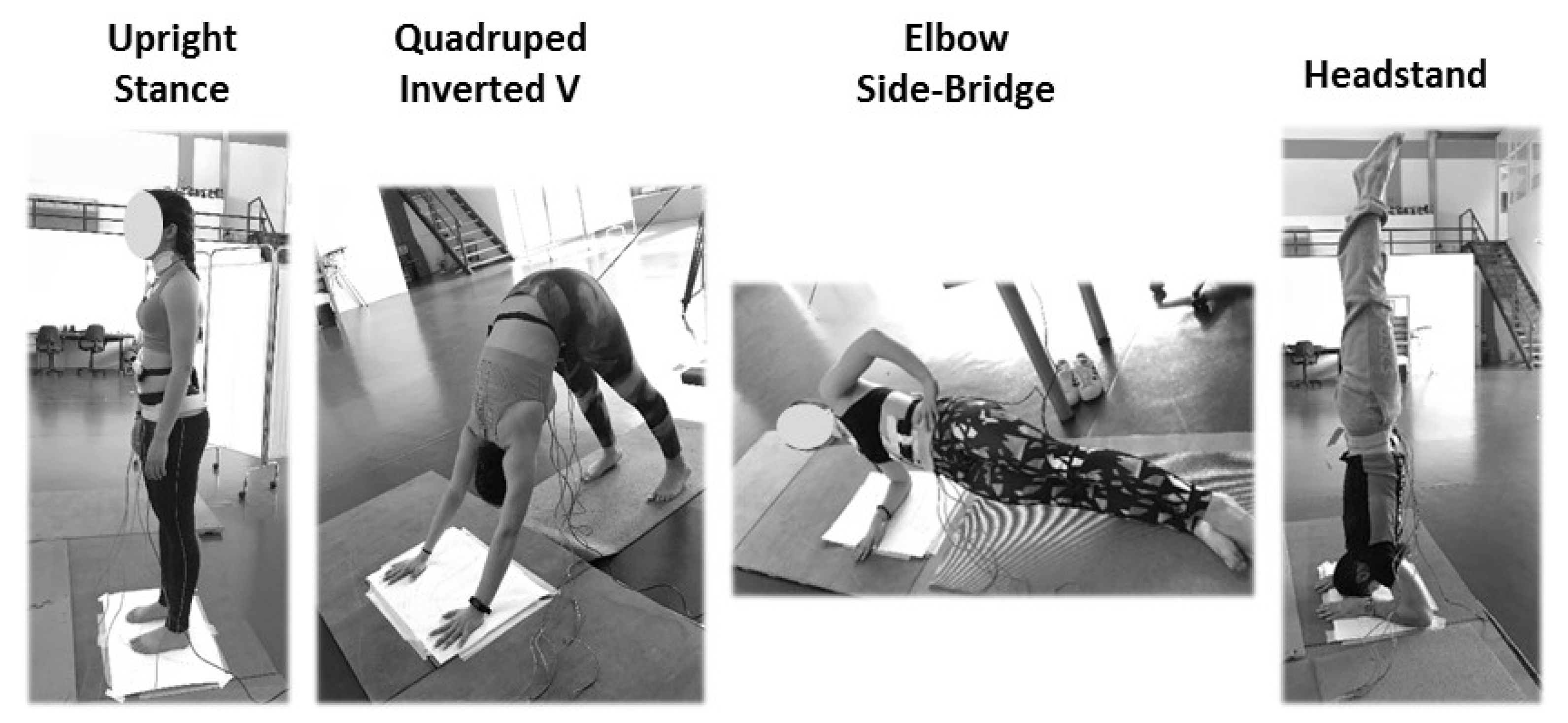
2.3. Experimental Procedure

The participants were instructed to maintain (40 s, barefoot) four body stances, namely, the Upright Stance, the Quadruped Inverted V, the Elbow Side-Bridge, and the Headstand (Figure 1). They maintained all body stances under two breathing conditions: regular and abdominal breathing, both at their preferred respiration frequency. The body stances as well as the breathing types were presented in a rotation order, aiming to obtain a similar number of participants in each body stance × breathing type combination. In all Headstand trials, a trained assistant was standing next to the participant to cushion a case of balance loss. Moreover, a thick mat was positioned in the potential falling area in the case of balance loss, during the Elbow Side-Bridge and the Headstand trials. A total of three successful trials were obtained per participant in each body stance × breathing type combination. One minute rest was allowed among trials of the same body stance, and 3 min rest before initiating the subsequent body stance trials.

2.4. Postural Stability: Data Collection and Data Procession

A force plate sampling at 100 Hz (Kistler Type 9286AA force plate, Bioware Data Acquisition and Analysis Software, version 3.2.6, Kistler Instrument, Winterthur, Switzerland) was used to record the 2D trajectory of the center of pressure (CoP) during all body stance × breathing type trials. A 10 Hz low pass filter (Bioware software filtering options) was applied to all CoP trajectories. To exclude the initiation phase, in all trials, the first 5 s of the CoP trajectories were not used. Thus, from the remaining 30 s, the following CoP variables were extracted (Bioware Software): the CoP Path (mm) and the CoP range (mm) in the anterior–posterior (AP-ax) and the medial–lateral (ML-ay) direction, respectively, as well as the CoP Area (mm/2). The OriginPro 9.0 software (SR1 b76) was used to calculate the CoP Sway area (90% confidence elliptical area) [41]. In continuance, for each postural stability variable, the three trial average (in each body stance × breathing type condition) consisted of the value inserted in statistical analysis.

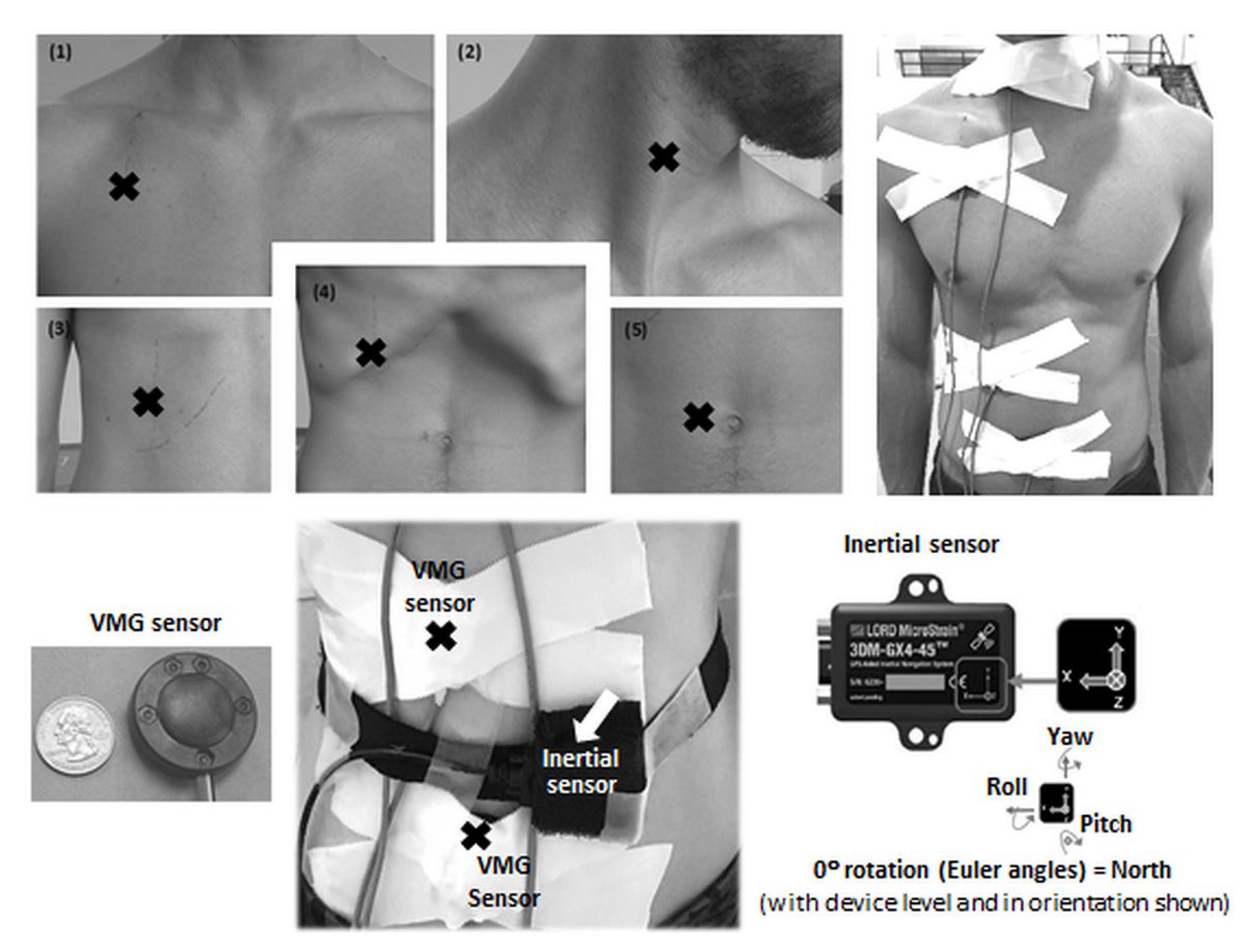
2.5. Respiratory Muscles’ Activation: Data Collection and Data Procession

In synchronization with the force plate recordings (simultaneous initialization of the software used for data collection), four vibromyography sensors (sampling at 2000 Hz, MEMS, MP150, AcqKnowledge software v. 5.0, Biopac Systems, Inc., Santa Barbara, CA, USA) [42] were used to record the respiratory muscles’ activation (Figure 2). On the basis of previous studies, two inspiratory and two expiratory muscles were tested. In specific, the two inspiratory muscles were the sternocleidomastoid (SCM) [43,44,45] and the external intercostal (Ext-IC) [44,45,46,47]. The two expiratory muscles were the Diaphragm [44,45,46,47,48,49,50] and the rectus abdominis (RA) [51]. The VMG sensors were secured with unelastic tape (one sensor per muscle) on the right side of the body (Figure 2) as follows: the SCM sensor was positioned over the middle third of the muscle’s length [46], the Ext-IC sensor was positioned between the second and third intercostal space at the midclavicular line [42,45], the Diaphragm sensor between the seventh and eighth anterior intercostal space at the midclavicular line [42,45,46], and the RA sensor at the umbilicus height and a 2 cm distance from the umbilicus [52].
Before variable extraction, the raw VMG signal was filtered (Matlab R2018A, Mathworks) (5–100 Hz bandpass filter with a cut-off threshold at three standard deviations of the mean value, followed by signal rectification and a final low-pass filter at a 6 Hz cut-off frequency). In continuance, for the 30 s VMG signal, the mean and the standard deviation of the VMG amplitude were calculated separately for each one of the respiratory muscles (expressed in VMG units, according to the AcqKnowledge Biopac software v.5.0). Thus, two variables were defined concerning the activation of each respiratory muscle, that is, the VMG intensity (mean value of the VMG amplitude) and the VMG variation (standard deviation of the VMG amplitude). In continuance, in each body stance × breathing type condition, the three trial average of the VMG intensity and the VMG variation was the value inserted in the statistical analysis.

2.6. Data Reliability

A concern of this study was the minimum number of trials per posture and breathing type that would allow reliable results while also minimizing a potential confounding effect due to fatigue. Thus, two reliability analyses were performed, one before and one after the experimental data collection procedure. There are several forms of the intraclass correlation coefficient (ICC), but considering that the reliability that is in question is in absolute agreement, the random effects, two-way, absolute agreement method (also known as ICC (2,1)) was decided as the most appropriate for the present study [53,54]. The ICC’s upper and lower bounds of their 95% confidence interval (95% CI) were also extracted.
In the reliability analysis before the experimental data collection (a total of five trials) of the ICCs concerning the CoP path were estimated for the trial accumulation from 1 to 2, 1 to 3, 1 to 4, and 1 to 5 across all body stance × breathing type conditions, and the Fleiss [55] classification was used to estimate the ICCs (poor if ICC < 0.40, fair to good if 0.40 ≤ ICC ≤ 0.75, high if ICC > 0.75). The reliability analysis before the experimental data collection procedure indicated three trials as sufficient to obtain a fair to good CoP path reliability (ICCs above 0.70) [55] for the CoP path variable, naming the fundamental postural stability variable. Thus, a total of three trials were decided for the experimental data collection procedure. The reliability analysis after the data collection procedure verified a high CoP (ICCs above 0.85), as well as a high VMG (ICCs above 0.90) reliability, across all body stance × breathing type conditions.

2.7. Statistical Analysis

To test the abdominal breathing effect, one-way ANOVA for repeated measures was applied on the postural stability variables as well as on the variables concerning the respiratory muscles’ activation, separately in each body stance. The level of significance was set at p ≤ 0.05 (SPSS version 26.0, IBM Statistics, Armonk, NY, USA).

3. Results

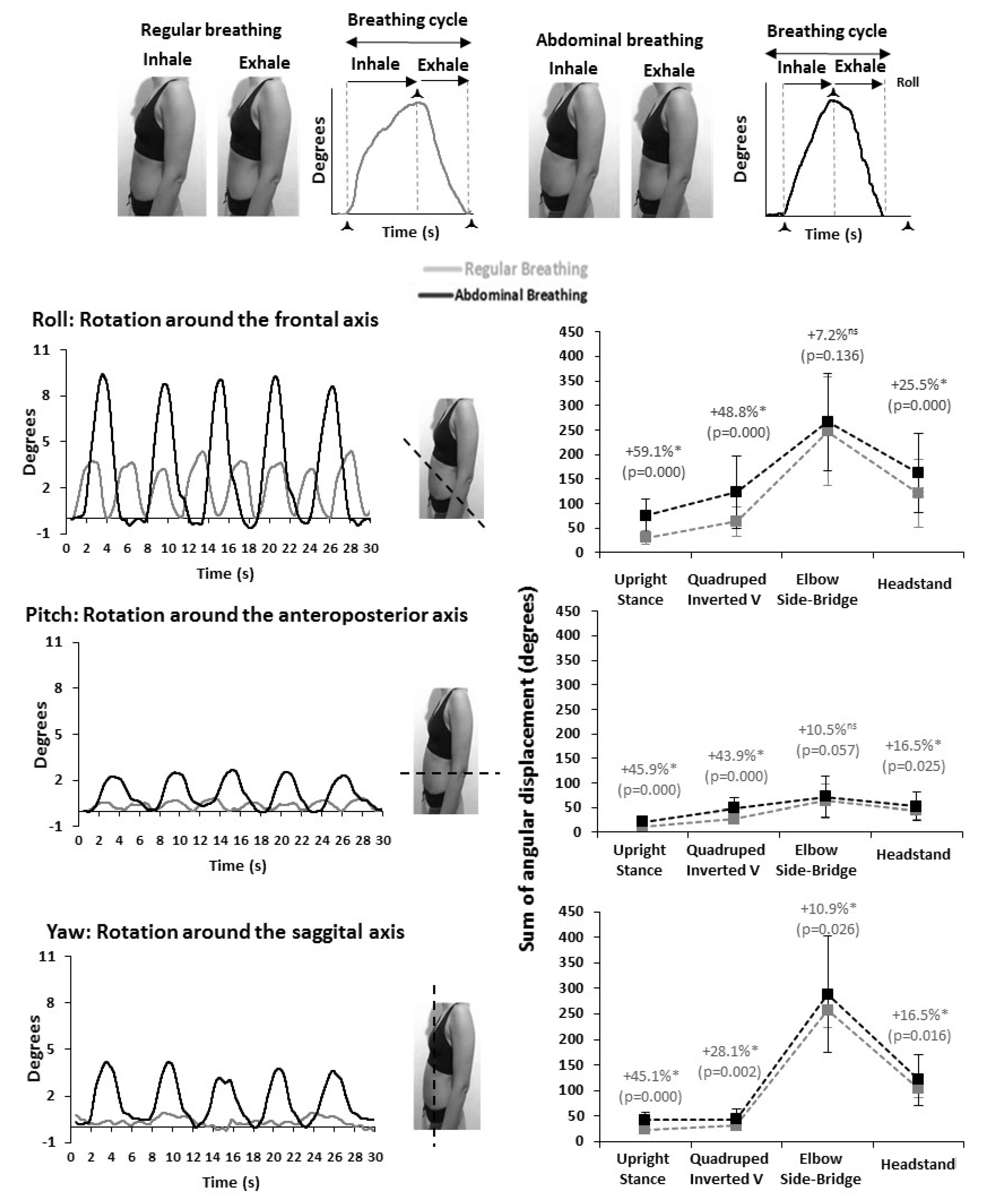
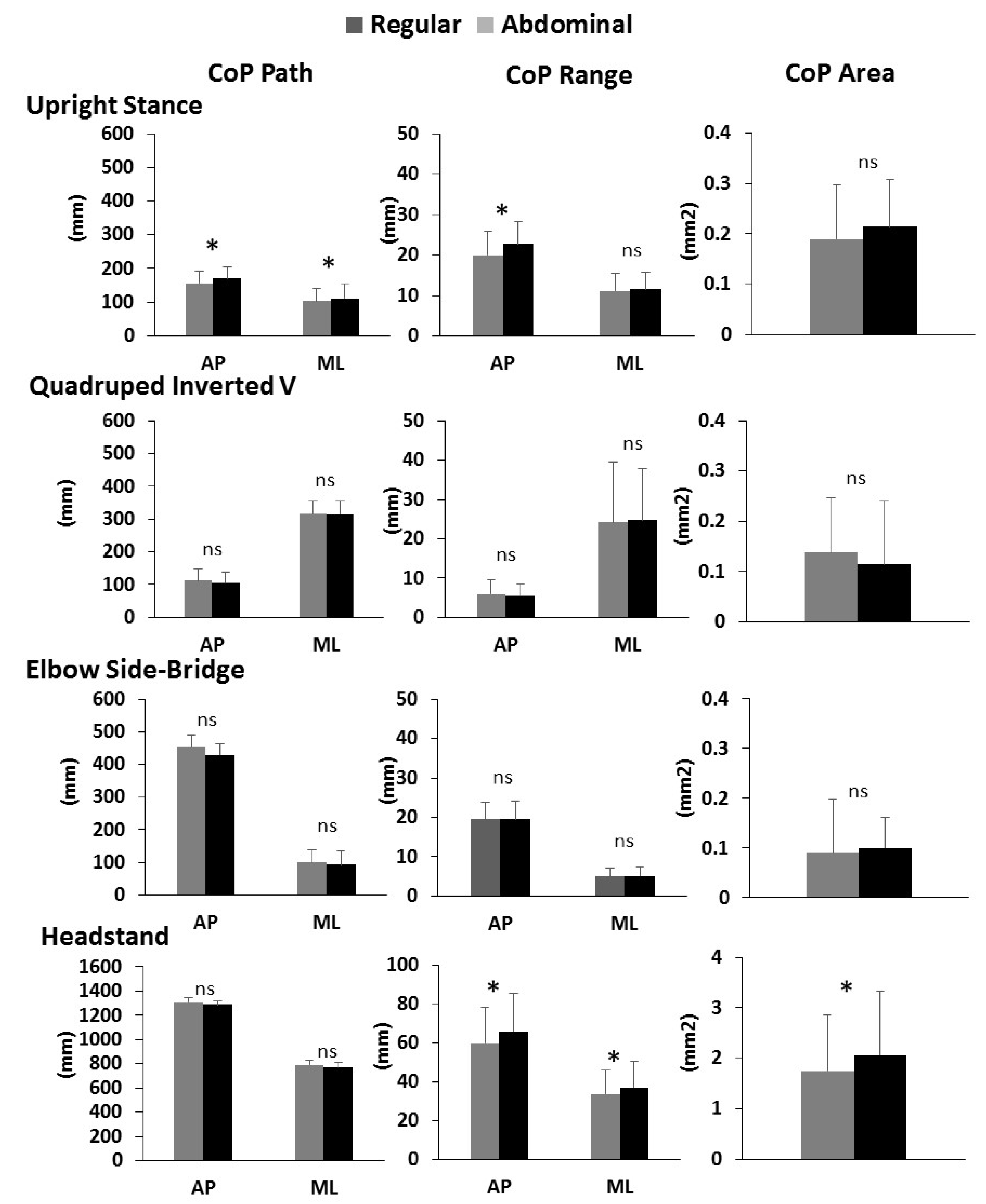
3.1. Abdominal Breathing Effect on Postural Stability

Abdominal breathing had a significant deteriorating effect on postural stability (evidenced as an increase in the CoP variables) in the Upright Stance and the Headstand (p ≤ 0.05); however, it did not affect postural stability in the Quadruped Inverted V and the Elbow Side-Bridge (Figure 4, Table 1). In specific, in the Upright Stance, the abdominal breathing increased significantly (p ≤ 0.05) in both the AP and ML CoP Path, the CoP Range (only in the AP direction), with no significant alteration in the CoP Area (p > 0.05) (Figure 4, Table 1). In the Headstand, the abdominal breathing increased significantly (p ≤ 0.05) the CoP Range (AP and ML direction) as well as the CoP Area, with no significant alteration in the CoP Path (p > 0.05) (Figure 4, Table 1).

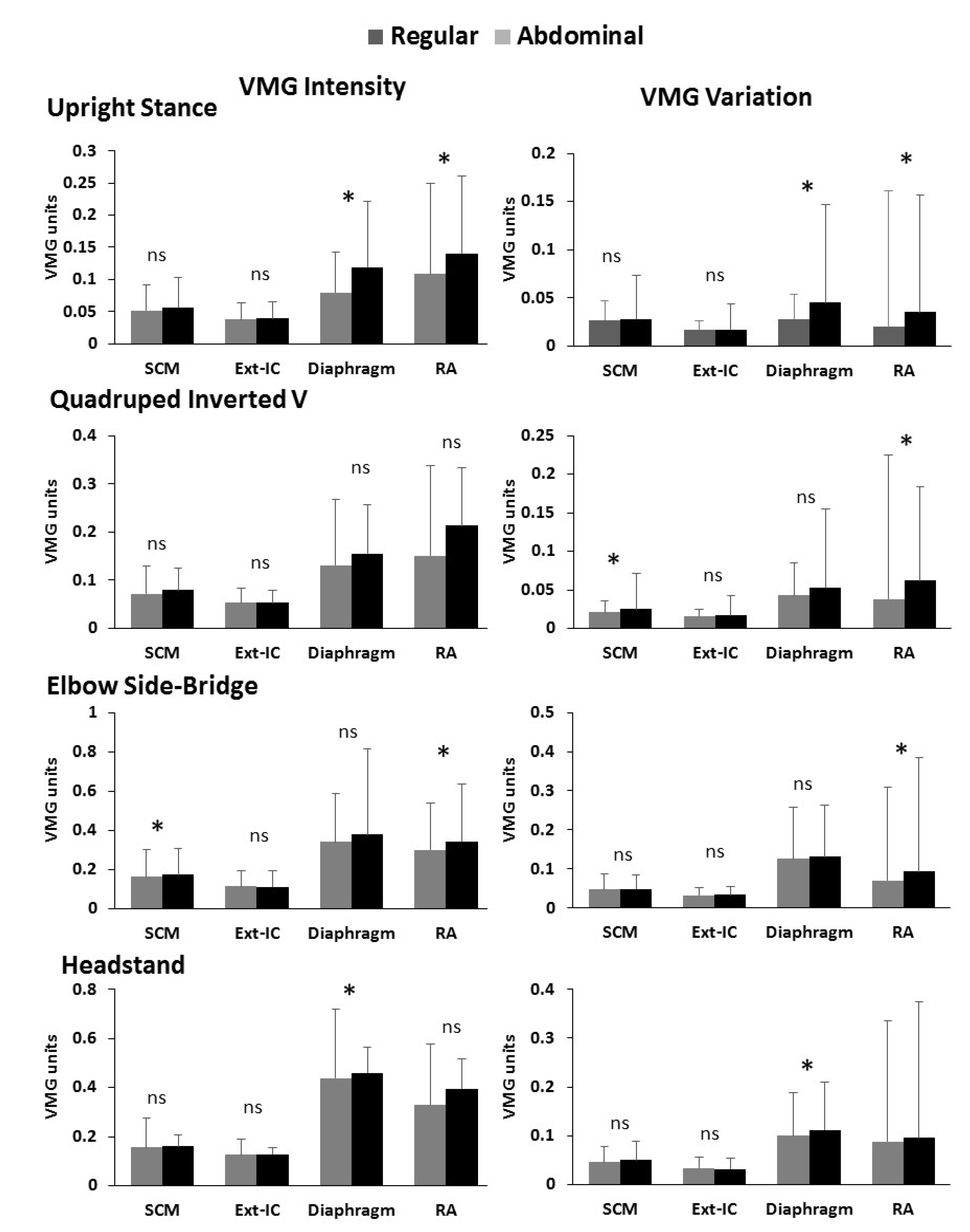
3.2. Abdominal Breathing Effect on the Respiratory Muscle’s Activation

Abdominal breathing had a significant increasing effect (p ≤ 0.05) on the respiratory muscles’ VMG intensity as well as on the VMG variation; however, this was not consistent across all body stances and all muscles (Figure 5, Table 2 and Table 3). In specific, during the Upright Stance, the abdominal breathing significantly increased the VMG intensity and the VMG variation of the Diaphragm and the RA (p ≤ 0.05), with no significant effect on the SCM and the Ext-IC muscles (p > 0.05) (Figure 5, Table 2 and Table 3). In the Quadruped Inverted V, the abdominal breathing significantly increased the VMG variation of the SCM and the RA (p ≤ 0.05), with no significant effect (p > 0.05) on either the VMG variation of the other two examined muscles or the VMG intensity of all four respiratory muscles (Figure 5, Table 2 and Table 3). In the Elbow Side-Bridge, the abdominal breathing significantly increased the VMG intensity of the SCM (p ≤ 0.05) and both the VMG intensity and the VMG variation of the RA (p ≤ 0.05), with no significant effect on the other two examined muscles (p > 0.05) (Figure 5, Table 2 and Table 3). In the Headstand, the abdominal breathing significantly increased the VMG intensity as well as the VMG variation of the Diaphragm (p ≤ 0.05), with no significant effect on the other three respiratory muscles (p > 0.05) (Figure 5, Table 2 and Table 3).

4. Discussion

The present study aimed to examine the abdominal breathing effect on postural stability and the respiratory muscles’ activation during stable as well as rather unstable body stances used in fitness modalities. The results of the study indicate that abdominal breathing had a significant deteriorating effect on postural stability (evidenced as an increase in the CoP variables) as well as an increasing effect on the respiratory muscles’ activation; however, this effect was not consistent across body stances. Furthermore, it was not consistent across the postural stability variables, as well as across all respiratory muscles.

4.1. Postural Stability

In specific, the deteriorating effect of abdominal breathing on postural stability was significant for the Upright Stance and the Headstand but not for the Quadruped Inverted V and the Elbow Side-Bridge. The significant abdominal breathing effect on postural stability of the Upright Stance and the Headstand was evidenced in both the AP and ML CoP directions, but it was not consistent across all CoP variables in both body stances.
In specific, when abdominal breathing was practiced during the Upright Stance, the CoP Path and the CoP Range were significantly increased but not the CoP area. However, when abdominal breathing was practiced during the Headstand, the CoP Range and the CoP Area were significantly increased but not the CoP Path. The results of our study are in agreement with Hernandez et al. [36], who found that the abdominal compared to regular breathing produced a significant increase in the examined CoP variables (CoP variability). During the Upright Stance, the increase in the CoP Path under abdominal breathing may be associated with an increase in the respiratory amplitude compared to regular breathing [17,19,56].
The breathing process itself is an endogenous source of postural stability disturbance [7,17,18,19,20,21] due to the anatomical displacements that take place. The respiration process causes changes in the projection of the center of gravity of the human body relative to the boundaries of the support base, which requires a counterbalancing response through the CoP trajectory so that body equilibrium is maintained as stable as possible [57]. Abdominal breathing is characterized by a slow and deep breathing pattern [5], during which the diaphragmatic contraction pushes the viscera downward and forward (i.e., diaphragm-related abdominal expansion) [6,19,57,58]. Abdominal breathing also appears to affect postural stability through its influence on the respiratory muscles’ activation [7,8] and specifically through the respiratory muscles’ effect on trunk stability [9,10,11,12].
During regular quiet breathing, expiration is performed by the passive recoil of the diaphragm [13]. However, in abdominal breathing, the higher activation of the abdominal muscles is associated with their purposeful contraction to increase the intra-abdominal pressure by pushing the diaphragm upwards, rather than the passive recoil of the diaphragm observed in regular breathing [13]. In specific, in coordination, the diaphragm and the abdominal muscles produce a hydraulic effect in the abdominal cavity that assists spinal stabilization by stiffening the lumbar spine through increased intra-abdominal pressure [9,10,11,12]. Moreover, during abdominal breathing, there is a shift in abdominal organs due to the pressure difference exerted by the diaphragm, which is accompanied by an increase in the elastance of the abdomen to allow the abdominal wall to project forward as the abdominal organs shift due to pressure [14]. Subsequently, the abdominal muscles contract to increase the intraabdominal pressure [13,15,16] and to act against the flattened diaphragm due to deep inspiration [15].

4.2. Respiratory Muscles’ Activation

In the present study, abdominal breathing appeared to significantly increase the activation of the diaphragm and the RA muscle, while the SCM and the Ext-IC showed no significant difference. It must be noted that the abdominal breathing effect on the respiratory muscles’ activation was examined for the overall breathing cycle, without distinction between the inspiration and the expiration phase. The inconsistent across-body stances effect of abdominal breathing on the respiratory muscles’ activation may be possibly associated with the findings of Nelson [59], who suggests that the type of breathing affects the way the respiratory muscles are used for postural stability as well as non-breathing movements. When the breathing effort increases, the increased inspiratory demand would inevitably reduce the respiratory muscles’ ability to perform their postural duties [12], which may lead to reduced postural control [8]. Thus, it is likely that the increase in the breathing effort may differentially affect postural stability in stable compared to rather unstable body stances as, for example, the inverted body stances. Thus, the different breathing mechanics (deep and slow inspiration as well as maximum expiration) in the abdominal compared to regular breathing may explain the increased activation of the diaphragm and the RA during the Upright Stance and the Elbow Side-Bridge. These results are consistent with previous studies demonstrating that during the Elbow Side-Bridge, the activation of the RA muscle was greater in maximum expiration compared with resting expiration [60]. The non-significant difference of the SCM and the Ext-IC activation between the regular and abdominal breathing across all body stances agrees with previous studies [61,62,63] and may be explained by the demand for a stable ribcage during abdominal breathing [19,64]. In specific, the Ext-IC activation was found to differ between breathing types that induce a ribcage movement and those that necessitate a still ribcage as the upper-costal [61] and mixed type (costo-diaphragmatic) [62] respiration.
A body stance specificity could explain the non-consistency of the abdominal breathing effect on the respiratory muscles’ activation. The Elbow Side-Bridge requires a high activation of the abdominal muscles throughout the respiratory cycle to maintain body stability [24] and to prevent the projection of the body’s center of gravity beyond the limits of the support base, making abdominal breathing rather non-feasible. In our study, the kinesiological contradiction for an intense relaxation of the abdominal muscles in the expiration phase during the elbow side bridge posture was evidenced in the inertially sensed similar angular displacements of the abdominal wall in both regular and abdominal breathing. Concerning the Quadruped Inverted V, the rather larger base of support and the rather central projection of the body’s center of gravity during the quadruped inverted V stance may most likely explain the inability of abdominal breathing to induce a postural stability difference compared to regular breathing [65]. The inverted stances are expected to induce postural instability and elicit compensatory motor control segmental strategies (enhanced motion at the distal segments of the legs and the elbow joint in order to minimize motion variability in the head and trunk) [26,27,28,29,30,31,32,33]. The isometric contraction of the abdominal muscles is among the segmental motor control strategies aiming at postural stability correction during inverted body stances (i.e., handstand) [29]. Furthermore, during the Headstand inverted stance, the neck area adopts the antigravity support role and sustains muscular and spinal loads that contrast or exaggerate those that the neck musculature is designed for (i.e., increased lateral deviations in the thoracic spine) [26,28,29]. As such, contrasting or exaggerating muscular and spinal loads, in combination with the resistance rather than assisted by gravitational force activation of the Ext-IC and the Diaphragm muscles, may also associate with the non-consistency of the significance of the abdominal breathing effect across muscles and body stances.
The results of the present work should be evaluated under the limitations of the participants’ characteristics who were selected to meet specific criteria concerning their ability for abdominal breathing and the maintenance of the Headstand. A gender bias could also be argued due to the disparity in the number of women and men participants. However, this disparity reflects the fact that women rather than men constitute the vast majority of the persons participating in fitness modalities where abdominal breathing is practiced while maintaining the body stances examined in the present study. One could also argue the adequacy of the sample size when reporting non-significant results. However, the sample size was decided with a priori G-Power analysis, on the basis of the previous relevant studies [7,8] as well as our preliminary data collection including 10 participants (6 women and 4 men). The strong correlation between the regular and the abdominal breathing variables in both the postural stability and the respiratory muscles’ activation (average value r = 0.70 and r = 0.74, for the preliminary and the actual data collection, respectively) allows for the assertion that a greater sample size is not likely to have altered the statistical power of the present results. One could also question the validity of abdominal breathing as the type of breathing was verified through inertial sensors and not through an invasive procedure [48,52]. Nevertheless, several studies apply a surface recording of diaphragm activity [42,45,46,47] instead of the invasive entry of a gastroesophageal catheter [48,65]. Furthermore, the use of inertial sensors as a research tool for examining the mechanics of breathing, as applied in the present study, has been documented in previous study protocols for kinetic changes of either the thoracic cavity [38,39,40] or the abdominal wall [39].
In conclusion, abdominal breathing had a significant deteriorating effect on postural stability and an increasing one on the respiratory muscles’ activation during the maintenance of non-everyday living body stances practiced in fitness modalities by physically active individuals. However, this was not consistent across body stances, indicating a body stance specificity of the abdominal breathing effect. Thus, before the request for abdominal breathing in popular fitness modalities, one should acknowledge the postural stability as well as the breathing demands of each particular stance, particularly for the inverted ones. In the present study, the Upright Stance, the Headstand, and the Elbow Side-Bridge demonstrated a 1.5, 1.7, and 3 times greater CoP Path, respectively, in the anterior–posterior than the medial–lateral direction, whereas, the Quadruped Inverted V demonstrates an about 4.5 times greater CoP Path in the medial–lateral than the anterior–posterior direction. Such directional differences in postural stability may stem from the segmental configuration of each particularly body stance, which ultimately defines not only the shape and the magnitude of the base of support but also the body weight percentage that is loaded on the segmental supports (for example, in the present study, about 38% and 66% of BW was supported by the hands and the forearm, in the Quadruped Inverted V and the Elbow Side-Bridge, respectively). Future studies could identify a body stance continuum concerning the postural stability and the respiratory muscles’ activation for everyday living body stances employed by sedentary individuals as well as body stances employed by physically active individuals during their participation in fitness modalities.

Author Contributions

Conceptualization, A.F. and E.R.; methodology, A.F. and E.R.; formal analysis, A.F. and E.R.; investigation, A.F. and E.R.; data curation, A.F., A.E., and E.R.; writing—original draft preparation, A.F. and E.R.; writing—review and editing, A.F., A.E., K.B. and E.R.; visualization, A.F., A.E. and E.R.; supervision, E.R. and K.B.; project administration, K.B. and E.R. All authors have read and agreed to the published version of the manuscript.

Funding

This research received no external funding.

Institutional Review Board Statement

The study was conducted in accordance with the Declaration of Helsinki and approved by the Institutional Ethics Committee on Research Ethics-Bioethics of the School of Physical Education and Sport Science, National and Kapodistrian University of Athens (approval number/date: 1046/14-02-2018).

Informed Consent Statement

Informed consent was obtained from all subjects involved in the study.

Acknowledgments

We acknowledge the National and Kapodistrian University of Athens, Greece, for the financial support concerning the publication fee of this paper in the Biomechanics, MDPI.

Conflicts of Interest

The authors declare no conflict of interest.

References

  1. Martarelli, D.; Cocchioni, M.; Scuri, S.; Pompei, P. Diaphragmatic breathing reduces exercise-induced oxidative stress. Evid. Based. Complement. Altern. Med. 2011, 2011, 932430. [Google Scholar] [CrossRef] [PubMed]
  2. Steurer-Stey, C.; Russi, E.W.; Steurer, J. Complementary and Alternative Medicine in Asthma: Do They Work? Swiss Med. Wkly. 2002, 132, 338–344. [Google Scholar] [PubMed]
  3. Dhruva, A.; Miaskowski, C.; Abrams, D.; Acree, M.; Cooper, B.; Goodman, S.; Hecht, F.M. Yoga Breathing for Cancer Chemotherapy-Associated Symptoms and Quality of Life: Results of a Pilot Randomized Controlled Trial. J. Altern. Complement. Med. 2012, 18, 473–479. [Google Scholar] [CrossRef] [PubMed]
  4. Ma, X.; Yue, Z.-Q.; Gong, Z.-Q.; Zhang, H.; Duan, N.-Y.; Shi, Y.-T.; Wei, G.-X.; Li, Y.-F. The Effect of Diaphragmatic Breathing on Attention, Negative Affect and Stress in Healthy Adults. Front. Psychol. 2017, 8, 1–12. [Google Scholar] [CrossRef]
  5. Wahbeh, H.; Elsas, S.-M.; Oken, B.S. Mind-Body Interventions: Applications in Neurology. Neurology 2008, 70, 2321–2328. [Google Scholar] [CrossRef]
  6. Perry, S.F.; Similowski, T.; Klein, W.; Codd, J.R. The Evolutionary Origin of the Mammalian Diaphragm. Respir. Physiol. Neurobiol. 2010, 171, 1–16. [Google Scholar] [CrossRef]
  7. Hamaoui, A.; Hudson, A.L.; Laviolette, L.; Nierat, M.C.; Do, M.C.; Similowski, T. Postural Disturbances Resulting from Unilateral and Bilateral Diaphragm Contractions: A Phrenic Nerve Stimulation Study. J. Appl. Physiol. 2014, 117, 825–832. [Google Scholar] [CrossRef]
  8. Kim, S.-H.; Shin, H.-J.; Cho, H.-Y. Impact of Types of Breathing on Static Balance Ability in Healthy Adults. Int. J. Environ. Res. Public Health 2022, 19, 1205. [Google Scholar] [CrossRef]
  9. Bordoni, B.; Zanier. Anatomic Connections of the Diaphragm Influence of Respiration on the Body System. J. Multidiscip. Healthc. 2013, 6, 281. [Google Scholar] [CrossRef]
  10. Hodges, P.W.; Butler, J.E.; McKenzie, D.K.; Gandevia, S.C. Contraction of the Human Diaphragm during Rapid Postural Adjustments. J. Physiol. 1997, 505, 539–548. [Google Scholar] [CrossRef]
  11. Hodges, P.W.; Gandevia, S.C. Changes in Intra-Abdominal Pressure during Postural and Respiratory Activation of the Human Diaphragm. J. Appl. Physiol. 2000, 89, 967–976. [Google Scholar] [CrossRef]
  12. Kocjan, J.; Adamek, M.; Gzik-Zroska, B.; Czyżewski, D.; Rydel, M. Network of Breathing. Multifunctional Role of the Diaphragm: A Review. Adv. Respir. Med. 2017, 85, 224–232. [Google Scholar] [CrossRef]
  13. Kim, E.; Lee, H. The Effects of Deep Abdominal Muscle Strengthening Exercises on Respiratory Function and Lumbar Stability. J. Phys. Ther. Sci. 2013, 25, 663–665. [Google Scholar] [CrossRef]
  14. De Troyer, A.; Boriek, A. Mechanics of the Respiratory Muscles. Compr. Physiol. 2011, 1, 1273–1300. [Google Scholar] [CrossRef]
  15. Kolář, P.; Šafářová, M. Dynamic Neuromuscular Stabilization. In Clinical Rehabilitation, 1st ed.; Kolář, P., Kobesová, A., Eds.; K. Vápence 16: Praha, Czech Republic, 2013; pp. 257–258. [Google Scholar]
  16. Sieck, G.C.; Ferreira, L.F.; Reid, M.B.; Mantilla, C.B. Mechanical Properties of Respiratory Muscles. In Comprehensive Physiology; John Wiley & Sons, Inc.: Hoboken, NJ, USA, 2013; Volume 3, pp. 1533–1567. [Google Scholar] [CrossRef]
  17. Bouisset, S.; Duchêne, J.L. Is Body Balance More Perturbed by Respiration in Seating than in Standing Posture? Neuroreport 1994, 5, 957–960. [Google Scholar] [CrossRef]
  18. Gurfinkel, V.S.; Kots, Y.M.; Paltsev, E.I.F.A. The Compensation of Respiratory Disturbances of the Erect Posture of Man as an Example of the Organisation of Interarticular Action. In Systems; Gurfinkel, V.S., Fomin, T.M.L.S., Eds.; MIT Press: Cambridge, MA, USA, 1971. [Google Scholar]
  19. Hamaoui, A.; Gonneau, E.; Le Bozec, S. Respiratory Disturbance to Posture Varies According to the Respiratory Mode. Neurosci. Lett. 2010, 475, 141–144. [Google Scholar] [CrossRef]
  20. Hunter, I.W.; Kearney, R.E. Respiratory Components of Human Postural Sway. Neurosci. Lett. 1981, 25, 155–159. [Google Scholar] [CrossRef]
  21. Stephens, R.J.; Haas, M.; Moore, W.L.; Emmil, J.R.; Sipress, J.A.; Williams, A. Effects of Diaphragmatic Breathing Patterns on Balance: A Preliminary Clinical Trial. J. Manip. Physiol. Ther. 2017, 40, 169–175. [Google Scholar] [CrossRef]
  22. Vera-Garcia, F.J.; Moreside, J.M.; McGill, S.M. MVC Techniques to Normalize Trunk Muscle EMG in Healthy Women. J. Electromyogr. Kinesiol. 2010, 20, 10–16. [Google Scholar] [CrossRef]
  23. Vera-Garcia, F.J.; Irles-Vidal, B.; Prat-Luri, A.; García-Vaquero, M.P.; Barbado, D.; Juan-Recio, C. Progressions of Core Stabilization Exercises Based on Postural Control Challenge Assessment. Eur. J. Appl. Physiol. 2020, 120, 567–577. [Google Scholar] [CrossRef]
  24. Lehman, G.J.; Hoda, W.; Oliver, S. Trunk Muscle Activity during Bridging Exercises on and off a Swiss Ball. Chiropr. Osteopat. 2005, 13, 14. [Google Scholar] [CrossRef]
  25. García-Vaquero, M.P.; Moreside, J.M.; Brontons-Gil, E.; Peco-González, N.; Vera-Garcia, F.J. Trunk Muscle Activation during Stabilization Exercises with Single and Double Leg Support. J. Electromyogr. Kinesiol. 2012, 22, 398–406. [Google Scholar] [CrossRef]
  26. Campos, M.H.; Giraldi, N.M.; Gentil, P.; de Lira, C.A.B.; Vieira, C.A.; de Paula, M.C. The Geometric Curvature of the Spine during the Sirshasana, the Yoga’s Headstand. J. Sports Sci. 2017, 35, 1134–1141. [Google Scholar] [CrossRef]
  27. Clément, G.; Rézette, D. Motor Behavior Underlying the Control of an upside-down Vertical Posture. Exp. Brain Res. 1985, 59, 478–484. [Google Scholar] [CrossRef]
  28. Hector, R.; Jensen, J.L. Sirsasana (Headstand) Technique Alters Head/Neck Loading: Considerations for Safety. J. Bodyw. Mov. Ther. 2015, 19, 434–441. [Google Scholar] [CrossRef]
  29. Hedbávný, P.; Sklena, J.; Hupka, D.; Kalichová, M. Balancing in Handstand on the Floor. Sci. Gymnast. J. 2013, 5, 69–80. [Google Scholar]
  30. Gautier, G.; Thouvarecq, R.; Chollet, D. Visual and Postural Control of an Arbitrary Posture: The Handstand. J. Sports Sci. 2007, 25, 1271–1278. [Google Scholar] [CrossRef]
  31. Omorczyk, J.; Bujas, P.; Puszcza\lowska-Lizis, E.; Biskup, L. Balance in Handstand and Postural Stability in Standing Position in Athletes Practicing Gymnastics. Acta. Bioeng. Biomech. 2018, 20, 139–147. [Google Scholar] [CrossRef]
  32. Slobounov, S.M.; Newell, K.M. Postural Dynamics in Upright and Inverted Stances. J. Appl. Biomech. 1996, 12, 185–196. [Google Scholar] [CrossRef]
  33. Sobera, M.; Siedlecka, B.; Piestrak, P.; Sojka-Krawiec, K.; Graczykowska, B. Maintaning Body Balance in Extreme Positions. Biol. Sport. 2007, 24, 81–88. [Google Scholar]
  34. Faul, F.; Erdfelder, E.; Lang, A.G.; Buchner, A. G*Power 3: A flexible statistical power analysis program for the social, behavioral, and biomedical sciences. Behav. Res. Methods 2007, 39, 175–191. [Google Scholar] [CrossRef] [PubMed]
  35. Faul, F.; Erdfelder, E.; Buchner, A.; Lang, A.G. Statistical power analyses using G*Power 3.1: Tests for correlation and regression analyses. Behav. Res. Methods 2009, 41, 1149–1160. [Google Scholar] [CrossRef] [PubMed]
  36. Hernandez, L.; Manning, J.; Zhang, S. Voluntary Control of Breathing Affects Center of Pressure Complexity during Static Standing in Healthy Older Adults. Gait Posture 2019, 68, 488–493. [Google Scholar] [CrossRef] [PubMed]
  37. Lee, H.-Y.; Cheon, S.-H.; Yong, M.-S. Effect of Diaphragm Breathing Exercise Applied on the Basis of Overload Principle. J. Phys. Ther. Sci. 2017, 29, 1054–1056. [Google Scholar] [CrossRef] [PubMed]
  38. Bates, A.; Ling, M.J.; Mann, J.; Arvind, D.K. Respiratory Rate and Flow Waveform Estimation from Tri-Axial Accelerometer Data. In Proceedings of 2010 International Conference on Body Sensor Networks, IEEE, Singapore, 7–9 June 2010. [Google Scholar] [CrossRef]
  39. Haescher, M.; Matthies, D.J.C.; Trimpop, J.; Urban, B. A Study on Measuring Heart- and Respiration-Rate via Wrist-Worn Accelerometer-Based Seismocardiography (SCG) in Comparison to Commonly Applied Technologies. In Proceedings of the iWOAR 2015—2nd International Workshop on Sensor-Based Activity Recognition and Interaction, Rostock Warnemade, Germany, 25–26 June 2015. [Google Scholar] [CrossRef]
  40. Phan, D.H.; Bonnet, S.; Guillemaud, R.; Castelli, E.; Pham Thi, N.Y. Estimation of Respiratory Waveform and Heart Rate Using an Accelerometer. In Proceedings of the 2008 30th Annual International Conference of the IEEE Engineering in Medicine and Biology Society, Vancouver, BC, Canada, 14 October 2018. [Google Scholar] [CrossRef]
  41. Schubert, P.; Kirchner, M. Ellipse Area Calculations and Their Applicability in Posturography. Gait Posture 2014, 39, 518–522. [Google Scholar] [CrossRef] [PubMed]
  42. Chien, M.-Y.; Wu, Y.-T.; Chang, Y.-J. Assessment of Diaphragm and External Intercostals Fatigue from Surface EMG Using Cervical Magnetic Stimulation. Sensors 2008, 8, 2174–2187. [Google Scholar] [CrossRef] [PubMed]
  43. De Mayo, T.; Miralles, R.; Barrero, D.; Bulboa, A.; Carvajal, D.; Valenzuela, S.; Ormeño, G. Breathing Type and Body Position Effects on Sternocleidomastoid and Suprahyoid EMG Activity. J. Oral. Rehabil. 2005, 32, 487–494. [Google Scholar] [CrossRef]
  44. Druz, W.S.; Sharp, J.T. Activity of Respiratory Muscles in Upright and Recumbent Humans. J. Appl. Physiol. Respir. Environ. Exerc. Physiol. 1981, 51, 1552–1561. [Google Scholar] [CrossRef]
  45. Santos, T.V.; Ruas, G.; Sande de Souza, L.A.P.; Volpe, M.S. Influence of Forward Leaning and Incentive Spirometry on Inspired Volumes and Inspiratory Electromyographic Activity during Breathing Exercises in Healthy Subjects. J. Electromyogr. Kinesiol. 2012, 22, 961–967. [Google Scholar] [CrossRef]
  46. Segizbaeva, M.O.; Pogodin, M.A.; Aleksandrova, N.P. Effects of Body Positions on Respiratory Muscle Activation during Maximal Inspiratory Maneuvers. In Advances in Experimental Medicine and Biology; Pokorski, M., Ed.; Springer: Dordrecht, The Netherlands, 2013; Volume 756, pp. 355–363. [Google Scholar] [CrossRef]
  47. Valenzuela, S.; Miralles, R.; Santander, H.; Bull, R.; Cordova, R.; Celhay, I.; Cavada, G.; Gutiérrez, M.F. Effects of Breathing Type on Electromyographic Activity of Respiratory Muscles at Different Body Positions. Cranio 2017, 35, 110–115. [Google Scholar] [CrossRef]
  48. Druz, W.S.; Sharp, J.T. Electrical and Mechanical Activity of the Diaphragm Accompanying Body Position in Severe Chronic Obstructive Pulmonary Disease. Am. Rev. Respir. Dis. 1982, 125, 275–280. [Google Scholar] [CrossRef]
  49. Hudson, A.L.; Joulia, F.; Butler, A.A.; Fitzpatrick, R.C.; Gandevia, S.C.; Butler, J.E. Activation of Human Inspiratory Muscles in an upside-down Posture. Respir. Physiol. Neurobiol. 2016, 226, 152–159. [Google Scholar] [CrossRef] [PubMed]
  50. Takazakura, R.; Takahashi, M.; Nitta, N.; Murata, K. Diaphragmatic Motion in the Sitting and Supine Positions: Healthy Subject Study Using a Vertically Open Magnetic Resonance System. J. Magn. Reson. Imaging 2004, 19, 605–609. [Google Scholar] [CrossRef] [PubMed]
  51. Kera, T.; Maruyama, H. The Effect of Posture on Respiratory Activity of the Abdominal Muscles. J. Physiol. Anthropol. Appl. Hum. Sci. 2005, 24, 259–265. [Google Scholar] [CrossRef] [PubMed]
  52. Maarsingh, E.J.; van Eykern, L.A.; Sprikkelman, A.B.; Hoekstra, M.O.; van Aalderen, W.M. Respiratory Muscle Activity Measured with a Noninvasive EMG Technique: Technical Aspects and Reproducibility. J. Appl. Physiol. 2000, 88, 1955–1961. [Google Scholar] [CrossRef] [PubMed]
  53. Koo, T.K.; Li, M.Y. A Guideline of Selecting and Reporting Intraclass Correlation Coefficients for Reliability Research. J. Chiropr. Med. 2016, 15, 155–163. [Google Scholar] [CrossRef] [PubMed]
  54. Pinsault, N.; Vuillerme, N. Test-Retest Reliability of Centre of Foot Pressure Measures to Assess Postural Control during Unperturbed Stance. Med. Eng. Phys. 2009, 31, 276–286. [Google Scholar] [CrossRef] [PubMed]
  55. Fleiss, J.L. Reliability of Measurement. In The Design and Analysis of Clinical Experiments; Fleis, J.L., Ed.; Wiley: New York, NY, USA, 1986; pp. 1–32. [Google Scholar]
  56. Rodrigues, G.D.; Gurgel, J.L.; Gonçalves, T.R.; Porto, F.; Paulo, P. Influence of Breathing Patterns and Orthostatic Stress on Postural Control in Older Adults. Geriatr. Gerontol. Int. 2018, 18, 692–697. [Google Scholar] [CrossRef] [PubMed]
  57. Kuznetsov, N.A.; Riley, M.A. Effects of Breathing on Multijoint Control of Center of Mass Position during Upright Stance. J. Mot. Behav. 2012, 44, 241–253. [Google Scholar] [CrossRef]
  58. Schmid, M.; Conforto, S.; Bibbo, D.; D’Alessio, T. Respiration and Postural Sway: Detection of Phase Synchronizations and Interactions. Hum. Mov. Sci. 2004, 23, 105–119. [Google Scholar] [CrossRef]
  59. Nelson, N. Diaphragmatic Breathing: The Foundation of Core Stability. Strength Cond. J. 2012, 34, 34–40. [Google Scholar] [CrossRef]
  60. Ishida, H.; Watanabe, S. Maximum Expiration Activates the Abdominal Muscles during Side Bridge Exercise. J. Back Musculoskelet. Rehabil. 2014, 27, 481–484. [Google Scholar] [CrossRef] [PubMed]
  61. Celhay, I.; Cordova, R.; Miralles, R.; Meza, F.; Erices, P.; Barrientos, C.; Valenzuela, S. Effect of upper Costal and Costo-Diaphragmatic Breathing Types on Electromyographic Activity of Respiratory Muscles. Cranio 2015, 33, 100–106. [Google Scholar] [CrossRef] [PubMed]
  62. Cordova, R.; Miralles, R.; Bull, R.; Gamboa, N.A.; Santander, H.; Valenzuela, S.; Fuentes, A.D.; Morales, N.S.; Gutierrez, M.F. Respiratory EMG Activity between Subjects with Costo-Diaphragmatic, Upper Costal or Mixed Breathing Type. J. Sleep. Disord. Ther. 2019, 8, 2167-0277. [Google Scholar] [CrossRef]
  63. Miralles, R.; Gamboa, N.A.; Gutiérrez, M.F.; Valenzuela, S.; Bull, R.; Fuentes, A.D. Effect of Breathing Type on Electromyographic Activity of Respiratory Muscles during Tooth Clenching at Different Decubitus Positions. Cranio 2019, 9634, 28–34. [Google Scholar] [CrossRef]
  64. Cahalin, L.P.; Braga, M.; Matsuo, Y.; Hernandez, E.D. Efficacy of Diaphragmatic Breathing in Persons with Chronic Obstructive Pulmonary Disease: A Review of the Literature. J. Cardiopulm. Rehabil. 2002, 22, 7–21. [Google Scholar] [CrossRef]
  65. Pollock, A.S.; Durward, B.R.; Rowe, P.J.; Paul, J.P. What Is Balance? Clin. Rehabil. 2000, 14, 402–406. [Google Scholar] [CrossRef]
Figure 1. The four body stances of the experimental protocol and the positioning on the force plate.
Biomechanics 02 00037 g001
Figure 2. Positioning of the VMG and inertial sensors: All VMG sensors were positioned on the right body side. The inertial sensor used to verify the abdominal breathing pattern was positioned on the left body side, 2 cm distance from the umbilicus and at 2 cm above the umbilicus height. All sensors were well secured using medical adhesive tape throughout all trials. (1) Sternocleidomastoid muscle (SCM): over the middle third of the muscle’s length. (2) External intercostal muscles (Ext-IC): between the second and third intercostal spaces. (3) Diaphragm: between the seventh and eighth anterior intercostal spaces, in the projection of the midclavicular line. (4) Diaphragm: a purposeful drawing of the abdominal wall was asked to facilitate the visualization of the sensor’s positioning. (5) Rectus abdominis (RA): at the umbilicus height and a 2 cm distance from the umbilicus.
Biomechanics 02 00037 g002
Figure 3. Abdominal breathing verification. Top and Left: Exemplary data for the inertially sensed angular displacements (Roll, Pitch, Yaw) of the abdominal wall during the regular (grey lines) and the abdominal (black lines) breathing in the Upright Stance. Right: Percentage of difference for the Roll, Pitch, and Yaw Euler angle with significance (*) of differences at p < 0.05 (paired t-test, SPSS 26.0). ns: non-significant abdominal breathing effect (p > 0.05).
Biomechanics 02 00037 g003
Figure 4. Abdominal breathing effect on postural stability: Mean (SD) of the postural stability variables (CoP Path, CoP Range, CoP Area) for the anterior–posterior (AP) and medial–lateral (ML) direction, in the Upright Stance, the Quadruped Inverted V, the Elbow Side-Bridge, and the Headstand, during the regular (grey bars) and the abdominal (black bars) breathing. * Significant abdominal breathing effect (p ≤ 0.05), ns: non-significant abdominal breathing effect (p > 0.05).
Biomechanics 02 00037 g004
Figure 5. Abdominal breathing effect on the respiratory muscles’ activation: VMG intensity and VMG variation during the regular (grey bars) and the abdominal (black bars) breathing in each body stance. SCM: sternocleidomastoid muscle, Ext-IC: external intercostal muscles, RA: rectus abdominis muscle. * Significant abdominal breathing effect (p ≤ 0.05), ns: non-significant abdominal breathing effect (p > 0.05).
Biomechanics 02 00037 g005
Table 1. Statistical indices of the abdominal breathing effect on postural stability variables (CoP Path, CoP Range, CoP Area) in each one of the four body stances.
FSig. (p Value)Observed PowerCohen’s f Effect Size
Upright Stance
Cop Path
AP6.80.014 *0.710.49
ML5.970.021 *0.660.46
CoP Range
AP9.590.004 *0.850.59
ML1.540.225 ns0.220.23
CoP Area2.980.095 ns0.390.33
Quadruped Inverted V
Cop Path
AP1.330.259 ns0.200.22
ML0.190.669 ns0.070.08
CoP Range
AP0.750.395 ns0.130.16
ML0.160.696 ns0.070.07
CoP Area0.480.495 ns0.100.13
Elbow Side-Bridge
Cop Path
AP3.90.058 ns0.480.37
ML0.980.330 ns0.160.19
CoP Range
AP0.040.850 ns0.050.00
ML0.290.596 ns0.080.04
CoP Area0.810.375 ns0.140.10
Headstand
Cop Path
AP0.220.643 ns0.070.09
ML0.640.431 ns0.120.15
CoP Range
AP9.240.005 *0.840.57
ML4.610.041 *0.550.41
CoP Area4.270.048 *0.510.39
AP: anterior–posterior direction, ML: medial–lateral direction. * Significant abdominal breathing effect (p ≤ 0.05), ns: non-significant abdominal breathing effect (p > 0.05). Cohens’ f effect size convention: small f = 0.10, medium f = 0.25, large f = 0.40.
Table 2. Statistical indices of the abdominal breathing effect on the VMG intensity of the respiratory muscles (SCM, Ext-IC, Diaphragm, RA) in each one of the four body stances.
FSig. (p Value)Observed PowerCohen’s f Effect Size
Upright Stance
SCM1.880.182 ns0.260.26
Ext-IC0.270.606 ns0.080.10
Diaphragm12.460.001 *0.930.67
RA9.970.004 *0.860.60
Quadruped Inverted V
SCM2.080.160 ns0.290.27
Ext-IC0.090.767 ns0.060.06
Diaphragm1.850.1840.260.26
RA3.030.093 ns0.390.33
Elbow Side-Bridge
SCM5.020.033 *0.580.42
Ext-IC0.090.773 ns0.060.05
Diaphragm2.610.117 ns0.350.31
RA10.050.004 *0.860.60
Headstand
SCM1.310.263 ns0.200.22
Ext-IC0.020.889 ns0.050.03
Diaphragm10.660.003 *0.880.62
RA0.930.343 ns0.150.18
SCM: sternocleidomastoid muscle, Ext-IC: external intercostal muscles, RA: rectus abdominis muscle. * Significant abdominal breathing effect (p ≤ 0.05), ns: non-significant abdominal breathing effect (p > 0.05). Cohen’s f effect size convention: small f = 0.10, medium f = 0.25, large f = 0.40.
Table 3. Statistical indices of the abdominal breathing effect on the VMG variation of the respiratory muscles (SCM, Ext-IC, Diaphragm, RA) in each one of the four body stances.
FSig. (p Value)Observed PowerCohen’s f Effect Size
Upright Stance
SCM0.310.580 ns0.080.11
Ext-IC0.440.514 ns0.100.12
Diaphragm20.220.000 *0.990.85
RA39.830.000 *1.001.19
Quadruped Inverted V
SCM6.960.013 *0.720.50
Ext-IC0.970.333 ns0.160.19
Diaphragm22.470.000 *1.000.90
RA13.820.001 *0.950.70
Elbow Side-Bridge
SCM0.280.604 ns0.080.10
Ext-IC0.170.688 ns0.070.08
Diaphragm0.140.714 ns0.070.07
RA9.080.005 *0.830.57
Headstand
SCM1.650.210 ns0.240.24
Ext-IC0.420.525 ns0.100.12
Diaphragm4.410.045 *0.530.40
RA1.150.293 ns0.180.20
SCM: sternocleidomastoid muscle, Ext-IC: external intercostal muscles, RA: rectus abdominis muscle. * Significant abdominal breathing effect (p ≤ 0.05), ns: non-significant abdominal breathing effect (p > 0.05). Cohen’s f effect size convention: small f = 0.10, medium f = 0.25, large f = 0.40.
Publisher’s Note: MDPI stays neutral with regard to jurisdictional claims in published maps and institutional affiliations.

