Case Report

Kiluluma ceratotherii (Nematoda: Strongylida) in a White Rhinoceros (Ceratotherium simum) from the United States: Case Report

by
Theresa A. Quintana
1,
Kamilyah R. Miller
1,
Kristen J. Hill-Thimmesch
1,2,
Grace Schieferecke
1,
William L. Johnson
1,
Brian H. Herrin
1,2 and
Jeba R. J. Jesudoss Chelladurai
1,2,*
1
Department of Diagnostic Medicine/Pathobiology, College of Veterinary Medicine, Kansas State University, Manhattan, KS 66506, USA
2
Kansas State Veterinary Diagnostic Laboratory, Manhattan, KS 66506, USA
*
Author to whom correspondence should be addressed.
Parasitologia 2023, 3(3), 241-247; https://doi.org/10.3390/parasitologia3030025
Submission received: 27 June 2023 / Revised: 24 July 2023 / Accepted: 25 July 2023 / Published: 6 August 2023

Abstract

:
Nematodes of the genus Kiluluma (Strongylidae, Cyathostominae) parasitize African rhinoceros. We describe the case of a one-year-old male white rhinoceros calf that presented with colonic inflammation and hemorrhage at necropsy. The animal had died following a neurological episode. We recovered and identified adult nematodes from the colon using morphology and ITS2 gene sequences as Kiluluma ceratotherii. We also generated nuclear ITS1, 5.8S, ITS2, and mitochondrial cox1 sequences for future studies and deposited them in GenBank (OR142644–OR142653). Since the animal was born in the same zoo and never transported, infection likely originated within the herd. This is the first report of this nematode from a white rhinoceros in the United States.

1. Introduction

Rhinoceroses are important keystone megaherbivores. There are five extant rhinoceros species in four genera, divided into two groups—African and Asian rhinoceroses. African rhinoceroses include the white rhinoceros (Ceratotherium simum) and the black rhinoceros (Diceros bicornis). Asian rhinoceroses include the Indian rhinoceros (Rhinoceros unicornis), Javan rhinoceros (Rhinoceros sondaicus), and Sumatran rhinoceros (Dicerorhinus sumatrensis). Rhinoceroses in zoological conservation programs serve several roles, including promoting education and outreach, facilitating research, and supporting ex situ and in situ breeding programs. Parasites that affect megaherbivores (rhinoceroses, elephants, hippopotami, and giraffes) in captivity are often understudied [1] because prospective necropsy studies cannot be carried out due to the threatened/endangered nature of the host species. Descriptive studies can only be carried out upon the death of the endangered animal from natural causes or illnesses.
Rhinoceroses can be infected by helminth parasites, protozoa, and arthropods [2]. Over 35 species of nematodes have been described from the two species of rhinoceros extant in Africa [2]. Previous reports of nematodes infecting rhinoceroses include members of the genera Kiluluma, Quilonia, Paraquilonia, Murshidia, Buissonia, Khalilia, Grammocephalus, Oxyuris, Habronema, and Parabronema [3]. Of these, Kiluluma, Quilonia, Paraquilonia, Murshidia, Buissonia, and Khalilia are strongyles that appear host-specific to and are reported from rhinoceroses and/or elephants [3]. However, few studies exist on the diversity of parasites found in and on rhinoceroses.
Morphological identification of nematodes can be reasonably performed to the species level for domestic animal hosts for which species-level identification keys exist. For wildlife hosts, species-level keys do not exist and original descriptions must be used to morphologically confirm identities. For reliable identification, morphological identification can be supplemented with DNA sequencing and analysis of barcoding genes.
We present the case of a nematode infection in a white rhinoceros calf using morphological and molecular techniques to reach a specific diagnosis of infection.

2. Results

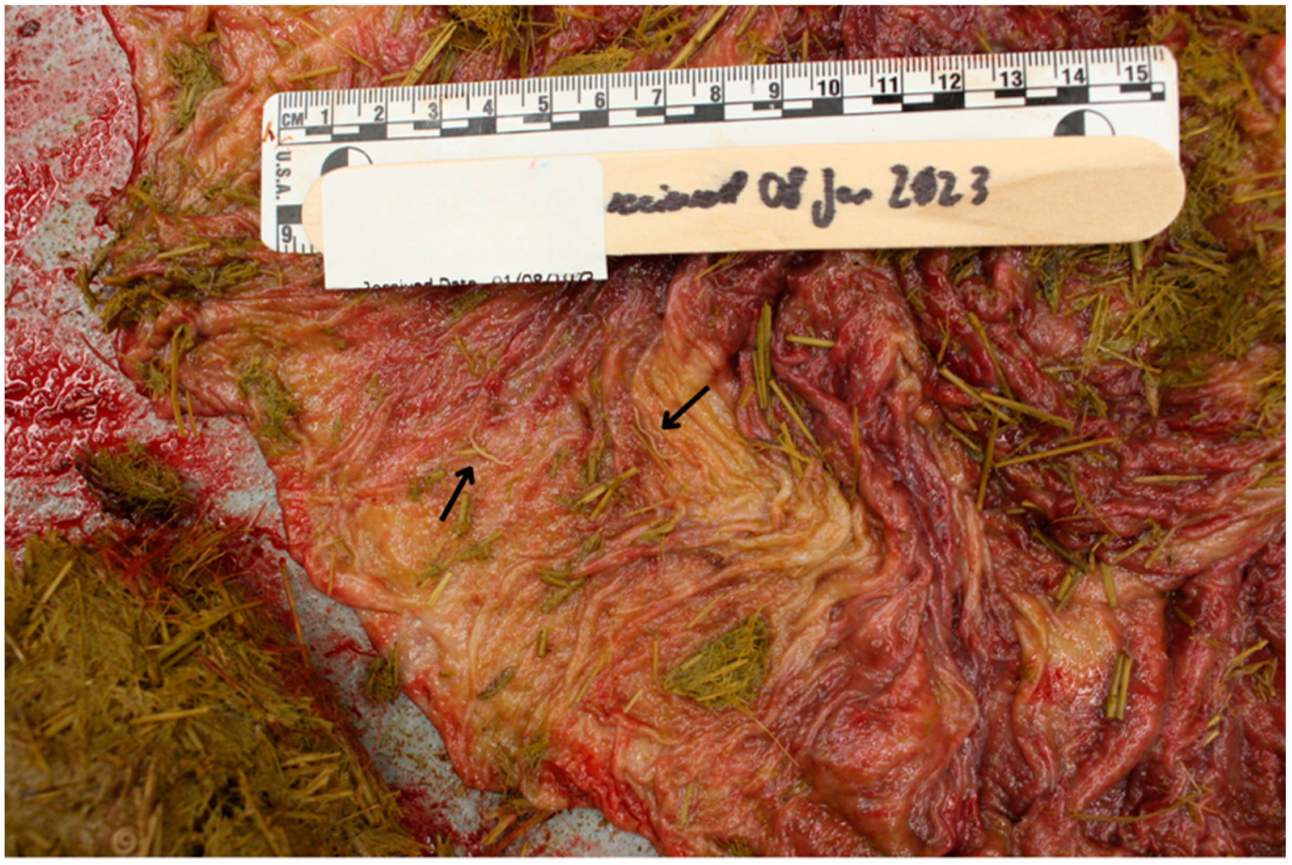
2.1. Case Presentation

A one-year-old male white rhinoceros (Ceratotherium simum) was submitted for necropsy to the Kansas State Veterinary Diagnostic Lab in January 2023. The animal had a four-day history of neurological disease and was found to be moribund. Supportive therapeutic interventions failed to control the clinical signs observed and the animal died. The animal had been born in December 2021 in a zoo in the midwestern United States and was housed in the same zoo. There was no history of animal movement.
At necropsy, the body condition was found to be adequate with moderate adipose tissue and muscling. Lesions including myocardial pallor, ulcers in the non-glandular stomach, and red mottling in the colon were observed. Colonic inflammation and hemorrhage were observed. Thin beige to translucent smooth nematodes were observed throughout the large colon (Figure 1). Nematodes were submitted to the parasitology section of the Kansas State Veterinary Diagnostic Lab for identification by a board-certified parasitologist. Adult male and female nematodes were cleared in lactophenol and imaged.

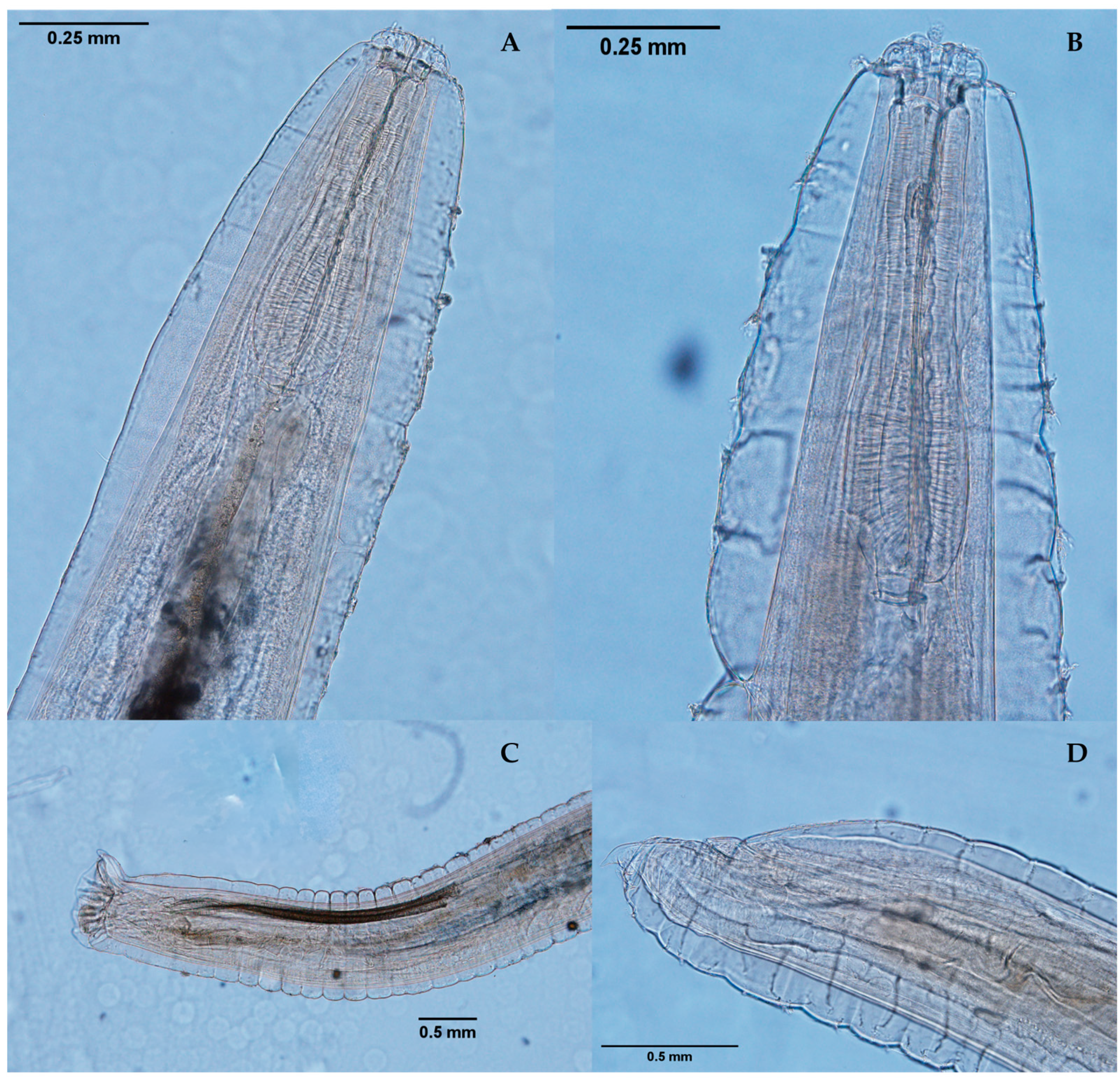
2.2. Morphological Diagnosis

Male and female nematodes were submitted. The bodies were elongated, cylindrical, and tapered at both ends. The cuticle was thick and separated from the body wall throughout the body; this separation was wider in the anterior region of the body than in the posterior end (Figure 2A,B). The body had widely spaced annulations. The anterior end comprised a mouth with a mouth collar that led to a broad yet shallow, thick-walled buccal capsule. No teeth were present in the buccal capsule. The mouth had four submedian papillae. Small, non-prominent internal leaf crowns were observed, with the tips extending past the lips. The esophagus was club-shaped with a distinct esophageal bulb. A prominent nerve ring was present. Females were on average 25.5 mm long and 1 mm wide, and males were on average 19 mm long and 0.8 mm wide. The posterior ends of the males had multiple lobes with wide expanded bursa supported by rays (Figure 2C). There were two spicules of equal length measuring on average 1695 µm, ±31.80 µm, with a posterior twist/coil forming an ala with fine striations. The female worms terminated in a straight, conical tail with the vulva immediately anterior to the anus (Figure 2D). The length from the tip of the tail to the anus measured 307.3 µm, ±48.56 µm. Anterior to the vulva was the uterus containing oval, thin-shelled, strongyle-type eggs measuring 73.44 µm long by 46.94 µm wide (range: length 73.44 µm, ±10.29 µm and width 46.94 µm, ±3.025 µm). Overall, the morphology of the worms was similar to cyathostomins (small strongyles) which affect horses. Primary descriptions of cyathostomin nematodes from megaherbivores were referred to for a preliminary genus-level identification.

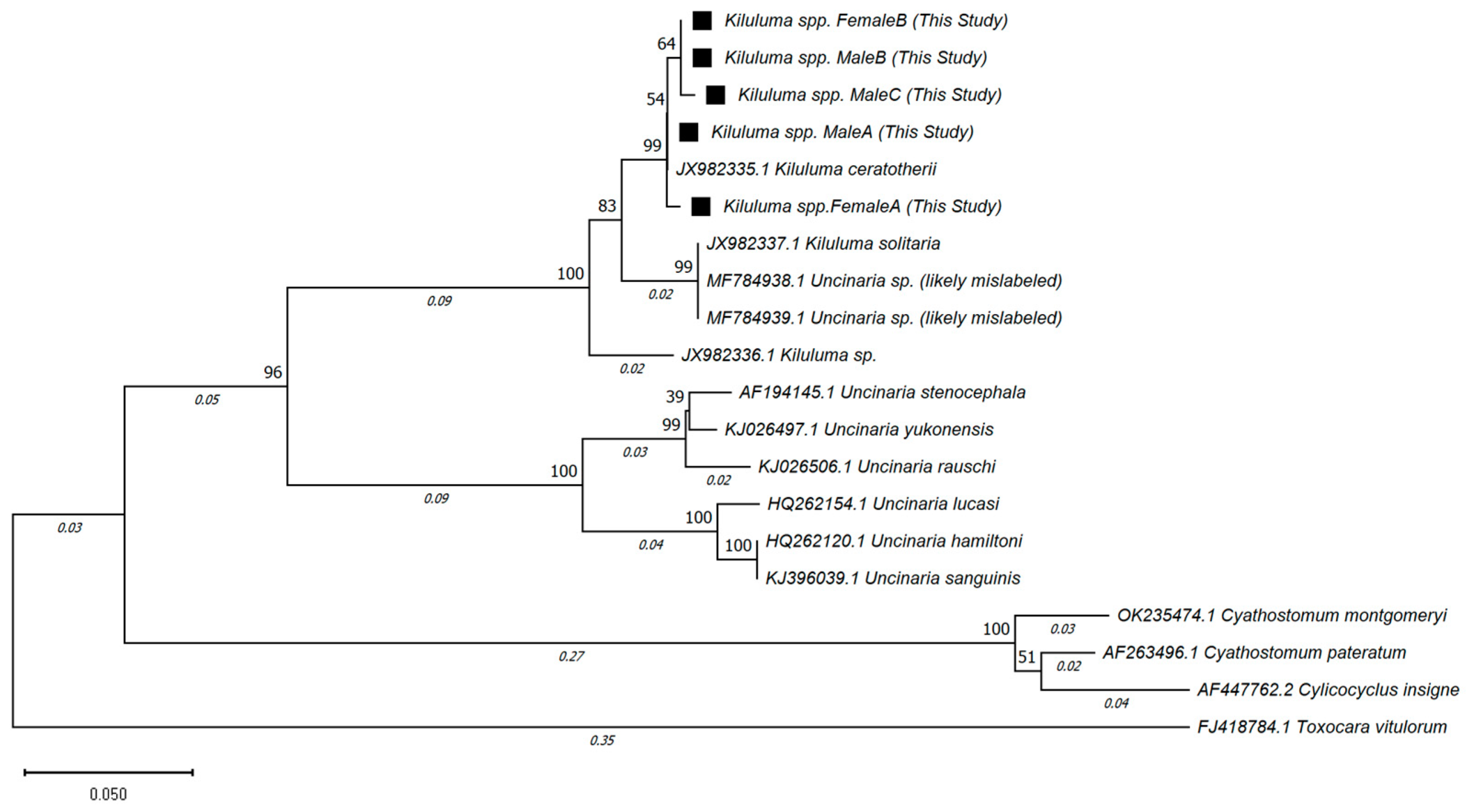
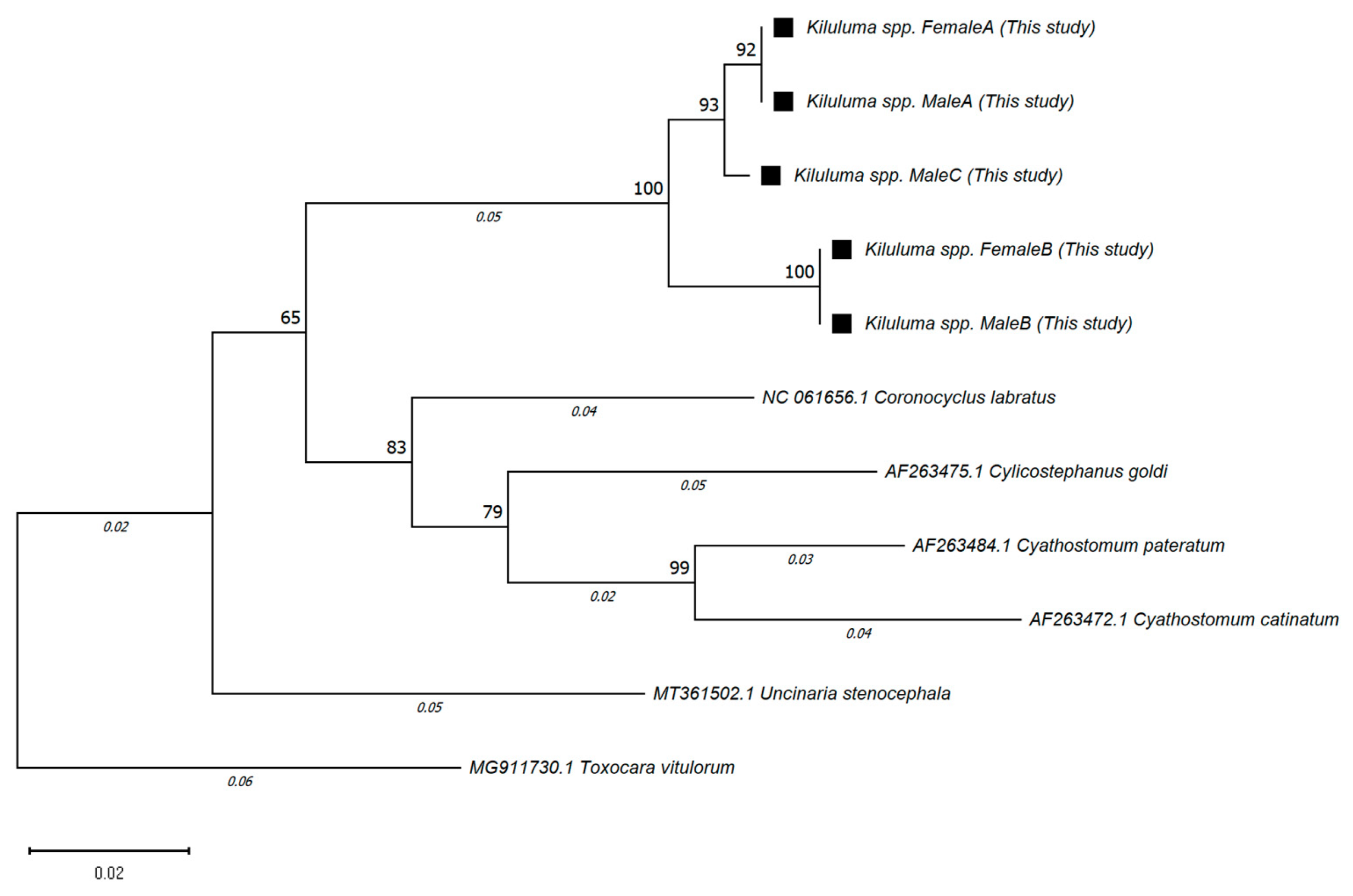
2.3. Results of Molecular Analysis

A ~400 bp region of the mitochondrial cytochrome oxidase 1 (cox1) gene, along with an ~870 bp region of the nuclear partial internal transcribed spacer (ITS) 1 and complete 5.8S ribosomal DNA and ITS2 region were amplified for species identification. However, due to limitations in available GenBank sequence data (lack of information about the ITS1 and 5.8S regions), only a 266 bp fragment of the ITS2 gene was used for analysis. ITS2 sequences from five individual nematodes in this study shared 99.64–100% identity with Kiluluma ceratotherii (GenBank Accession: JX982335.1) described from a white rhinoceros at the Western Plains Open Range Zoo in Dubbo, New South Wales, Australia [4]. The ITS2 sequences obtained in this study were deposited into GenBank (accession number: OR142649–OR142653). A neighbor-joining phylogenetic tree for ITS2 was constructed using the sequences from this study and from additional nematode sequences derived from GenBank (Figure 3). ITS2 sequences derived from this study formed a cluster with the previously described Kiluluma spp., specifically, K. ceratotherii [3] with 100% and 99% bootstrap support, respectively. Moreover, we discovered two sequences in GenBank labeled as Uncinaria spp. originating from a white rhinoceros that appeared to be mislabeled. This is evident because these sequences formed a cluster with Kiluluma spp. with 100% bootstrap support.
Additionally, no cox1 sequence data existed previously for Kiluluma spp. When compared to other cyathostomins, cox1 sequences in this study shared 88.9% identity with Coronocyclus labratus (GenBank accession: NC_061656.1) and 86.92% identity with Cylicostephanus goldi (GenBank accession: AF263475.1). A neighbor-joining phylogenetic tree for cox1 was constructed using the sequences from this study and additional nematode sequences derived from GenBank (Figure 4). Sequences derived from this study formed their own cluster separate from other Cyathostomins with 100% bootstrap support. The cox1 sequences obtained in this study were deposited into GenBank (accession number: OR142644–OR142648).

3. Discussion

We present a case of a white rhinoceros calf infected with K. ceratotherii Beveridge, 2013 (Nematoda: Strongylida) in the United States. Nematodes of the genus Kiluluma belong to the subfamily Cyathostominae of the family Strongylidae. Kiluluma spp. have only been reported from rhinoceroses and appear to be host-specific.
The genus Kiluluma was first instituted by Skrjabin [7] in 1916 with the description of the species K. stylosa. Six new species in the genus were described by Thapar in 1924 [8] and four more in 1925 [9]. Several of these species were subsequently synonymized by Taylor in 1925 [10] on the basis of non-significant differences in small morphological features and variations caused by the fixation of samples. However, 12 species were recognized by Zumpt [3]. Recently, three species of Kiluluma were described from white rhinoceroses: K. ceratotherii [4], a redescription of K. solitaria [4] and a new Kiluluma spp. which was later renamed to K. ornata [11]. The number and diversity of species in this genus are still unknown.
Cyathostomins are common strongyle parasites of equids and related animals. Similarities in appearance among cyathostomin species make the morphological identification of these parasites to the species level difficult. In the case of Kiluluma, species-level keys do not exist. Several characteristics described in the original records of the species have very small differences in measurements and size [10]. Some morphological features like the internal leaf crowns, which are pliable, can be fixed in different positions, altering the conclusions reached from morphological identification [10].
Morphological identification, combined with molecular characterization of nuclear and/or mitochondrial genes, provides a more accurate approach to species identification. We obtained both nuclear ITS1, 5.8S, ITS2, and mitochondrial cox1 sequences from five nematodes in this study. Based on comparison with ITS2 sequences provided by Beveridge et al. [4], the nematodes in this study were identified as K. ceratotherii. Since cox1 sequences from Kiluluma spp. were not available for comparison, we performed comparisons with equine cyathostomins and have submitted the generated sequences to GenBank to enable future comparative studies on taxonomy.
The pathogenesis of Kiluluma spp. in rhinoceroses has not been studied. In this case, the presence of large numbers of adult nematodes in the colon was the likely cause of colonic hemorrhage and inflammation. Thus, it appears that heavy infections of K. ceratotherii in susceptible young rhinoceroses can cause colonic damage and inflammation. However, these lesions were an incidental finding and unrelated to the neurological cause of death.
The presence of nematodes in a juvenile animal without any prior travel history outside of its birthplace at the zoo is noteworthy. Infected cohort members housed with the juvenile were the likely source of infection since Kiluluma spp. appears to be host-specific. To reduce parasitic worm burdens and potential adverse health events associated with infections, zoological parks should perform regular diagnostic testing of animals, especially as a screening measure during quarantine.

4. Materials and Methods

A deceased one-year-old male white rhinoceros calf was submitted for necropsy to the Kansas State Veterinary Diagnostic Lab. Nematodes were isolated and submitted in tap water within a few hours of isolation. They were washed and transferred to 70% ethanol. Two male and two female adult nematodes were cleared in lactophenol and observed under 40x–400x magnification.
The middle third of three male and two female adult nematodes were isolated and used for DNA extraction. Anterior and posterior ends were retained as vouchers for identification. DNA was extracted from the midsections using the DNeasy Blood and Tissue Kit (Qiagen, Valencia, CA, USA) according to the manufacturer’s protocol and eluted DNA was stored at −20 °C. PCR amplification of the partial ITS1, complete 5.8S ribosomal DNA, and ITS2 sequence was performed as previously described [12]. Briefly, PCR was performed in 25 µL reactions using 2 µL of DNA, 1.5 units Gotaq Flexi DNA Polymerase (Promega, USA), 10 µM of each primer (NC5: 5′-GTAGGTGAACCTGCGGAAGGATCATT-3′ and NC2: 5′-TTAGTTTCT TTTCCTCCGCT-3′), 3 mM of MgCl2, 10 µM dNTPs, and 1x PCR buffer. Thermocycler conditions were initial denaturation at 95 °C for 2 min, followed by 34 cycles of 98 °C for 25 s, at 62 °C for 30 s, and at 72 °C for 30 s, with a final extension at 72 °C for 2 min.
PCR amplification of cox1 was performed as previously described [13]. Briefly, PCR reactions were performed in 25 µL reactions using 2 µL of DNA, 1.5 units Gotaq Flexi DNA Polymerase (Promega, Waltham, MA), 10 µM of each primer (JB3 5′-TTTTTTGGGCATCCTGAGGTTTAT-3′ and JB4.5 5′-TAAAGAAAGAACATAATGAAAATG-3′), 3 mM of MgCl2, 10 µM dNTPs, and 1x PCR buffer. Thermocycler conditions were initial denaturation at 94 °C for 5 min, followed by 30 cycles at 94 °C for 30 s, 55 °C for 30 s, and 72 °C for 30 s, with a final extension at 72 °C for 5 min.
The presence of amplicons was visualized using agarose gel electrophoresis on 0.8% agarose gels. PCR products were purified with ExoSAP-IT Express (ThermoFisher Scientific, Vilinus, Lithuania) according to manufacturer instructions and sequenced in both directions using Sanger sequencing (Eurofins Genomics, Louisville, KY, USA). Neighbor-joining phylogenic trees were constructed in MEGA11 [14].

5. Conclusions

In this study, we morphologically and molecularly identified K. ceratotherii in a white rhinoceros, C. simum, from the United States. Sequences obtained in this study have been deposited in GenBank (accession number: OR142644–OR142653).

Author Contributions

Conceptualization: T.A.Q. and J.R.J.J.C.; data curation: T.A.Q. and J.R.J.J.C.; formal analysis: T.A.Q. and J.R.J.J.C.; funding acquisition: J.R.J.J.C.; investigation: T.A.Q., K.R.M., G.S., W.L.J., K.J.H.-T. and J.R.J.J.C.; supervision: B.H.H. and J.R.J.J.C.; visualization: T.A.Q. and J.R.J.J.C.; writing—original draft: T.A.Q. and J.R.J.J.C.; writing—review and editing: T.A.Q., K.R.M., G.S., W.L.J., B.H.H., K.J.H.-T. and J.R.J.J.C. All authors have read and agreed to the published version of the manuscript.

Funding

This work was supported by start-up funds provided to J.R.J.J.C. by the College of Veterinary Medicine, Kansas State University.

Institutional Review Board Statement

Not applicable.

Informed Consent Statement

Not applicable.

Data Availability Statement

All data from this study have been deposited in GenBank under accession numbers (OR142644–OR142653).

Acknowledgments

We would like to acknowledge the Kansas State University Veterinary Diagnostic Laboratory for their collaborative efforts.

Conflicts of Interest

The authors declare no conflict of interest.

References

  1. Titcomb, G.C.; Pansu, J.; Hutchinson, M.C.; Tombak, K.J.; Hansen, C.B.; Baker, C.C.; Kartzinel, T.R.; Young, H.S.; Pringle, R.M. Large-herbivore nemabiomes: Patterns of parasite diversity and sharing. Proc. R. Soc. B 2022, 289, 20212702. [Google Scholar] [CrossRef] [PubMed]
  2. Knapp, S.E.; Krecek, R.C.; Horak, I.G.; Penzhorn, B.L. Helminths and arthropods of black and white rhinoceroses in southern Africa. J. Wildl. Dis. 1997, 33, 492–502. [Google Scholar] [CrossRef] [PubMed]
  3. Zumpt, F. Parasites of the white and black rhinoceroses. Lammergeyer 1964, 3, 59–70. [Google Scholar]
  4. Beveridge, I.; Jabbar, A. New species of Kiluluma Skrjabin, 1916 (Nematoda: Strongylida) from the white rhinoceros Ceratotherium simium (Burchell), with a redescription of K. solitaria Thapar, 1924. Syst. Parasitol. 2013, 85, 131–145. [Google Scholar] [CrossRef] [PubMed]
  5. Saitou, N.; Nei, M. The neighbor-joining method: A new method for reconstructing phylogenetic trees. Mol. Biol. Evol. 1987, 4, 406–425. [Google Scholar] [CrossRef] [PubMed]
  6. Felsenstein, J. Confidence limits on phylogenies: An approach using the bootstrap. Evolution 1985, 39, 783–791. [Google Scholar] [CrossRef] [PubMed]
  7. Skrjabin, K. Parasitic trematodes and nematodes collected by the expedition of Prof. V. Dogiel and I. Sokolov in British East Africa. In Scientific Results of the Zoological Expedition to British East Africa and Uganda Made by Prof. V. Dogiel and I. Sokolov in the Year 1914; Forgotten Books: London, UK, 1916; Volume 1, pp. 99–157. [Google Scholar]
  8. Thapar, G.S. On Kiluluma Skriabin, a genus of strongylid nematodes parasitic in the African rhinoceros. J. Helminthol. 1924, 2, 209–238. [Google Scholar] [CrossRef]
  9. Thapar, G.S. On some new members of the genus Kiluluma from the African rhinoceros. J. Helminthol. 1925, 3, 63–80. [Google Scholar] [CrossRef]
  10. Taylor, E. The genus Kiluluma. Ann. Trop. Med. Parasitol. 1925, 19, 53–55. [Google Scholar] [CrossRef]
  11. Beveridge, I. A new species of Kiluluma Skrjabin, 1916 (Nematoda: Strongyloidea) from the white rhinoceros, Ceratotherium simum (Burchell) from South Africa. Syst. Parasitol. 2018, 95, 841–847. [Google Scholar] [CrossRef] [PubMed]
  12. Chelladurai, J.J.; Bader, C.; Snobl, T.; Magstadt, D.; Cooper, V.; Brewer, M.T. Toxocara vitulorum infection in a cohort of beef calves in Iowa. Vet. Parasitol. 2015, 214, 96–99. [Google Scholar] [CrossRef] [PubMed]
  13. Quintana, T.A.; Johnson, W.L.; Ritchie, D.; Smith, V.; Martin, K.A.; McMahan, K.; Brewer, M.T.; Jesudoss Chelladurai, J.R.J. Genetic characterization of the zoonotic parasite Ancylostoma caninum in the central and eastern United States. J. Helminthol. 2023, 97, e37. [Google Scholar] [CrossRef] [PubMed]
  14. Tamura, K.; Stecher, G.; Kumar, S. MEGA11: Molecular Evolutionary Genetics Analysis Version 11. Mol. Biol. Evol. 2021, 38, 3022–3027. [Google Scholar] [CrossRef] [PubMed]
Figure 1. Nematode parasites, indicated by black arrows, found during necropsy in the colon of a white rhinoceros.
Parasitologia 03 00025 g001
Figure 2. Anterior end of adult male (A) and female Kiluluma ceratotherii (B). Posterior end of adult male (C) and female Kiluluma ceratotherii (D).
Parasitologia 03 00025 g002
Figure 3. Neighbor-joining tree of the internal transcribed spacer (ITS) 2 sequences. Black squares indicate sequences obtained from this study. Evolutionary history was inferred using the neighbor-joining method [5]. The optimal tree is shown. The percentage of trees in which the associated taxa clustered together in the bootstrap test (1000 replicates) are shown above the branches [6]. The tree is drawn to scale, with branch lengths (below the branches) in the same units as those of the evolutionary distances used to infer the phylogenetic tree. The tree was rooted with ITS2 from Toxocara vitulorum (accession no. FJ418784.1).
Parasitologia 03 00025 g003
Figure 4. Neighbor-joining tree of partial mitochondrial cytochrome oxidase (cox1) gene sequences. Black squares (∎) indicate sequences obtained in this study. Evolutionary history was inferred using the neighbor-joining method [5]. The optimal tree is shown. The percentage of replicate trees in which the associated taxa clustered together in the bootstrap test (1000 replicates) are shown above the branches [6]. The tree is drawn to scale, with branch lengths shown below the branches in the same units as those of the evolutionary distances used to infer the phylogenetic tree. The tree was rooted with the mitochondrial cox1 gene from Toxocara vitulorum (accession no. MG911730.1).
Parasitologia 03 00025 g004
Disclaimer/Publisher’s Note: The statements, opinions and data contained in all publications are solely those of the individual author(s) and contributor(s) and not of MDPI and/or the editor(s). MDPI and/or the editor(s) disclaim responsibility for any injury to people or property resulting from any ideas, methods, instructions or products referred to in the content.

