Article

Blastocystis sp. Infection: Prevalence and Clinical Aspects among Patients Attending to the Laboratory of Parasitology–Mycology of Fann University Hospital, Dakar, Senegal

1
Département de Parasitologie-Mycologie, Faculté de Médecine, Pharmacie et Odontologie, Université Cheikh Anta Diop de Dakar, Dakar 10700, Senegal
2
Laboratoire de Parasitologie-Mycologie, Centre National Hospitalier Universitaire Fann Dakar, Dakar 10700, Senegal
3
Service de Parasitologie-Mycologie, UFR Sciences de la Santé, Université Gaston Berger de Saint-Louis, Saint-Louis 32000, Senegal
*
Author to whom correspondence should be addressed.
Parasitologia 2022, 2(4), 292-301; https://doi.org/10.3390/parasitologia2040024
Submission received: 8 September 2022 / Revised: 19 October 2022 / Accepted: 20 October 2022 / Published: 22 October 2022

Abstract

:
Introduction: Blastocystis sp. is a unicellular obligate anaerobic protozoa found in the human intestinal tract. Its role in human health is widely discussed because of the high proportion of asymptomatic carriers. In sub-Saharan Africa, the prevalence of the disease is underestimated. This study was performed to describe the epidemiological, clinical, and parasitological aspects of Blastocystis sp. infection in patients attending to Fann University Hospital. Material and Methods: We carried out a retrospective and descriptive study among patients attending to the laboratory of Parasitology and Mycology of Fann University Hospital from January 2016 to December 2020. All stool samples collected were examined using direct examination, a formal ether concentration method, and a modified Zeilh–Nielsen staining method. A descriptive analysis was performed with Stata MP 16 software. The significance level was set at 5%. Results: Overall, 447 cases of Blastocystis sp. were reported in our study, representing a prevalence rate of 13.7% ((447/3264) (95% CI: 12.5–15.5)). The mean age of the patients was 26 ± 20.7 years. Subjects over 45 years of age were more affected, with a frequency of 14.7%. Blastocystis sp. carriage was more common in males, at 14.6%. The symptomatology was mainly represented by diarrhea, abdominal pain, and dyspeptic disorders. In asymptomatic patients, the frequency of Blastocystis sp. was 33.3%. Mono-infection was found in 78.6% of cases. In total, 96 patients were carriers of at least two parasites (21.5%). Blastocystis sp. was most associated with Entamoeba coli (8.1%) and Endolimax nanus (4.03%). The association with helminths was noted in 5 patients (3 Ascaris lumbricoides, Trichuris trichiura, and Taenia). Conclusion: These results show the frequency of Blastocystis sp. infection with a large proportion of asymptomatic carriers. The presence of the parasite in the stool, associated with digestive disorders or with the association of other intestinal parasites, could justify the initiation of an anti-parasitic treatment.

1. Introduction

Blastocystis sp., which is commonly found in the gastro-intestinal tract, has been recognized as a non-pathogenic organism for a few decades. The parasite has a worldwide distribution. A high prevalence has been reported in developing countries (22.1–100%). In industrialized countries, the prevalence ranges from 0.5% to 23.1% [1,2,3,4,5]. The high prevalence noted in developing countries is related to poor hygiene, low access to safe water and food, and a lack of standard sanitary facilities [6,7].
The pathogenicity of Blastocystis is controversial because most patients carrying this parasite are asymptomatic. Several studies have considered Blastocystis sp. as a commensal micro-organism, while other results have showed the pathogenicity of the parasite. In addition, several epidemiological studies have reported a high prevalence of Blastocystis in patients with irritable bowel syndrome (IBS) [8]. The presence of the parasite is often linked to symptoms such as nausea, fever, urticaria, vomiting, anorexia, diarrhea, cramps, flatulence, discomfort, and abdominal pain [9,10]. The illness may be acute or chronic, with symptoms persisting for several years [11].
However, most studies have reported that Blastocystis sp. was frequently found in immunocompromised patients such as kidney transplant patients, patients with hematologic malignancies, and HIV patients with a CDT4 <200/mm3 [12,13,14].
The data available in Africa concerning Blastocystis are still limited. In Zambia, 53.8% prevalence of Blastocystis sp. was observed by Thaddeus et al. [15]. The most common parasite noted in Gabon was Blastocystis sp., with 48.6% prevalence [16]. These results are similar with general distribution of Blastocystis in developing countries.
In Senegal, low prevalence rates of Blastocystis sp. were reported of 4.8% and 3.7% in Dakar (capital city) and rural areas, respectively [17,18].
In order to improve the management of Blastocystis infection, an update of the data is necessary. It was in this context that we conducted this study, with the objective of evaluating the prevalence of Blastocystis sp.

2. Results

2.1. Socio-Demographic Characteristics of Study Participants

A total of 3264 patients were included in the study. The mean age of the study participants was 26.3 ± 20.7 years. Patients under 15 years of age were most represented (41.8%), followed by patients with ages ranging from 15 to 30 years (21.5%). Patients in the age group of 30–45 years and those over 45 years represented 16.7% and 20% of the total, respectively. According to gender, the study population was predominantly male (50.9%). The sex ratio was 1.04. The numbers of samples received were higher in 2018 and 2017 at 24.6% and 24.5%, respectively. Regarding the season, the number of samples was higher in the dry season at 78.8% compared to 24.2% in the rainy season (Table 1).

2.2. Clinical Characteristics of Study Patients

According to the symptoms, 53.2% (1737/3264) of the patients were symptomatic, while 46.8% of the study participants were asymptomatic. Among the symptomatic patients, diarrhea, abdominal pain, fever, and dyspeptic disorders were the most frequently noted symptoms. However, several types of diarrhea were observed, such as acute febrile diarrhea (4.7%), acute non-febrile diarrhea (16.4%), chronic diarrhea in HIV-positive patients (9.1%), and chronic diarrhea in HIV-negative patients (23%). The other symptoms were represented by abdominal pain (20.9%), fever (8.2%), dyspeptic disorders (8.2%), anal pruritus (0.8%), dysenteric syndrome (3.5%), urticaria (1.2%), and vomiting (0.8) (Table 2).

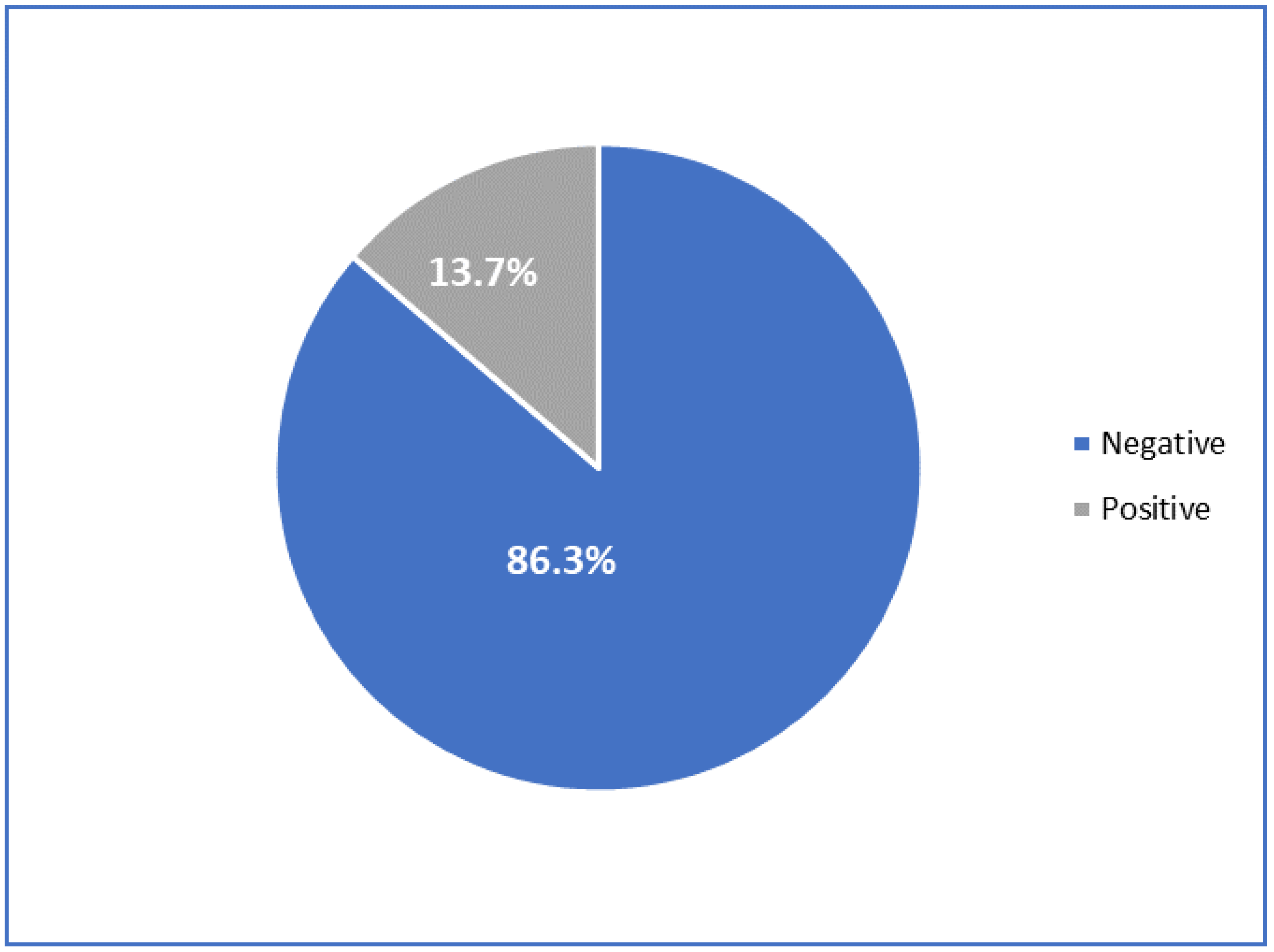
2.3. Prevalence of Blastocystis sp. Infection

Overall, 13.7% (447/3264) of the study participants were found to be infected by Blastocystis sp. (Figure 1). Mono-infection was noted in 351 patients (78.6%). Di-parasitism (presence of two parasites) and poly-parasitism (more than two parasites) were found in 21.5% and 3.6%, respectively. In 98.6% of cases the protozoa were isolated alone, and in 1.6% of cases they were associated with helminths. The vacuolar form was the most common form, representing 87.7% of Blastocystis sp. isolates, while the granular form was noted in 12.3% of cases (Table 3).
The main associations of the species were Blastocystis sp. + Cyst Entamoeba coli (8.1%), Blastocystis sp. + Cyst Endolimax Nana (4%), Blastocystis sp. + Trophozoite Entamoeba coli (2.9%), and Blastocystis sp. + Ascaris lumbricoïdes (0.8%) (Table 4).
According to the macroscopic aspect of the stool specimens, in patients with consistent stools, the Blastocystis sp. prevalence was 14.7%. In patients who had watery stools, it was 14.5%. The prevalence of Blastocystis sp. infection was 12.1% in patients with watery stools with blood and mucus (Figure 2).

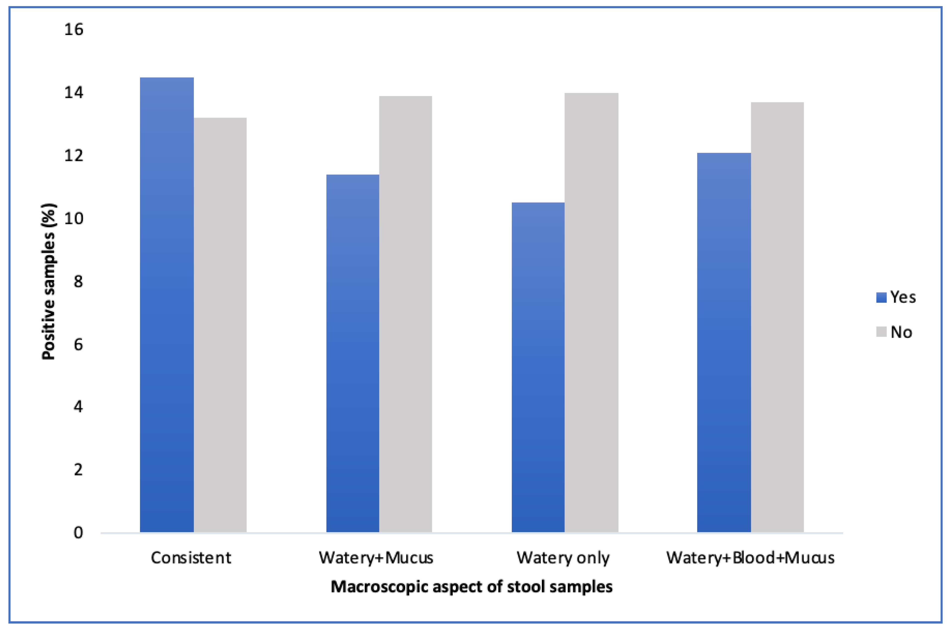
2.4. Prevalence of Blastocystis sp. according to Socio-Demographic and Clinical Characteristics of Study Participants

Stratifying through the year of sample collection, the Blastocystis sp. infection rates were more frequent in 2017 and 2020 at 18.7% and 16.9%, respectively. A low positive rate was noted in 2016 (8.9%). The frequency of Blastocystis sp. was more important in patients aged over 45 years (14.7%). In patients under 15 years of age, the Blastocystis sp. prevalence was 13.9%. This prevalence was lower in the 30-45-year-old age group (12.5%). However, the differences were not significative between age groups (p = 0.65). Infection with Blastocystis sp. was more frequent in male patients (14.6%) than women (12.7) (p value = 0.12). According to the season, Blastocystis sp. was more common in the rainy season (15.7%) than in the dry season (13.9%) (p = 0.05). In total, 16.1% of asymptomatic patients had Blastocystis sp. in their stool samples versus 11.6% of symptomatic patients, with a significant difference (p < 10−3) (Table 5).
In symptomatic patients, the prevalence of Blastocystis sp. was 9.76% in patients with acute febrile diarrhea and 8.71% in patients with acute non-febrile diarrhea. In patients with chronic diarrhea, Blastocystis sp. was observed in HIV-positive and -negative patients at rates of 9.75% and 5.1%, respectively. High positivity rates were noted in patients with abdominal pain (15.9%) and those with dyspeptic disorders (17.2). In patients with fever and dysenteric syndrome, the prevalence rates were 14.7% and 8.3%, respectively.

3. Discussion

Blastocystis sp. is the most common anaerobic parasite to colonize the gastrointestinal tracts of humans as well as many animals. It can be associated with gastro-intestinal disorders that are not specific to it and seems to be involved in some functional disorders, especially in patients suffering from irritable bowel syndrome and in immunocompromised subjects. In order to improve the management of patients and to update the epidemiology of the disease, this study was conducted among patients attending to the Laboratory of Parasitology of Fann University Hospital.
The results of this study showed 13.7% prevalence of Blastocystis sp. infection. Previous studies conducted in Senegal have described the frequency of blastocystosis. Sow et al., when assessing the prevalence rates of Blastocystis sp. infection from 2011 to 2013 in the same laboratory, showed 4.8% prevalence [17]. The same trends were described by Sylla et al. when evaluating the epidemiological profile of intestinal parasitic infection among preschool and schoolchildren living in a rural community, where 3.7% prevalence was observed [16]. These lower frequencies compared to our study could be explained by the long period of our study and the sample size. Low prevalence was also noted in Tunisia by Trabelsi et al., who showed a prevalence rate of Blastocystis of 7.2% [19].
Other studies conducted at the community level have noted high prevalence rates of Blastocystis infection. Niang et al., when evaluating the performance of the KOPCOLOR kit for the detection of intestinal parasites in November 2012 in 117 children living in the Thiès region (Central part of Senegal), found a prevalence rate of 87.1% for intestinal parasites, with Blastocystis sp (58.1%) as the main species observed [20]. El Safadi et al., in evaluating Blastocystis sp. via molecular method in 93 children aged 6–10 years in 2011 in the Senegal River Basin, showed a prevalence rate of 100% for Blastocystis sp. [21].
In Zambia, 53.8% prevalence of Blastocystis sp. was observed by Thaddeus et al. [15]. The most common parasite noted in Gabon was Blastocystis sp. with 48.6% prevalence [16].
Several studies using molecular biology or other techniques as diagnostic tools have shown higher frequencies. For example, in a study comparing the Flotac dual 400 technique with the conventional concentration technique in 108 patients between May and June 2009, Becker et al. observed a prevalence of 20.4% [22]. Poulsen et al. showed 84% prevalence of Blastocystis sp. in a molecular biology study in 199 Nigerian children in 2013 [23].
In Tanzania, Forsell et al., when studying the distribution of Blastocystis sp. subtypes and the prevalence of intestinal parasites using molecular methods and microscopy, showed 61% prevalence of Blastocystis via PCR. The prevalence revealed via microscopy was lower (20%) [24].
Other studies have demonstrated that Blastocystis is an emergent pathogen, with frequency rates ranging from 3% to 14.5% in France, and from 0.5% in Japan to 60% in Malaysia [2,25,26,27].
The results from this study showed that the frequency of Blastocystis sp. was more important in patients under 15 years old and those over 45 years (13.9% and 14.7%, respectively). Sow et al. found similar results, with higher prevalence in subjects under 15 years and in the age group of 5–30 years [18]. A high prevalence (84%) of Blastocystis sp. infection was noted in in children aged 2–14 years in Nigeria [23]. In Libya, Abdul Salam et al. found that adults (aged ≥ 18 years) were more affected than other age groups (< 18 years) (29.4% vs 9.9%; p < 0.001) [28]. A study conducted in Angola by Dacal et al. in Benguala province in 2015 showed that Blastocystis sp. is one the most common protozoa parasites detected (25.6%; 95% CI: 21.18–30.2%). Children of all ages are affected [29]. The last two studies used molecular methods for the detection of the parasite.
According to gender, men were more affected (14.6%) by Blastocystis sp. than women (12.7%) in our study. Our results are in line with what were found by Angolo and Malaysia [1,30]. In the study conducted by Sow et al. in Senegal, Blastocystis sp. infection was more frequent in women [17]. However, several studies have noted that gender has no significant influence on the carriage of Blastocystis sp. [31,32].
The pathogenicity of Blastocystis sp. remains highly controversial due to the presence of asymptomatic carriers. In our study, the prevalence rates of Blastocystis sp. in asymptomatic patients were 16.1% and 11.6% in symptomatic patients (p < 10−3). Similar results were previously described. In the study performed by Sow et al., 50% of patients were asymptomatic and 32.1% were symptomatic [18]. Otherwise, in several studies, the presence of gastro-intestinal symptoms has been related to the presence of Blastocystis sp. This was demonstrated by Abdulsalam et al., who showed that the prevalence of gastrointestinal symptoms is higher in subjects with Blastocystis infection compared to those without Blastocystis infection (35.3% vs. 13.2%, p value < 0.001) [28]. The same trends were observed in another previous study conducted in Italia [33].
The main symptoms noted in our study were diarrhea, abdominal pain, and dyspeptic disorders (flatulence). This is in line with results found by Abdulsalam et al., who noted that the most common symptoms in their study were abdominal pain (76.4%), flatulence (41.1%), and diarrhea (21.5%) [29]. Sebaa et al., in their study, noted that the most frequent clinical signs were abdominal pain (39.1%), diarrhea (6.5%), and nausea (3.3%) [34]. Other authors have noted that Blastocystis sp. is directly responsible for gastrointestinal disorders and is considered the etiological agent responsible for a certain type of persistent diarrhea, abdominal pain, and vomiting [2,21,35,36,37]. In immunocompetent subjects, the presence of Blastocystis is not correlated with gastrointestinal symptoms. This was previously demonstrated by Leder et al. [38]. Otherwise, immunocompromised subjects (cancer, HIV) seem to be more receptive to the parasite [39,40,41].
Even if the previous studies could not draw conclusions regarding the pathogenicity of Blastocystis sp., it is important to look for it and to report it to the clinicians, who will decide whether to treat the patient or not according to the clinical symptoms.
According to the results of this study, Blastocystis sp. prevalence was higher in the rainy season (15.7%) compared to dry season (13.1%). This is consistent with the data from literature showing that Blastocystis sp. infestation is more common in summer. Suresh et al., when comparing the methods for detecting Blastocystis sp., showed that infections were more prevalent in summer than in winter or spring [42].

4. Materials and Methods

4.1. Study Design and Population

We carried out a retrospective and descriptive study at the Parasitology and Mycology Laboratory at Fann University Hospital in Dakar. From January 2016 to December 2020, all patients attending to the laboratory for a parasitological examination of stool samples were included in the study.

4.2. Data Collection

Data on the sociodemographic, clinical, and biological aspects from patients were collected using the laboratory records. The following variables were collected: age, gender, year, season, clinical indication, macroscopic aspect of sample, and parasitological results. Age was defined as 4 categories: less than 15 years, [15–30 years], [30–45 years], and more than 45 years. The season was defined as either the dry season (October to June) or the rainy season (July to September).

4.3. Parasitological Examination

Fresh fecal specimens were collected into a wide-mouth screw-cap clean containers. The stool samples were examined macroscopically for color, consistency, blood, mucus, pus, and large worms. A portion of each of the stool samples was processed, with a direct examination by light microscopy to detect cysts, trophozoites, eggs, and larvae. The remaining parts of the stool samples were examined using a modified Ritchie technique and modified Zielh–Neelsen technique.

4.4. Data Analysis

After data collection, the data were entered in Excel software and the analysis was performed using Stata software version 16. A descriptive analysis was performed. The quantitative variables were described in terms of the means and standard deviations. For the descriptive data, a description in terms of the frequency with a 95% confidence interval was used. The significance level of the different tests was set to 5% for two sides.

4.5. Ethics Considerations

This study was conducted according to the Declaration of Helsinki and existing national legal and regulatory requirements. To respect their confidentiality, an identification code was given to each participant

5. Conclusions

The results of this study have demonstrated the frequency of Blastocystis sp. infection in patients attending to Fann Hospital in Dakar (Senegal). Children under 15 years and adults over 45 years old were more affected. Gender has no influence in Blastocystis sp. carriage. Blastocystis was found in symptomatic and asymptomatic patients. The main clinical symptoms were diarrhea, abdominal pain, and dyspeptic disorders. The presence of the parasite in the stool, associated with digestive disorders or with the association of other intestinal parasites, could justify the initiation of an anti-parasitic treatment. Other studies assessing risk factors and using molecular methods will be necessary to better understand the dynamics of the transmission and to better characterize the parasite.

Author Contributions

K.S. conceived and designed the study. K.S. supervised the data collection. K.S. analyzed the data and wrote the first draft of the manuscript. D.S., S.L., T.D., R.C.T., B.F. read and approved the final manuscript. All authors have read and agreed to the published version of the manuscript.

Funding

This study was not funded. The funding for the data collection was covered by the Laboratory of Parasitology–Mycology, Fann Teaching Hospital.

Institutional Review Board Statement

Data are routinely collected from patients who attend to the hospital for biomedical testing. Permission to use the data for publication was requested from the administration of Fann University Hospital, which a is national reference hospital. A code was given to each enrolled participants and data on their sociodemographic characteristics, clinical aspects, and parasitological findings were collected from the laboratory records.

Informed Consent Statement

Informed consent was not required.

Data Availability Statement

The data used for this research article are available from the corresponding author upon request.

Acknowledgments

We would like to acknowledge the entire study population and the staff of the Laboratory of Parasitology at Fann Teaching Hospital.

Conflicts of Interest

The authors declare that they have no competing interests.

References

  1. Nithyamathi, K.; Chandramathi, S.; Kumar, S. Predominance of Blastocystis sp. infection among school children in Peninsular Malaysia. PLoS ONE 2016, 25, e0136709. [Google Scholar] [CrossRef]
  2. Osman, M.; El Safadi, D.; Cian, A.; Benamrouz, S.; Nourrisson, C.; Poirier, P.; Pereira, B.; Razakandrainibe, R.; Pinon, A.; Lambert, C.; et al. Prevalence and risk factors for intestinal protozoan infections with Cryptosporidium, Giardia, Blastocystis and Dientamoeba among schoolchildren in Tripoli, Lebanon. PLoS Negl. Trop. Dis. 2016, 10, e0004496. [Google Scholar]
  3. El Safadi, D.; Gaayeb, L.; Meloni, D.; Cian, A.; Poirier, P.; Wawrzyniak, I.; Delbac, F.; Dabboussi, F.; Delhaes, L.; Seck, M.; et al. Children of Senegal River Basin show the highest prevalence of Blastocystis sp. ever observed worldwide. BMC Infect. Dis. 2014, 25, 164. [Google Scholar]
  4. Stensvold, C.R.; Christiansen, D.B.; Olsen, K.E.; Nielsen, H.V. Blastocystis sp. subtype 4 is common in Danish Blastocystis- positive patients presenting with acute diarrhea. Am. J. Trop. Med. Hyg. 2011, 84, 883–885. [Google Scholar] [CrossRef] [PubMed]
  5. Abu-Madi, M.; Aly, M.; Behnke, J.M.; Clark, C.G.; Balkhy, H. The distribution of Blastocystis subtypes in isolates from Qatar. Parasit Vectors 2015, 17, 465. [Google Scholar] [CrossRef] [Green Version]
  6. Walker, C.L.; Aryee, M.J.; Boschi-Pinto, C.; Black, R.E. Estimating diarrhea mortality among young children in low and middle income countries. PLoS ONE 2012, 7, 29151. [Google Scholar]
  7. Keogh, M.B.; Castro-Alférez, M.; Polo-López, M.I.; Calderero, I.F.; Al-Eryani, Y.A.; Joseph-Titus, C.; Fernandez-Ibanez, P. Capability of 19-L polycarbonate plastic water cooler containers for efficient solar water disinfection (SODIS): Field case studies in India, Bahrain and Spain. Sol. Energy 2015, 116, 1–11. [Google Scholar] [CrossRef]
  8. Poirier, P.; Wawrzyniak, I.; Vivarès, C.P.; Delbac, F.; El Alaoui, H. New insights into Blastocystis spp.: A potential link with irritable bowel syndrome. PLoS Pathog. 2012, 8, 1002545. [Google Scholar] [CrossRef] [Green Version]
  9. Ocana-Losada, C.; Cuenca-Gomez, J.A.; Cabezas-Fernandez, M.T.; Vazquez-Villegas, J.; Soriano-Perez, M.J.; Cabeza-Barrera, I.; Salas-Coronas, J. Clinical and epidemiological characteristics of intestinal parasite infection by Blastocystis hominis. Rev. Clínica Española 2018, 218, 115–120. [Google Scholar] [CrossRef]
  10. Stensvold, C.R.; Lewis, H.C.; Hammerum, A.M.; Porsbo, L.J.; Nielsen, S.S.; Olsen, K.E.; Arendrup, M.C.; Nielsen, H.V.; Mølbak, K. Blastocystis: Unravelling potential risk factors and clinical significance of a common but neglected parasite. Epidemiol. Infect. 2009, 137, 1655–1663. [Google Scholar] [CrossRef] [PubMed] [Green Version]
  11. Yaicharoen, R.; Sripochang, S.; Sermsart, B.; Pidetcha, P. Prevalence of Blastocystis hominis infection in asymptomatic individuals from Bangkok, Thailand. Southeast Asian J. Trop Med. Public Health 2005, 36, 17–20. [Google Scholar]
  12. Stensvold, C.R.; Suresh, G.K.; Tan, K.S.; Thompson, R.C.; Traub, R.J.; Viscogliosi, E.; Yoshikawa, H.; Clark, C.G. Terminology for Blastocystis subtypes-a consensus. Trends Parasitol. 2007, 23, 93–96. [Google Scholar] [CrossRef]
  13. Ghosh, K.; Ayyaril, M.; Nirmala, V. Acute GVHD involving the gastrointestinal tract and infestation with Blastocystis hominis in a patient with chronic myeloid leukaemia following allogeneic bone marrow transplantation. Bone Marrow Transplant. 1998, 22, 1115–1117. [Google Scholar] [CrossRef] [Green Version]
  14. Rao, K.; Sekar, U.; Iraivan, K.T.; Abraham, G.; Soundararajan, P. Blastocystis hominis-an emerging cause of diarrhoea in renal transplant recipients. J. Assoc. Physicians India 2003, 51, 719–721. [Google Scholar]
  15. Graczyk, T.K.; Shiff, C.K.; Tamang, L.; Munsaka, F.; Beitin, A.M.; Moss, W.J. The association of Blastocystis hominis and Endolimax nana with diarrheal stools in Zambian school-age children. Parasitol. Res. 2005, 98, 38–43. [Google Scholar] [CrossRef]
  16. M’bondoukwé, N.P.; Kendjo, E.; Mawili-Mboumba, D.P.; Koumba Lengongo, J.V.; Offouga Mbouoronde, C.; Nkoghe, D.; Touré, F.; Bouyou-Akotet, M.K. Prevalence of and risk factors for malaria, filariasis, and intestinal parasites as single infections or co-infections in different settlements of Gabon, Central Africa. Infect. Dis. Poverty 2018, 30, 6. [Google Scholar] [CrossRef] [Green Version]
  17. Sylla, K.; Tine, R.K.; Sow, D.; Lelo, S.; Ndiaye, L.A.; Faye, B.T.; Ndiaye, M.; Dieng, T.; Faye, B.; Gaye, O. Epidemiological profile of intestinal parasitic infection among preschool and school children living in a rural community in Senegal: A cross sectional survey. J. Bacteriol. Parasitol. 2018, 9, 2. [Google Scholar] [CrossRef]
  18. Sow, D.; Sylla, K.; Dieng, T.; Tine, R.C.; Ndiaye, M.; Faye, B.; Ndiaye, J.L.; Ndiaye, D.; Gaye, O.; Dieng, Y. Infection par Blastocystis hominis au Sénégal: Aspects épidémiologiques, cliniques et parasitologiques des cas diagnostiqués au CHNU de Fann à Dakar. Rev. CAMES Santé 2015, 3, 2. [Google Scholar]
  19. Trabelsi, S.; Ali, I.B.; Khaled, S. Clinical and epidemiological characteristics of blastocystis hominis. Tunis Med. 2010, 88, 190–192. [Google Scholar]
  20. Niang, T.M. Evalution d’un Nouveau Kit Pour la Detection de Parasites Intestinaux. Ph.D. Thesis, University Cheikh Anta Dakar, Dakar, Senegal, 2013; 140p. Available online: http://bibnum.ucad.sn/viewer.php?c=thm&d=THM%5f48541 (accessed on 19 October 2022).
  21. El Safadi, D.; Cian, A.; Nourrisson, C.; Pereira, B.; Morelle, C.; Bastien, P.; Bellanger, A.P.; Botterel, F.; Candolfi, E.; Desoubeaux, G.; et al. Prevalence, risk factors for infection and subtype distribution of the intestinal parasite Blastocystis sp. from a large-scale multi-center study in France. BMC Infect. Dis. 2016, 26, 451. [Google Scholar] [CrossRef] [Green Version]
  22. Becker, S.L.; Lohourignon, L.K.; Speich, B.; Rinaldi, L.; Knopp, S.; N’goran, E.K.; Cringoli, G.; Utzinger, J. Comparison of the Flotac-400 dual technique and the formalin-ether concentration technique for diagnosis of human intestinal protozoon infection. J. Clin. Microbiol. 2011, 49, 2183–2190. [Google Scholar] [CrossRef] [Green Version]
  23. Poulsen, C.S.; Efunshile, A.M.; Nelson, J.A.; Stensvold, C.R. Epidemiological Aspects of Blastocystis Colonization in Children in Ilero, Nigeria. Am. J. Trop. Med. Hyg. 2016, 95, 175–179. [Google Scholar] [CrossRef] [Green Version]
  24. Forsell, J.; Granlund, M.; Samuelsson, L.; Koskiniemi, S.; Edebro, H.; Evengard, B. High occurrence of Blastocystis sp. subtypes 1–3 and Giardia intestinalis assemblage B among patients in Zanzibar, Tanzania. Parasit. Vectors 2016, 9, 370. [Google Scholar] [CrossRef] [Green Version]
  25. Pinel, C.; Réjasse, C.; Picot, S.; Brenier-Pinchart, M.P.; Grillot, R.; Ambroise-Thomas, P. Blastocystis hominis: Epidemiological and clinical remarks from more than 3500 stool examinations. Ann. Biol. Clin. 1999, 57, 601–604. [Google Scholar]
  26. Poirier, P.; Wawrzyniak, I.; Albert, A.; El Alaoui, H.; Delbac, F.; Livrelli, V. Development and evaluation of a real-time PCR assay for detection and quantification of blastocystis parasites in human stool samples: Prospective study of patients with hematological malignancies. J. Clin. Microbiol. 2011, 49, 975–983. [Google Scholar] [CrossRef] [Green Version]
  27. Wong, K.H.; Ng, G.C.; Lin, R.T.; Yoshikawa, H.; Taylor, M.B.; Tan, K.S. Predominance of subtype 3 among Blastocystis isolates from a major hospital in Singapore. Parasitol. Res. 2008, 102, 663–670. [Google Scholar] [CrossRef]
  28. Abdulsalam, A.M.; Ithoi, I.; Al-Mekhlafi, H.M.; Khan, A.H.; Ahmed, A.; Surin, J.; Mak, J.W. Prevalence, predictors and clinical significance of Blastocystis sp. in Sebha, Libya. Parasit. Vectors 2013, 6, 86. [Google Scholar] [CrossRef] [Green Version]
  29. Dacal, E.; Saugar, J.M.; de Lucio, A.; Hernández-de-Mingo, M.; Robinson, E.; Köster, P.C.; Aznar-Ruiz-de-Alegría, M.L.; Espasa, M.; Ninda, A.; Gandasegui, J.; et al. Prevalence and molecular characterization of Strongyloides stercoralis, Giardia duodenalis, Cryptosporidium spp., and Blastocystis spp. isolates in school children in Cubal, Western Angola. Parasit. Vectors 2018, 11, 67. [Google Scholar] [CrossRef]
  30. Bart, A.; Wentink-Bonnema, E.M.; Gilis, H.; Verhaar, N.; Wassenaar, C.J.; van Vugt, M.; Goorhuis, A.; Van Gool, T. Diagnosis and subtype analysis of Blastocystis sp. in 442 patients in a hospital setting in the Netherlands. BMC Infect. Dis. 2013, 23, 389. [Google Scholar]
  31. Kain, K.C.; Noble, M.A.; Freeman, H.; Barteluk, R.L. Epidemiology and clinical features associated with Blastocystis hominis infection. Diagn. Microbiol. Infect. Dis. 1987, 8, 235–244. [Google Scholar] [CrossRef]
  32. Cirioni, O.; Giacometti, A.; Drenaggi, D.; Ancarani, F.; Scalise, G. Prevalence and clinical relevance of Blastocystis hominis in diverse patient cohorts. Eur. J. Epidemiol. 1999, 15, 389–393. [Google Scholar] [CrossRef]
  33. Sebaa, S. Contribution A L’etude De La Place Des Parasites Protozoaires Dans L’etiologie Des Enterites Chez La Population Humaine En Milieu Hospitalier Et Non Hospitalier Issues Des Zones Urbaines Et Rurales Via Les Sources Animales. Ph.D Thesis, University de Djelfa, Djelfa, Algeria, 2020; 231p. Available online: http://dspace.univ-djelfa.dz:8080/xmlui/handle/123456789/3403 (accessed on 19 October 2022).
  34. Al-Fellani, M.A.; Khan, A.H.; Al-Gazoui, R.M.; Zaid, M.K.; Al-Ferjani, M.A. Prevalence and Clinical Features of Blastocystis hominis Infection among Patients in Sebha, Libya. Sultan Qaboos Univ. Med. J. 2007, 7, 35–40. [Google Scholar]
  35. Beyhan, Y.E.; Yilmaz, H.; Cengiz, Z.T.; Ekici, A. Clinical significance and prevalence of Blastocystis hominis in Van, Turkey. Saudi Med. J. 2015, 36, 1118–1121. [Google Scholar] [CrossRef]
  36. Vassalos, C.M.; Spanakos, G.; Vassalou, E.; Papadopoulou, C.; Vakalis, N. Differences in clinical significance and morphologic features of Blastocystis sp subtype 3. Am. J. Clin. Pathol. 2010, 133, 251–258. [Google Scholar] [CrossRef] [Green Version]
  37. Leder, K.; Hellard, M.E.; Sinclair, M.I.; Fairley, C.K.; Wolfe, R. No correlation between clinical symptoms and Blastocystis hominis in immunocompetent individuals. J. Gastroenterol. Hepatol. 2005, 20, 1390–1394. [Google Scholar] [CrossRef]
  38. Kumarasamy, V.; Roslani, A.C.; Rani, K.U.; Kumar Govind, S. Advantage of using colonic washouts for Blastocystis detection in colorectal cancer patients. Parasit. Vectors 2014, 3, 162. [Google Scholar] [CrossRef] [Green Version]
  39. Agholi, M.; Hatam, G.R.; Motazedian, M.H. HIV/AIDS-associated opportunistic protozoal diarrhea. AIDS Res. Hum. Retrovir. 2013, 29, 35–41. [Google Scholar] [CrossRef] [Green Version]
  40. Rivero-Rodriguez, Z.; Hernandez, A.; Bracho, A.; Salazar, S.; Villalobos, R. Prevalence of intestinal microsporidia and other intestinal parasites in HIV positive patients from Maracaibo, Venezuela. Biomedica 2013, 33, 538–545. [Google Scholar]
  41. Taşova, Y.; Sahin, B.; Koltaş, S.; Paydaş, S. Clinical significance and frequency of Blastocystis hominis in Turkish patients with hematological malignancy. Acta Med. Okayama 2000, 54, 133–136. [Google Scholar]
  42. Suresh, K.; Smith, H. Comparison of methods for detecting Blastocystis hominis. Eur. J. Clin. Microbiol. Infect. Dis. 2004, 23, 509–511. [Google Scholar] [CrossRef]
Figure 1. Prevalence of Blastocystis sp. infection.
Parasitologia 02 00024 g001
Figure 2. Prevalence of Blastocystis sp. according to the macroscopic aspect of stool specimens.
Parasitologia 02 00024 g002
Table 1. Socio-demographic characteristics of study participants (N = 3264).
VariableFrequency (n)Percentage (%)95% CI
Year
201667220.619.1–22.2
201780124.522.8–26.3
201880424.622.9–26.3
201961618.817.4–20.4
202037111.410.2–12.6
Age group
<15 years136541.839.6–44.1
[15–30]70321.519.9–23.2
[30–45]54516.715.3–18.2
≥45 years6512018.4–21.5
Gender
Female160049.146.6–51.5
Male166450.948.5–53.5
Season
Rainy247578.872.8–78.8
Dry78924.222.5–25.9
Total3264100
Table 2. Clinical symptoms of study participants (N = 1737).
Clinical SymptomFrequency (n)Percentage (%)
Constipation160.9
Acute febrile diarrhea824.7
Acute non-febrile diarrhea28516.4
Chronic diarrhea (HIV-positive)1579.1
Chronic diarrhea (HIV-negative)40023
Abdominal pain36420.9
Hypereosinophily50.3
Malnutrition60.3
Clinical anemia251.4
Anal pruritus150.8
Dysenteric syndrome603.5
Fever1438.2
Dyspeptic disorders1458.2
Urticaria201.2
Vomiting140.8
Total1737100
Table 3. Parasitological aspects of Blastocystis sp. infection.
Frequency (n)Percentage (%)95% CI
Parasitism
Mono-parasitism35178.670.5–87.2
Di-parasitism9621.517.4–26.2
Poly-parasitism163.62.1–5.8
Type of parasite
Protozoa44198.689.6–99.9
Protozoa—Helminth61.60.4–2.9
Parasite form
Vacuolar39287.779.2–96.8
Granular5512.39.3–16
Table 4. Intestinal parasite species associated with Blastocystis sp.
Parasites SpeciesFrequency (n)Percentage (%)
Di-parasitism
Blastocystis sp. + Cyst Entamoeba coli368.1
Blastocystis sp. + Cyst Endolimax nana184
Blastocystis sp. + Trophozoite Entamoeba coli132.9
Blastocystis sp. + Cyst Giardia intestinalis122.7
Blastocystis sp. + Ascaris lumbricoïdes30.8
Blastocystis sp. + Trichuris trichiura10.2
Blastocystis sp. + Teania10.2
Polyparasitism
Blastocystis sp. + Trophozoite E. coli + Cyst E. coli61.3
Blastocystis sp. + Cyst E. nana + Cyst E. coli20.4
Blastocystis sp. + Pseudolimax Butschili + Cyst E. coli20.5
Blastocystis sp. + Trophozoite E. coli + Cyst E. coli + Trichomonas. intestinalis10.2
Blastocystis sp. + Cyst G. intestinalis + Cyst E. coli10.2
Blastocystis sp. + T. intestinalis + Cyst E. coli10.2
Table 5. Prevalence rates of Blastocystis sp. according to socio-demographic and clinical characteristics of study participants (N = 447).
VariableFrequency (n)Percentage (%)95% CIp Value
Year
2016808.96.8–11.5
201715018.715.8–21.9
20188911.18.8–13.6
20198613.911.2–17.2
20206216.712.8–21.4<10−3
Age group
<15 years19113.912.1–16.1
[15–30]9213.110.5–16.1
[30–45]6812.59.6–15.8
≥45 years9614.711.9–180.65
Gender
Female20412.711.1–14.6
Male24314.612.8–15.50.12
Season
Rainy12415.713.1–18.7
Dry32313.111.6–15.50.06
Symptom
Asymptomatic24616.114.2–18.2
Symptomatic20111.610–13.2<10−3
Publisher’s Note: MDPI stays neutral with regard to jurisdictional claims in published maps and institutional affiliations.

