Communication

Prevalence and Financial Losses of Cystic Echinococcosis in Slaughtered Goats at Gumbo Slab in Juba County, South Sudan

1
Department of Clinical Studies, College of Veterinary Science, University of Bahr El-Ghazal, Wau 91113, South Sudan
2
Department of Preventive Medicine, College of Veterinary Medicine, University of Upper Nile, Malakal 71111, South Sudan
3
School of Veterinary Medicine, University of Juba, Juba 81111, South Sudan
4
The Royal Veterinary College, University of London, Hawkshead Lane, North Mymms, Hatfield AL9 7TA, UK
5
College of Veterinary Medicine, Sudan University of Science and Technology, P.O. Box 204 Hilat Kuku, Khartoum North 13321, Sudan
*
Author to whom correspondence should be addressed.
Parasitologia 2022, 2(2), 54-62; https://doi.org/10.3390/parasitologia2020006
Submission received: 13 February 2022 / Revised: 23 March 2022 / Accepted: 25 March 2022 / Published: 29 March 2022

Abstract

:
A cross-sectional study was conducted at Gumbo slaughter slab in Juba, Central Equatoria State (CES) in South Sudan, between October and December 2015, to determine the prevalence of cystic echinococcosis (CE) and to estimate the direct economic losses associated with condemnation of infected organs in slaughtered goats. A thorough postmortem examination was conducted on carcasses of a total of 1126 goats selected by systematic random sampling for detection of CE in the lungs, liver, heart, kidneys, and the spleen. Moreover, CE-related direct financial losses were estimated by using a standardized formula. The overall prevalence of CE was found to be 281 (24.9%, 95% CI 22.5–27.6). Furthermore, old age (OR = 2.61, p value < 0.001), Mubende breed (OR = 2.97, p value < 0.001), and Ugandan geographical origin (OR = 2.97, p value < 0.001) were associated with higher prevalence of CE. The lungs were the most affected organ with a prevalence of 55.2% followed by liver (44.1%), and the spleen (0.07%). A direct CE-related monetary loss of 78,820 South Sudanese Pounds (SSP) per year was estimated. This loss is equivalent to USD 43,788. In conclusion, CE is considerably prevalent and is associated with significant direct financial losses among goats slaughtered at Gumbo slaughter slab in Juba, CES, South Sudan.

1. Introduction

Cystic echinococcosis (CE) or hydatidosis is a major parasitic infection of public health concern in humans and animals, including sheep and goats [1,2,3,4]. While the larval stage of the tapeworm Echinococcus granulosus sensu stricto (G1–3), E. equinus (G4), E. ortleppi (G5), E. intermedius, and E. canadensis (G6–7, G8, G10) is responsible for causing CE, adult parasites are found in the small intestine of dogs and other carnivores [1,2,3,4,5,6]. CE is known as one of the most important widespread parasitic infections in livestock with considerable high fatality and poor prognosis in humans if left unmanaged [2,7,8]. Moreover, the disease is hyperendemic in some countries of Africa, such as Kenya, South Sudan, Sudan, and Uganda [2,3]. The transmission cycle of CE in animals depends on a complex interaction of several epidemiological factors (i.e., environmental, pathogenic, and host factors) [9]. A comprehensive account of the epidemiology of CE is inadequate and its true burden is underestimated in many low-middle income countries (LMICs). Thus, understanding the epidemiology and risk factors of CE in endemic settings will contribute for the development and implementation of effective control and management strategies.
Besides to its public health importance, infections due to Echinococcus species have severe economic implications in small ruminants industry [9]. Most of these financial losses are associated with condemnation of infected organs along with costs of treatment and control which have been estimated to be over USD 3 billion annually worldwide [9]. In Africa, direct financial losses to the livestock sector were found to be between $5869.8 and $152,002 per year in Ethiopia and Kenya [10,11,12]. In South Sudan, however, studies reporting the financial losses of CE are very limited [13,14].
CE was first reported in South Sudan during the early 1900s in dogs [15]. Since then, a few studies have reported the disease in humans and in different domestic animal species with a varying prevalence [15,16,17]. Nevertheless, apart from the work of Mousa et al. [13] and Ochi et al. [14], no studies explored the direct financial losses of CE in animals. Therefore, this study aimed to investigate the prevalence of CE in slaughtered goats and its associated direct financial losses in Juba, South Sudan.

2. Results

A total of 1126 slaughtered goats at Gumbo slaughter slab were examined for presence of CE. Overall, it was found in 24.9% (n = 281) of the inspected carcasses with a 95% confidence interval (CI) ranging from 22.5 to 27.6 (Table 1).
Among the examined carcasses, 33.8% (n = 188) of the old goats (i.e., >4 years old) had significantly higher CE than young goats (i.e., ≤4 years old) (16.3%, n = 93). Furthermore, the prevalence of CE among the carcasses of the Mubende goats which are originating from Uganda (35.7%, n = 179) was significantly higher than the prevalence of CE in the carcasses of Toposa goats that are originating from Greater Kapoeta in South Sudan (n = 102). However, the proportions of male (23.1%, n = 113) and female (26.4%, n = 168) goats infected with CE were not statistically different.
Univariate analysis, using Chi squared test, further confirmed that no difference between the prevalence of CE in males and females (X2 = 1.66, OR = 1.19 and p value 0.197) as shown in Table 1. On the other hand, goats of the older age group (i.e., >4 years old) had 2.61 increased odds of being infected with CE when compared to goats of the younger age group (X2 = 45.54, p value < 0.001). Similarly, Mubende goats originating from Uganda had 2.85 increased odds of being infected with CE when compared to Toposa breed (X2 = 55.9, p value < 0.001) (Table 1).
A logistic regression analysis was done to obtain adjusted odds ratios for sex, age, breed, and origin as shown in Table 2. The OR for age, breed, and origin remained significant.
Among the detected 281 CE positive cases, 155 (55.2%) were detected in the lungs, 124 (44.1%) in the liver, and 2 (0.7%) in the spleen (Table 3). Moreover, a total of 253 cysts were observed in the lungs, 276 in livers, and 2 in the spleen of the infected goats. However, cysts were neither found in any of the investigated hearts nor in kidneys.
To estimate the direct monetary losses due to organ condemnation, the prices of 1 kg of the lungs, liver, heart, kidneys, and spleen were obtained from butcheries in the study area and were 10, 15, 5, 3 and 3 South Sudanese pounds (SSP), respectively, during the period of the study. Using the standardized formula [14], the direct total monetary losses due to organ condemnations was found to be 78,820 SSP which is equivalent to 43,788 USD at Gumbo slaughtering slab per year.

3. Discussion

CE is of vital importance to the livestock production sector and public health in many developing countries [9,18]. This importance is due to its huge resultant economic losses and its zoonotic nature [9,18,19,20,21]. Moreover, it has been reported in different parts of the world with varying prevalence rates [9,18]. Therefore, it is important to understand the epidemiology of CE in both animals and humans and to explore its socio-economic impact in hyperendemic areas, such as South Sudan.
The current study revealed a prevalence of 24.9% of CE in carcasses of slaughtered goats at Gumbo animal slaughtering slab in Juba, South Sudan. This prevalence was higher than reports of former studies where CE was detected in up to 7.1% of the carcasses of slaughtered animals in South Sudan [14,18,22] but similar to the prevalence observed in Jimma in Ethiopia (a neighboring country to South Sudan) (24.8%) [23]. Moreover, while the prevalence of CE reported herein was less than the one reported in Arusha in Tanzania (35.0%) [24], it was higher than the one reported by Addy et al. [25] in Maasailand in Kenya (10.0%). In General, CE prevalence in livestock was as high as 55.9% in Western African countries, 63.8% in Eastern African countries, and 10.8% in Southern African countries with up to 75.0–94.5% fertile cysts [18,26].
Grosso et al. [26] indicated that the occurrence and extend of spread of CE depend on many factors that provide the optimum conditions for the parasite’s life cycle completion, perpetuation, and transmission. These factors, include suitable environmental/ecological conditions, presence and abundance of the intermediate and definitive hosts, livestock husbandry or animal production system (e.g., nomadic, or semi-nomadic vs. intensive farming), stocking rate, nature of the pasture, and grazing patterns [26]. Breed, sex, and age of the animal besides to the parasite’s strain/genotype are also important factors that should be considered [27,28,29]. Variations of these factors by region/country could probably explain the variations of CE prevalence. This notion is supported by the findings of this study as there were significant differences in CE prevalence between animals by age group, breed, and origin. Carcasses of goats of the older age group were having 2.61 increased odds of being infected with CE in comparison with carcasses of goats of the young age group. Furthermore, carcasses of the goats of the Mubende breed that are originating from Uganda were having 2.85 increased odds of being infected with CE when compared to carcasses of Toposa breed goats from Greater Kapoeta in South Sudan. Similar findings have been reported elsewhere [27,28,29,30]. The high prevalence in adult animals as compared to young animals was probably due to the higher possibilities and frequency of exposure to infection in addition to the social norms of slaughtering older animals in South Sudan. Besides, the diminishing or the compromised immune response in older animals could be another possible explanation for the high prevalence of CE in animals of this age group [29]. CE-prevalence between sexes (i.e., male and female goats) was not statistically different and this might be related to the same pathophysiology of both male and female animals [14,22].
Former studies have reported CE cysts in different anatomical sites of the body of infected animals [24,30,31]. Herein, almost all CE cysts were detected in the lungs and liver except two cysts which were found in the spleen. Nevertheless, CE cysts were neither found in the hearts nor in the kidneys of any of the examined carcasses. Similar findings showing high prevalence of pulmonary and hepatic CE have been reported previously [24]. A plausible explanation for this is that the lungs and the liver have a great capillary field as compared to other peripheral organs. When the oncospheres (hexacanth embryo) penetrate the intestinal wall and enter the portal blood/lymph, they are transported passively throughout the body to the major filtering organs, mainly the liver, where they develop into larval hydatid cysts [30]. In this study, the frequency of pulmonary CE was higher than that of hepatic CE. This might be due to co-infection with other parasites, like Fasciola hepatica. Stoore et al. [31] have noted an antagonistic relationship between E. granulosus sensu stricto and F. hepatica in cattle where F. hepatica affected the instating of CE cysts in the liver and displaced them towards the lungs [31]. Furthermore, host and parasite characteristics that may affect host-parasite interaction, such as genotype, can be the reason behind the high frequency of CE cysts in the lungs.
CE is known to lead to huge economic deficit because of production losses (i.e., less milk and meat productivity and decreased fecundity), treatment and control costs, and condemnation of infected organs [9,18]. In this study, the direct economic loss due to CE in goats slaughtered at Gumbo slab in Juba is estimated to be 78,820 SSP which is equivalent to 43,788 USD per year. This direct economic loss because of condemnation of organs appeared to be significant and may reflect the erratic veterinary and extension delivery services to key stakeholders in the country. In neighboring countries, such as Ethiopia and Kenya, the direct financial losses to the livestock production sector due to CE were found to be between $5869.8 and $152,002 per year, respectively [10,11,12,32]. Variations in retail market price, the size of each condemned organ, and carcass weight loss can be explain the differences of the amount of the lost money [14,22].
In conclusion, the findings of this study showed that CE is widely spread among slaughtered goats at Gumbo slab in Juba. Lungs and liver were the most affected organs. Moreover, the disease is responsible for causing substantial financial losses to producers due to organ condemnation. Efforts should be made to break the transmission cycle of the disease. Further epidemiological studies involving different species of livestock, dogs, wildlife, and human are warranted in South Sudan. Additionally, determining of the genotypes of prevalent parasite in South Sudan by using molecular techniques would be of high epidemiological and public health value.

4. Materials and Methods
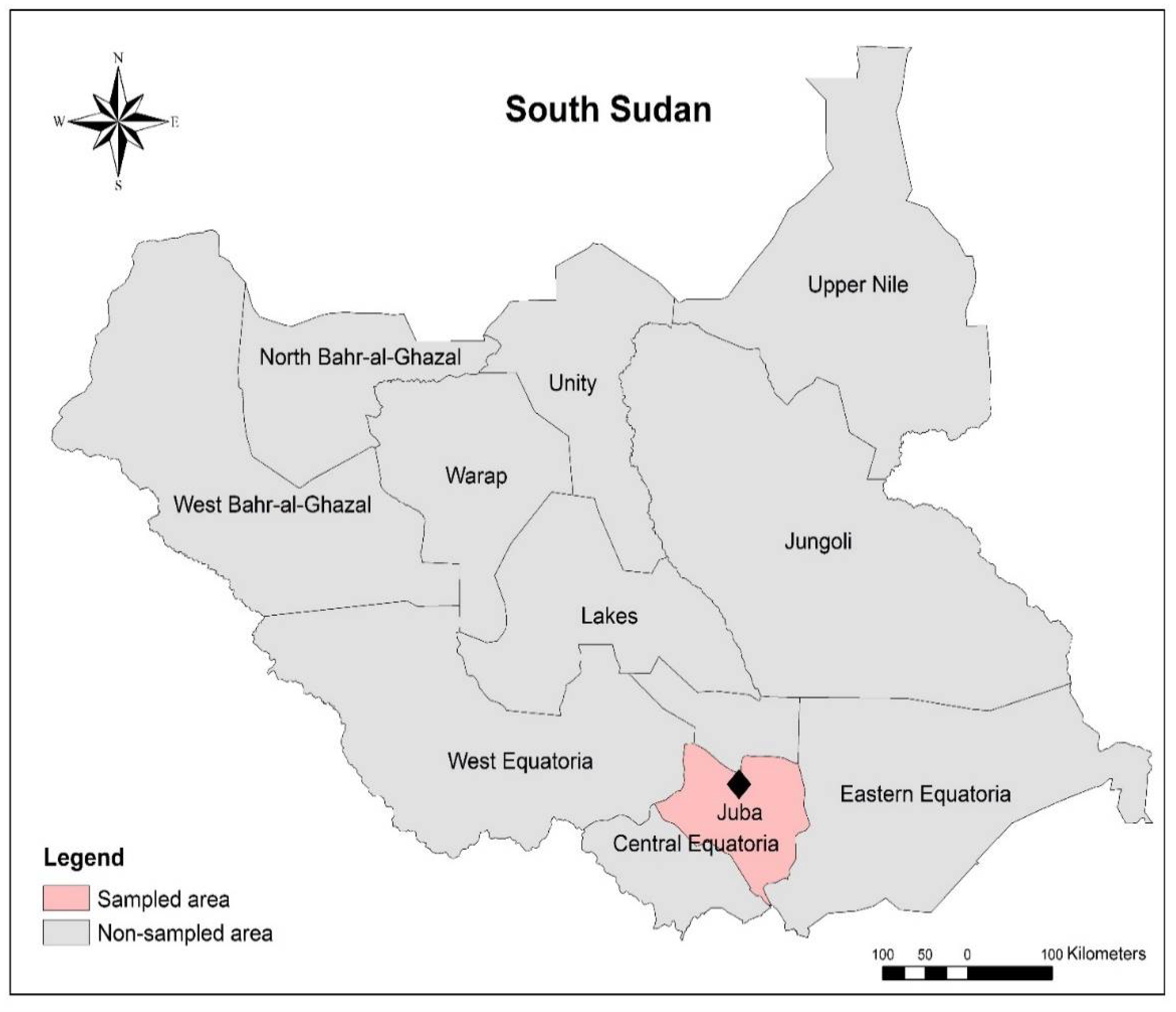
4.1. Study Area

This study was conducted in Juba County which is one of the counties of Central Equatoria State (area equals 43,033 square kilometers) in South Sudan (Figure 1). The county lies between latitude 4°51′33.7068 N and longitude 31°3416.5036 E at an elevation of 460 m above sea level and falls in the tropical savanna climate zone of Africa (i.e., African savanna climate) with an average annual ambient temperature ranging from 24.7 °C to 34.5 °C and a mean relative humidity that stands at 55%. The rainfall season usually lasts for nearly nine months. It commences in March and ends in December and is around 800–1100 mm per year. Livestock are brought to Juba daily for marketing from various locations, such as Rumbek, Terekaka, Bor, and Pibor, and from neighboring countries, such as Uganda, as well [13].

4.2. Study Design

A cross-sectional study was conducted for three months, October to December 2015, Juba County, Central Equatoria State, South Sudan [33]. This county was purposively selected as it has five slaughtering slabs and therefore the largest number of slaughtered goats compared with other counties. On average, around 5550 head of animals are slaughtered per month in the five slaughtering slabs of Juba, of which about 500 cattle, 400 sheep, and 1300 goats are slaughtered at Gumbo slaughtering slab [34]. This slaughtering slab was conveniently included in this study [33].

4.3. Sample Size and Sampling

The sample size (n) was calculated based on the standard formula of Thrusfield [33]. The parameters of the formula were as follows: 95% confidence level, ±5% desired level of precision, and the expected prevalence of CE in Juba County [14,33]. The formula was n = (1.96)2 × Pexp × (1 − Pexp)/d2 where n was the required sample size, (1.96)2 the constant, Pexp the expected prevalence rate of 3.99% [14], and d the desired precision level of ±5%. The required sample size n was determined to be 58.9 goats. This number was inflated by 20 times to account for the effect of randomness and representativeness [33]. Thus, the total n was 1178 goats. Nevertheless, 1,126 carcasses were investigated during the study.
Systematic random sampling was used to select individual study goats, which were identified using permanent markers, kept in a separate place, and released for slaughter one after another [33]. Sampling interval was obtained by dividing the total number of slaughtered goats on that day by the estimated daily sample size [33].
The age of each animal was determined by examination of the dentition of the animal and/or by asking the animal’s owner.

4.4. Antemortem and Postmortem Inspection

Antemortem inspection was carried out by visual screening to identify goats with clear clinical signs and symptoms of disease or any noticeable abnormality. Diseased goats that were deemed not fit for human consumption were excluded from slaughter.
Postmortem inspection was performed as described by Shuaib et al. [35] and Woldemariyam et al. [36]. It included visual inspection, palpation, and in situ slicing of lymph nodes, organs of the thoracic and abdominal cavities, and the head. Additionally, lymph nodes of the fore and hind limbs and the inguinal region were also inspected.
To search for CE, special attention was given to the liver, lungs, spleen, kidneys, and the heart during postmortem examination. These organs were systematically inspected by sight, palpation, and in situ slicing for presence of CE [36].

4.5. Economic Losses Due to CE

The direct economic loss due to CE because of organ condemnation was evaluated by considering the following parameters: 1- information on the mean retail market price of organs (lungs, liver, heart, spleen, and kidney) in Juba obtain from butchers during the study period, 2- the average annual rate of slaughtered goats at Gumbo slaughter slab which was obtained from the retrospective data of the last two years at the State Ministry of Animal Resources and Fisheries. The loss from organs condemned was calculated by using the following formula [14]:
LOC = (NAS × Ph × Plu × Cplu) + (NAS × Ph × Phr × Cphr) + (NAS × Ph × Pli × Cpli) + (NAS × Ph × Psp × Cpsp) + (NAS × Ph × Pkid × Cpkid)
where:
  • LOC = Loss due to organ condemnation.
  • NAS = Average number of goats slaughter annually
  • Ph = Prevalence of hydatidosis
  • Plu = Percent involvement of lung
  • Cplu = Current mean retail price of lung
  • Phr = Percent involvement of heart
  • Cphr = Current mean retail price of heart
  • Pli = Percent involvement of liver
  • Cpli = Current mean retail price of liver
  • Psp = Percent involvement of spleen
  • Cpsp = Current mean retail price of spleen
  • Pkid = Percent involvement of kidney
  • Cpkid = Current mean retail price of kidney

4.6. Statistical Analyses

SPSS version 20.0 and Stata/IC version 16.1 were used for all appropriate statistical analyses. First, descriptive statistics of the variables, including frequencies and proportions, were obtained and then differences between groups by using the χ2 or Fisher exact test and logistic regression with a p value of ≤0.05 denoting to statistical significance.

Author Contributions

Conceptualization, K.L.S.N. and Y.A.S.; methodology, K.L.S.N.; formal analysis, Y.A.S.; investigation, K.L.S.N.; resources, B.T.J., D.L.L. and E.P.L.; data curation, K.L.S.N. and Y.A.S.; writing—original draft preparation, K.L.S.N. and Y.A.S.; writing—review and editing, K.L.S.N., B.T.J., D.L.L., E.P.L., A.Y.O. and Y.A.S.; visualization, A.Y.O. and Y.A.S.; supervision, Y.A.S. All authors have read and agreed to the published version of the manuscript.

Funding

This research received no external funding.

Institutional Review Board Statement

Not applicable.

Informed Consent Statement

Not applicable.

Data Availability Statement

Data is contained within the article.

Acknowledgments

The authors would like to express our special gratitude to the State Ministry of Animal Resources and Fisheries, Central Equatoria State, staff of Juba Malakia Clinic for their support. AYO acknowledges support from the EU European & Developing Countries Clinical Trials Partnership-funded Pan-African Network for Rapid Research, Response, Relief and Preparedness for infectious diseases epidemics (PANDORA-ID-NET).

Conflicts of Interest

The authors declare no conflict of interest.

References

  1. Soulsby, E.J.L. Helminths, Arthropods and Protozoa and Domesticated Animals, 7th ed.; Bailliere Tindall: London, UK, 1982. [Google Scholar]
  2. McManus, D.P.; Zhang, W.; Li, J.; Bartley, P.B. Echinococcosis. Lancet 2003, 362, 1295–1304. [Google Scholar] [CrossRef]
  3. Eckert, J.; Deplazes, P. Biological, epidemiological, and clinical aspects of echinococcosis, a zoonosis of increasing concern. Clin. Microbiol. Rev. 2004, 17, 107–135. [Google Scholar] [CrossRef] [Green Version]
  4. Agudelo, H.N.I.; Brunetti, E.; McCloskey, C. Cystic Echinococcosis. J. Clin. Microbiol. 2016, 54, 518–523. [Google Scholar] [CrossRef] [PubMed] [Green Version]
  5. Alvarez, C.A.; Romig, T.; Lightowlers, M.W. Echinococcus granulosus sensu lato genotypes infecting humans—Review of current knowledge. Int. J. Parasitol. 2014, 44, 9–18. [Google Scholar] [CrossRef]
  6. Shahabi, S.; Sarkari, B.; Barazesh, A. Echinococcus granulosus sensu stricto G1 is the predominant genotype in human and livestock isolates from Turkey and Iran, based on mitochondrial nad5 gene differentiation. Parasites Vectors 2021, 14, 369. [Google Scholar] [CrossRef] [PubMed]
  7. da Silva, A.M. Human echinococcosis: A neglected disease. Gastroenterol. Res. Pract. 2010, 2010, 583297. [Google Scholar] [CrossRef]
  8. Craig, P.S.; Budke, C.M.; Schantz, P.M.; Li, T.; Qiu, J.; Yang, Y.; Zeyhle, E.; Rogan, M.T.; Ito, A. Human Echinococcosis: A Neglected Disease? Trop. Med. Health 2007, 35, 283–292. [Google Scholar] [CrossRef] [Green Version]
  9. Budke, C.M.; Deplazes, P.; Torgerson, P.R. Global socioeconomic impact of cystic echinococcosis. Emerg. Infect. Dis. 2006, 12, 296–303. [Google Scholar] [CrossRef] [PubMed]
  10. Biressaw, S.; Dulo, J. Hydatidosis: Prevalence and financial loss of bovine hydatidosis from cattle slaughtered at Adama Municipal Abattoir, South Eastern Ethiopia. J. Parasitol. Vector Biol. 2017, 9, 8–12. [Google Scholar]
  11. Kere, O.J.; Joseph, E.; Jessika, B.L.; Maina, K.J. Prevalence and monetary loss due to cystic Echinococcosis in slaughter house livestock: A case study of Migori County, Kenya. Parasite Epidemiol. Control 2019, 5, e00105. [Google Scholar] [CrossRef]
  12. Getaw, A.; Beyene, D.; Ayana, D.; Megersa, B.; Abunna, F. Hydatidosis: Prevalence and its economic importance in ruminants slaughtered at Adama municipal abattoir, Central Oromia, Ethiopia. Acta Trop. 2010, 113, 221–225. [Google Scholar] [CrossRef] [PubMed]
  13. Mousa, A.A.; Malik, K.H.; Ochi, E.B. Prevalence and Monetary Loss due to Bovine Fasciolosis in Juba Slaughter House South Sudan. Nat. Sci. 2013, 11, 145–148. [Google Scholar]
  14. Ochi, E.B. Prevalence and Economic Loss due to Hydatidosis in Slaughtered Animals in Juba South. Sudan. Int. J. Res. Stud. Biosci. 2015, 3, 177–182. [Google Scholar]
  15. Eisa, A.M.; Mustafa, A.A.; Soliman, K.N. Preliminary Report on Cysticercosis and hydatidosis in Southern Sudan. Sudan J. Vet. Sci. 1962, 3, 97–108. [Google Scholar]
  16. Magambo, J.K.; Hall, C.; Zeyle, E.; Wachira, T.M. Prevalence of human hydatid disease in southern Sudan. Afr. J. Health Sci. 1996, 3, 154–156. [Google Scholar]
  17. Magambo, J.K.; Zeyhle, E.; Wachira, T. Hydatid disease in Toposa land southern Sudan. Afr. J. Health Sci. 1997, 5, 129–132. [Google Scholar]
  18. Wahlers, K.; Menezes, C.N.; Wong, M.L.; Zeyhle, E.; Ahmed, M.E.; Ocaido, M.; Stijnis, C.; Romig, T.; Kern, P.; Grobusch, M.P. Cystic echinococcosis in sub-Saharan Africa. Lancet Infect. Dis. 2012, 12, 871–880. [Google Scholar] [CrossRef]
  19. Kia, E.B.; Ouma, F.F.; Mulambalah, C.S.; Okoth, P.K. The Burden of Cystic Echinococcosis in Kenya: A Review Article. Iran. J. Parasitol. 2019, 14, 502–509. [Google Scholar]
  20. Miambo, R.D.; Afonso, S.M.S.; Noormahomed, E.V.; Pondja, A.; Mukaratirwa, S. Echinococcosis in humans and animals in Southern Africa Development Community countries: A systematic review. Food Waterborne Parasitol. 2020, 20, e00087. [Google Scholar] [CrossRef]
  21. Gessese, A.T. Review on Epidemiology and Public Health Significance of Hydatidosis. Vet. Med. Int. 2020, 2020, 8859116. [Google Scholar] [CrossRef]
  22. Ochi, E.B.; Akol, D.A.; Lukaw, Y.S. A Review on Epidemiology of Hydatidosis in Livestock and Humans in South Sudan. Int. J. Res. Stud. Biosci. 2016, 4, 4–10. [Google Scholar]
  23. Kumsa, B.; Mohammedzein, A. Prevalence, organ distribution, risk factors, and financial losses of hydatid cysts in sheep and goats slaughtered in restaurants in Jimma, south western Oromia. Comp. Clin. Pathol. 2012, 23, 333–339. [Google Scholar] [CrossRef]
  24. Ernest, E.; Nonga, H.E.; Kassuku, A.A.; Kazwala, R.R. Hydatidosis of slaughtered animals in Ngorongoro district of Arusha region, Tanzania. Trop. Anim. Health Prod. 2009, 41, 1179–1185. [Google Scholar] [CrossRef] [PubMed]
  25. Addy, F.; Alakonya, A.; Wamae, N.; Magambo, J.; Mbae, C.; Mulinge, E.; Zeyhle, E.; Wassermann, M.; Kern, P.; Romig, T. Prevalence and diversity of cystic echinococcosis in livestock in Maasailand, Kenya. Parasitol. Res. 2012, 111, 2289–2294. [Google Scholar] [CrossRef]
  26. Grosso, G.; Gruttadauria, S.; Biondi, A.; Marventano, S.; Mistretta, A. Worldwide epidemiology of liver hydatidosis including the Mediterranean area. World J. Gastroenterol. 2012, 18, 1425–1437. [Google Scholar] [CrossRef]
  27. Otero-Abad, B.; Torgerson, P.R. A systematic review of the epidemiology of echinococcosis in domestic and wild animals. PLoS Negl. Trop. Dis. 2013, 7, e2249. [Google Scholar] [CrossRef]
  28. Haleem, S.; Niaz, S.; Qureshi, N.A.; Ullah, R.; Alsaid, M.S.; Alqahtani, A.S.; Shahat, A.A. Incidence, risk factors, and epidemiology of cystic echinococcosis: A complex socioecological emerging infectious disease in Khyber Pakhtunkhwa, Province of Pakistan. Biomed Res. Int. 2018, 2018, 5042430. [Google Scholar] [CrossRef]
  29. Torgerson, P.R.; Burtisurnov, K.K.; Shaikenov, B.S.; Rysmukhambetova, A.T.; Abdybekova, A.M.; Ussenbayev, A.E. Modelling the transmission dynamics of Echinococcus granulosus in sheep and cattle in Kazakhstan. Vet. Parasitol. 2003, 114, 143–153. [Google Scholar] [CrossRef]
  30. Abdel-Baki, A.S.; Almalki, E.; Al-Quarishy, S. Prevalence and characterization of hydatidosis in Najdi sheep slaughtered in Riyadh city, Saudi Arabia. Saudi J. Biol. Sci. 2018, 25, 1375–1379. [Google Scholar] [CrossRef]
  31. Stoore, C.; Andrade, C.; Hidalgo, C.; Corrêa, F.; Jiménez, M.; Hernandez, M.; Paredes, R. Echinococcus granulosus hydatid cyst location is modified by Fasciola hepatica infection in cattle. Parasites Vectors 2018, 11, 542. [Google Scholar] [CrossRef]
  32. Melaku, A.; Lukas, B.; Bogale, B. Cyst Viability, Organ Distribution and Financial Losses due to Hydatidosis in Cattle Slaughtered at Dessie Municipal Abattoir, North-eastern Ethiopia. Vet. World 2012, 5, 213. [Google Scholar] [CrossRef]
  33. Thrusfield, M. Veterinary Epidemiology, 3rd ed.; Wiley-Blackwell Science: London, UK, 2007. [Google Scholar]
  34. Ministry of Animal Resources and Fisheries. Number of Slaughtered Livestock in Juba County; Ministry of Animal Resources and Fisheries: Juba, South Sudan, 2014. [Google Scholar]
  35. Shuaib, Y.A.; Niemann, S.; Khalil, E.; Schaible, U.; Wieler, L.H.; Bakheit, M.A.; Mohamed-Noor, S.E.; Abdalla, M.a.; Richter, E. Mycobacterial infections in carcasses of ruminants slaughtered at the two slaughterhouses of Kassala, Sudan. Rev. Élev. Méd. Vét. Pays Trop. 2018, 70, 131–136. [Google Scholar] [CrossRef] [Green Version]
  36. Woldemariyam, F.T.; Markos, T.; Shegu, D.; Abdi, K.D.; Paeshuys, J. Evaluation of Postmortem Inspection Procedures to Diagnose Bovine Tuberculosis at Debre Birhan Municipal Abattoir. Animals 2021, 11, 2620. [Google Scholar] [CrossRef] [PubMed]
Figure 1. Map of study area.
Parasitologia 02 00006 g001
Table 1. Univariate analysis (association of sex, age, breed, and origin with CE positivity in slaughtered goat carcasses), Gumbo slaughtering slab, October to December 2015.
Risk FactorNo. of TestedNo. of Positive (%)χ2dfCrude ORp Value95% CI
Sex 1.66211.190.1970.91–1.58
Male490113 (23.1)
Female636168 (26.4)
Age 45.5412.61<0.0011.95–3.48
Young 56993 (16.3)
Old557188 (33.8)
Breed * 55.9312.85<0.0012.15–3.79
Toposa625102 (16.3)
Mubende501179 (35.7)
Origin * 55.9312.85<0.0012.15–3.79
GK625102 (16.3)
Uganda501179 (35.7)
Total1126281 (24.9)22.5–27.6
No. = number, df = degree of freedom, OR = odds ratio, CI = confidence interval, GK = Greater Kapoeta in South Sudan, * = all Toposa goats were from Greater Kapoeta in South Sudan while all Mubende goats were from Uganda, therefore, the outcomes for the variables breed and origin were identical.
Table 2. Multivariate analysis (association of sex, age, breed, and origin with CE positivity in slaughtered goat carcasses), Gumbo slaughtering slab, October to December 2015.
Risk FactorNo. of TestedNo. of Positive (%)Positive Outcome OR Adjusted p Value95% CI
CrudeAdjusted
Sex 1.191.300.0780.97–1.73
Male490113 (23.1)
Female636168 (26.4)
Age 2.612.64<0.0011.98–3.54
Young56993 (16.3)
Old557188 (33.8)
Breed * 2.852.97<0.0012.22–3.96
Toposa625102 (16.3)
Mubende501179 (35.7)
Origin * 2.852.97<0.0012.22–3.96
GK625102 (16.3)
Uganda501179 (35.7)
No. = number, OR = odds ratio, CI = confidence interval, GK = Greater Kapoeta in South Sudan, * = all Toposa goats were from Greater Kapoeta in South Sudan while all Mubende goats were from Uganda, therefore, the outcomes for the variables breed and origin were identical.
Table 3. Number of cysts found in the lungs, liver, heart, kidney, and spleen at Gumbo slaughtering slab, October to December 2015.
OrgansNo. of Positive PrevalenceNo. of Hydatid Cysts
Lung15555.2%253
Liver12444.1%276
Heart00.00%0
Kidney00.00%0
Spleen20.70% 2
Total281100%531
Publisher’s Note: MDPI stays neutral with regard to jurisdictional claims in published maps and institutional affiliations.

