Article

A New Aulacoseira Thwaites (Bacillariophyta) Species from Wyoming, USA

by
Jeremy Greifenstein
,
Rachel Shea
and
John Patrick Kociolek
*
Department of Ecology and Evolutionary Biology, Museum of Natural History, University of Colorado, Boulder, CO 80309, USA
*
Author to whom correspondence should be addressed.
Taxonomy 2022, 2(2), 236-243; https://doi.org/10.3390/taxonomy2020018
Submission received: 5 May 2022 / Revised: 28 May 2022 / Accepted: 2 June 2022 / Published: 8 June 2022
(This article belongs to the Special Issue Exclusive Paper Collection of Editorial Board Members of Taxonomy)

Abstract

:
A new species, Aulacoseira wyomingensis, is described from a small seep near Casper, Wyoming, USA, using light and scanning electron microscopy, in which it was the dominant diatom taxon. Valves have large areolae that cover the valve face. Spines, when present, are short and shield-like, precluding chain formation. Unlike most members of the genus, A. wyomingensis appears to lack rimoportulae. While we assign this species to the genus Aulacoseira within the Aulacoseiraceae based on the radial nature of its valve, its cingulum composed of fine ligulate bands, and presence of a ringleiste and collum, it does not appear to be easily assigned to any of the morphological groups within the genus.

1. Introduction

Freshwater members of the Aulacoseiraceae include the genera Aulacoseira Thwaites (1848), Alveolophora Moisseeva and Nevretdinova (1990), Eosira Wolfe and Edlund (2005) and Miosira Schiller et al. (in Krammer et al., 1997). Among these, all have elongated valve mantles with many species also having spines that interdigitate with valves from opposing frustules to form chains [1]. Eosira, Miosira and Alveolphora are exclusively fossil genera, known from the Eocene [2], Miocene [3] and Miocene/Pliocene [4,5], respectively. They are differentiated from Aulacoseira by having specialized external or internal morphological features [2,3,4,5]. The family was treated taxonomically recently [6]. Aulacoseira has a long geological history, extending from the Cretaceous [7] to Recent (e.g., [8,9]). While Aulacoseira is worldwide in its distribution, Alveolophora is found only in the northern hemisphere [4,10], Miosira is known only from Europe [3] and Eosira is known only from North America [2].
Fossil and recent species of Aulacoseira species have been described from across North America. Several extant Aulacoseira species have been described from acidophilic habitats in the eastern and midwestern USA [11,12]. Siver and Hamilton [13] described three new species from acidic habitats along the Atlantic Coastal Plain. Kociolek et al. [14] described five new species from sediments from several lakes in Oregon and Idaho. The systematics of the genus Aulacoseira has been studied by Edgar and Theriot [15], who suggested there were five groupings within Aulacoseira, and that to maintain the monophyly of Aulacoseira as it is currently envisioned, Miosira would need to be included in the genus.
The purpose of the present report is to describe a new Aulacoseira from a wetland near the town of Casper, WY, USA and to comment on its systematic position within the genus.

2. Materials and Methods
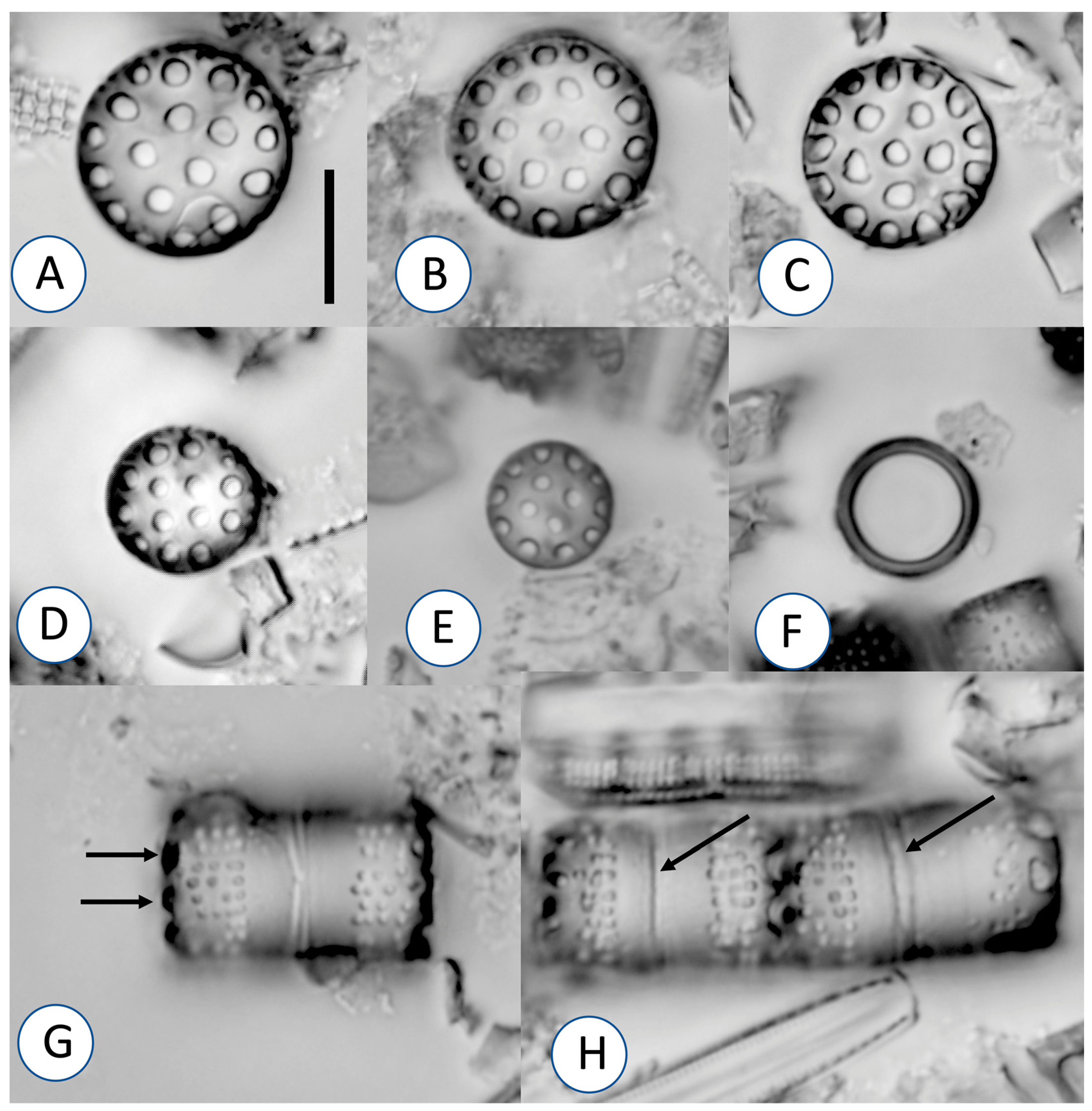
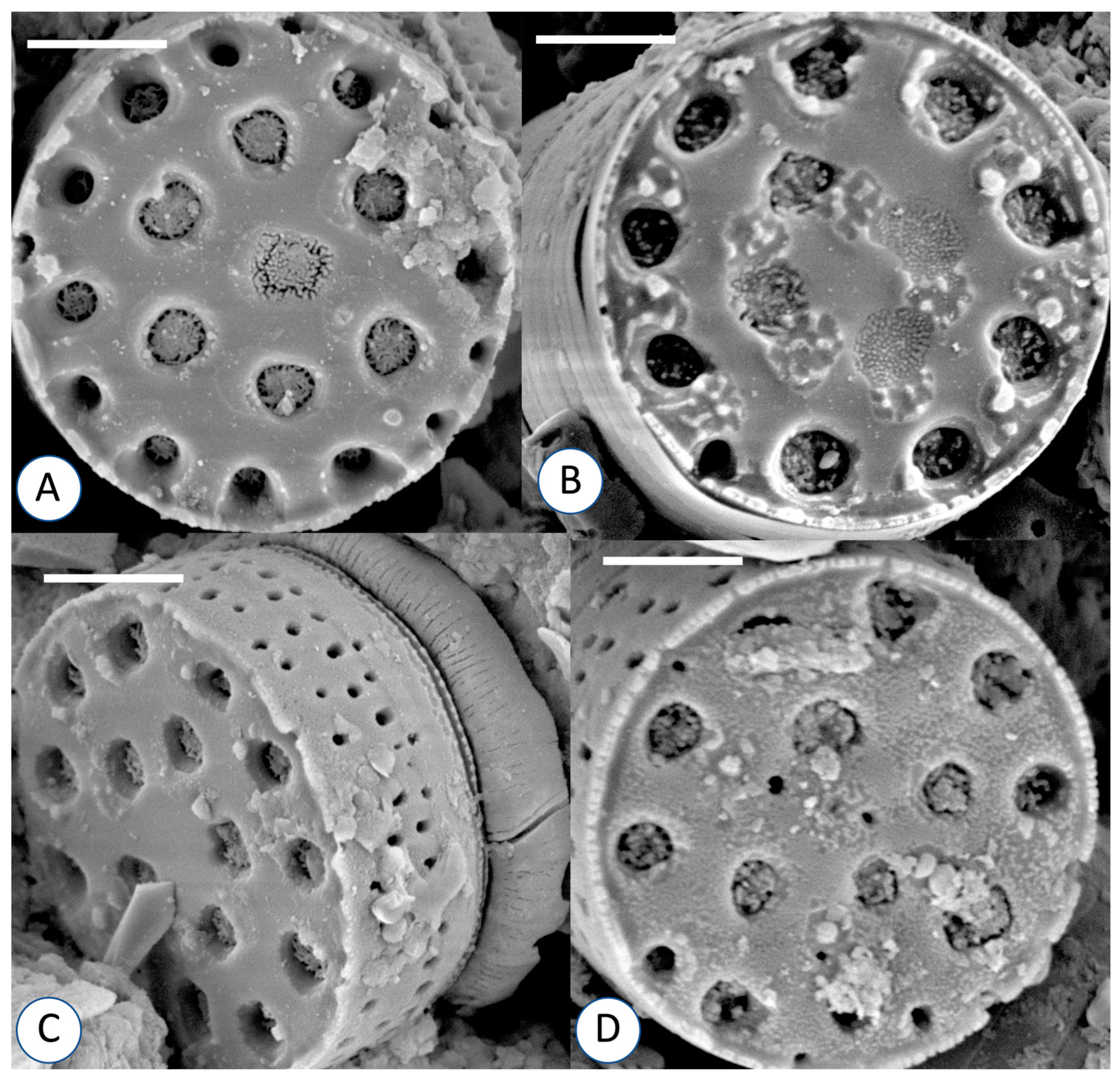
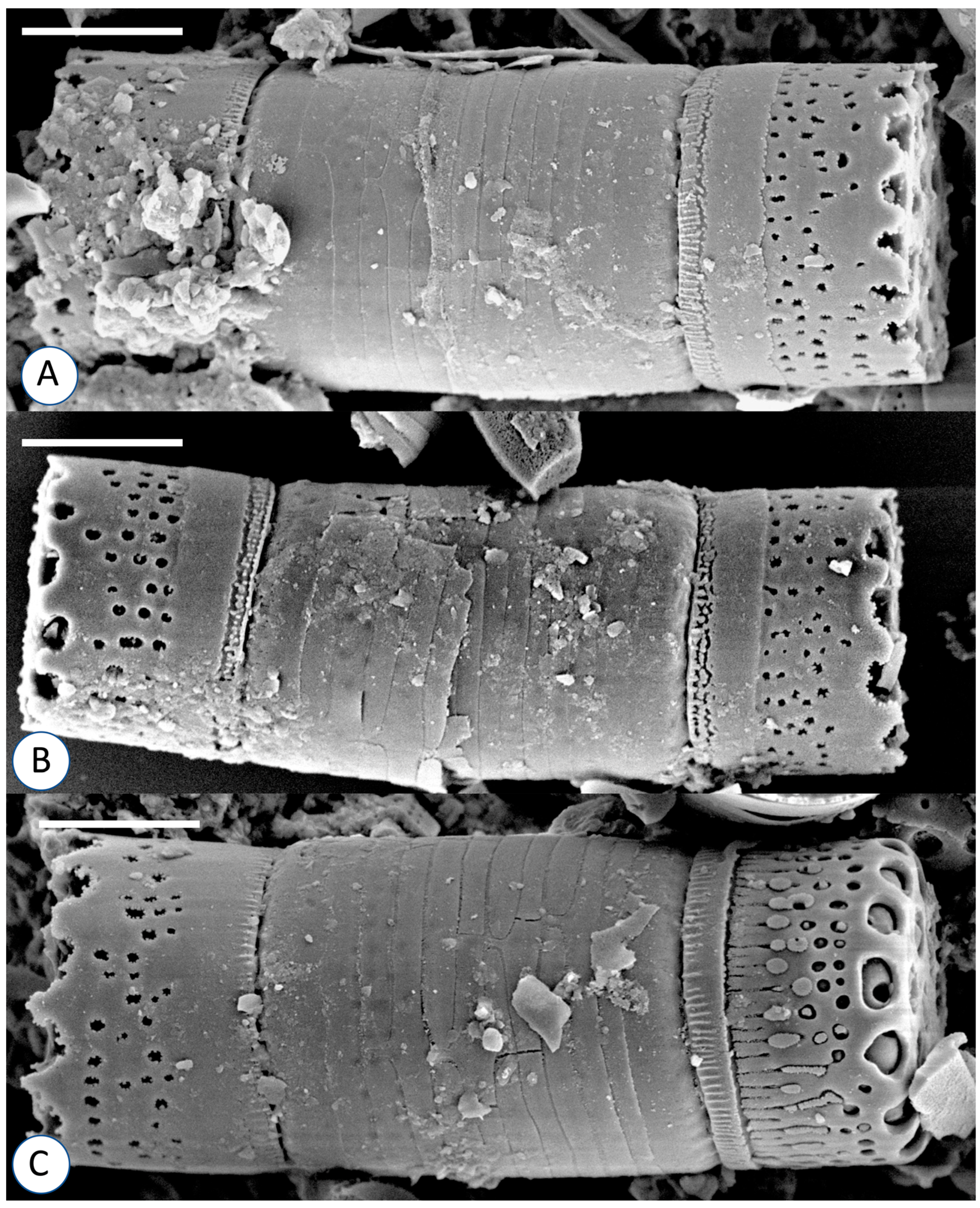
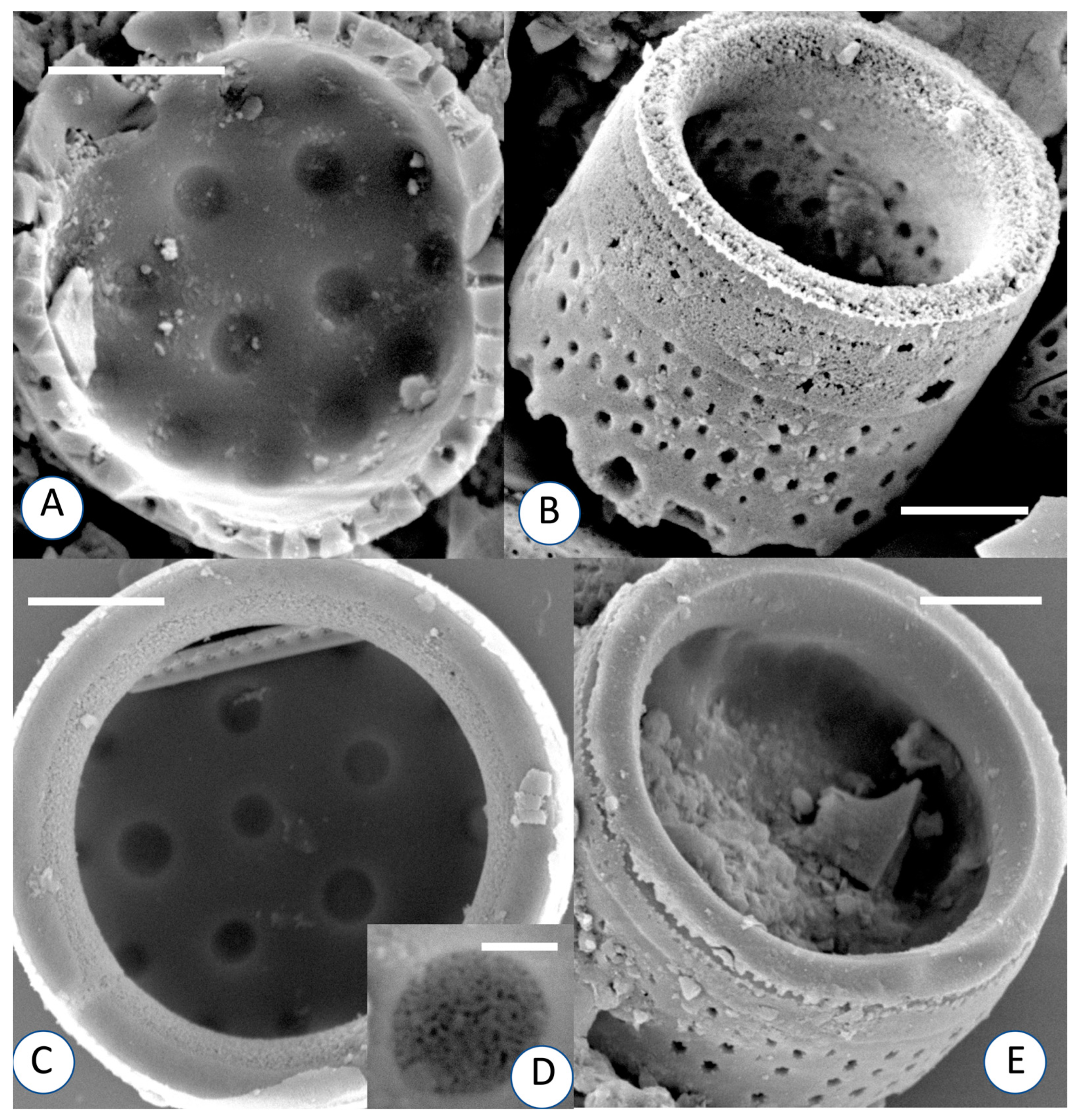
A composite sample from a still seep near Casper, Wyoming (42.7125323 N, 106.8828493 W), USA, was collected on 22 August 2021. Except for locality and date information, no other data were collected with the sample. Approximately 25 mL of the sample was cleaned with concentrated (70%) nitric acid. After cleaning, the sample was rinsed five times with deionized water, allowing at least 24 h between rinses. Portions of the fully-settled sample were mixed with deionized water and air-dried on coverslips. Coverslips with the dried material were mounted on microscope slides using Naphrax (refractive index 1.74). Light microscope observations were made with a BX-51 light microscope with DIC optics (100× objective with 1.42NA) and images were taken with a DP-71 digital camera (Olympus Corporation of the Americas, Breinigsville, PA, USA).
For SEM, cleaned material was dried onto coverslips which were mounted on aluminum stubs. Stubs with the cleaned material were sputter-coated with 10 nm of platinum using a Cressington sputter coater. The coated material was viewed with a Hitachi SU 3500 SEM at a working distance of 5.7 mm and accelerating voltage set at 5.0 kV at the Colorado Shared Instrumentation in the Nanofabrication and Characterization (COSINC-CHR) facilities at the University of Colorado Boulder.
To provide an ecological context to the community present in the sample studied, 600 diatom valves were identified to species.

3. Results

  • Division Bacillariophyta Haeckel 1878.
  • Class Coscinodiscophyceae Round and Crawford 1990.
  • Order Aulacoseirales Crawford 1990.
  • Family Aulacoseiraceae Crawford 1990.
  • Genus Aulacoseira Thwaites 1848.
Aulacoseira wyomingensis Greifenstein, Shea and Kociolek, sp. nov. Figure 1, Figure 2, Figure 3 and Figure 4.
Holotype: Accession number 14073, JPK Collection at COLO. Holotype slide: 652059 in the JPK Collection at COLO. Isotype slides: To be deposited at ANSP and BM. Numbers to be issued upon acceptance of the manuscript.
Etymology: Named for the U.S. state in which it was found.
Description: Valves are disc-shaped (Figure 1A–E), cylindrical in girdle view (Figure 1G,H) 7–14 µm in diameter, and with a valve mantle height of 3–9 µm (n = 100). Frustules are single, rarely forming chains; if so, never comprising more than two frustules. Valve face has large areolae (ca. 1 µm diameter) that are present across its entirety. In girdle view, short spine-like protrusions are evident around the valve margin. Mantle striae are straight, areolae are disorganized within a stria, two to six areolae are present per stria. Stria density on the mantle is 10–12/10 µm. Striae are usually interrupted along their length. Striae extend from the valve margin to only ½ the height of the mantle, extending to the wide collum. Areolar density on the mantle is 12–16/10 µm. Collum is evident. Ringleiste is relatively narrow (Figure 1F).
In the SEM, externally, the areolae on the valve face are seen as forming depressions occluded by volae (Figure 2A–D). Short, blunt, spine-like protrusions originate from the mantle (Figure 2C and Figure 3A–C), shield-like, but absent from some valves. The cingulum is comprised of many narrow, ligulate bands (Figure 3A–C). The collum is ornamented with siliceous ridges (Figure 3A–C). Internally, individual areolae are covered by fine, hymenate occlusions (Figure 4A,C,D). Rimoportulae have not been observed (Figure 4A–C,E).

4. Discussion

Many species of Aulacoseira have only a few areolae scattered across the valve face (e.g., A. pusilla (F.Meister) Tuji and Houki; [16,17]) or concentrated around the periphery (e.g., A. lirata (Ehrenberg) R.Ross in Hartley; [11,18,19]). Aulacoseira wyomingensis is among the Aulacoseira species with areolae across the entire valve face. In Table 1, A. wyomingensis is compared and contrasted with other species of the genus having the valve face covered with areolae. In short, the few, large areolae on the valve face, shield-like, small spines that do not interdigitate with opposing valves (and thus lack of a filamentous growth habit), lack of rimoportulae and the disorganized nature of the striae on the valve mantle all distinguish this new species from other, previously described taxa. Aulacoseira wyomingensis does share the feature of not forming filaments with taxa such as A. singulara Bennett and Kociolek [14] and A. tenella (Nygaard) Simonsen [20], A. chockii Siver [9] among others, but differs from these taxa in the other features described above.
Aulacoseira wyomingensis sp. nov. was the dominant taxon in the sample from which it is described, having a relative abundance of 57.8%. Other common taxa in the sample include Epithemia cf. adnata (11.0%), Denticula kuetzingii Grunow (7.8%), D. valida (Pedicino) Grunow (7.2%), Encyonopsis microcephala (Grunow) Krammer (4.2%), Adlafia bryophila (J.B.Petersen) Moser et al. (3.2%), Staurosirella sp. (2.7%) and Epithemia cf. sorex (2.5%). Other ecological data for this sample are wanting. To date, it has only been found in this Recent collection.
We assign A. wyomingensis to Aulacoseira, based on its expanded valve mantle and that it lacks features of most of the genera currently in Aulacoseiraceae. For example, A. wyomingenesis lacks the diagnostic features of internal struts found in Miosira and Alveolophora [3,4,28] and lacks the long spathulate spines found in Eosira [2]. Within Aulacoseira, five groups can be recognized (according to [15] using a total evidence approach for understanding phylogenetic relationships. Synapomorphies for these different groups have not been recognized, so it is difficult to align their phylogenetic relationships with specific morphological features. Species of Aulacoseira that have the valve face covered in areolae occur in three of their five groups, including A. pfaffiana in group 1 (with A. crenulata (Ehrenberg) Thwaites, the generitype), A. perglabra in group 4 (with A. subarctica and allies) and A. islandica in group 5. The tremendous morphological diversity seen amongst taxa currently assigned to Aulacoseira, both Recent and fossil [9,25,29,30], suggests further research and, perhaps, an analysis of the phylogenetic relationships of the genus as well as the family is warranted.

Author Contributions

Conceptualization, J.G., R.S. and J.P.K.; sample processing, J.G. and R.S.; observations, J.G., R.S. and J.P.K.; data analysis, J.G. and J.P.K.; writing, review and editing J.G., R.S. and J.P.K.; visualization J.G. and J.P.K. All authors have read and agreed to the published version of the manuscript.

Funding

This research received no external funding.

Institutional Review Board Statement

Not applicable.

Informed Consent Statement

Not applicable.

Data Availability Statement

Specimens were deposited in publicly accessible collections.

Acknowledgments

These data were collected in partnership with the Colorado Natural Heritage Program at Colorado State University as part of the Bureau of Land Management’s Riparian and Wetland Assessment, Inventory, and Monitoring (AIM) program. Any use of trade, firm, or product names is for descriptive purposes only and does not imply endorsement by the U.S. Government. Collections from Wyoming were made by Jordan Swenson, Evan Cox and Teresa Wolf.

Conflicts of Interest

The authors declare no conflict of interest.

References

  1. Round, F.E.; Crawford, R.M.; Mann, D.G. The Diatoms. Biology and Morphology of the Genera; Cambridge University Press: Cambridge, UK, 1990; 747p. [Google Scholar]
  2. Wolfe, A.P.; Edlund, M.B. Taxonomy, phylogeny, and paleoecology of Eoseira wilsonii gen. et sp. nov., a Middle Eocene diatom (Bacillariophyceae: Aulacoseiraceae) from lake sediments at Horsefly, British Columbia, Canada. Can. J. Earth Sci. 2005, 42, 243–257. [Google Scholar] [CrossRef] [Green Version]
  3. Krammer, K.; Lange-Bertalot, H.; Schiller, W. Miosira rhoenana n. gen. n. sp. (Bacillariophyceae) from Miocene limnic sediments of the Rhoen mountains in comparison with other centric diatoms. Paläontologische Z. 1997, 71, 5–18. [Google Scholar] [CrossRef]
  4. Usoltseva, M.; Kociolek, J.P.; Khursevich, G.K. Three new species of Alveolophora (Aulacoseiraceae) from Miocene fossil deposits in western North America. Phycologia 2013, 52, 109–117. [Google Scholar] [CrossRef]
  5. Khursevich, G.K.; Kociolek, J.P. A Preliminary, Worldwide Inventory of the Extinct, Freshwater Fossil Diatoms from the Orders Thalassiosirales, Stephanodiscales, Paraliales, Aulacoseirales, Melosirales, Coscindiscales, and Biddulphiales. Nova Hedwig. Beih. 2012, 141, 315–364. [Google Scholar]
  6. Houk, V.; Klee, R.; Tanaka, H. Atlas of freshwater centric diatoms with a brief key and descriptions. Second emended edition of Part I and II. Melosiraceae, Liparogyraceae, Paraliaceae and Aulacoseiraceae. Fottea 2007, 1 (Suppl. S17), 1–616. [Google Scholar]
  7. Siver, P.A.; Velez, M.; Cliveti, M.; Binda, P. Early freshwater diatoms from the Upper Cretaceous Battle Formation in western Canada. Palaios 2018, 33, 525–534. [Google Scholar] [CrossRef]
  8. Krebs, W.N.; Bradbury, J.P.; Theriot, E.C. Neogene and Quaternary lacustrine diatom biochronology, Western USA. Palaios 1987, 2, 505–513. [Google Scholar] [CrossRef] [Green Version]
  9. Siver, P.A. Aulacoseira chockii sp. nov., an early freshwater centric diatom from the Eocene bearing a unique morphology. Diatom Res. 2021, 36, 253–263. [Google Scholar] [CrossRef]
  10. Moisseeva, A.I.; Nevretdinova, T.L. Novye semeistvo i rod presnovodnykh diatomovykh vodoroslei (Bacillariophyta). Bot. Zhurnal 1990, 75, 539–544. [Google Scholar]
  11. Camburn, K.E.; Kingston, J.C. The genus Melosira from soft-water lakes with special reference to northern Michigan, Wisconsin and Minnesota. In Diatoms and Lake Acidity; Smol, J.P., Battarbee, R.W., Davis, R.B., Meriläinen, J., Eds.; Dr. W. Junk Publishers: Dordrecht, The Netherlands, 1986; pp. 17–34. [Google Scholar]
  12. Camburn, K.E.; Charles, D.F. Diatoms of Low-Alkalinity Lakes in the Northeastern United States. (Academy of Natural Sciences of Philadelphia Special Publication); Academy of Natural Sciences: Philadelphia, PA, USA, 2000; Volume 18, pp. 1–152. [Google Scholar]
  13. Siver, P.A.; Hamilton, P.B. The Diatoms of North America: The Freshwater Flora of Waterbodies on the Atlantic Coastal Plain. Iconogr. Diatomol. 2011, 22, 1–916. [Google Scholar]
  14. Kociolek, J.P.; Laslandes, B.; Bennett, D.; Thomas, E.; Brady, M.; Graeff, C. Diatoms of the United States 1: Taxonomy, Ultrastructure and Descriptions of New Species and Other Rarely Reported Taxa from Lake Sediments in the Western, U.S.A. Bibl. Diatomol. 2014, 61, 1–188. [Google Scholar]
  15. Edgar, S.M.; Theriot, E.C. Phylogeny of Aulacoseira (Bacillariophyta) based on molecules and morphology. J. Phycol. 2004, 40, 772–788. [Google Scholar] [CrossRef]
  16. Tuji, A.; Houki, A. Taxonomy, ultrastructure, and biogeography of the Aulacoseira subarctica species complex. Bull. Natl. Sci. Mus. Ser. B 2004, 30, 35–55. [Google Scholar]
  17. Tuji, A.; Williams, D.M. Type examination of the freshwater centric diatom Aulacoseira pusilla (F. Meister) Tuji et Houki. Diatom 2006, 22, 70–73. [Google Scholar]
  18. Florin, M.-B. The taxonomy of some Melosira species, a comparative morphological study. II. In Proceedings of the 6th Symposium on Recent and Fossil Diatoms, Budapest, Hungary, 1–5 September 1980; International Society of Diatioms: Koeltz, Germany; Koenigstein, Germany, 1981; pp. 43–74. [Google Scholar]
  19. Krammer, K. Morphology and taxonomy of some taxa in the genus Aulacoseira Thwaites (Bacillariophyceae). I. Aulacoseira distans and similar taxa. Nova Hedwig. 1991, 52, 89–112. [Google Scholar]
  20. Potapova, M. Aulacoseira tenella. Diatoms of North America. 2010. Available online: https://diatoms.org/species/aulacoseira_tenella (accessed on 5 May 2022).
  21. Tremarin, P.I.; Ludwig, T.A.V.; Torgan, L.C. Ultrastructure of Aulacoseira brasiliensis sp. nov. (Coscinodiscophyceae) and comparison with related species. Fottea 2012, 12, 171–188. [Google Scholar] [CrossRef]
  22. English, J.; Potapova, M. Aulacoseira pardata sp. nov., A. nivalis comb. nov., A. nivaloides comb. nov et stat. nov., and their occurrences in western North America. Proc. Acad. Nat. Sci. Phila. 2009, 158, 37–48. [Google Scholar] [CrossRef]
  23. English, J.; Potapova, M. Aulacoseira humilis. Diatoms of North America. 2010. Available online: https://diatoms.org/species/aulcaoseira_humilis (accessed on 5 May 2022).
  24. Tuji, A. Examination of type material of Melosira nivalis W.Sm. (Bacillariophyceae) and its synonymy with Aulacoseira pfaffiana (Reinsch) Krammer. Bull. Natl. Mus. Nat. Sci. Ser. B 2010, 36, 161–164. [Google Scholar]
  25. Krammer, K.; Lange-Bertalot, H. Bacillariophyceae, 3 Teil, Centrales, Fragilariaceae, Eunotiaceae. In Süsswasserflora von Mitteleuropa, Band 2; Ettl, H., Gerloff, J., Heynig, H., Mollenhauer, D., Eds.; Gustav Fischer: Stuttgart/Jena, Germany, 1991; pp. 1–576. [Google Scholar]
  26. Genkal, S.I.; Poposkaya, G.I. New data on the diatom morphology of Aulacoseira islandica (Bacillariophyta). Diatom Res. 1991, 6, 255–266. [Google Scholar] [CrossRef]
  27. Tremarin, P.I.; Ludwig, T.A.V.; Torgan, L.C. Four new Aulacoseira species (Coscinodiscophyceae) from Matogrossense Pantanal, Brazil. Diatom Res. 2014, 29, 183–199. [Google Scholar] [CrossRef]
  28. Usoltseva, M. Morphological variability of Alveolophora jouseana (Bacillariophyta) from type material. Phytotaxa 2022, 533, 194–204. [Google Scholar] [CrossRef]
  29. Lee, Y.; Yoon, S.-K. Morphologic accounts of the diatom genus Aulacoseira, the dominant of the Han River. Korean J. Phycol. 1994, 9, 135–143. [Google Scholar]
  30. Houk, V. Aulacoseira hibschii (Reichelt) Houk comb. nov. (Bacillariophyceae, Centrales) from the type locality in Varnsdorf (Czech Republic). Bull. Geosci. 2007, 82, 419–428. [Google Scholar] [CrossRef] [Green Version]
Figure 1. (AH) Aulacoseira wyomingensis, sp. nov. Light microscopy. (AE) Valve views. Size diminution series. (F) Ringleiste. (G,H) girdle views. Arrows indicate small, spine-like protrusions (G) and collum (H). All specimens are from the holotype slide. Scale bar = 10 µm for all images.
Taxonomy 02 00018 g001
Figure 2. (AD) Aulacoseira wyomingensis, sp. nov. Scanning electron microscopy. External valve views. Valve face is composed of areolae in deep depressions on the valve face, with covering at the base of each areola. In some cases, the areolae appear as depressions, without communication to the valve interior. Spines are mostly lacking. Scale bars = 2.5 µm.
Taxonomy 02 00018 g002
Figure 3. (AC) Aulacoseira wyomingensis, sp. nov. Scanning electron microscopy. External girdle views of entire frustules. Valves have striae that are disorganized. Column has small ridges. Cingulum is composed of numerous ligulate elements. Spines are small in length and shield-like. Valve on the right in (C) appears to be incompletely formed. Scale bars = 5 µm.
Taxonomy 02 00018 g003
Figure 4. (AC) Aulacoseira wyomingensis, sp. nov. Scanning electron microscopy. Internal views. (A) Valve view showing areolae and interior of valve. Scale bar = 2.5 µm. (B) Side view showing part of valve interior and exterior. Scale bar = 2.5 µm. (C) Valve view of interior showing ringleiste. Scale bar = 2.5 µm. (D) High magnification view of single areola showing fine hymenate occlusion over opening. Scale bar = 0.3 µm. (E) Side view showing part of the valve interior and exterior. Scale bar = 2.5 µm.
Taxonomy 02 00018 g004
Table 1. Comparison of morphometric data of some Aulacoseira species with valve face covered with areolae.
TaxonDiameter (µm)Valve Mantle Height (µm)Striae/10 µmAreolae/10 µmReferences
A. brasiliensis8–244–1010–1610–15[21]
A. tenella 5–12 2–5 20–24 (face)
ca. 20 (mantle)
[22]
A. humilis5–92–512–2017 (face)
24–40 (mantle)
[23]
A. nivalis6–18 2.5–6.012–169–10 (mantle)
7–14 (face)
[22,24]
A. nivaloides4–114–6 16–2215–21 (mantle)
ca. 18
[22]
A. islandica3–284–2111–1612–15[25,26]
A. minuscula3.0–4.52.0–3.5 16–20No data[27]
A. imparis7–13 7–1023–25No data[13]
A. lancea7–155–1022–25No data[13]
A. occulata7–177–1022–25No data [13]
A. perglabra8–172–420–2828–80[25]
A. pfaffiana4–23 3.0–10.512–15 16–18 [25]
A. delicatula3–62–3No data No data[14]
A. singularia2.0–5.51–213–1520[14]
A. wyomingensis6–15 3–7 4–6 (face)
9–12 (mantle)
12–16 (mantle)This Study
Publisher’s Note: MDPI stays neutral with regard to jurisdictional claims in published maps and institutional affiliations.

