Review

Combined Therapies with Taxane-Based Chemotherapeutic Drugs in Prostate Cancer: Novel Insights and Future Directions

by
Rafaella S. Coelho
,
Sandra M. Rocha
and
Cláudio J. Maia
*
CICS-UBI-Health Sciences Research Center, University of Beira Interior, 6200-506 Covilhã, Portugal
*
Author to whom correspondence should be addressed.
These authors contributed equally to this work.
BioChem 2023, 3(3), 118-141; https://doi.org/10.3390/biochem3030009
Submission received: 18 April 2023 / Revised: 2 August 2023 / Accepted: 25 August 2023 / Published: 29 August 2023

Abstract

:
Oncologic disease is a significant global health issue that causes thousands of deaths annually, and it has a significant impact on the quality of life of patients. Prostate cancer (PCa) is the second most diagnosed cancer and the fourth leading cause of cancer-related death in men in the Western world. Delineation of pathogenetic pathways and key driver molecular alterations involved in PCa development has provided a roadmap for the evaluation of biomarkers in predicting disease outcome and to identify potential therapeutic targets. Chemotherapeutic agents introduced from the 1990s include the taxanes (paclitaxel, docetaxel, and cabazitaxel), which are the anticancer drugs used most frequently for PCa treatment. This review presents the current knowledge about the onset and development of PCa, the state of the art of the use of taxane-based therapy, and their combination with targeting different transmembrane oncoproteins in PCa. The silencing of some transmembrane proteins can improve taxane sensitivity, and therefore may be a mechanism to improve the effectiveness of these drugs in PCa treatment. This combined therapy needs to be explored as a potential therapeutic agent for reducing cell proliferation, migration, and invasiveness in PCa.

1. Introduction

The burden of cancer incidence and mortality is rapidly growing worldwide, and expectations for 2020 showed approximately 19.3 million new cancer cases and 10.0 million cancer deaths [GLOBOCAN, https://gco.iarc.fr/, accessed on 9 March 2023]. Prostate cancer (PCa) is currently the second most common cancer in men and represents the fourth leading cause of cancer-related mortality. In 2020, 1.4 million new cases of PCa were diagnosed worldwide and approximately 375,000 associated deaths were reported by the World Health Organization [1]. The increased number of PCa cases can be explained by the lack of comprehensive national control programs that contributes to substantial disparities in the early detection of cancer and management of these patients, with a three-fold higher incidence rates in countries with high human development when compared to countries with low human development (37.5 and 11.3 per 100,000 habitants, respectively), although mortality rates are less variable (8.1 and 5.9 per 100,000 habitants, respectively) [2,3]. Moreover, the etiology of PCa is multifactorial and remains largely unknown when compared to other types of cancer. Epidemiologic evidence has identified several biological and genetic factors, but environmental and lifestyle factors have also been shown to contribute to the appearance and progression of PCa, such as advanced age, family history and genetic predisposition, ethnicity, smoking and alcohol consumption, obesity and metabolic syndrome, physical inactivity, diet and nutrition, medications, sexual activity and vasectomy, hormones, infection, inflammation, and chemokines [4,5]. However, age is considered the highest risk factor for the development of PCa. The peak of incidence is found in older men approximately 70–74 years of age [6].
Currently, several agents have received FDA approval and have been associated with beneficial effects in improving survival and life quality in patients with this pathology, including abiraterone, enzalutamide, apalutamide, and darolutamide (inhibitors of the androgen axis); paclitaxel, docetaxel, and cabazitaxel (which target microtubules by inhibiting depolymerization or promoting polymerization); radium-223 (radioactive agents targeting bone metastasis); and sipuleucel-T (triggers cellular immune mechanisms) [7]. From those agents, appropriate drug selection is carried out according to clinical usage for the treatment of PCa. Several cancers are treated with drug combinations, but PCa has remained an exception [8]. Transmembrane proteins are involved in many crucial cell processes, including signaling transduction pathways, transport of ions and molecules, protein targeting and intracellular transport, and membrane trafficking [9]. Moreover, since membrane proteins are involved in essential cellular pathways, they are often recognized in the pathophysiology of many diseases and are major targets for pharmaceutical agents, with more than 60% of drug targets being transmembrane proteins [10]. Hence, developing an effective combination of drugs and targeting some transmembrane proteins can provide insights concerning new therapeutic strategies for advanced stages of PCa. This review provides an overview of the development of PCa, with a special focus on the taxane-based therapy currently used. Furthermore, we reviewed the scientific literature concerning the combined action of taxane-based chemotherapeutic drugs with inhibition of transmembrane oncoproteins within the paradigm of PCa.

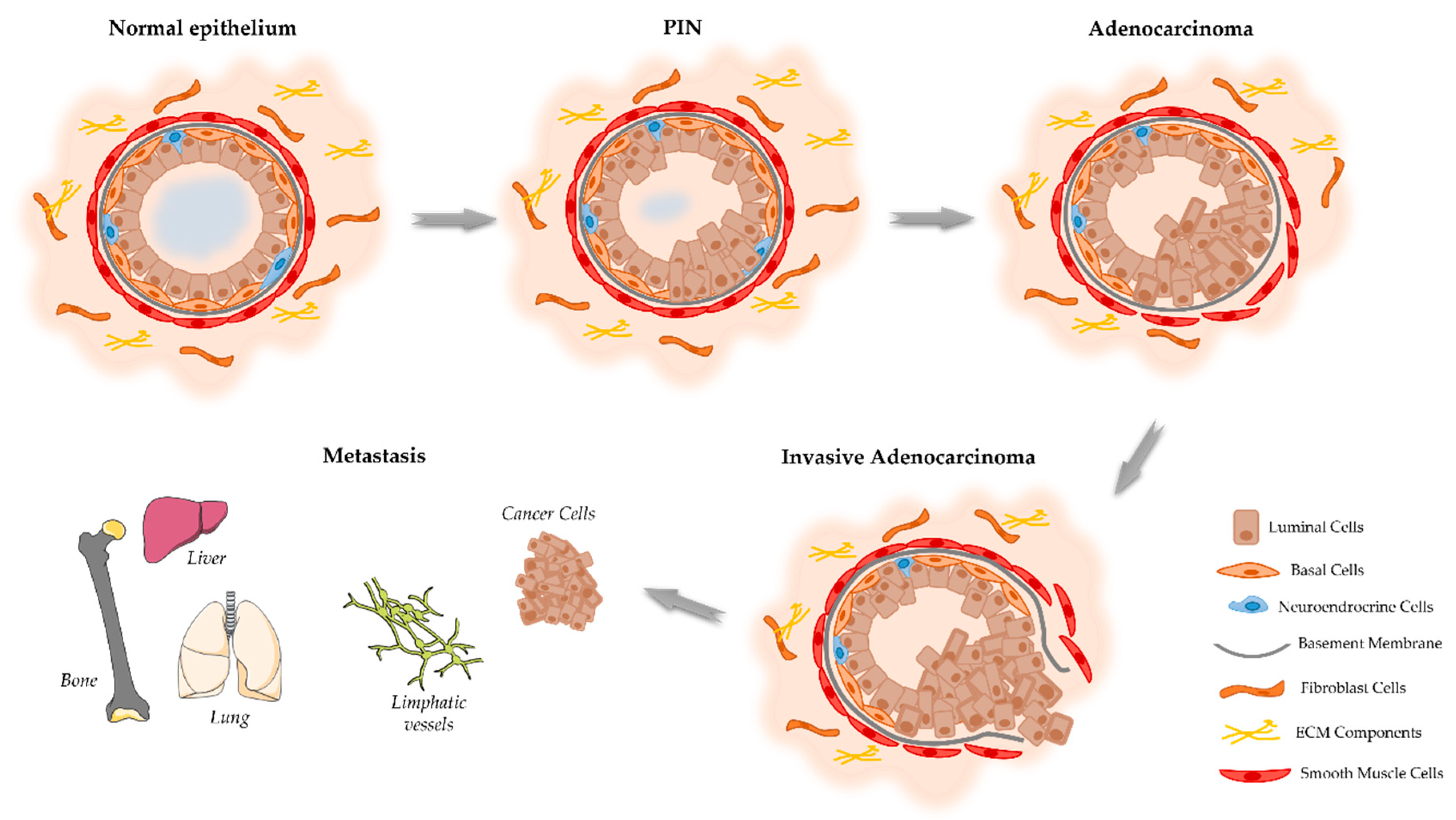
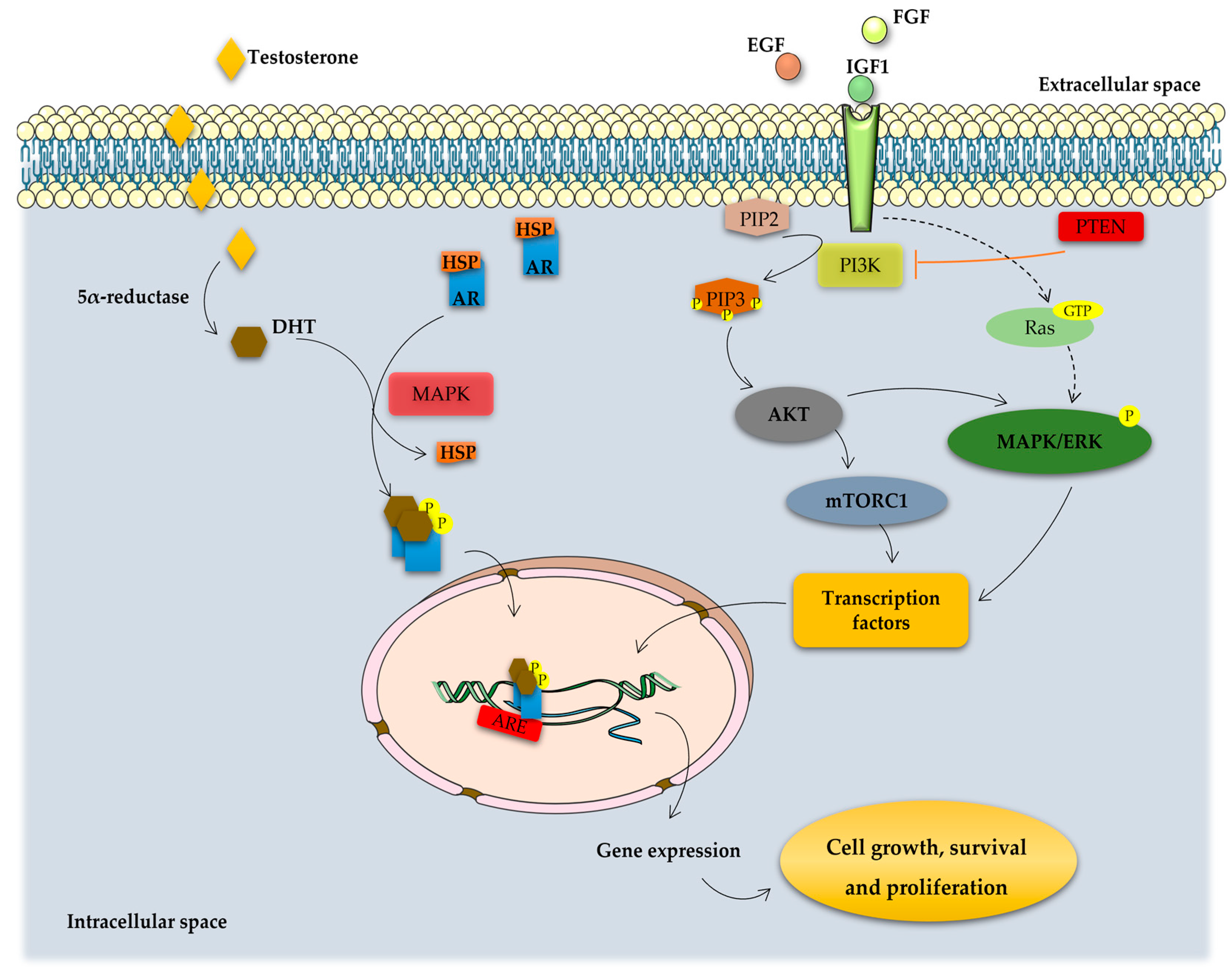
2. Onset and Development of PCa

The human prostate gland is the major accessory gland of the male reproductive system, located frontal to the rectum and immediately below the urinary bladder, surrounding the prostatic urethra and the ejaculatory ducts [11,12]. Normal prostate tissue consists of prostatic ducts lined with epithelial cells surrounded by fibromuscular stroma [13,14]. Homeostasis of normal prostate tissue is maintained by the crosstalk between epithelial cells and the surrounding stromal components [15,16]. The glandular prostatic epithelium is a well-organized tissue composed of acini and ducts consisting of three types of cells: luminal, basal, and neuroendrocrine cells (Figure 1). Luminal cells are columnar epithelial cells specialized in the production of prostatic secretions, including prostate-specific antigen (PSA), and are responsible for the main prostate function [17]. Basal cells adhere to the basement membrane and have the ability to produce several components essential to the maintenance of cell growth [18,19]. Neuroendocrine cells comprise less than 1% of the prostatic epithelium and express chromogranin A, synaptophysin, enolase 2, and CD56, which promote the growth of the prostate [20]. Interactions between the epithelium and basement membrane are fundamental to maintain epithelial cell polarity involving apical and basal surfaces, which represent the well-differentiated cell state [13]. The non-epithelial tissue of the prostate, referred to as the stroma, is composed essentially by fibroblasts, smooth muscle cells, and extracellular matrix (ECM) proteins (Figure 1) [15]. The ECM forms a dynamic and structured mixture of collagens, proteoglycans, thrombospondin, and hyaluronic acid, which responds to tissue injuries and allows its regeneration [16].
Considering the onset of PCa, there is good agreement that this cancer develops from prostate epithelial cells [14]. However, conflicting evidence exists regarding if the oncogenic transformation in PCa arises from basal [19,21] or luminal epithelial cells [22,23]. In addition, it also has been hypothesized that PCa arising from luminal cells is more aggressive than that arising from basal cells [21]. The prostatic epithelium can be damaged and drive the carcinogenesis of the prostate due to several factors, including inflammation, infections, genetic/epigenetic changes, persistent activation by androgens, exposure to carcinogens, and/or genetic factors [14,24]. The first identifiable histologic alteration in prostate malignant transformation is so-called prostatic intraepithelial neoplasia (PIN) (Figure 1) [25]. PIN lesions can be divided into two grades: low-grade PIN (LGPIN) and high-grade PIN (HGPIN). HGPIN lesions are considered the most likely precursors of PCa [26,27], but they do not appear to raise serum PSA concentration [28]. Characteristically, HGPIN lesions contain a basal cell layer around their periphery, although it is thin and often discontinuous. This is an important diagnostic feature because preservation of the basal cell layer can help to differentiate PIN from prostatic adenocarcinoma in which the basal cells are absent [24,29].
Prostatic adenocarcinoma mostly arises in the peripheral zone of the prostate and initially is represented as a small foci of intraductal dysplasia, that with time differentiates and progresses into an invasive adenocarcinoma (Figure 1) [30]. The tumor foci lead to a disruption of prostate tissue and a decrease in glandular activity and prostatic fluid production [31]. Histologically, PCa is characterized by the destruction of the basal cell layer, derangement of the basement membrane, decreased epithelial cell polarity, and lack of connection of the glandular acini formed by the prostate epithelial cells [32]. As the tumor progresses, neoplastic cells increase the production of proteolytic enzymes, which cause degradation of the basement membrane, allowing the spread to adjacent tissues and the development of a metastatic disease [33]; firstly, to the lymph nodes and then to distant organs, including the bones, liver, and lungs, with bone as the most common site of metastasis [34]. In fact, in the context of epithelial neoplasia, the prostate stroma induces alterations in the tumor microenvironment, being the so-called reactive stroma. This phenotypic histological change leads to a loss of well-differentiated smooth muscle cells, increase of fibroblast population, and increase of secretion and deposition of ECM components, such as matrix metalloproteinase (MMP). All these changes can lead to epithelial cell depolarization and formation of conduits favoring neoplastic cell migration [16,35]. All these histological changes cause a thousand-fold increased release of PSA from prostate neoplastic cells into the blood [32].
Androgens play a central role in the control of the normal prostate, as well as PCa cell growth and proliferation [14]. Androgens are the primary regulators of the proliferation/apoptosis ratio, stimulating proliferation and inhibiting apoptosis of prostate cells, and, thus, inducing the development of PCa [14,36]. The major circulating androgen, testosterone, can be converted into DHT by the activity of 5α-reductase enzyme. Both testosterone and DHT exert their actions through binding to the AR. PCa growth and disease progression is initially dependent on AR activation. The main mechanism of action leads to the nuclear translocation of the ligand–receptor complex and subsequent binding to the androgen response elements (AREs), which initiates the transcription of genes that regulate cellular differentiation, proliferation, and apoptosis (Figure 2) [27,36,37].
In primary PCa, the action of AR retains the same role as in a normal prostate; for example, synthesis of PSA and modulating lipid metabolism [22]. However, it also triggers other events that promote epithelial cell growth, such as the induction of the type II transmembrane serine protease (TMPRSS2):ETS fusion [26,38]. The TMPRSS2 is an androgen-regulated gene overexpressed in PCa, which encodes a protein belonging to the serine protease family that functions in prostate carcinogenesis and relies on gene fusion with ETS transcription factors, such as the ETS-related gene (ERG) and ETV1. The TMPRSS2:ETS fusion is considered the most common chromosomal rearrangement in PCa and drives the overexpression of ETS oncogenes, previously identified as the most expressed proto-oncogenes present on malignant epithelial prostate cells [38,39,40]. ARs also have two active functional domains (AFs) that initiate transcription when activated. AF-1 is present in the NTD and its activation is androgen-independent. AF-2 is located in the LBD and is ligand-dependent [41]. AF-1 may enable cross-coupling between androgenic and growth factor signaling pathways [36,42]. Therefore, these AFs are deemed clinically important as they could provide the key to understand the development of castration-resistant PCa (CRPC). At early stages of disease, PCa growth is androgen-dependent; the so-called androgen-sensitive PCa. However, with continuous tumor development, PCa cells became androgen-insensitive, and the disease progresses to the so-called CRPC [36].
Patients that acquire resistance to the use of androgen-deprivation therapy (ADT) inevitably develop CRPC, a more lethal form of PCa. The role of AR in PCa progression and development of CRPC has been attributed to several factors, such as AR gene amplification, activating mutations, and aberrant expression of co-activators [37,43,44]. These alterations lead to an increased AR expression, activation of AR by non-androgenic ligands, broadened ligand specificity and sensitivity, and increased AR transactivation, which ultimately contribute to tumor cell growth in a low androgen-environment [36,44,45]. AR mutations in primary PCa are rare, but these mutations are prevalent in about 50% of CRPC [46,47]. These mutations lead to alterations that improve the functional activity of the receptor, such as increased AR sensitivity to low levels of ligand, non-androgen ligand binding, ligand-independent activation, and AR-independent pathways [41,46,47]. Furthermore, recent data indicate that an increased expression of constitutively active AR splice variants follows castration and is associated with poor prognostics and a rapid recurrence of PCa [48,49]. The reduction in AR activation by endogenous androgen ligands leads to hypersensitization of other pathways of AR activation through ligand-independent mechanisms [44,50].
Various growth factors, cytokines, kinases, and other proteins have been shown to interact with and activate AR in a ligand-independent manner, including insulin-like growth factor (IGF1), fibroblast growth factor (FGF), and epidermal growth factor (EGF) [51,52]. These growth factors activate tyrosine receptor kinases, which results in the activation of phosphatidylinositol 3-kinase (PI3K) and subsequently the PI3K/AKT pathway (Figure 2) [53]. The serine/threonine protein kinase (AKT), also known as protein kinase B (PKB), is one of the major downstream effectors of PI3K. Binding of ligands to the membrane growth factor receptors initiates a cascade of events that activate PI3K, which converts phosphatidylinositol 4,5-bisphosphonate (PIP2) to phosphatidylinositol 3–5-triphosphate (PIP3). PI3K activation stimulates AKT, which recruits proteins to the luminal cell cytoplasm [53,54]. Downstream targets of AKT, namely the mammalian target of rapamycin complex 1 (mTORC1), forkhead box protein O1, and the mitogen-activated protein kinase/extracellular signal-regulated kinase (MAPK/ERK) cascade, activate several transcription factors, such as c-myc, which induces the expression of proteins associated with cell survival and proliferation, cell cycle progression, migration, and angiogenesis, and thus contribute to the progression of PCa [44,53,55].

3. Current Use of Chemotherapy in PCa

Treatment approaches for PCa differ depending on the stage of the disease. Several types of therapeutic options are available, such as surgery, cryosurgery, radiation therapy, hormone therapy, chemotherapy, vaccine treatment, immunotherapy, and bone-directed treatment [56]. Active surveillance is the recommended treatment option for low-risk PCa, monitoring its progression while not undergoing definitive therapy [57]. Therapeutic approaches based on surgery often are used in combination with therapeutic approaches based on drugs, namely hormone therapy and chemotherapy. As the non-neoplastic prostate cells, PCa cells need androgens to grow and survive, making ADT an effective first-line therapy. This therapy can involve two approaches: surgical castration (i.e., orchiectomy) or, more commonly, chemical castration with drugs targeting AR signaling regulated by the hypothalamic pituitary gonadal axis (e.g., GnRH agonists, AR antagonists, and CYP17A1 inhibitors). This castration reduces tissue androgen levels and also reduces the expression of several androgen-regulated genes [34]. However, several adverse effects of ADT are known, such as decreased bone mineral density, metabolic changes, hot flashes, and sexual dysfunction [58]. Although most men show positive outcomes for 1 to 2 years with ADT, clinical progression occurs with the disease entering the stage of CRPC [36]. When PCa is considered castration-resistant, different treatment options are needed, which include chemotherapy [57]. However, CRPC can progress and metastasize without currently having an effective therapy, and only palliative care is provided [59].
As the disease progresses to the CRPC stage, treatment involves the use of chemotherapeutic drugs (Figure 3). Mitoxantrone was the first cytotoxic chemotherapy approved by the FDA for metastatic PCa [60]. Next, other therapeutic agents for the treatment of CRPC were included, such as the chemotherapeutic taxanes paclitaxel and docetaxel. After the discovery of the mechanism of action of paclitaxel, which involves tubulin binding and enhanced microtubule polymerization resulting in mitotic arrest [61], other taxanes were explored, and their synthetic and semisynthetic analogues with the best properties and improved water solubility were produced [62]. The most successful semisynthetic analogue of paclitaxel is docetaxel, which is a taxane derivative that induces microtubule stabilization, arresting cells in the G2/M phase of the cell cycle, and it induces bcl-2 phosphorylation, promoting a cascade of events that leads to apoptotic cell death (Figure 4) [63].
Some studies using docetaxel as a single agent or in combination with other drugs showed objective response rates in up to 38% of patients, PSA declines in more than 50% of patients with hormone refractory PCa, and increased overall survival in metastatic PCa patients in approximately 24 months [60,64,65]. However, both paclitaxel and docetaxel drugs have a high affinity for multidrug resistance proteins [66]. Cabazitaxel is a novel third-generation semisynthetic analogue of docetaxel, and it is a promising treatment for docetaxel-resistant CRPC [67]. Like paclitaxel and docetaxel, cabazitaxel binds to tubulin and promotes its assembly into microtubules, while simultaneously inhibiting disassembly. This leads to the stabilization of microtubules, which results in the interference of mitotic and interphase cellular functions. The cell is then unable to progress further into the cell cycle, being stalled at the metaphase, thus triggering apoptosis of the cancer cell [62]. In recent years, several studies have shown cabazitaxel to be more effective in improving the life quality of metastatic CRPC patients. Cabazitaxel induced molecular changes in favor of killing PCa cells when compared with other taxanes [68], showing a reduction of 30% of PSA levels in PCa patients [69], and cabazitaxel markedly improved the prognostic outcomes of metastatic CRPC patients [69,70].
Multiple prospective randomized clinical trials have been designed to evaluate the efficacy and toxicity of therapies, and diverse combinations have been attempted [71,72,73]. The CHAARTED (Chemohormonal Therapy versus Androgen Ablation Randomized Trial for Extensive Disease in PCa) and STAMPEDE (Systemic Therapy in Advancing or Metastatic PCa: Evaluation of Drug Efficacy) trials showed a remarkable overall survival benefit when combining ADT with docetaxel, as well as increased time to progression to castration-resistant status [74,75]. In the FIRSTANA (Cabazitaxel Versus Docetaxel Both With Prednisone in Patients With Metastatic CRPC) trial, cabazitaxel showed no superiority versus docetaxel for overall survival of PCa patients as a first-line treatment [76]. Although docetaxel and cabazitaxel have similar efficacy, there are differences in their toxicity profiles. Low doses of cabazitaxel are associated with lesser overall toxicity than docetaxel [77]. However, the CARD study (registered at ClinicalTrials.gov as NCT02485691) showed that high doses of cabazitaxel significantly improved a number of clinical outcome, compared with the androgen-signaling-targeted inhibitor (abiraterone or enzalutamide), in patients with metastatic CRPC who had been previously treated with docetaxel and the alternative androgen-signaling-targeted agent (abiraterone or enzalutamide) [78]. These results provide evidence of a survival benefit with taxane treatment in CRPC patients. Furthermore, patient preference studies have increased in significance in recent years for evidence-based medicine [79]. Therefore, the most recent clinical trial aimed to evaluate patient preference between docetaxel and cabazitaxel: the CABADOC study (registered at ClinicalTrials.gov as NCT02044354) [80]. This study showed that a significantly higher proportion of chemotherapy-naïve men with metastatic CRPC who received both taxanes preferred cabazitaxel over docetaxel. Less fatigue and better quality of life were the two main reasons driving patient choice [80].
It is evident that taxanes are constantly being upgraded both in terms of mechanistic and clinical aspects, and their success in the treatment of PCa (castration-sensitive and castration-resistant settings) lies in the continued development of rational combination therapy strategies with the explicit goal of improving overall survival [73]. However, a persisting obstacle in taxane administration is the ability of tumors to acquire resistance. This further opens the way for the exploration of new combinations to improve efficacy and anticancer activity.

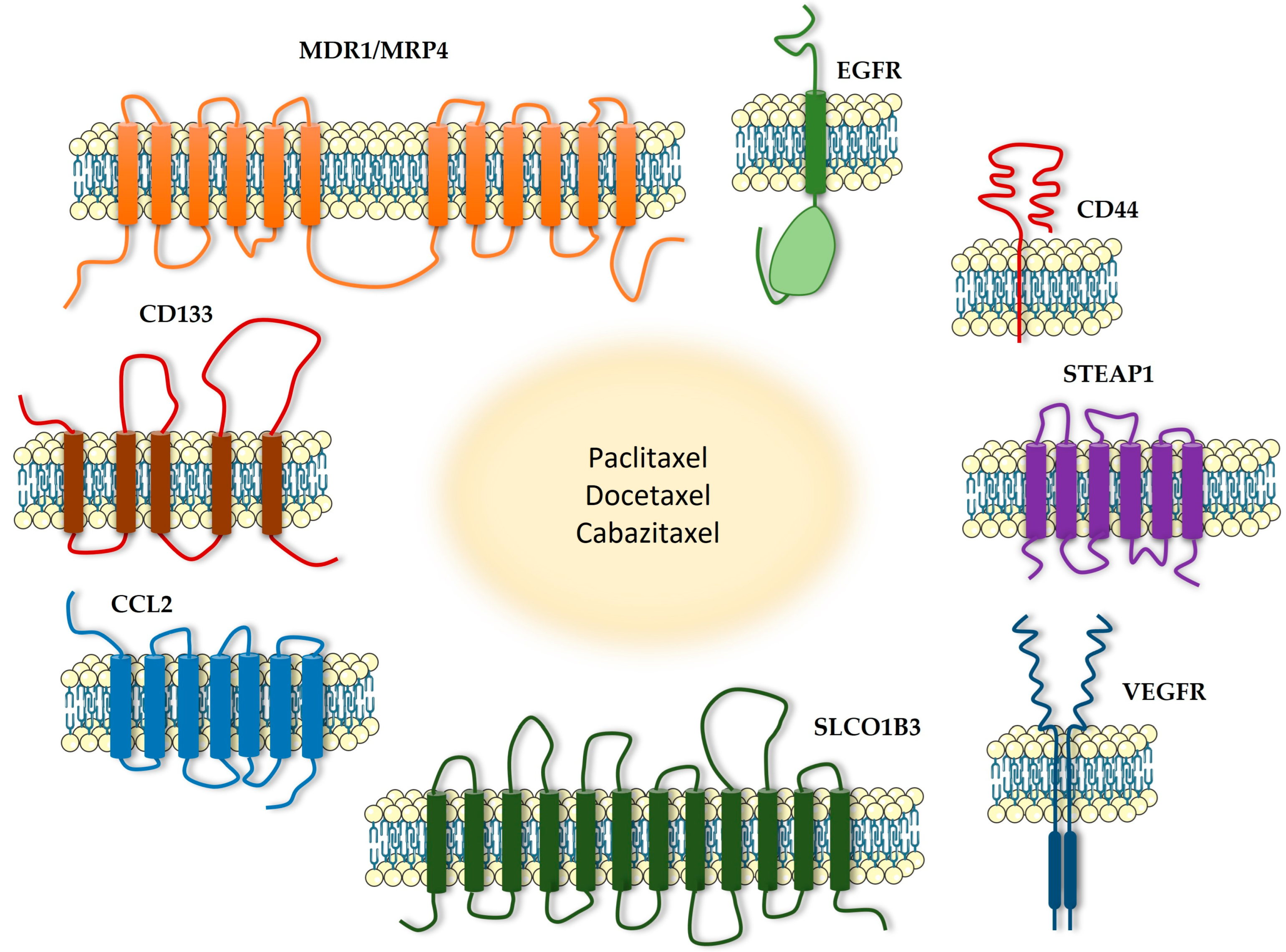
4. Transmembrane Proteins as a Potential Therapeutic Target in Combination with Taxanes

A transmembrane protein is a type of protein located either in the lipid bilayer of the plasma membrane or in the membrane of organelles [81]. Different from monotopic proteins, transmembrane protein structure completely crosses the membrane [82]. Representing approximately 30% of the genome, transmembrane proteins are essential for many cellular processes [83]. These proteins are responsible for cell–cell and cell–environment communication, through signal transduction, the binding of receptors to hormones and neurotransmitters, and the transport of substances across the membrane [82,83]. There are two types of transmembrane proteins regarding their structure: either alpha-helical proteins or beta-barrel proteins. They can also be categorized according to their protein topology, referring to the position of the N- and C-terminal domains [81,82].
Several studies have shown a link between different transmembrane proteins and cancer, due to their functions related to cancer progression, metastasis, and patient survival, and additionally, can also be used as therapeutic targets and/or biomarkers [81,82]. Studies supporting the potential for targeting transmembrane proteins in taxane drug resistance in PCa are summarized in Table 1.

4.1. MDR1

The efflux pump Multidrug Resistance Protein 1 (MDR1), also called p-glycoprotein, is a protein composed of 12 transmembrane domains and a single monomer of 170 kDa (Figure 5) [84]. This protein is part of the ATP-binding cassette (ABC) transporter family and is encoded by the p-glycoprotein (ABCB1) gene, located in the region 7q21 [84]. The overexpression of MDR1 has been shown to be partially responsible for drug resistance in PCa, due to higher drug efflux [85]. Regarding p-glycoprotein expression, Kawai et al. reported that both PCa and normal prostate epithelial cells are positive for the expression of the MDR1 gene [86]. Using monoclonal antibodies to detect the presence of p-glycoprotein, the same study confirmed that this protein is asymmetrically expressed in the inner and outer zones of nonmalignant prostate glands [86]. Moreover, the inner zone showed a higher level of protein expression [86].
To investigate whether the presence of p-glycoprotein in blood exosomes could be a marker to diagnose docetaxel resistance in PCa, Kato et al. tested the susceptibility to docetaxel and cabazitaxel drugs in parental and docetaxel-resistant PC3 cell lines considering p-glycoprotein expression [87]. It was demonstrated that docetaxel-sensitive PC3 cells showed little or no expression of this protein, while docetaxel-resistant PC3 cells showed high expression of p-glycoprotein [87]. The knockdown of the ABCB1 gene was also performed in docetaxel-resistant PC3 cells. The results indicated an improvement in docetaxel sensitivity when compared with the negative control. These findings confirm the relationship between p-glycoprotein expression and docetaxel resistance [87]. Additionally, another study on PC3 cells, after demonstrating that the E26 transformation specific sequence (ETS1) transcription factor had a role in regulating the expression of the MDR1 gene, it was assessed how the downregulation of ETS1 could impact cell sensitivity to paclitaxel [88]. The results showed that the combined treatment of paclitaxel exposure and knockdown of ETS1 induces a decrease in cell viability in paclitaxel-resistant PC3 cell line, improving the resistance to paclitaxel [88]. In a further study using the C4-2B cell line, it was tested the association between the MDR1 protein and the retinoic acid receptor-related orphan receptor g (RORg) [89]. First, they established that MDR1 is regulated upstream by RORg, since the knockdown of the retinoic receptor decreased the expression of MDR1, while ectopic RORg increased it [89]. Also, both RORg antagonists, SR2211 and GSK805, have led to the inhibition of MDR1 expression in taxane-resistant C4-2B cells [89]. Next, this study demonstrated that the knockdown alone and the use alone of RORg antagonists have led to a significant decrease in cell viability and growth in both taxane-resistant and non-resistant C4-2B cell lines [89]. Furthermore, the combination of a partial RORg and a low concentration of docetaxel (20 nmol/L) led to a reduction of cell growth from 96.1% in control to 72.2% in treated cells. Likewise, the use of 1.25 mmol/L SR2211 combined with 12.5 nmol/L docetaxel reduced the viability of taxane-resistant C4-2B to 33.2%, suggesting that the downregulation of RORg can sensitize taxane-resistant CRPC cells to taxane treatment [89].

4.2. MRP4

Similarly to the MDR1 protein, the MRP4 protein (Figure 5), also known as multidrug resistance protein 4, is part of the ABC transporter family [90]. This transmembrane protein is present in almost all tissues in the body, such as the brain, kidney, liver, erythrocytes, platelets, adrenal gland, and pancreas [91]. MRP4 is responsible for the transportation of prostaglandins E1 and E2 (PGE1 and PGE2), as well as cAMP and cGMP [92]. The MRP4 protein was reported as being highly overexpressed in docetaxel-resistant C4-2B cells, while no expression of MRP4 was detected in docetaxel-sensitive C4-2B cells [93]. To assess if the overexpression of MRP4 leads to docetaxel resistance, a combined treatment of MRP4 knockdown plus docetaxel exposure was applied to the docetaxel-resistant C4-2B cell line. The results showed diminished cell viability, indicating a re-sensitization to docetaxel treatment [93]. Furthermore, researchers assessed the hypothesis that androgens are responsible for MRP4 overexpression in docetaxel-resistant cells [93]. For this, C4-2B cells were exposed to DHT or bicalutamide followed by the quantification of MRP4 mRNA and protein levels [93]. After treatment with DHT, both mRNA and protein levels were increased, displaying a dose-dependent manner [93]. However, the exposure to bicalutamide prevented the upregulation of MRP4 [93]. These data show that MRP4 can be upregulated by androgen and downregulated by anti-androgen treatment [93]. Considering that the data point towards sensitization to docetaxel in cells knocked down for MRP4, further assessing whether pharmacological MRP4 inhibitors would exhibit a similar synergistic effect with taxanes could shed light on their future clinical use for PCa treatment. One potential candidate is Ceefourin-1, a selective inhibitor of MRP4, which has already been investigated for use in other cancer types [94].

4.3. CD44

CD44 is a non-kinase cell surface transmembrane glycoprotein (Figure 5) [95]. This important hyaluronate receptor is overexpressed in cancer stem cells and is involved in cellular adhesion and communication, lymphopoiesis, myelopoiesis, and angiogenesis [95]. In regard to cancer, CD44 is implicated in metastasis, cellular growth, proliferation, migration, and invasion [95]. There are several isoforms for the CD44 protein and some of them have been associated with PCa, namely the CD44s, CD44v6, and CD44v7-10 isoforms [95]. Furthermore, CD44 is also overexpressed in this type of cancer and is associated with aggressive biological behavior and a poor prognosis [95]. CD44 expression is upregulated by transforming growth factor-beta 1 (TGF-β1) in PCa cells [95]. CD44 is expressed in PC3 cells and it was demonstrated that this receptor regulates glucose metabolism, intracellular reactive oxygen species (ROS), and cell proliferation in these cells; however, CD44 is not expressed in LNCaP cells [90]. Collected data also point to the regulation of proliferation, invasion, and migration via PDK1 and PFKFB4, which are enzymes that regulate glucose metabolism and are modulated by CD44 [96]. Li et al. reported that the use of docetaxel treatment combined with SB-3CT, a possible inhibitor of CD44 cleavage, decreases the viability of PC3 cells in comparison with the docetaxel-only treatment [96]. Researchers also assessed the combination index using CompuSyn software, showing that mild-to-moderate synergistic effects were observed for an SB-3CT concentration of 20μmol/L in combination with docetaxel [96]. Lai et al. also reported that docetaxel-resistant PC3 and DU145 cells have a higher migration and invasion rate than the parental cells [97]. In addition, when analyzed for the CD44+ population, both docetaxel-resistant cell lines showed higher numbers than the parental cells [97]. The knockdown of CD44 reduced cell migration in both docetaxel-resistant cell lines, while invasion was suppressed only in docetaxel-resistant PC3 cells [97].

4.4. CD133

The pentaspan transmembrane glycoprotein prominin-1, also known as CD133 (Figure 5), is a protein mostly found in the microvilli of different epithelial cells but is also expressed in numerous types of cancer, such as breast, ovarian, and PCa and other non-epithelial cell types [98,99]. CD133 is frequently used as a biomarker for the detection of cancer stem cells [99]. The molecular function of this glycoprotein has not been yet fully clarified, but there is strong evidence pointing towards a role in membrane organization, due to its preferred location on the microvilli, and a role in spermatozoa biogenesis and photoreceptor disc formation [98]. Regarding the photoreceptor disc formation, it is known that a mutation on the CD133 gene is the cause of a type of macular degeneration called Stargardt disease [98]. CD133 is also important in angiogenesis through the regulation of the expression of vascular endothelial growth factor (VEGF) [98]. Concerning the expression of CD133 in PCa cell lines, flow cytometric analysis performed by Wang et al. found that CD133+ cells were only present in the DU145 cell line, and indetectable in PC3 and LNCaP cell lines, when cultured in normal conditions [100]. However, when cultured in a serum-free medium, the PC3 cell line was able to present an increased proportion of CD133+ cells [100]. In LNCaP cells, the presence of CD133+ remained non-observable [100]. Nonetheless, Aghajani et al. evaluated the CD133 mRNA expression levels in the same PCa cell lines and discovered that CD133 is expressed in low amounts in all three cell lines, although with higher expression levels in the LNCaP cell line [101]. Additionally, Wang et al. assessed the possibility of enriching the proportion of CD133+ cells via chemotherapy, for which a docetaxel-containing medium was used in DU145 cell culture [100]. An increase of 9.8% in the proportion of CD133+ cells was observed after treatment, corroborating that these cells are chemo-resistant [100]. Through studying the knockdown alone of CD133 and in combination with paclitaxel, Aghajani et al. reported that, in LNCaP cells, the downregulation alone did not alter cell proliferation and viability when compared to the control group [101]. However, the combination with the paclitaxel treatment led to a decrease in survival rate compared to the LNCaP cells that were uniquely treated with paclitaxel [101]. Regarding migration and invasiveness, both knockdown of CD133 or paclitaxel treatment alone was able to reduce it, while the combination of treatments led to a synergistic decrease [101]. Also, the combination CD133-siRNA/paclitaxel significantly reduced the metastatic potential due to a lower expression of vimentin and MMP9 [101]. Finally, an apoptosis study using the LNCaP cells showed that the knockdown of CD133 may increase the sensitivity to paclitaxel [101].

4.5. SLCO1B3

Belonging to the Solute Carriers superfamily, SLCO1B3 (Figure 5), also called organic anion-transporting polypeptide (OATP) [102], is a sodium-independent transporter of both endogenous substrates, such as bilirubin, bile salts, steroid conjugates, bromosulfophthalein (BSP), and Taurocholate (TCA) [102,103], and exogenous substrates, such as antihistamines, blood-glucose-lowering drugs, statins, heart medications, and docetaxel and paclitaxel [102,104].
Konig et al. confirmed that, under normal conditions, SLCO1B3 is exclusively expressed in hepatocytes, with its subcellular location on the basolateral plasma membrane of those cells [105]. Additionally, a preferred lobular zonation was also observed, where the hepatocytes near the central vein showed a higher expression of this protein when compared to other locations within the liver [105]. Meanwhile, several studies have confirmed the abnormal expression of SLCO1B3 in tumorous tissue, including PCa [106]. Wright et al. demonstrated a significantly higher expression of the gene SLCO1B3 in CRPC metastasis in comparison to untreated primary PCa [102]. In addition, a higher risk for PCa-specific mortality was connected to the single-nucleotide polymorphism (SNP) SLCO1B3 rs4149117 [107]. Moreover, SLCO1B3 mRNA levels were found in 62% of the PCa samples, but no expression was detected in normal prostate [104]. The same study also indicated a clear positive association between the Gleason score and SLCO1B3 expression [104].
Regarding the effects of taxanes, a study evaluated patient-derived xenografts (PDXs) of PCa and discovered that docetaxel-resistant PDX tumors presented a significant downregulation of SLCO1B3 [108]. Along with this result, the PDXs presented reduced intratumorally docetaxel concentrations. To assess if the downregulation of SLCO1B3 was responsible for the low concentration of docetaxel, the silencing of SLCO1B3, as well as other docetaxel transporters, was performed [108]. Only cells that were transfected with the SLCO1B3 siRNA presented a significant reduction in docetaxel uptake. To further investigate the role of SLCO1B3, SLCO1B3-negative PDXs were transfected with SLCO1B3 and later exposed to docetaxel and cabazitaxel. The outcome pointed toward a higher sensitivity to both taxane drugs treatments among SLCO1B3-overexpressing cells [108]. SLCO1B3 expression has been shown to be enhanced, in hepatoma cell lines, by a substance called Chenodeoxycholic acid (CDCA) [109]. CDCA is a natural bile acid produced in the liver which is also used clinically to treat gallbladder stones [110]. This acid is a farnesoid X receptor (FXR) agonist, which is a transcription factor known for modulating SLCO1B3 [109]. Since this drug is already available for clinical use in humans, further assessment of its potential use in PCa, alongside taxanes, would be beneficial.

4.6. EGFR

The transmembrane glycoproteins epidermal growth factor receptor (EGFR), together with HER-2/neu (erbB-2), HER-3 (erbB-3), and HER-4 (erbB-4), belong to the HER (erbB) family of membrane receptors (Figure 5) [111]. All these receptors are expressed in both normal and malignant cells, playing important roles in cell proliferation and differentiation [112]. All four family members have a very similar structure, consisting of three regions: the first is an extracellular ligand-binding region, which, in the case of EGFR, is the binding region for the epidermal growth factor (EGF), transforming growth factor-a (TGF-a), amphiregulin (AR), Heparin-binding EGF-like growth factor (HB-EGF), and betacellulin (BTC) [111,112]. HER2 dimerizes with EGFR [113] and has no exclusive natural ligand [111]. The second region, a transmembrane domain, consists of a single hydrophobic anchor sequence that crosses the cell membrane only once [112]. Lastly, the third region acts as a binding site for intracellular substrates, and therefore activates signaling pathways [112]. The intracellular domain has tyrosine kinase activity [111]. Rossini et al. confirmed that DU145 and PC3 cell lines express the activated form of the EGFR and HER-2 receptors [114]. LNCaP and C4-2B cell lines also express EGFR, being higher in C4-2B cells [115]. In in vivo studies, EGFR was confirmed as overexpressed in both metastatic and CRCP, as well as moderately expressed in localized primary PCa [115]. Furthermore, the assessment of EGFR expression on circulating tumor cells from the blood of patients with metastatic disease demonstrated that 90% of patients presented circulating tumor cells positive for EGFR [115]. Vicentini et al. studied the use of ZD1839, a selective EGFR tyrosine kinase inhibitor, in both androgen-sensitive cell lines (ND1, LNCaP, and ALVA-31), as well as androgen-independent cell lines (PC3, DU145, and TSU-Pr1) [116]. First, it was reported by the authors that higher levels of EGFR and its ligands were present in the androgen-receptor-negative cell lines. However, ZD1839 treatment resulted in reduced cell proliferation in all cell lines tested [116]. Furthermore, an in vivo study assessed the tumor mass response to the blockade of EGFR and HER2 [114]. In order to do so, subcutaneous DU145 or PC-3 tumors were established on male mice, and tumor volume was quantified before, during, and after treatments [114]. It was demonstrated that Cetuximab and Trastuzumab (blockers of EGFR and HER2, respectively) in combination with docetaxel treatment induced a significant tumor regression when compared to the respective control group [114]. Furthermore, 80% of mice that were given the triple combination became tumor-free. However, even though docetaxel alone, cetuximab alone, and in combination with Trastuzumab showed significant tumor growth inhibition, tumor regrowth was observed [114]. In another study, Monteverde et al. used the tyrosine kinase inhibitor Vandetanib to target EGFR in sensitive and docetaxel-resistant PC3 cell lines [117]. The study showed that the docetaxel-resistant PC3 cells presented 3 times the amount of EGFR mRNA and 12 times the amount of EGFR protein when compared to sensitive PC3 cells. Additionally, the treatment with docetaxel alone produced an increase in the pEGFR/EFGR ratio, while the combination with Vandetanib had the opposite effect [117]. In docetaxel-resistant PC3 cells, no treatment altered the pEGFR/EFGR ratio [117]. Regarding the effect in cell proliferation, a maximum of 90% inhibition was observed in response to docetaxel treatment alone in both sensitive and resistant PC3 cells. To attain this inhibition, a 2 × 10⁻⁹ M concentration of docetaxel was necessary for the sensitive cell line and a 0.9 × 10⁻⁷ M concentration for the resistant line [117]. Vandetanib alone also displayed inhibition effects, but the strongest cytotoxic effects were observed when vandetanib was combined with low concentrations of docetaxel (0.061–0.246 nM), for which the combination index value was 0.49–0.71 [117]. However, for resistant PC3 cells, there were different results depending on whether the treatment was administrated in sequence (vandetanib followed by docetaxel) or together [117]. In the first case, the combination index value of 0.55–0.90 indicated a synergetic effect, but for the treatment given together a combination index of 1.22–1.73 was found, indicating a possible antagonism [117]. Supplementarily, other tyrosine kinase inhibitors should be evaluated for their possible role in taxane sensitization. Afatinib, for example, was launched in the market in 2013 by Roche [118], but it remains without data on potential combined use with taxanes on PCa.

4.7. STEAP1

STEAP1, together with STEAP2-4, is part of the six-transmembrane epithelial antigen of prostate (STEAP) family of proteins (Figure 5) [119]. The STEAP1 protein is overexpressed in several human cancers, including prostate, bladder, colon ovary, breast, and cervical cancer [120]. Although its function remains unclear, some studies have pointed out that STEAP1 is involved in metal reductase activity, and also in the transport of ions such as Na+, Ca2+, and K+ [121]. STEAP1 is highly expressed in LNCaP cells and also at significant levels in the C4-2B cell line [122]. Regarding the effect of STEAP1 knockdown in LNCaP cells, reduced cell viability was observed in comparison to the control group [123]. This result was supported by the cell proliferation index, showing a 0.3-fold decrease in LNCaP cells knocked down for STEAP1. In addition to its effect on the inhibition of cell proliferation, the STEAP1 knockdown increased the number of apoptotic cells [123]. The same study also evaluated the behavior of LNCaP cells knocked down for STEAP1 in response to DHT, and the result was that the effect of STEAP1 gene silencing was not reversed after exposure to DHT [123]. Recently, another study reported the effect of paclitaxel, docetaxel, and cabazitaxel on STEAP1 expression in LNCaP and C4-2B cells [122]. It was observed that paclitaxel or cabazitaxel treatment increased the STEAP1 protein expression when compared with the control group, but no differences were observed in C4-2B cells [122]. Furthermore, it was reported that STEAP1 knockdown alone decreased the cell viability in both cell lines, as well as all taxane-based treatments when administered alone. However, the combination of STEAP1 knockdown and exposure to taxane-based therapy led to an increase in cell viability/proliferation and diminished levels of apoptosis [122]. Although more studies are required, these data suggest that the combination of taxane-based drugs with STEAP1 knockdown may lead to PCa progression.

4.8. CCL2/CCR2

A member of the CC beta chemokine family, monocyte chemoattractant protein 1 (MCP-1 or CCL2) is a monomeric polypeptide (Figure 5) [124,125]. CCL2 is an agonist for its main receptor, the transmembrane protein CCR2, and also an agonist for the CCR4 and CCR5 receptors [125]. Moreover, other chemokines act as agonists on the CCR2 receptor, such as CCL7 and CCL8; therefore, there is an overlap of ligands and receptors [125]. It is found in many cell types, such as endothelium, epithelium, and bone marrow; CCL2’s main function is recruiting immune cells, such as monocytes, T lymphocytes, and natural killer (NK) cells [124,125].
It has been demonstrated that in the tumor–bone microenvironment of metastasis collected from patients diagnosed with PCa, several cytokines were upregulated, namely CCL2, which was expressed four times more on the tumor than on the normal tissue adjacent to the tumor [126]. Furthermore, a correlation between CCL2 serum levels and PCa progression can be found, indicating that elevated CCL2 serum levels are associated with bone metastasis [127]. The genetic variation found in CCL2 also supports its role in cancer progression and development; three SNPs of the CCL2 gene are linked to higher Gleason scores [128]. Regarding in vitro culture of PCa cells, it is known that several cell lines expressed different levels of the CCR2 receptor, but PC3 and VcaP cell lines showed the highest levels of expression [126]. CCR2 expression has also been correlated to the Gleason score and pathological stage [124,127]. Furthermore, researchers performed the knockdown of CCR2 using shRNA in C4-2B and PC3 cell lines and the results support the importance of the MCP-1/CCR2 axis in PCa bone metastasis. It was found that both cell lines, when silenced for CCR2, showed a significant decrease in invasion [127]. In vitro tests also confirmed that CCR2 knockdown significantly diminished the PCa osteoclast formation and bone resorption [127]. However, the cell proliferation was only slightly affected by CCR2 silencing [127]. Finally, it was demonstrated that when PC3 cells silenced for CCR2 were transferred into mice, there was a reduction in the numbers of tumor-induced osteoclasts at the tumor and bone interface, and the tumor growth was also inhibited [127].
There is evidence pointing towards CCL2 as having an important role in cell migration. Using PC3 cells, the treatment with human recombinant CCL2 (hrCCL2) showed that cells present higher migration compared to control, and that the effect is dose-dependent [126]. In the same way, the presence of either an anti-CCR5 neutralizing antibody or anti–human CCL2 and anti–mouse CCL2/JE neutralizing antibodies led to a decrease in cell migration [126]. Furthermore, in PC3 cells, Akt phosphorylation is stimulated in a dose-dependent manner by CCL2 [126].
To investigate the results of CCL2 inhibition in PCa, several assays were performed using the DU145 cell line [129]. Three experimental groups were delineated: a chemosensitive cell line (DU145), a paclitaxel-resistant cell line (DU145- TxR), and a paclitaxel- and cabazitaxel-resistant cell line (DU145- TxR/CxR). Using cDNA microarray analysis data, it was confirmed that CCL2 gene expression was 70-fold higher in DU145-TxR cells when compared to DU145 cells, and 43-fold higher in DU145-TxR/CxR cells [129]. The level of CCL2 in the cell medium was also measured, indicating a higher level in the DU145- TxR/CxR cell culture and the lowest in the DU145 cell culture [129]. The exposure to cabazitaxel was not able to alter the CCR2 receptor expression in any cell line [129]. For the apoptosis assay, both DU145-TxR and DU145-TxR/CxR cell lines, when treated with CCR2 antagonist alone, showed little to no increase in apoptosis, and the effect of cabazitaxel alone was similar to that observed in DU145-TxR/CxR [129]. However, a combination of a CCR2 antagonist and cabazitaxel resulted in a great increase in apoptosis. Finally, this study injected these cell lines into mice and treated them with either a CCR2 antagonist alone, cabazitaxel alone, or both a CCR2 antagonist and cabazitaxel [129]. Similarly to what was seen in cell culture, neither cabazitaxel alone nor CCR2 antagonist alone was able to significantly inhibit tumor growth; only the combination of treatments showed positive results [129].
Qian et al. reported that in LNCaP and LAPC4 cell lines treated with docetaxel, the mRNA expression of CCL2 displayed a dose-dependent increase [130]. Additionally, both intracellular and extracellular CCL2 protein levels were also increased when cells were exposed to docetaxel. The upregulation of CCL2 in PCa cells was also linked to the JNK and NF-kB pathways [130]. Using a JNK signaling inhibitor, the SP600125, and the NF-kB inhibitor, parathenolide, in a co-treatment with docetaxel in the LNCaP and LAPC4 cell lines, led to the inhibition of the docetaxel-induced CCL2 expression [130]. Additionally, the knockdown of CCL2 expression in the LNCaP cell line resulted in a significant decrease in viability relative to the control group, and the combination of CCL2 knockdown plus docetaxel showed an even greater reduction [130]. To further validate the role of CCL2 in docetaxel response, an LNCaP cell line that overexpresses CCL2 was established. When this cell line was exposed to docetaxel, higher viability was observed compared to the control group [129].
An in vivo study aiming to assess the efficacy of CCL2 knockdown in combination with docetaxel in the bone environment performed an intra-tibial injection in mice using C4-2B cells [131]. CCL2 blockade alone and docetaxel treatment alone resulted in significantly lower PSA levels, and the combined treatment was responsible for even larger decreases in PSA [131]. Regarding animal survival, an increase was observed when CCL2 blockade was used alone as well as in combination with docetaxel [131]. An AR staining of tumors was also performed and indicated a decrease in nuclear AR in the animal group that was treated with CCL2 blockade [131]. Bone mineral density (BMD) measurements were performed and the results showed a 28.6% increase in BMD in those animals that received CCL2 blockade in combination with docetaxel, while CCL2 blockade alone results in a 20.8% increase, and docetaxel alone in a 7.8% increase when compared to the control group [131].
Lobert et al. used PC3 cells stimulated with hrCCL2 to determine the potential of CCL2 inhibition [124]. Two types of antibodies were used, an anti-human CCL2 neutralizing antibody (CNTO888) and an anti-mouse CCL2/JE neutralizing antibody (C1142). Exposure to CNTO888 resulted in a decrease in hrCCL2-induced proliferation and hrCCL2-induced migration when compared to the human IgG control antibody and the C1142 antibody [124]. The activation of Akt, p70S6 kinase, and p44/p42 mitogen-activated protein stimulated by hrCCL2 was also attenuated by CNTO888 [124]. This same study used an in vivo model of PCa, in which the administration of CNTO888 led to a 47% decrease in overall tumor burden and an 87% reduction in tibia-specific tumor burden, and the anti-mouse CCL2/JE (C1142) led to a 96% and a 95% reduction, respectively [124]. In this case, the inhibition of either the host stromal-derived mouse CCL2 or the tumor-derived human CCL2 resulted in a decreased bone metastasis formation. Furthermore, they compared docetaxel as a single-agent treatment to CNTO888 and C1142 as single agents [124]. Neither C1142 nor C1142 + CNTO888 were as effective as docetaxel, which showed a 96% decrease in total tumor burden [124]. Moreover, researchers also observed that when docetaxel treatment alone was administrated and stopped, mice subsequently developed additional tumor burden [124]. This scenario did not occur when they combined docetaxel with anti-CCL2 antibodies. After the docetaxel treatment was discontinued, and only the administration of antibodies continued, a further decrease in tumor burden was observed, compared with mice that did not receive antibody therapy. However, after antibody treatment was discontinued, the tumor burden also increased [124].
Another in vivo study used the same antibodies, C1142 and CNTO888, to evaluate the treatment response and survival of animals injected with PC3 cells [132]. The combined treatment of docetaxel and anti-CCL2 led to a significant inhibition in tumor growth and a greater rate of inhibition compared to docetaxel alone and anti-CCL2 alone [132]. The same was observed for the survival rate of animals. While the combination therapy resulted in a 22-week survival period, docetaxel alone showed a 15-week period and anti-CCL2 antibodies alone an 11-week period, but the survival period in the control group was only 6 weeks [132].
Since several pre-clinical studies showed promising results when targeting CCL2, a clinical phase 2 trial was performed [133]. Carlumab is a human monoclonal antibody that binds with high affinity and specificity to human CCL2. This agent was administered to 46 patients diagnosed with metastatic CRPC that were part of the clinical trial [133]. All of them had been treated previously with docetaxel without success. Carlumab was administered as a single-agent treatment and unfortunately, the results were not as good as expected: the levels of free CCL2 only decreased following the first round of calumab administration, and after that, the increase of free serum CCL2 was not suppressed by treatment. Furthermore, researchers did not detect any significant inhibition of tumor progression [133]. Even though the results of this clinical trial did not mirror what was observed in pre-clinical studies, it is important to notice that carlumab was used as a single agent and not in combination with any taxane. A further investigation of the synergistic effect of CCL2 blockage together with taxane treatment in humans could elucidate the potential use of carlumab, or other CCL2 inhibitors, in PCa treatment.

4.9. VEGFR

The Vascular Endothelial Growth Factor (VEGF) family of ligands and its receptors (VEGFR) are responsible for the development of both blood and lymphatic vascular networks [134]. The receptors are VEGFR1, VEGFR2, and VEGFR3. All receptors are comprised of an extracellular domain containing seven immunoglobulin homology domain repeats, a transmembrane domain, and a tyrosine kinase domain (Figure 5) [134]. Although there is a similar structure, different mechanisms of activation, signal transduction pathways, and biological effects are attributed to each of the different receptors [134]. Regarding expression, the LNCaP cell line is known to exhibit low expression of VEGFR2, while DU145, PC3, and PC3DR2 cells showed significant expression levels [135]. It has been reported that, in the PC3 cells, docetaxel treatment can enhance mRNA and protein expression of VEGFR2 [136].
Lu et al. engineered a novel anti-VEGFR2 fully human Ab and applied it to PCa [137]. Using a PC3 xenograft PCa model, researchers assessed the therapeutic potential of this Ab in comparison to ramucirumab and docetaxel [137]. Mice were divided into the following treatment options: ramucirumab, anti- VEGFR2, docetaxel, anti-VEGFR2 plus docetaxel, or ramucirumab plus docetaxel. Regarding tumor growth, the best treatment was anti-VEGFR2 plus docetaxel, with a 90% reduction, followed by a combination of ramucirumab plus docetaxel that reduced 82% of tumor growth. Docetaxel alone, anti-VEGFR2, and ramucirumab resulted in a reduction of 70%, 52%, and 45%, respectively [137]. Furthermore, it was observed that anti-VEGFR2 reduced tumor vascular density and enhanced cancer cell apoptosis [137].
Already approved by the FDA for gastrointestinal stromal tumors, Sunitinib is an oral multi-tyrosine kinase inhibitor, which includes an effect against VEGFR2 [135]. Studies on PCa cell lines demonstrated that Sunitinib had an antitumor effect that was dose- and time-dependent [135]. A phase I/II clinical trial was also performed using Sunitinib in a combined treatment with docetaxel and prednisone [138]. Overall, 39% percent of the 55 enrolled patients had a partial response, and in 56%, the PSA levels declined. However, 36 patients had to discontinue therapy, some of them due to disease progression, and others due to adverse events [138]. Another phase II trial administrated Sunitinib to patients with CRPC, who had not been yet exposed to chemotherapy, and to patients with metastatic, docetaxel-resistant PCa [139]. PSA levels, biochemical markers of bone turnover, and safety were the parameters assessed in the study. The results were not positive, showing no significant clinical benefits in the administration of sunitinib to metastatic CRPC patients [139].
Cediranib (AZD-2171) is an inhibitor of all three VEGFRs, and its effect is being studied in several types of cancer, including PCa [140]. In the PC3 and DU145 cell lines, Cediranib as a sole treatment decreased cell survival, induced apoptosis, and cell motility [140]. Furthermore, the combination with docetaxel can enhance sensitivity to the same taxane [140]. Another study used Cediranib to treat mice that were injected with a generated DU145 cell line, which overexpresses platelet-derived growth factor D (PDGFD) and is known to upregulate the VEGF/VEGFR axis [141]. The treatment with Cediranib alone or in combination with docetaxel significantly reduced tumor volume and stabilized the disease progression, and even led to regression in some mice [141]. A preclinical study established bone and brain metastasis in mice using a DU145 cells, in which an activated Ras effector mutant (RasV12G37) was introduced, since the parental DU145 does not form metastasis in xenograft models [142]. Researchers administered Cediranib alone to mice, which led to multifaceted responses. First, mice in the control group were more likely to develop rapidly growing and expansive solid brain metastases [142]. When it comes to tumor vasculature, exposure to Cediranib results in regression of the vessels in the center of large tumors. However, there was no regression in the tumor vasculature of the invasive tumors nor of the tumor cells at the rim of the large expansive tumors [142]. Another contrasting finding was that histologic analysis of the brain metastases found increased invasive projections after treatment with Cediranib [142]. However, mice were observed to display an increased survival and decreased metastatic tumor burden. Lastly, different sensitivities to Cediranib were observed between brain- and bone-resident tumor cells. After treatment with Cediranib, only brain metastases showed a rebound growth, while inhibition of brain metastases was observed [142].
Cediranib has already reached clinical studies. In a phase I study, AZD2171 was administrated to 19 patients with CRPC [143]. Unfortunately, none of them achieved a ≥50% decline in PSA levels [143]. A phase II trial also included CRPC patients, who had already been exposed to docetaxel, and their disease continued to progress [138]. In this study, besides the PSA levels, further criteria such as clinical or radiographic evidence were assessed. Twenty-three patients with measurable diseases were enrolled, and some of them presented tumor shrinkage after treatment [138].

5. Conclusions

Taxane-based chemotherapeutic drugs are currently the main approach when it comes to PCa treatment. Even though this type of therapy has good results in improving patient survival, the development of resistance to chemotherapeutic drugs remains a great obstacle. In this review, we have covered the state of the art on the use of taxane-based therapies combined with targeting different transmembrane oncoproteins in PCa. The knockdown of transmembrane oncoproteins can improve, in some cases, taxane sensitivity, and therefore, might be a mechanism to improve the efficacy of taxane drugs. However, it should be taken into account that some combinations may even trigger harmful effects, such as the knockdown of STEAP1. Besides the proteins described in this article, there are many more transmembrane oncoproteins whose specific role in PCa and association with taxane resistance requires further elucidation.
Despite some studies showing a promising use of taxane treatment in combination with inhibitors of transmembrane oncoproteins, additional studies are still needed to support a translation for clinical practice. Most of the scientific studies are focused on cell lines, which present several limitations. Therefore, it is necessary to perform studies using animal models in order to find good combinations to evaluate in clinical trials.

Author Contributions

Conceptualization, C.J.M.; methodology, S.M.R. and C.J.M.; investigation, R.S.C. and S.M.R.; writing—original draft preparation, R.S.C. and S.M.R.; writing—review and editing, S.M.R. and C.J.M.; visualization, C.J.M.; supervision, C.J.M.; All authors have read and agreed to the published version of the manuscript.

Funding

This research received no external funding.

Conflicts of Interest

The authors declare no conflict of interest.

References

  1. Bray, F.; Ferlay, J.; Soerjomataram, I.; Siegel, R.L.; Torre, L.A.; Jemal, A. Global cancer statistics 2018: GLOBOCAN estimates of incidence and mortality worldwide for 36 cancers in 185 countries. CA Cancer J. Clin. 2018, 68, 394–424. [Google Scholar] [CrossRef] [PubMed]
  2. Rawla, P. Epidemiology of Prostate Cancer. World J. Oncol. 2019, 10, 63. [Google Scholar] [CrossRef]
  3. Oar, A.; Moraes, F.Y.; Romero, Y.; Ilbawi, A.; Yap, M.L. Core elements of national cancer control plans: A tool to support plan development and review. Lancet Oncol. 2019, 20, e645–e652. [Google Scholar] [CrossRef] [PubMed]
  4. Ng, K.L. The Etiology of Prostate Cancer. In Prostate Cancer; Exon Publications: Brisbane, Australia, 2021; pp. 17–28. [Google Scholar]
  5. Malik, S.S.; MPhil Batool, R.; Honors, B.S.; Masood, N.; Yasmin, A. Risk factors for prostate cancer: A multifactorial case-control study. Curr. Probl. Cancer 2018, 42, 337–343. [Google Scholar] [CrossRef]
  6. Leitzmann, M.F.; Rohrmann, S. Risk factors for the onset of prostatic cancer: Age, location, and behavioral correlates. Clin. Epidemiol. 2012, 4, 1–11. [Google Scholar] [CrossRef]
  7. Swami, U.; McFarland, T.R.; Nussenzveig, R.; Agarwal, N. Advanced Prostate Cancer: Treatment Advances and Future Directions. Trends Cancer 2020, 6, 702–715. [Google Scholar] [CrossRef]
  8. Corn, P.G.; Agarwal, N.; Araujo, J.C.; Sonpavde, G. Taxane-based Combination Therapies for Metastatic Prostate Cancer. Eur. Urol. Focus 2019, 5, 369–380. [Google Scholar] [CrossRef]
  9. Cournia, Z.; Allen, T.W.; Andricioaei, I.; Antonny, B.; Baum, D.; Brannigan, G.; Buchete, N.V.; Deckman, J.T.; Delemotte, L.; Del Val, C.; et al. Membrane Protein Structure, Function and Dynamics: A Perspective from Experiments and Theory. J. Membr. Biol. 2015, 248, 611–640. [Google Scholar] [CrossRef]
  10. Overington, J.P.; Al-Lazikani, B.; Hopkins, A.L. How many drug targets are there? Nat. Rev. Drug. Discov. 2006, 5, 993–996. [Google Scholar] [CrossRef]
  11. Lee, C.H.; Akin-Olugbade, O.; Kirschenbaum, A. Overview of prostate anatomy, histology, and pathology. Endocrinol. Metab. Clin. N. Am. 2011, 40, 565–575. [Google Scholar] [CrossRef]
  12. Ittmann, M. Anatomy and Histology of the Human and Murine Prostate. Cold Spring Harb. Perspect. Med. 2018, 8, a030346. [Google Scholar] [CrossRef] [PubMed]
  13. Leong, K.G.; Wang, B.-E.; Johnson, L.; Gao, W.-Q. Generation of a prostate from a single adult stem cell. Nature 2008, 456, 804–808. [Google Scholar] [CrossRef] [PubMed]
  14. Packer, J.R.; Maitland, N.J. The molecular and cellular origin of human prostate cancer. Biochim. Biophys. Acta 2016, 1863 Pt A, 1238–1260. [Google Scholar] [CrossRef]
  15. Corn, P.G. The tumor microenvironment in prostate cancer: Elucidating molecular pathways for therapy development. Cancer Manag. Res. 2012, 4, 183–193. [Google Scholar] [CrossRef] [PubMed]
  16. Levesque, C.; Nelson, P.S. Cellular Constituents of the Prostate Stroma: Key Contributors to Prostate Cancer Progression and Therapy Resistance. Cold Spring Harb. Perspect. Med. 2018, 8, a030510. [Google Scholar] [CrossRef] [PubMed]
  17. Yadav, N.; Heemers, H.V. Androgen action in the prostate gland. Minerva Urol. Nefrol. 2012, 64, 35–49. [Google Scholar]
  18. Long, R.M.; Morrissey, C.; Fitzpatrick, J.M.; Watson, R.W. Prostate epithelial cell differentiation and its relevance to the under-standing of prostate cancer therapies. Clin. Sci. 2005, 108, 1–11. [Google Scholar] [CrossRef]
  19. Lawson, D.A.; Zong, Y.; Memarzadeh, S.; Xin, L.; Huang, J.; Witte, O.N. Basal epithelial stem cells are efficient targets for prostate cancer initiation. Proc. Natl. Acad. Sci. USA 2010, 107, 2610–2615. [Google Scholar] [CrossRef]
  20. Parimi, V.; Goyal, R.; Poropatich, K.; Yang, X.J. Neuroendocrine differentiation of prostate cancer: A review. Am. J. Clin. Exp. Urol. 2014, 2, 273–285. [Google Scholar]
  21. Wang, Z.A.; Mitrofanova, A.; Bergren, S.K.; Abate-Shen, C.; Cardiff, R.D.; Califano, A.; Shen, M.M. Lineage analysis of basal epithelial cells reveals their unexpected plasticity and supports a cell-of-origin model for prostate cancer heterogeneity. Nature 2013, 15, 274–283. [Google Scholar] [CrossRef]
  22. Wang, Z.A.; Toivanen, R.; Bergren, S.K.; Chambon, P.; Shen, M.M. Luminal Cells Are Favored as the Cell of Origin for Prostate Cancer. Cell Rep. 2014, 8, 1339–1346. [Google Scholar] [CrossRef]
  23. Wang, X.; Kruithof-de Julio, M.; Economides, K.D.; Walker, D.; Yu, H.; Halili, M.V.; Hu, Y.-P.; Price, S.M.; Abate-Shen, C.; Shen, M.M. A luminal epithelial stem cell that is a cell of origin for prostate cancer. Nature 2009, 461, 495–500. [Google Scholar] [CrossRef]
  24. Shen, M.M.; Abate-Shen, C. Molecular genetics of prostate cancer: New prospects for old challenges. Genes Dev. 2010, 24, 1967–2000. [Google Scholar] [CrossRef] [PubMed]
  25. Ayala, A.G.; Ro, J.Y. Prostatic intraepithelial neoplasia: Recent advances. Arch. Pathol. Lab. Med. 2007, 131, 1257–1266. [Google Scholar] [CrossRef]
  26. Joshua, A.; Evans, A.; Van der Kwast, T.; Zielenska, M.; Meeker, A.; Chinnaiyan, A.; Squire, J. Prostatic preneoplasia and beyond. Biochim. Biophys. Acta 2008, 1785, 156–181. [Google Scholar] [CrossRef] [PubMed]
  27. Murray, T.B.J. The Pathogenesis of Prostate Cancer. In Prostate Cancer; Exon Publications: Brisbane, Australia, 2021; pp. 29–42. [Google Scholar]
  28. Alexander, E.E.; Qian, J.; Wollan, P.C.; Myers, R.P.; Bostwick, D.G. Prostatic intraepithelial neoplasia does not appear to raise serum prostate-specific antigen concentration. Urology 1996, 47, 693–698. [Google Scholar] [CrossRef] [PubMed]
  29. Bostwick, D.G.; Brawer, M.K. Prostatic Intra-Epithelial Neoplasia and Early Invasion in Prostate Cancer. Cancer 1987, 59, 788–794. [Google Scholar] [CrossRef]
  30. Schiebler, M.L.; E Tomaszewski, J.; Bezzi, M.; Pollack, H.M.; Kressel, H.Y.; Cohen, E.K.; Altman, H.G.; Gefter, W.B.; Wein, A.J.; Axel, L. Prostatic carcinoma and benign prostatic hyperplasia: Correlation of high-resolution MR and histopathologic findings. Radiology 1989, 172, 131–137. [Google Scholar] [CrossRef]
  31. Schiebler, M.L.; Schnall, M.D.; Pollack, H.M.; Lenkinski, R.E.; E Tomaszewski, J.; Wein, A.J.; Whittington, R.; Rauschning, W.; Kressel, H.Y. Current role of MR imaging in the staging of adenocarcinoma of the prostate. Radiology 1993, 189, 339–352. [Google Scholar] [CrossRef]
  32. Ulmert, D.; O’Brien, M.F.; Bjartell, A.S.; Lilja, H. Prostate kallikrein markers in diagnosis, risk stratification and prognosis. Nat. Rev. Urol. 2009, 6, 384–391. [Google Scholar] [CrossRef]
  33. Duffy, M.J. The role of proteolytic enzymes in cancer invasion and metastasis. Clin. Exp. Metastasis 1992, 10, 145–155. [Google Scholar] [CrossRef] [PubMed]
  34. Wang, G.; Zhao, D.; Spring, D.J.; DePinho, R.A. Genetics and biology of prostate cancer. Genes Dev. 2018, 32, 1105–1140. [Google Scholar] [CrossRef] [PubMed]
  35. Tuxhorn, J.A.; Ayala, G.E.; Smith, M.J.; Smith, V.C.; Dang, T.D.; Rowley, D.R. Reactive stroma in human prostate cancer: Induction of myofibroblast phenotype and extracellular matrix remodeling. Clin. Cancer Res. 2002, 8, 2912–2923. [Google Scholar]
  36. Shafi, A.A.; Yen, A.E.; Weigel, N.L. Androgen receptors in hormone-dependent and castration-resistant prostate cancer. Pharmacol. Ther. 2013, 140, 223–238. [Google Scholar] [CrossRef]
  37. Culig, Z.; Klocker, H.; Bartsch, G.; Hobisch, A. Androgen receptors in prostate cancer. Endocr. Relat. Cancer 2002, 9, 155–170. [Google Scholar] [CrossRef]
  38. Knuuttila, M.; Mehmood, A.; Mäki-Jouppila, J.; Ryberg, H.; Taimen, P.; Knaapila, J.; Ettala, O.; Boström, P.J.; Ohlsson, C.; Venäläinen, M.S.; et al. Intratumoral androgen levels are linked to TMPRSS2-ERG fusion in prostate cancer. Endocr.-Relat. Cancer 2018, 25, 807–819. [Google Scholar] [CrossRef] [PubMed]
  39. Nicholas, T.R.; Strittmatter, B.G.; Hollenhorst, P.C. Oncogenic ETS Factors in Prostate Cancer. Adv. Exp. Med. Biol. 2019, 1210, 409–436. [Google Scholar] [CrossRef] [PubMed]
  40. Wei, T.; Lu, J.; Ma, T.; Huang, H.; Kocher, J.-P.; Wang, L. Re-Evaluate Fusion Genes in Prostate Cancer. Cancer Inform. 2021, 20, 11769351211027592. [Google Scholar] [CrossRef]
  41. Davey, R.A.; Grossmann, M. Androgen Receptor Structure, Function and Biology: From Bench to Bedside. Clin. Biochem. Rev. 2016, 37, 3–15. [Google Scholar]
  42. Bevan, C.; Parker, M. The Role of Coactivators in Steroid Hormone Action. Exp. Cell Res. 1999, 253, 349–356. [Google Scholar] [CrossRef]
  43. Tan, M.H.E.; Li, J.; Xu, H.E.; Melcher, K.; Yong, E.-L. Androgen receptor: Structure, role in prostate cancer and drug discovery. Acta Pharmacol. Sin. 2015, 36, 3–23. [Google Scholar] [CrossRef] [PubMed]
  44. Saraon, P.; Drabovich, A.P.; Jarvi, K.A.; Diamandis, E.P. Mechanisms of Androgen-Independent Prostate Cancer. EJIFCC 2014, 25, 42–54. [Google Scholar] [PubMed]
  45. Devlin, H.-L.; Mudryj, M. Progression of prostate cancer: Multiple pathways to androgen independence. Cancer Lett. 2009, 274, 177–186. [Google Scholar] [CrossRef] [PubMed]
  46. Gottlieb, B.; Beitel, L.K.; Nadarajah, A.; Paliouras, M.; Trifiro, M. The androgen receptor gene mutations database: 2012 update. Hum. Mutat. 2012, 33, 887–894. [Google Scholar] [CrossRef] [PubMed]
  47. Beltran, H.; Yelensky, R.; Frampton, G.M.; Park, K.; Downing, S.R.; MacDonald, T.Y.; Jarosz, M.; Lipson, D.; Tagawa, S.T.; Nanus, D.M.; et al. Targeted Next-generation Sequencing of Advanced Prostate Cancer Identifies Potential Therapeutic Targets and Disease Heterogeneity. Eur. Urol. 2013, 63, 920–926. [Google Scholar] [CrossRef]
  48. Chan, S.C.; Li, Y.; Dehm, S.M. Androgen Receptor Splice Variants Activate Androgen Receptor Target Genes and Support Aberrant Prostate Cancer Cell Growth Independent of Canonical Androgen Receptor Nuclear Localization Signal. J. Biol. Chem. 2012, 287, 19736–19749. [Google Scholar] [CrossRef]
  49. Sun, S.; Sprenger, C.C.; Vessella, R.L.; Haugk, K.; Soriano, K.; Mostaghel, E.A.; Page, S.T.; Coleman, I.M.; Nguyen, H.M.; Sun, H.; et al. Castration resistance in human prostate cancer is conferred by a frequently occurring androgen receptor splice variant. J. Clin. Investig. 2010, 120, 2715–2730. [Google Scholar] [CrossRef]
  50. Jenster, G. Ligand-independent activation of the androgen receptor in prostate cancer by growth factors and cytokines. J. Pathol. 2000, 191, 227–228. [Google Scholar] [CrossRef]
  51. Culig, Z.; Hobisch, A.; Cronauer, M.V.; Radmayr, C.; Trapman, J.; Hittmair, A.; Bartsch, G.; Klocker, H. Androgen receptor activation in prostatic tumor cell lines by insulin-like growth factor-I, keratinocyte growth factor, and epidermal growth factor. Cancer Res. 1994, 54, 5474–5478. [Google Scholar] [CrossRef]
  52. Zhu, M.-L.; Kyprianou, N. Androgen receptor and growth factor signaling cross-talk in prostate cancer cells. Endocr.-Relat. Cancer 2008, 15, 841–849. [Google Scholar] [CrossRef]
  53. Shorning, B.Y.; Dass, M.S.; Smalley, M.J.; Pearson, H.B. The PI3K-AKT-mTOR Pathway and Prostate Cancer: At the Crossroads of AR, MAPK, and WNT Signaling. Int. J. Mol. Sci. 2020, 21, 4507. [Google Scholar] [CrossRef] [PubMed]
  54. Martini, M.; De Santis, M.C.; Braccini, L.; Gulluni, F.; Hirsch, E. PI3K/AKT signaling pathway and cancer: An updated review. Ann. Med. 2014, 46, 372–383. [Google Scholar] [CrossRef]
  55. Labbé, D.P.; Brown, M. Transcriptional Regulation in Prostate Cancer. Cold Spring Harb. Perspect. Med. 2018, 8, a030437. [Google Scholar] [CrossRef] [PubMed]
  56. Schatten, H. Brief Overview of Prostate Cancer Statistics, Grading, Diagnosis and Treatment Strategies. Adv. Exp. Med. Biol. 2018, 1095, 1–14. [Google Scholar] [CrossRef] [PubMed]
  57. Litwin, M.S.; Tan, H.J. The diagnosis and treatment of prostate cancer: A review. JAMA—J. Am. Med. Assoc. 2017, 317, 2532–2542. [Google Scholar] [CrossRef] [PubMed]
  58. Liao, C.H.; Li, H.Y.; Chung, S.D.; Chiang, H.S.; Yu, H.J. Significant association between serum dihydrotestosterone level and prostate volume among Taiwanese men aged 40–79 years. Aging Male 2012, 15, 28–33. [Google Scholar] [CrossRef]
  59. Catt, S.; Matthews, L.; May, S.; Payne, H.; Mason, M.; Jenkins, V. Patients’ and partners’ views of care and treatment provided for met-astatic castrate-resistant prostate cancer in the UK. Eur. J. Cancer Care 2019, 28, e13140. [Google Scholar] [CrossRef]
  60. Tannock, I.F.; Osoba, D.; Stockler, M.R.; Ernst, D.S.; Neville, A.J.; Moore, M.J.; Armitage, G.R.; Wilson, J.J.; Venner, P.M.; Coppin, C.M.; et al. Chemotherapy with mitoxantrone plus prednisone or pred-nisone alone for symptomatic hormone-resistant prostate cancer: A Canadian randomized trial with palliative end points. J. Clin. Oncol. 1996, 14, 1756–1764. [Google Scholar] [CrossRef]
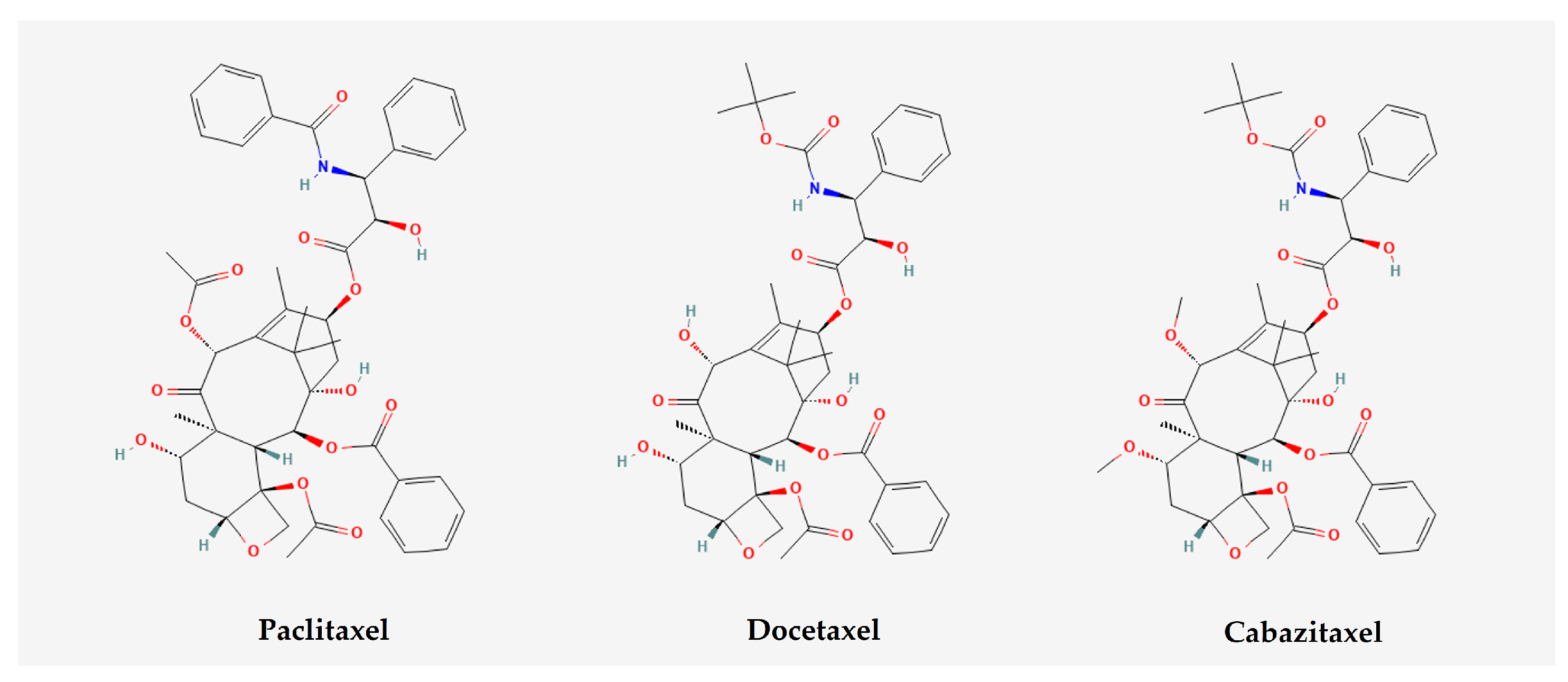
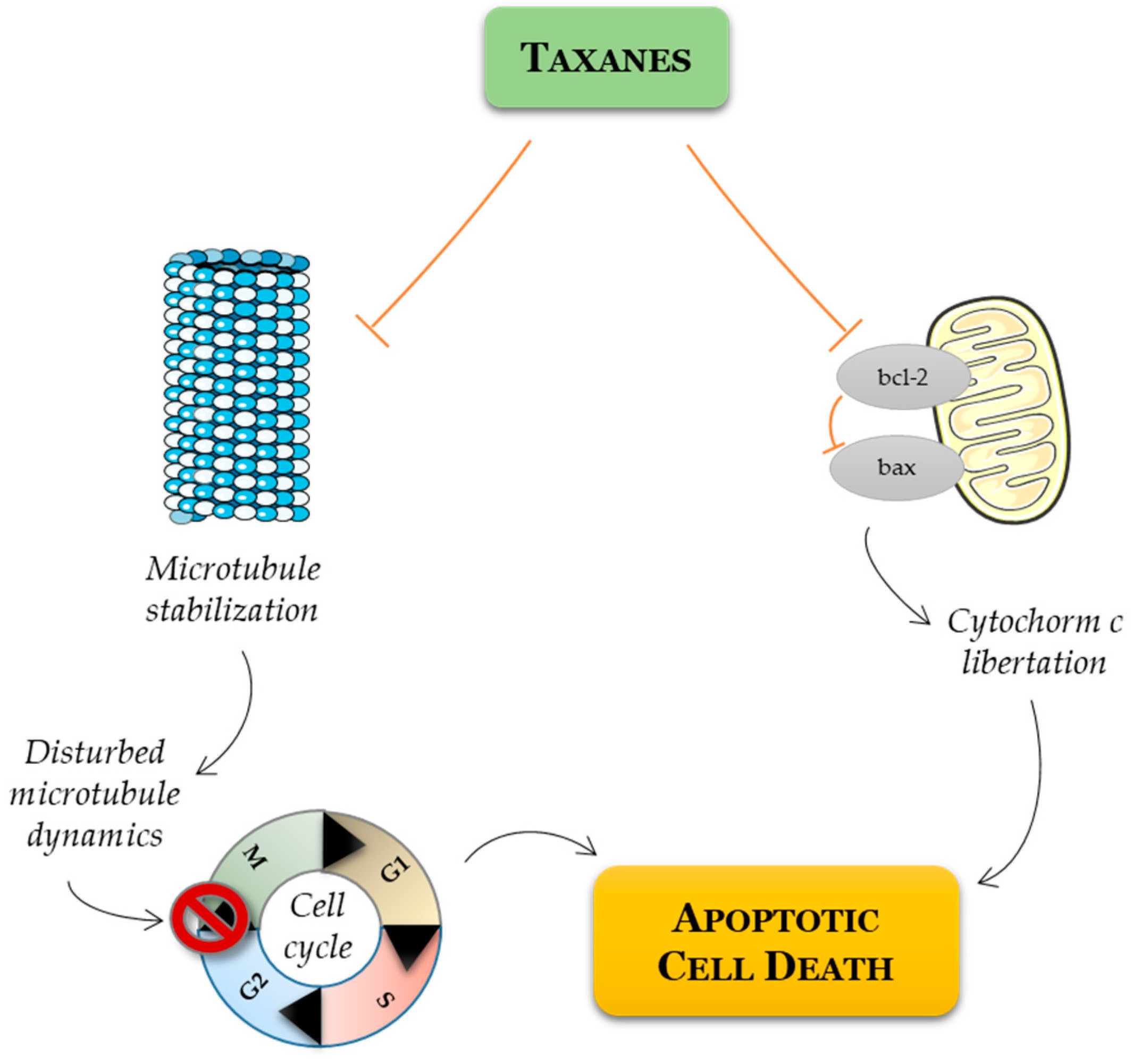
  61. Long, H.J. Paclitaxel (Taxol): A Novel Anticancer Chemotherapeutic Drug. Mayo Clin. Proc. 1994, 69, 341–345. [Google Scholar] [CrossRef]
  62. Kubník, J.; Pavlíčková, V.; Ruml, T.; Rimpelová, S. Current Perspectives on Taxanes: Focus on Their Bioactivity, Delivery and Combination Therapy. Plants 2021, 10, 569. [Google Scholar] [CrossRef]
  63. Pienta, K.J. Preclinical mechanisms of action of docetaxel and docetaxel combinations in prostate cancer. Semin. Oncol. 2001, 28 (Suppl. 15), 3–7. [Google Scholar] [CrossRef] [PubMed]
  64. Roumiguié, M.; Paoletti, X.; Neuzillet, Y.; Mathieu, R.; Vincendeau, S.; Kleinclauss, F.; Mejean, A.; Guy, L.; Timsit, M.O.; Lebret, T. Apalutamide, darolutamide and enzalutamide in nonmetastatic castration-resistant prostate cancer: A meta-analysis. Futur. Oncol. 2021, 17, 1811–1823. [Google Scholar] [CrossRef] [PubMed]
  65. Rathkopf, D.E.; Morris, M.J.; Fox, J.J.; Danila, D.C.; Slovin, S.F.; Hager, J.H.; Rix, P.J.; Maneval, E.C.; Chen, I.; Gönen, M.; et al. Phase I study of ARN-509, a novel antiandrogen, in the treatment of castration-resistant prostate cancer. J. Clin. Oncol. 2013, 31, 3525–3530. [Google Scholar] [CrossRef]
  66. Madan, R.A.; Pal, S.K.; Sartor, O.; Dahut, W.L. Overcoming Chemotherapy Resistance in Prostate Cancer. Clin. Cancer Res. 2011, 17, 3892–3902. [Google Scholar] [CrossRef]
  67. Watanabe, H.; Kawakami, A.; Sato, R.; Watanabe, K.; Matsushita, Y.; Miyake, H. Molecular Mechanism Mediating Cytotoxic Activity of Cabazitaxel in Docetaxel-resistant Human Prostate Cancer Cells. Anticancer Res. 2021, 41, 3753–3758. [Google Scholar] [CrossRef] [PubMed]
  68. Cevik, O.; Acidereli, H.; Turut, F.A.; Yildirim, S.; Acilan, C. Cabazitaxel exhibits more favorable molecular changes compared to other taxanes in androgen-independent prostate cancer cells. J. Biochem. Mol. Toxicol. 2020, 34, e22542. [Google Scholar] [CrossRef] [PubMed]
  69. Takai, M.; Kato, S.; Nakano, M.; Fujimoto, S.; Iinuma, K.; Ishida, T.; Taniguchi, M.; Tamaki, M.; Uno, M.; Takahashi, Y.; et al. Efficacy of cabazitaxel and the influence of clinical factors on the overall survival of patients with castration-resistant prostate cancer: A local experience of a multicenter retrospective study. Asia-Pac. J. Clin. Oncol. 2021, 17, 238–244. [Google Scholar] [CrossRef]
  70. Miyake, H.; Sato, R.; Watanabe, K.; Matsushita, Y.; Watanabe, H.; Motoyama, D.; Ito, T.; Sugiyama, T.; Otsuka, A. Prognostic significance of third-line treatment for patients with metastatic castration-resistant prostate cancer: Comparative assessments between cabazitaxel and other agents. Int. J. Clin. Oncol. 2021, 26, 1745–1751. [Google Scholar] [CrossRef]
  71. Van Soest, R.J.; De Wit, R. Irrefutable evidence for the use of docetaxel in newly diagnosed metastatic prostate cancer: Results from the STAMPEDE and CHAARTED trials. BMC Med. 2015, 13, 304. [Google Scholar] [CrossRef]
  72. Damodaran, S.; Lang, J.M.; Jarrard, D.F. Targeting Metastatic Hormone Sensitive Prostate Cancer: Chemohormonal Therapy and New Combinatorial Approaches. J. Urol. 2019, 201, 876–885. [Google Scholar] [CrossRef]
  73. Huebner, N.A.; Shariat, S.F.; Resch, I.; Gust, K.; Kramer, G. The role of taxane-based chemotherapy in the treatment of prostate cancer. Curr. Opin. Urol. 2020, 30, 527–533. [Google Scholar] [CrossRef] [PubMed]
  74. James, N.D.; Sydes, M.R.; Clarke, N.W.; Mason, M.D.; Dearnaley, D.P.; Spears, M.R.; Ritchie, A.W.S.; Parker, C.C.; Russell, J.M.; Attard, G.; et al. Addition of docetaxel, zoledronic acid, or both to first-line long-term hormone therapy in prostate cancer (STAMPEDE): Survival results from an adaptive, multiarm, multistage, platform randomised controlled trial. Lancet 2016, 387, 1163–1177. [Google Scholar] [CrossRef] [PubMed]
  75. Clarke, N.W.; Ali, A.; Ingleby, F.C.; Hoyle, A.; Amos, C.L.; Attard, G.; Brawley, C.D.; Calvert, J.; Chowdhury, S.; Cook, A.; et al. Addition of docetaxel to hormonal therapy in low- and high-burden metastatic hormone sensitive prostate cancer: Long-term survival results from the STAMPEDE trial. Ann. Oncol. Off. J. Eur. Soc. Med. Oncol. 2019, 30, 1992–2003. [Google Scholar] [CrossRef]
  76. Oudard, S.; Fizazi, K.; Sengeløv, L.; Daugaard, G.; Saad, F.; Hansen, S.; Hjälm-Eriksson, M.; Jassem, J.; Thiery-Vuillemin, A.; Caffo, O.; et al. Cabazitaxel Versus Docetaxel As First-Line Therapy for Patients With Metastatic Castration-Resistant Prostate Cancer: A Randomized Phase III Trial—FIRSTANA. J. Clin. Oncol. 2017, 35, 3189–3197. [Google Scholar] [CrossRef]
  77. Eisenberger, M.; Hardy-Bessard, A.-C.; Kim, C.S.; Géczi, L.; Ford, D.; Mourey, L.; Carles, J.; Parente, P.; Font, A.; Kacso, G.; et al. Phase III Study Comparing a Reduced Dose of Cabazitaxel (20 mg/m2) and the Currently Approved Dose (25 mg/m2) in Postdocetaxel Patients With Metastatic Castration-Resistant Prostate Cancer—PROSELICA. J. Clin. Oncol. 2017, 35, 3198–3206. [Google Scholar] [CrossRef] [PubMed]
  78. de Wit, R.; de Bono, J.; Sternberg, C.N.; Fizazi, K.; Tombal, B.; Wülfing, C.; Kramer, G.; Eymard, J.C.; Bamias, A.; Carles, J.; et al. Faculty Opinions recommendation of Cabazitaxel versus Abiraterone or Enzalutamide in Metastatic Prostate Cancer. N. Engl. J. Med. 2019, 381, 2506–2518. [Google Scholar] [CrossRef]
  79. Menges, D.; Piatti, M.C.; Cerny, T.; A Puhan, M. Patient Preference Studies for Advanced Prostate Cancer Treatment Along the Medical Product Life Cycle: Systematic Literature Review. Patient Prefer. Adherence 2022, 16, 1539–1557. [Google Scholar] [CrossRef]
  80. Baciarello, G.; Delva, R.; Gravis, G.; Tazi, Y.; Beuzeboc, P.; Gross-Goupil, M.; Bompas, E.; Joly, F.; Greilsamer, C.; Hon, T.N.T.; et al. Patient Preference Between Cabazitaxel and Docetaxel for First-line Chemotherapy in Metastatic Castration-resistant Prostate Cancer: The CABADOC Trial. Eur. Urol. 2022, 81, 234–240. [Google Scholar] [CrossRef]
  81. Schmit, K.; Michiels, C. TMEM Proteins in Cancer: A Review. Front. Pharmacol. 2018, 9, 1345. [Google Scholar] [CrossRef]
  82. Ryu, H.; Fuwad, A.; Yoon, S.; Jang, H.; Lee, J.C.; Kim, S.M.; Jeon, T.-J. Biomimetic Membranes with Transmembrane Proteins: State-of-the-Art in Transmembrane Protein Applications. Int. J. Mol. Sci. 2019, 20, 1437. [Google Scholar] [CrossRef]
  83. Marx, S.; Dal Maso, T.; Chen, J.W.; Bury, M.; Wouters, J.; Michiels, C.; Le Calvé, B. Transmembrane (TMEM) protein family members: Poorly charac-terized even if essential for the metastatic process. Semin. Cancer Biol. 2020, 60, 96–106. [Google Scholar] [PubMed]
  84. Bossennec, M.; Di Roio, A.; Caux, C.; Ménétrier-Caux, C. MDR1 in immunity: Friend or foe? Oncoimmunology 2018, 7, e1499388. [Google Scholar] [PubMed]
  85. Ganju, A.; Yallapu, M.M.; Khan, S.; Behrman, S.W.; Chauhan, S.C.; Jaggi, M. Nanoways to overcome docetaxel resistance in prostate cancer. Drug Resist. Updat. 2014, 17, 13–23. [Google Scholar] [CrossRef]
  86. Kawai, K.; Sakurai, M.; Sakai, T.; Misaki, M.; Kusano, I.; Shiraishi, T.; Yatani, R. Demonstration of MDR1 P-glycoprotein isoform expression in benign and malignant human prostate cells by isoform-specific monoclonal antibodies. Cancer Lett. 2000, 150, 147–153. [Google Scholar] [CrossRef] [PubMed]
  87. Kato, T.; Mizutani, K.; Kameyama, K.; Kawakami, K.; Fujita, Y.; Nakane, K.; Kanimoto, Y.; Ehara, H.; Ito, H.; Seishima, M.; et al. Serum exosomal P-glycoprotein is a potential marker to di-agnose docetaxel resistance and select a taxoid for patients with prostate cancer. Urol. Oncol. 2015, 33, e15–e385. [Google Scholar]
  88. Kato, T.; Fujita, Y.; Nakane, K.; Kojima, T.; Nozawa, Y.; Deguchi, T.; Ito, M. ETS1 promotes chemoresistance and invasion of paclitaxel-resistant, hormone-refractory PC3 prostate cancer cells by up-regulating MDR1 and MMP9 expression. Biochem. Biophys. Res. Commun. 2012, 417, 966–971. [Google Scholar] [CrossRef] [PubMed]
  89. Wang, Y.; Huang, Z.; Chen, C.Z.; Liu, C.; Evans, C.P.; Gao, A.C.; Zhou, F.; Chen, H.-W. Therapeutic Targeting of MDR1 Expression by RORγ Antagonists Resensitizes Cross-Resistant CRPC to Taxane via Coordinated Induction of Cell Death Programs. Mol. Cancer Ther. 2020, 19, 364–374. [Google Scholar] [CrossRef]
  90. Hardy, D.; Bill, R.M.; Jawhari, A.; Rothnie, A.J. Functional Expression of Multidrug Resistance Protein 4 MRP4/ABCC4. SLAS Discov. 2019, 24, 1000–1008. [Google Scholar]
  91. Ravna, A.W.; Sager, G. Molecular model of the outward facing state of the human multidrug resistance protein 4 (MRP4/ABCC4). Bioorg. Med. Chem. Lett. 2008, 18, 3481–3483. [Google Scholar]
  92. Sauna, Z.E.; Nandigama, K.; Ambudkar, S.V. Multidrug resistance protein 4 (ABCC4)-mediated ATP hydrolysis: Effect of transport substrates and characterization of the post-hydrolysis transition state. J. Biol. Chem. 2004, 279, 48855–48864. [Google Scholar]
  93. Li, Y.-F.; Ji, H.-H.; Zhang, Z.-L.; Zhang, T.-T.; Gan, W.; Zhang, S.-F. Targeting MRP4 expression by anti-androgen treatment reverses MRP4-mediated docetaxel resistance in castration-resistant prostate cancer. Oncol. Lett. 2017, 14, 1748–1756. [Google Scholar] [CrossRef] [PubMed]
  94. Becerra, E.; Berumen, L.; Soto-Ontiveros, V.; García-Alcocer, G. Specific MRP4 Inhibitor Ceefourin-1 Enhances Apoptosis Induced by 6-Mercaptopurine in Jurkat Leukemic Cells, but Not in Normal Lymphoblast Cell Line CRL-1991. Medicina 2022, 58, 695. [Google Scholar] [CrossRef] [PubMed]
  95. Mesrati, M.H.; Syafruddin, S.E.; Mohtar, M.A.; Syahir, A. CD44: A Multifunctional Mediator of Cancer Progression. Biomolecules 2021, 11, 1850. [Google Scholar] [CrossRef]
  96. Li, W.; Qian, L.; Lin, J.; Huang, G.; Hao, N.; Wei, X.; Wang, W.; Liang, J. CD44 regulates prostate cancer proliferation, invasion and migration via PDK1 and PFKFB4. Oncotarget 2017, 8, 65143–65151. [Google Scholar] [CrossRef]
  97. Lai, C.-J.; Lin, C.-Y.; Liao, W.-Y.; Hour, T.-C.; Wang, H.-D.; Chuu, C.-P. CD44 Promotes Migration and Invasion of Docetaxel-Resistant Prostate Cancer Cells Likely via Induction of Hippo-Yap Signaling. Cells 2019, 8, 295. [Google Scholar] [CrossRef] [PubMed]
  98. Behrooz, A.B.; Syahir, A.; Ahmad, S. CD133: Beyond a cancer stem cell biomarker. J. Drug Target. 2019, 27, 257–269. [Google Scholar] [CrossRef]
  99. Glumac, P.M.; LeBeau, A.M. The role of CD133 in cancer: A concise review. Clin. Transl. Med. 2018, 7, 18. [Google Scholar] [CrossRef] [PubMed]
  100. Wang, L.; Huang, X.; Zheng, X.; Wang, X.; Li, S.; Zhang, L.; Yang, Z.; Xia, Z. Enrichment of Prostate Cancer Stem-Like Cells from Human Prostate Cancer Cell Lines by Culture in Serum-Free Medium and Chemoradiotherapy. Int. J. Biol. Sci. 2013, 9, 472–479. [Google Scholar] [CrossRef]
  101. Aghajani, M.; Mokhtarzadeh, A.; Aghebati-Maleki, L.; Mansoori, B.; Mohammadi, A.; Safaei, S.; Asadzadeh, Z.; Hajiasgharzadeh, K.; Khaze Shahgoli, V.; Baradaran, B. CD133 suppression increases the sen-sitivity of prostate cancer cells to paclitaxel. Mol. Biol. Rep. 2020, 47, 3691–3703. [Google Scholar]
  102. Sun, R.; Ying, Y.; Tang, Z.; Liu, T.; Shi, F.; Li, H.; Guo, T.; Huang, S.; Lai, R. The Emerging Role of the SLCO1B3 Protein in Cancer Resistance. Protein Pept. Lett. 2020, 27, 17–29. [Google Scholar] [CrossRef]
  103. Liu, S.; Peng, T.; Wang, Z.; Li, Y.; Zhang, H.; Gui, C. Effect of rare coding variants of charged amino acid residues on the function of human organic anion transporting polypeptide 1B3 (SLCO1B3). Biochem. Biophys. Res. Commun. 2021, 557, 1–7. [Google Scholar] [CrossRef] [PubMed]
  104. Pressler, H.; Sissung, T.M.; Venzon, D.; Price, D.K.; Figg, W.D. Expression of OATP Family Members in Hormone-Related Cancers: Potential Markers of Progression. PLoS ONE 2011, 6, e20372. [Google Scholar] [CrossRef] [PubMed]
  105. König, J.; Cui, Y.; Nies, A.T.; Keppler, D. Localization and Genomic Organization of a New Hepatocellular Organic Anion Transporting Polypeptide. J. Biol. Chem. 2000, 275, 23161–23168. [Google Scholar] [CrossRef]
  106. Thakkar, N.; Kim, K.; Jang, E.R.; Han, S.; Kim, K.; Kim, D.; Merchant, N.; Lockhart, A.C.; Lee, W. A Cancer-Specific Variant of the SLCO1B3 Gene Encodes a Novel Human Organic Anion Transporting Polypeptide 1B3 (OATP1B3) Localized Mainly in the Cytoplasm of Colon and Pancreatic Cancer Cells. Mol. Pharm. 2013, 10, 406–416. [Google Scholar] [CrossRef]
  107. Wright, J.L.; Kwon, E.M.; Ostrander, E.A.; Montgomery, R.B.; Lin, D.W.; Vessella, R.; Stanford, J.L.; Mostaghel, E.A. Expression of SLCO transport genes in castration re-sistant prostate cancer and impact of genetic variation in SCLO1B3 and SLCO2B1 on prostate cancer outcomes. Cancer Epi-Demiol Biomark. Prev. 2011, 20, 619–627. [Google Scholar] [CrossRef]
  108. de Morrée, E.S.; Böttcher, R.; van Soest, R.J.; Aghai, A.; de Ridder, C.M.; A Gibson, A.; Mathijssen, R.H.; Burger, H.; Wiemer, E.A.; Sparreboom, A.; et al. Loss of SLCO1B3 drives taxane resistance in prostate cancer. Br. J. Cancer 2016, 115, 674–681. [Google Scholar] [CrossRef] [PubMed]
  109. Malagnino, V.; Hussner, J.; Issa, A.; Midzic, A.; zu Schwabedissen, H.E.M. OATP1B3-1B7, a novel organic anion transporting polypeptide, is modulated by FXR ligands and transports bile acids. Am. J. Physiol. Liver Physiol. 2019, 317, G751–G762. [Google Scholar] [CrossRef]
  110. Fiorucci, S.; Distrutti, E. Chenodeoxycholic Acid: Update Its Ther. Applications. Handb. Exp. Pharmacol. 2019, 256, 265–282. [Google Scholar] [CrossRef]
  111. Bellezza, I.; Bracarda, S.; Caserta, C.; Minelli, A. Targeting of EGFR tyrosine kinase by ZD1839 (“Iressa”) in androgen-responsive prostate cancer in vitro. Mol. Genet. Metab. 2006, 88, 114–122. [Google Scholar] [CrossRef]
  112. Rude Voldborg, B.; Damstrup, L.; Spang-Thomsen, M.; Skovgaard Poulsen, H. Epidermal growth factor receptor (EGFR) and EGFR mutations, function and possible role in clinical trials. Ann. Oncol. 1997, 8, 1197–1206. [Google Scholar] [CrossRef]
  113. Jathal, M.K.; Steele, T.M.; Siddiqui, S.; Mooso, B.A.; D’abronzo, L.S.; Drake, C.M.; Whang, Y.E.; Ghosh, P.M. Dacomitinib, but not lapatinib, suppressed progression in castration-resistant prostate cancer models by preventing HER2 increase. Br. J. Cancer 2019, 121, 237–248. [Google Scholar] [CrossRef]
  114. Rossini, A.; Giussani, M.; Ripamonti, F.; Aiello, P.; Regondi, V.; Balsari, A.; Triulzi, T.; Tagliabue, E. Combined targeting of EGFR and HER2 against prostate cancer stem cells. Cancer Biol. Ther. 2020, 21, 463–475. [Google Scholar] [CrossRef]
  115. Day, K.C.; Hiles, G.L.; Kozminsky, M.; Dawsey, S.J.; Paul, A.; Broses, L.J.; Shah, R.; Kunja, L.P.; Hall, C.; Palanisamy, N.; et al. HER2 and EGFR Overexpression Support Metastatic Progression of Prostate Cancer to Bone. Cancer Res. 2017, 77, 74–85. [Google Scholar] [CrossRef]
  116. Vicentini, C.; Festuccia, C.; Gravina, G.L.; Angelucci, A.; Marronaro, A.; Bologna, M. Prostate cancer cell proliferation is strongly reduced by the epidermal growth factor receptor tyrosine kinase inhibitor ZD1839 in vitro on human cell lines and primary cultures. J. Cancer Res. Clin. Oncol. 2003, 129, 165–174. [Google Scholar] [CrossRef]
  117. Monteverde, M.; Tonissi, F.; Fischel, J.-L.; Etienne-Grimaldi, M.-C.; Milano, G.; Merlano, M.; Nigro, C.L. Combination of docetaxel and vandetanib in docetaxel-sensitive or resistant PC3 cell line. Urol. Oncol. Semin. Orig. Investig. 2013, 31, 776–786. [Google Scholar] [CrossRef]
  118. Jiao, Q.; Bi, L.; Ren, Y.; Song, S.; Wang, Q.; Wang, Y.-S. Advances in studies of tyrosine kinase inhibitors and their acquired resistance. Mol. Cancer 2018, 17, 36. [Google Scholar] [CrossRef]
  119. Xu, M.; Evans, L.; Bizzaro, C.L.; Quaglia, F.; Verrillo, C.E.; Li, L.; Stieglmaier, J.; Schiewer, M.J.; Languino, L.R.; Kelly, W.K. STEAP1–4 (Six-Transmembrane Epithelial Antigen of the Prostate 1–4) and Their Clinical Implications for Prostate Cancer. Cancers 2022, 14, 4034. [Google Scholar] [CrossRef]
  120. Gomes, I.M.; Maia, C.J.; Santos, C.R. STEAP proteins: From structure to applications in cancer therapy. Mol. Cancer Res. 2012, 10, 573–587. [Google Scholar] [CrossRef]
  121. Zhao, C.; Xiong, K.; Ji, Z.; Liu, F.; Li, X. The Prognostic Value and Immunological Role of STEAP1 in Pan-Cancer: A Result of Da-ta-Based Analysis. Oxid. Med. Cell Longev. 2022, 2022, 8297011. [Google Scholar] [CrossRef]
  122. Rocha, S.M.; Nascimento, D.; Coelho, R.S.; Cardoso, A.M.; Passarinha, L.A.; Socorro, S.; Maia, C.J. STEAP1 Knockdown Decreases the Sensitivity of Prostate Cancer Cells to Paclitaxel, Docetaxel and Cabazitaxel. Int. J. Mol. Sci. 2023, 24, 6643. [Google Scholar] [CrossRef]
  123. Gomes, I.M.; Rocha, S.M.; Gaspar, C.; Alvelos, M.I.; Santos, C.R.; Socorro, S.; Maia, C.J. Knockdown of STEAP1 inhibits cell growth and induces apoptosis in LNCaP prostate cancer cells counteracting the effect of androgens. Med. Oncol. 2018, 35, 40. [Google Scholar] [CrossRef] [PubMed]
  124. Loberg, R.D.; Ying, C.; Craig, M.; Day, L.L.; Sargent, E.; Neeley, C.; Wojno, K.; Snyder, L.A.; Yan, L.; Pienta, K.J. Targeting CCL2 with systemic delivery of neutralizing an-tibodies induces prostate cancer tumor regression in vivo. Cancer Res. 2007, 67, 9417–9424. [Google Scholar] [CrossRef] [PubMed]
  125. Kadomoto, S.; Izumi, K.; Mizokami, A. Roles of CCL2-CCR2 Axis in the Tumor Microenvironment. Int. J. Mol. Sci. 2021, 22, 8530. [Google Scholar] [CrossRef]
  126. Loberg, R.D.; Day, L.L.; Harwood, J.; Ying, C.; John, L.N.S.; Giles, R.; Neeley, C.K.; Pienta, K.J. CCL2 is a Potent Regulator of Prostate Cancer Cell Migration and Proliferation. Neoplasia 2006, 8, 578–586. [Google Scholar] [CrossRef]
  127. Lu, Y.; Chen, Q.; Corey, E.; Xie, W.; Fan, J.; Mizokami, A.; Zhang, J. Activation of the MCP-1/CCR2 axis promotes prostate cancer growth in bone. Clin. Exp. Metastasis 2009, 26, 161–169. [Google Scholar] [CrossRef] [PubMed]
  128. Sun, T.; Mary, L.G.-S.; Oh, W.K.; Freedman, M.L.; Pomerantz, M.; Pienta, K.J.; Kantoff, P.W. Inherited Variants in the Chemokine CCL2 Gene and Prostate Cancer Aggressiveness in a Caucasian Cohort. Clin. Cancer Res. 2015, 17, 1546–1552. [Google Scholar] [CrossRef]
  129. Natsagdorj, A.; Izumi, K.; Hiratsuka, K.; Machioka, K.; Iwamoto, H.; Naito, R.; Makino, T.; Kadomoto, S.; Shigehara, K.; Kadono, Y.; et al. CCL2 induces resistance to the antiproliferative effect of cabazitaxel in prostate cancer cells. Cancer Sci. 2019, 110, 279–288. [Google Scholar] [CrossRef]
  130. Qian, D.Z.; Rademacher, B.L.; Pittsenbarger, J.; Huang, C.-Y.; Myrthue, A.; Higano, C.S.; Garzotto, M.; Nelson, P.S.; Beer, T.M. CCL2 is induced by chemotherapy and protects prostate cancer cells from docetaxel-induced cytotoxicity. Prostate 2010, 70, 433–442. [Google Scholar] [CrossRef]
  131. Kirk, P.S.; Koreckij, T.; Nguyen, H.M.; Brown, L.G.; Snyder, L.A.; Vessella, R.L.; Corey, E. Inhibition of CCL2 Signaling in Combination with Docetaxel Treatment Has Profound Inhibitory Effects on Prostate Cancer Growth in Bone. Int. J. Mol. Sci. 2013, 14, 10483–10496. [Google Scholar] [CrossRef]
  132. Rozel, S.; Galbán, C.J.; Nicolay, K.; Lee, K.C.; Sud, S.; Neeley, C.; Snyder, L.A.; Chenevert, T.L.; Rehemtulla, A.; Ross, B.D.; et al. Synergy between anti-CCL2 and docetaxel as determined by DW-MRI in a metastatic bone cancer model. J. Cell. Biochem. 2009, 107, 58–64. [Google Scholar] [CrossRef]
  133. Pienta, K.J.; Machiels, J.-P.; Schrijvers, D.; Alekseev, B.; Shkolnik, M.; Crabb, S.J.; Li, S.; Seetharam, S.; Puchalski, T.A.; Takimoto, C.; et al. Phase 2 study of carlumab (CNTO 888), a human monoclonal antibody against CC-chemokine ligand 2 (CCL2), in metastatic castration-resistant prostate cancer. Investig. New Drugs 2013, 31, 760–768. [Google Scholar] [CrossRef]
  134. Secker, G.A.; Harvey, N.L. Regulation of VEGFR Signalling in Lymphatic Vascular Development and Disease: An Update. Int. J. Mol. Sci. 2021, 22, 7760. [Google Scholar] [CrossRef] [PubMed]
  135. Park, H.S.; Hong, S.K.; Oh, M.M.; Yoon, C.Y.; Jeong, S.J.; Byun, S.S.; Cheon, J.; Lee, S.E.; Moon, D.G. Synergistic antitumor effect of NVP-BEZ235 and sunitinib on docetaxel-resistant human castration-resistant prostate cancer cells. Anticancer. Res. 2014, 34, 3457–3468. [Google Scholar] [PubMed]
  136. Ortholan, C.; Durivault, J.; Hannoun-Levi, J.-M.; Guyot, M.; Bourcier, C.; Ambrosetti, D.; Safe, S.; Pagès, G. Bevacizumab/docetaxel association is more efficient than docetaxel alone in reducing breast and prostate cancer cell growth: A new paradigm for understanding the therapeutic effect of combined treatment. Eur. J. Cancer 2010, 46, 3022–3036. [Google Scholar] [CrossRef] [PubMed]
  137. Lu, R.; Chiu, C.; Liu, I.; Chang, Y.; Liu, Y.; Wu, H. Novel human Ab against vascular endothelial growth factor receptor 2 shows therapeutic potential for leukemia and prostate cancer. Cancer Sci. 2019, 110, 3773–3787. [Google Scholar] [CrossRef]
  138. Adesunloye, B.A.; Karzai, F.H.; Dahut, W.L. Angiogenesis Inhibitors in the Treatment of Prostate Cancer. Chem. Immunol. Allergy 2014, 99, 197–215. [Google Scholar] [CrossRef]
  139. Michaelson, M.D.; Regan, M.M.; Oh, W.K.; Kaufman, D.S.; Olivier, K.; Michaelson, S.Z.; Spicer, B.; Gurski, C.; Kantoff, P.W.; Smith, M.R. Phase II study of sunitinib in men with advanced prostate cancer. Ann. Oncol. 2009, 20, 913–920. [Google Scholar] [CrossRef]
  140. Momeny, M.; Sankanian, G.; Hamzehlou, S.; Yousefi, H.; Esmaeili, F.; Alishahi, Z.; Karimi, B.; Zandi, Z.; Shamsaiegahkani, S.; Sabourinejad, Z.; et al. Cediranib, an inhibitor of vascular endothelial growth factor receptor kinases, inhibits proliferation and invasion of prostate adenocarcinoma cells. Eur. J. Pharmacol. 2020, 882, 173298. [Google Scholar] [CrossRef]
  141. Najy, A.J.; Jung, Y.S.; Won, J.J.; Conley-LaComb, M.K.; Saliganan, A.; Kim, C.J.; Heath, E.; Cher, M.L.; Bonfil, R.D.; Kim, H.-R.C. Cediranib inhibits both the intraosseous growth of PDGF D-Positive prostate cancer cells and the associated bone reaction. Prostate 2012, 72, 1328–1338. [Google Scholar] [CrossRef]
  142. Yin, J.J.; Zhang, L.; Munasinghe, J.; Linnoila, R.I.; Kelly, K. Cediranib/AZD2171 inhibits bone and brain metastasis in a preclinical model of advanced prostate cancer. Cancer Res. 2010, 70, 8662–8673. [Google Scholar] [CrossRef]
  143. Ryan, C.J.; Stadler, W.M.; Roth, B.; Hutcheon, D.; Conry, S.; Puchalski, T.; Morris, C.; Small, E.J. Phase I dose escalation and pharmacokinetic study of AZD2171, an inhibitor of the vascular endothelial growth factor receptor tyrosine kinase, in patients with hormone refractory prostate cancer (HRPC). Investig. New Drugs 2007, 25, 445–451. [Google Scholar] [CrossRef] [PubMed]
Figure 1. Schematic representation of the proposed model of the cellular events associated with the development and progression of PCa. The prostate epithelium is composed of the luminal cells responsible for the production of prostatic secretions and the basal cells that are on the base of the epithelium in contact with the basement membrane. Located among the epithelial cells also exist neuroendocrine cells that are involved in the regulation of secretory activity and prostate cell growth. Prostate epithelial cells maintain contact with the stroma, including smooth muscle cells, fibroblast cells, and components of the extracellular matrix (ECM). Damage in the prostate normal epithelium induces the development of pre-neoplastic lesions called prostatic intraepithelial neoplasia (PIN). This stage progresses to localized prostate adenocarcinoma where the basal cell layer is lost, which then becomes invasive adenocarcinoma when the basement membrane is degraded, and neoplastic cells can invade the lymphatic system and other organs including the liver, lungs, and bones.
Biochem 03 00009 g001
Figure 2. Overview of the molecular pathways associated with the development of CRPC. In the cytoplasm, activity of AR is regulated by ligand binding and heat-shock proteins (HSP). Testosterone is transported into the cytoplasm of androgen-receptive cells and is converted to 5α-dihydrotestosterone (DHT) by the enzyme 5α-reductase. DHT binding leads to dissociation of AR from HSP and its phosphorylation by the mitogen-activated protein kinase (MAPK), which is followed by receptor dimerization and translocation into the nucleus, where it binds to the androgen response elements (AREs) in the DNA, activating the transcription of genes essential for cell growth, survival, and proliferation. On the other hand, PCa cell fate is controlled by receptor tyrosine kinases (RTKs) activated by several growth factors, such as insulin-like growth factor (IGF1), fibroblast growth factor (FGF), and epidermal growth factor (EGF). RTK activation leads to the stimulation of phosphatidylinositol 3-kinase (PI3K) that phosphorylates phosphatidylinositol 4,5-bisphosphonate (PIP2) into phosphatidylinositol 3–5-triphosphate (PIP3). This process is inhibited by the tumor suppressor phosphatase and tensin homolog (PTEN). PIP3 activates, which subsequently removes the inhibition on the mTOR/Raptor complex (also known as mTORC1), thus leading to mTORC1 activation. mTORC1 is pivotal in the translation of proteins for protein synthesis and activation of transcription factors that translocate to the nucleus, inducing the expression of pro-proliferation and anti-apoptotic genes. Other intracellular pathways also converging on the mTORC1 complex are constituted by the Ras-dependent pathway. Activated Ras (a small GTPase) phosphorylates and activates the mitogen-activated protein kinase/extracellular signal-regulated kinase (MAPK/ERK) cascade, regulating the activity of several transcription factors that are important for the cell cycle and proliferation. The activation of these signaling pathways inhibits apoptosis and induces the proliferation, invasion, and migration of PCa cells, also being implicated in tumor metastization.
Biochem 03 00009 g002
Figure 3. Chemical structures of chemotherapy drugs, namely paclitaxel, docetaxel, and cabazitaxel (image extracted by PubChem on 13 July 2023).
Biochem 03 00009 g003
Figure 4. Schematic representation of mode of action of taxanes on cancer cell. Taxanes have been described as exerting their antitumor efficacy via distinct modes of action: mitotic and apoptotic action. Taxanes bind to microtubules and thereby prevent their disassembly, resulting in G2/M cell cycle arrest and apoptosis. Alternatively, taxanes may inhibit the expression of antiapoptotic Bcl-2, favoring apoptotic cell death through the relief of BAX-mediated cytochrome c release.
Biochem 03 00009 g004
Figure 5. Schematic representation of the structure of the transmembrane proteins referred in Table 1.
Biochem 03 00009 g005
Table 1. Identification of transmembrane proteins with the combined effect of taxanes in PCa.
ProteinFunctionEffect of Knockdown AloneEffect of Knockdown + Taxane Treatment
MDR1Efflux pump-Improvement in docetaxel sensitivity
MRP4Efflux pump-Resensitization to docetaxel treatment
CD44Hyaluronate receptorReduced cell migrationDecrease viability of PC3 cells
CD133Membrane organizationNo alteration in cell proliferation and viabilityDecrease in survival rate of cell, reduced metastatic potential, sensibilization to paclitaxel
SLCO1B3Sodium-independent transporter Reduction in cellular uptake of docetaxel -
EGFRMembrane receptorReduce cell proliferationTumor regression
STEAP1MetalloreductaseReduce cell viability and proliferationIncrease cell viability
CCL2/CCR2Immune cell recruitmentDecreased cell viability in culture and decreased tumor burden in animalsIncreased survival period of animals and tumor growth inhibition
VEGFRDevelopment of blood and lymphatic vascular networksDecreased tumor growthEnhanced sensitivity to docetaxel
Disclaimer/Publisher’s Note: The statements, opinions and data contained in all publications are solely those of the individual author(s) and contributor(s) and not of MDPI and/or the editor(s). MDPI and/or the editor(s) disclaim responsibility for any injury to people or property resulting from any ideas, methods, instructions or products referred to in the content.

