Communication

DMSO Alleviates LPS-Induced Inflammatory Responses in RAW264.7 Macrophages by Inhibiting NF-κB and MAPK Activation

1
Jeju Inside Agency and Cosmetic Science Center, Department of Chemistry and Cosmetics, Jeju National University, Jeju 63243, Republic of Korea
2
Biology Education, College of Education, Jeju National University, Jeju 63243, Republic of Korea
*
Author to whom correspondence should be addressed.
BioChem 2023, 3(2), 91-101; https://doi.org/10.3390/biochem3020007
Submission received: 30 January 2023 / Revised: 31 March 2023 / Accepted: 12 June 2023 / Published: 14 June 2023

Abstract

:
Dimethyl sulfoxide (DMSO), an amphipathic molecule composed of one highly polar sulfinyl group and two nonpolar methyl groups, is considered an excellent solvent due to its capability to dissolve many polar and nonpolar compounds. Therefore, DMSO is widely used to solubilize drugs for therapeutic applications. DMSO is reported to possess anti-inflammatory, anticancer, and antioxidative capacities, and the anti-inflammatory efficacy of DMSO has been intensively studied in various cell lines and animal models. An in vitro model of mouse macrophage RAW 264.7 cells has been widely used, among several experimental designs, for evaluation during the development of new anti-inflammatory drugs. DMSO, which is used to dissolve samples, is also prone to experimental errors because of its anti-inflammatory properties. Therefore, we systematically confirmed the cytotoxic and anti-inflammatory effects of DMSO and the related signaling pathways in RAW 264.7 cells. The results show that DMSO at 0.25% to 1.5% did not result in cellular toxicity, with results comparable to the control group where DMSO is absent; at concentrations 2.0%, however, it inhibited the viability of RAW264.7 cells (13.25%). The results demonstrate that pretreatment with DMSO profoundly attenuates the lipopolysaccharide (LPS)-stimulated levels of nitric oxide (NO) and prostaglandin (PG)E2, as well as the levels of pro-inflammatory cytokines, cyclooxygenase-2 (COX-2) protein, and inducible nitric oxide synthase (iNOS). Collectively, the DMSO pretreatments appear to notably alleviate LPS-induced damage by reducing phosphorylation of p38, c-Jun N-terminal kinase (JNK), and extracellular signal-regulated kinase proteins (ERKs), nuclear factor-kappa-B (NF-κB) in addition to NF-κB/p65 nuclear translocation. Taken together, the results clearly show that DMSO attenuates the inflammatory response in LPS-induced RAW264.7 cells by regulating the activation of the MAPK and NF-κB signaling pathways. These results contribute to potentially reducing experimental errors or misjudgments when using the LPS-induced RAW 264.7 macrophage cell model for evaluation during the development of new anti-inflammatory drugs.

1. Introduction

Dimethyl sulfoxide (DMSO), a polar aprotic compound with high water affinity, is commonly used as a solvent in biological experiments because it is low in toxicity, can dissolve both polar and nonpolar substances, and easily penetrates hydrophobic barriers such as plasma membranes. These properties make it an ideal vehicle for in vivo and in vitro experiments, especially for studies on pharmacological compounds acting at the intracellular level [1]. DMSO has been used for cryopreservation of cells in research and clinical applications over many years because it reduces cell death by preventing ice crystal formation [2]. DMSO is also reported to possess anticancer, anti-inflammatory, and hepatoprotective activities [3,4]. Conversely, DMSO has detrimental effects on the morphology and viability of mesenchymal stem cells, with toxic effects due to reduced collagen I expression in stem cells derived from intraoral areas [5]. Bini et al. suggested that treatment with DMSO can be used to modulate the expression of NF-κB and heat shock protein 70 after hemorrhagic shock (HS) in rats. This modulation may have potential effects on HS through inhibition of the NF-κB-dependent production of pro-inflammatory mediators [6]. Kelly et al. reported that DMSO decreases the level of NF-κB activation in J774 macrophage-like cells, correlating with the decreased expression of cytokine mRNAs and tumor necrosis factor bioactivity [7]. Further research has shown that a low concentration of DMSO significantly accelerates skin wound healing by Akt/mTOR-mediated cell proliferation and migration in diabetic mice [8]. Alongside its various applications, DMSO is most commonly used as a chemical solvent, and it is known to be miscible in a wide range of organic solvents, as well as in water.
Macrophages, known as one of the immune cell types, secrete a variety of pro-inflammatory cytokines, including tumor necrosis factor-α (TNF-α), interleukin-1β (IL-1β), and IL-6, as well as inflammatory mediators such as nitric oxide (NO) and prostaglandin E2 (PGE2), through phosphorylation and activation of the transcription factors nuclear factor-kappa B (NF-κB) and mitogen-activated protein kinase (MAPK) [9]. At appropriate levels, pro-inflammatory cytokines and mediators secreted by macrophages act to protect the body from harmful external factors, but in excess, pro-inflammatory cytokines and mediators are known to cause chronic inflammation associated with human inflammatory diseases such as diabetes, inflammatory bowel diseases (IBD), asthma, rheumatoid arthritis, psoriasis, chronic hepatitis, cardiovascular diseases, and various cancers [10]. Macrophages play a crucial role in host defense and homeostasis during the inflammatory response, as they are activated by bacterial lipopolysaccharides (LPS), which are cell wall components of Gram-negative bacteria. Upon activation, macrophages express various inflammatory cytokines and mediators. Recent studies on inflammation have overwhelmingly identified that RAW264.7 cells induced by LPS represent the most popular model for in vitro studies. RAW264.7, a monocyte/macrophage-like cell line, is also most commonly used in in vitro studies for screening the anti-inflammatory activity of natural compounds [11]. Therefore, the regulation of pro-inflammatory cytokines and mediators in LPS-induced macrophage RAW 264.7 cells is regarded as a complementary strategy and model for studying inflammatory diseases in humans. However, as mentioned above, since DMSO itself has an anti-inflammatory effect, its use may result in experimental errors or erroneous scientific judgments. Therefore, the objective of this study is to evaluate the effects of DMSO on macrophage activation parameters. We investigate the anti-inflammatory effects of DMSO on pro-inflammatory responses and cytokine signaling pathways, which are activated in RAW264.7 cells through the LPS challenge. The levels of NF-κB- and MAPK-dependent activation, as well as TNFα, IL-6, and 1L-β induced by LPS, were determined. To the best of our knowledge, the impact of DMSO on anti-inflammatory effects in the RAW264.7 cell model has not been systematically and comprehensively investigated. In this study, we utilized LPS stimulation of RAW264.7 cells to create an inflammatory model, which was then used to evaluate the effectiveness of DMSO as an anti-inflammatory agent and investigate the underlying mechanisms.

2. Results and Discussion

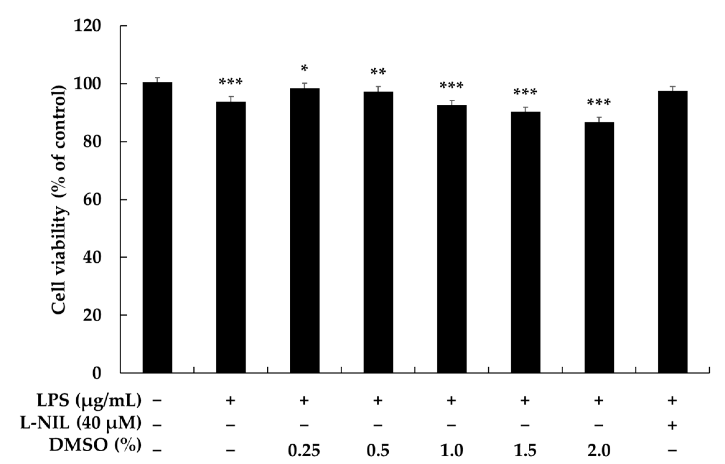
2.1. Effects of DMSO on Cell Viability

DMSO toxicity was assessed in terms of the cell viability of RAW 264.7 macrophages determined by MTT assay. As displayed in Figure 1, when the concentration of DMSO is increased from 0.25% to 1.5%, the cell viability of the RAW 264.7 macrophages is marginally affected, indicating that DMSO can be safely used within these concentrations (0.25–1.5%). However, when the DMSO concentration was increased to 2.0%, the cell viability of RAW 264.7 macrophages decreased to 86.75%; thus, DMSO concentrations of 0.25–1.5% are considered safe [12]. In the interests of determining precise effects related to specific concentrations, DMSO concentrations of 0.25%, 0.5%, 1.0%, and 1.5% were tested.
Kloesch et al. [13] verified that C-28/I2 human chondrocyte cultures treated with different DMSO concentrations for 12 h showed reduced expression of IL-6 and IL-8 at concentrations higher than 1% v/v. Hollebeeck et al. [14] also reported that in vitro stimulation with an inflammatory cocktail and treatment with low concentrations of DMSO (0.1 to 0.5% v/v) reduces the expression of IL-6 and IL-1 in intestinal Caco-2, which does not align with the toxicity results in our present study. Wedner et al. [15] discovered that treatment of human peripheral blood lymphocytes with 40% v/v DMSO in vitro increased selectin expression and phosphorylation of proteins involved in intracellular activation pathways. Kloverpris et al. [16] found that up to 10% v/v DMSO could be used for a one-hour treatment of human peripheral blood mononuclear cell cultures without affecting culture viability. These studies suggest that different cell types respond differently to DMSO concentrations and stimulation conditions. However, a full understanding of the physiological and pharmacological effects of DMSO is still lacking, and further investigation is required to determine safe and reliable concentrations for its use.

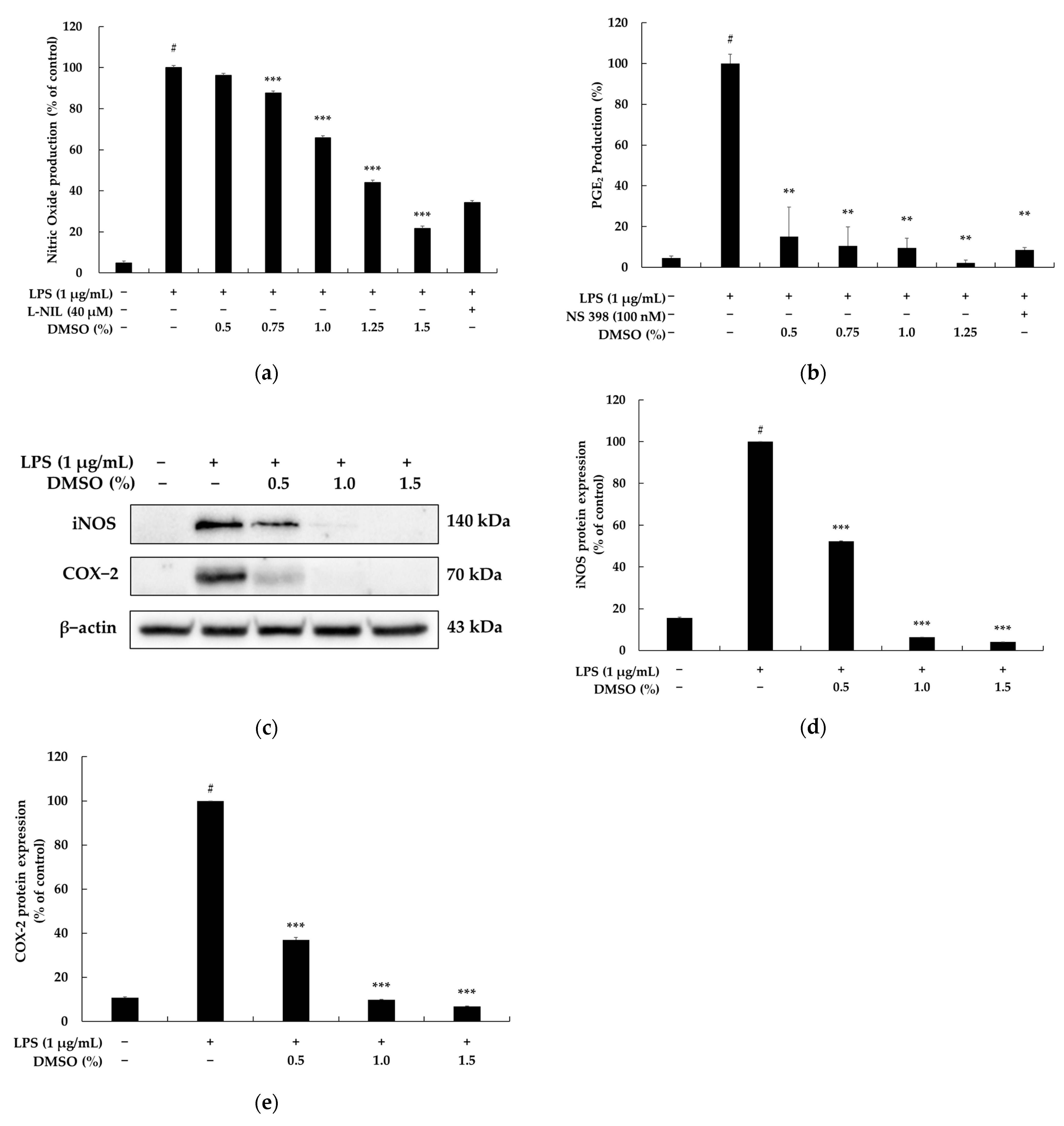
2.2. Effects of DMSO on NO Release, PGE2 Production, and Protein Expression in LPS-Stimulated RAW 264.7 Macrophages

LPS and RAW 264.7 cells are commonly used as in vitro models for studying immune function. LPS, an endotoxin derived from Gram-negative bacteria, is a natural stimulator of immune and inflammatory responses. It activates signaling pathways that trigger the release of cytokines, NO, and PGE2 from cells [17,18,19]. To assess the pro-inflammatory response, the amount of nitrite, a stable NO metabolite, was measured in the cell medium. PGE2, an important lipid mediator, is synthesized in response to various stimuli and can act as an inflammatory mediator. The inhibition of PGE2 synthesis is considered an anti-inflammatory strategy. Thus, the level of pro-inflammatory mediators was measured using Griess reagent and ELISA kits in LPS-induced RAW 264.7 cells in this study.
We initially assessed the effects of DMSO on nitrite production in RAW264.7 cells induced by LPS using the Griess assay. We found that DMSO at 0.5, 0.75, 1.0, 1.25, and 1.5% reduced the amount of nitrite by 3.69, 12.44, 34.20, 55.89, and 78.27%, respectively, in the LPS-activated RAW264.7 macrophages (Figure 2a). DMSO at 0.5, 0.75, 1.0, and 1.25% reduced PGE2 production by 84.96, 89.56, 90.45, and 97.74%, respectively, in the LPS-activated RAW264.7 macrophages (Figure 2b). In addition, DMSO also reduced the expression levels of COX-2 and iNOS in the LPS-activated RAW264.7 macrophages (Figure 2c). Therefore, the above results confirm that DMSO possesses an anti-inflammatory action via the downregulation of COX and iNOS protein expression. Similar to our results, Elisia et al. [2] discovered that DMSO significantly suppressed the expression of various pro-inflammatory cytokines/chemokines and PGE2 in Human Blood Cells at concentrations ranging from 0.5% to 2%. Hollebeeck et al. [13] also reported that low concentrations (0.1–0.5%) of DMSO inhibited COX-2 expression in the Caco-2 cell model. However, it is important to note that the effective DMSO concentration varies for each cell line.

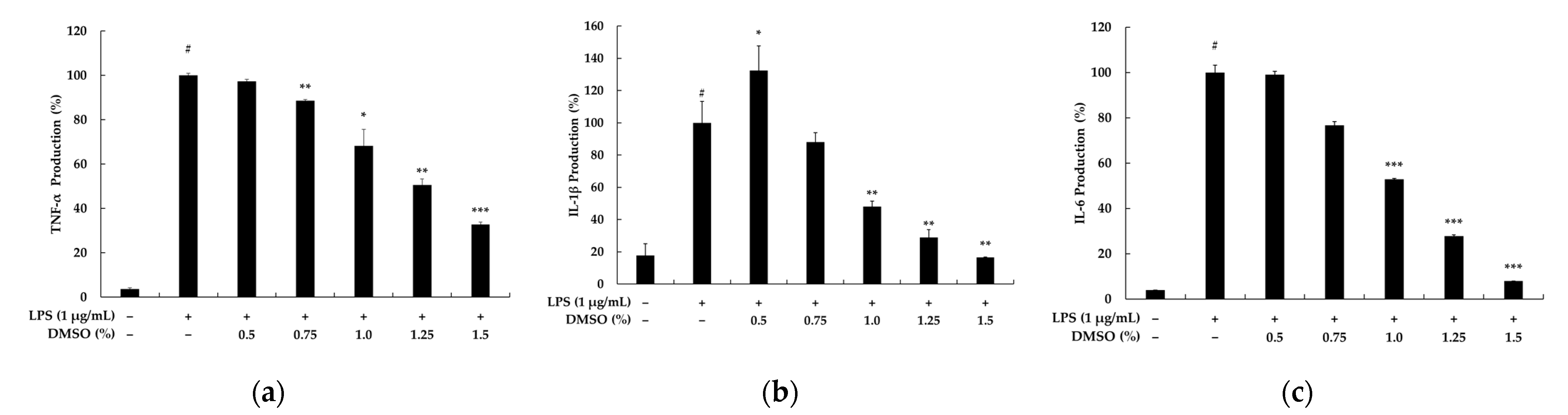
2.3. Effects of DMSO on Pro-Inflammatory Cytokine Secretion in LPS-Stimulated RAW 264.7 Cells

TNF-α is a pro-inflammatory cytokine that induces inflammation and triggers signaling events involved in cell necrosis and apoptosis, as well as the release of other cytokines such as IL-6 and IL-1β. Reducing levels of these cytokines can attenuate the immune response and decrease inflammation in inflammation models [20]. To assess the anti-inflammatory potential of DMSO, TNF-α secretion by macrophage cells was measured. Figure 3 demonstrates that LPS exposure increases TNF-α production significantly compared to untreated cells, while treatment with DMSO inhibits TNF-α secretion. Additionally, the inflammatory mediator IL-1β contributes to the initiation and escalation of inflammatory responses, leading to intestinal damage [21]. Figure 3a shows that TNF-α levels in the LPS group reached 27.0 times higher in the control group, while the TNF-α levels in the DMSO group were significantly inhibited. In addition, elevated IL-1β levels are considered a typical sign of an acute inflammatory response. LPS stimulated a significant increase in IL-1β production compared with the control group. As shown in Figure 3b,c, under LPS stimulation, the content of both IL-1β and IL-6 increased by 5.7-fold and 25.1-fold, respectively. Compared with the LPS group, the treatment with the DMSO inhibited LPS stimulation of IL-1β and IL-6 production in RAW 264.7 cells. At DMSO concentrations of 0.5–1.5%, IL-1β and IL-6 production in RAW264.7 cells was significantly lower than that of the LPS group. Taken together, these results suggest that DMSO exerts an anti-inflammatory action, which decreases the production of the pro-inflammatory cytokines.

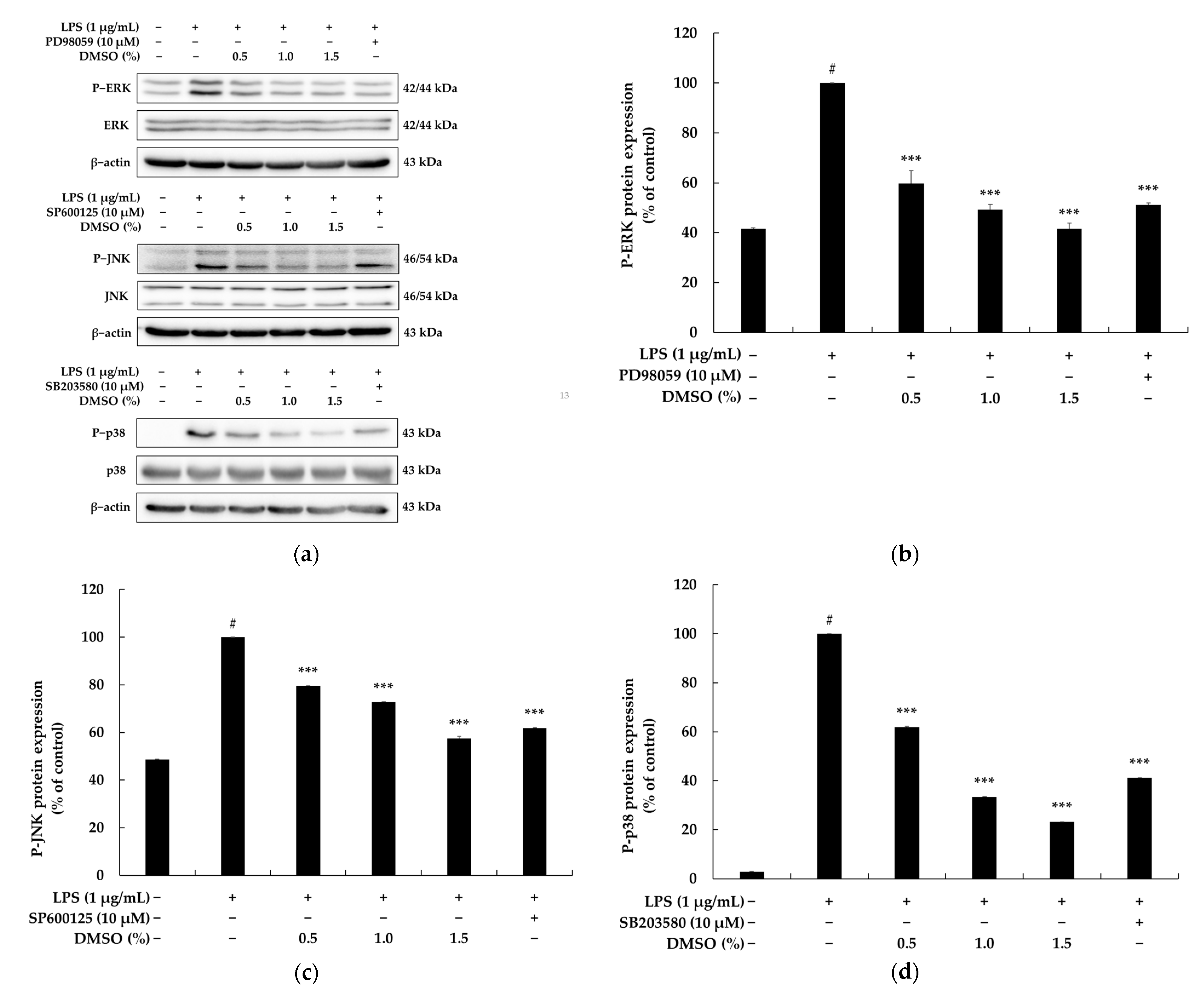
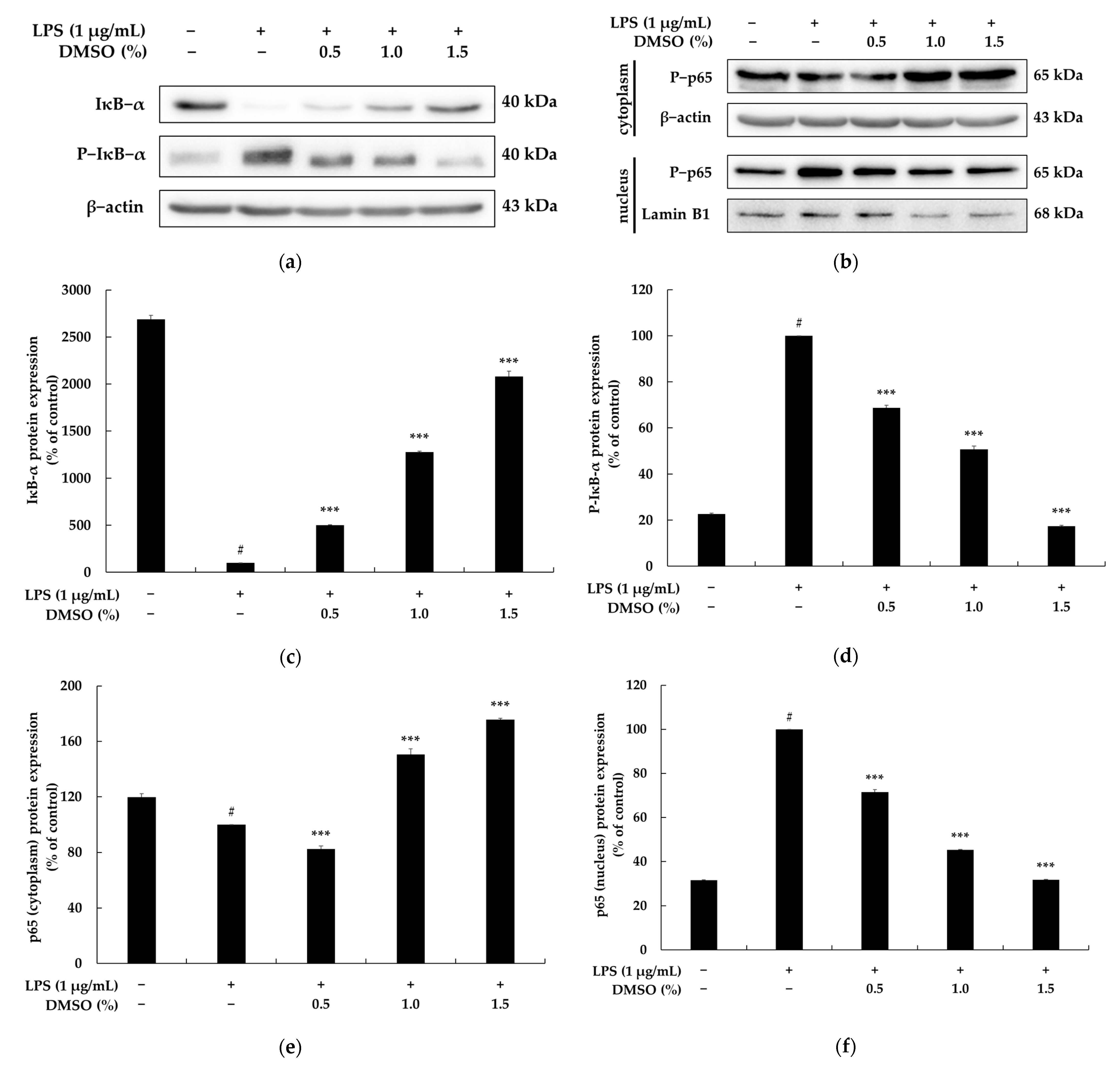
2.4. Effects of DMSO on the NF-κB and MAPK Signaling Pathways

Pro-inflammatory mediators and cytokines are produced through the activation of the NF-κB and MAPK signaling pathways, with the latter being important for regulating the inflammatory response and activating NF-κB. These pathways mainly involve ERK, JNK, and p38-MAPK and are crucial for the TLR4-mediated immune response-associated gene expression [22]. After TLR4 recognizes the antigen, the activated TLR4 recruits the adapter protein MyD88, which in turn triggers a subsequent response by activating the MAPK signaling pathway [23]. To investigate whether the downregulation of TLR4, induced by DMSO, results in the inactivation of the MAPK signaling pathway, cells were treated with varying concentrations of DMSO, and the total phosphorylation levels of JNK, ERK, and p38 were assessed using Western blot analysis. As depicted in Figure 4, upon stimulation with LPS (1 μg/mL) alone, there was a significant upregulation of phosphorylation levels of p38, ERK, and JNK compared to the control group. However, intriguingly, pretreatment with DMSO led to a concentration-dependent downregulation of LPS-induced phosphorylation levels of p38, ERK, and JNK. The above results suggest that DMSO can suppress the phosphorylation of MAPKs in RAW264.7 macrophages stimulated by LPS.
The key transcription factor NF-κB p65 regulates the transcription of several genes that are involved in inflammation, including those that encode pro-inflammatory enzymes such as iNOS and COX-2, as well as cytokines such as TNF-α, IL-6, and IL-1β. IκBα acts as an endogenous inhibitor of NF-κB and binds to cytosolic NF-κB p65 in an unstimulated state. However, upon stimulation, IκBα undergoes phosphorylation and subsequent degradation via the ubiquitin-proteasome pathway. Thereafter, NF-κBp65 is again released, translocated from the cytoplasm to the nucleus, and then accumulates in the nucleus to regulate the expression of target genes [24]. Furthermore, as a significant downstream component of the MAPK signaling pathway, the phosphorylated form of NF-κB p65, induced by MAPK activation, stimulates the expression of genes encoding pro-inflammatory cytokines. Therefore, further investigation explored whether the inactivation of the MAPK signaling pathway, induced by DMSO, could contribute to the inactivation of the NF-κB signaling pathway. As shown in Figure 5a,c,d, after the RAW264.7 cells were incubated with DMSO, the expression levels of IκB-α increased remarkably, and those of p-IκB decreased significantly. Furthermore, an increased level of cytosolic p65 and a decreased level of p65 in the nucleus were observed, which suggests that DMSO treatment inhibits the nuclear translocation of p65 in RAW264.7 cells (Figure 5b,e,f).

3. Materials and Methods

Thermo Fisher Scientific (Waltham, MA, USA) supplied DMEM and penicillin-streptomycin (P/S), while Merck Millipore (Burling, VT, USA) supplied FBS for cell culture. Lipopolysaccharide (LPS) from Escherichia coli O111:B4 and Griess reagent used for cell experiments were purchased from Sigma–Aldrich (St. Louis, MO, USA). Biosesang (Seongnam, Gyeonggi-do, Republic of Korea) supplied MTT, DMSO, PBS, TBS, SDS, RIPA buffer, and ECL kit. Thermo Fisher Scientific (Waltham, MA, USA) provided the nuclear/cytoplasmic extraction reagents, BCA protein assay kit, and 0.5% trypsin−ethylenediaminetetraacetic acid (10×), while Bio-Rad (Hercules, CA, USA) supplied Tween 20 and 2× Laemmli sample buffer. Skim milk was obtained from BD Difco (Sparks, MD, USA), and ELISA kits for PGE2 were purchased from Abcam (Cambridge, EN, UK), while the primary antibody COX-2, cytokine kits (IL-1β, IL-6, and TNF-α) were purchased from BD Biosciences (Franklin Lakes, NJ, USA). Cell Signaling Technology (Danvers, MA, USA) provided the primary antibody iNOS, p-ERK, ERK, p-p38, p38, p-JNK, JNK, p-IκB-α, IκB-α, p65, lamin B, and β-actin antibodies, as well as the anti-mouse and anti-rabbit secondary antibodies. Additionally, Cayman (Ann Arbor, Michigan) provided L-NIL and NS 398.

3.1. Cell Culture

We obtained the RAW 264.7 murine macrophage cells from the Korea Cell Line Bank in Seoul, Republic of Korea. The cells were cultured in Dulbecco’s modified Eagle’s medium (DMEM) supplemented with 10% fetal bovine serum (FBS) and 1% penicillin-streptomycin under a humidified atmosphere containing 5% CO2 at a temperature of 37 °C.

3.2. Cell Viability

Cytotoxicity was assessed by MTT assay. RAW 264.7 macrophage cells were plated at a density of 1.5 × 10⁵ cells/well in a 24-well plate and allowed to adhere overnight. The cells were treated with DMSO at different concentrations (0.25, 0.5, 1.0, 1.5, and 2.0%) and LPS (1 μg/mL) for 24 h [25,26]. Following this, the medium was completely removed and replaced with MTT dye (0.2 mg/mL) and incubated for 3 h, after which the insoluble formazan crystals were solubilized in dimethyl sulfoxide. Absorbance was measured at 570 nm using a microplate reader (Biotek; Winooski, VT, USA).

3.3. Determination of NO Production

The Griess assay is utilized to quantify the levels of nitrite, which is the ultimate outcome of the NO oxidation process. The levels of nitrite were evaluated in the cell culture supernatant, and the Griess reaction was used to determine them as an indicator of NO production. Initially, RAW 264.7 macrophage cells were seeded in a 24-well plate at a density of 1.5 × 10⁵ cells/well and permitted to adhere overnight before the analysis. The cells were treated with DMSO at different concentrations (0.5, 0.75, 1.0, 1.25, and 1.5%) and LPS (1 μg/mL) for 24 h [25,26]. We used the iNOS-specific inhibitor L-NIL (40 μM) for the positive control group. Cell culture supernatants were mixed with an equal volume (100 μL) of Griess reagent and incubated in a 96-well plate for 10 min at room temperature. Absorbance was measured at 540 nm using a microplate reader (Biotek; Winooski, VT, USA).

3.4. Determination of the PGE2, IL-1β, IL-6, and TNF-α Levels

We determined the levels of PGE2 and cytokines (IL-1β, IL-6, and TNF-α) in culture supernatants using cytokine-detection ELISA kits, following the manufacturer’s instructions. RAW 264.7 macrophage cells were plated at a density of 1.5 × 10⁵ cells/well in a 24-well plate and allowed to adhere overnight. The cells were treated with DMSO at different concentrations (0.5, 0.75, 1.0, 1.25, and 1.5%) and LPS (1 μg/mL) for 24 h. We used the COX-2 specific inhibitor NS 398 (100 nM) as a positive control for measuring PGE2 production. Protein levels were determined by measuring absorbance at 405 or 450 nm using a microplate reader (Biotek; Winooski, VT, USA).

3.5. Preparation of Nuclear and Cytoplasmic Extracts

Nuclear and cytoplasmic extracts were isolated using reagent extraction kits. RAW 264.7 macrophage cells were plated at a density of 6.0 × 10⁵ cells/dish in 60 mm cell culture dishes and allowed to adhere overnight. The cells were treated with DMSO at different concentrations (0.5, 1.0, and 1.5%) and LPS (1 μg/mL) for 15 min. After incubation, the nuclear extract was obtained according to the manufacturer’s protocols of the extraction reagents kit.

3.6. Western Blotting

We plated RAW 264.7 macrophage cells at a density of 6.0 × 10⁵ cells/dish in 60 mm cell culture dishes and allowed them to adhere overnight. The cells were treated with the DMSO at different concentrations (0.5, 1.0, and 1.5%) and LPS (1 μg/mL) for each protein expression time point. To prepare the cells for analysis, they were washed with a solution of 1× PBS, and then treated with a lysis buffer containing RIPA and a protease inhibitor cocktail at a temperature of 4 °C for a duration of 20 min. The resulting lysate was then centrifuged at a speed of 15,000 rpm and a temperature of −8 °C for 20 min to obtain the supernatant, which was then quantified using a BCA protein assay kit to adjust the protein concentration to 30 μg/mL. To prepare loading samples for further analysis, the protein was mixed with 2× Laemmli sample buffer in a 1:1 ratio and heated at a temperature of 100 °C for a duration of 5 min. The samples were then separated by size using SDS-polyacrylamide gels through a process called electrophoresis. Once separated, the proteins were transferred to a PVDF membrane and blocked for 2 h in a solution of 5% skim milk dissolved in TBS-T (Tris-buffered saline with 1% Tween 20). The membrane was washed with 1× TBS−T and then incubated in a solution of primary antibody, diluted at a ratio of 1:2000, overnight at 4 °C for reaction. After washing the primary antibody, horseradish peroxidase (HRP)-conjugated secondary antibody (1:1000) was added onto the membrane for interaction with the respective primary antibody for 2 h at room temperature. After washing the antibody, the protein band signals were visualized with the ECL chemiluminescence reagents and fusion solo S (24 rue de Lamirault, 77090 Collégien, France). The band images were analyzed using ImageJ software (NIH, Bethesda, MD, USA).

3.7. Statistical Analyses

The experimental results are presented as the mean ± standard deviation (SD) of three repeated experiments. Statistical significance was assessed using Student’s t-test, with p-values indicated as follows: # p < 0.001 vs. unstimulated control group; * p < 0.05, ** p < 0.01, *** p < 0.001 vs. LPS alone.

4. Conclusions

DMSO can dissolve both polar and nonpolar substances, and it is therefore used as a solvent for anti-inflammatory drugs during their evaluation and development. However, considering that DMSO itself has an anti-inflammatory effect in various cell lines, erroneous research results may be obtained when samples are dissolved at high concentrations. Therefore, we characterized the cytotoxic and anti-inflammatory effects of different concentrations of DMSO in RAW264.7 macrophages activated with LPS, a widely used model in anti-inflammatory drug research. In conclusion, this study confirms that the cell viability was reduced by 9.98% and 13.25% for 1.5% and 2% DMSO concentrations in LPS-indued macrophage, respectively, and that DMSO inhibits NO, PGE2, and other cytokines through the inhibition of both NF-κB activation and MAPK phosphorylation in LPS-stimulated RAW 264.7 cells.

Author Contributions

Conceptualization, C.-G.H.; methodology, H.H.; software, H.H.; validation, H.H.; formal analysis, J.-K.K.; investigation, C.-G.H.; data curation, K.J.A.; writing—original draft preparation, C.-G.H.; writing—review and editing, C.-G.H.; visualization, H.H.; project administration, C.-G.H.; funding acquisition, C.-G.H. All authors have read and agreed to the published version of the manuscript.

Funding

This research was supported by the Ministry of Trade, Industry, and Energy (MOTIE), Korea Institute for Advancement of Technology (KIAT) through the Demand-Customized R&D Project for National Innovation Convergence Complex (P0015357).

Institutional Review Board Statement

Not applicable.

Informed Consent Statement

Not applicable.

Data Availability Statement

Not applicable.

Conflicts of Interest

The authors declare no conflict of interest.

References

  1. Deol, P.; Yang, J.; Morisseau, C.; Hammock, B.D.; Sladek, F.M. Dimethyl Sulfoxide Decreases Levels of Oxylipin Diols in Mouse Liver. Front. Pharmacol. 2019, 10, 580. [Google Scholar] [CrossRef] [Green Version]
  2. Elisia, I.; Nakamura, H.; Lam, V.; Hofs, E.; Cederberg, R.; Cait, J.; Hughes, M.R.; Lee, L.; Jia, W.; Adomat, H.H.; et al. DMSO Represses Inflammatory Cytokine Production from Human Blood Cells and Reduces Autoimmune Arthritis. PLoS ONE 2016, 11, e0152538. [Google Scholar] [CrossRef] [PubMed] [Green Version]
  3. Hajighasemi, F.; Tajik, S. Assessment of Cytotoxicity of Dimethyl Sulfoxide in Human Hematopoietic Tumor Cell Lines. Iran. J. Blood Cancer 2017, 9, 48–53. [Google Scholar]
  4. de Abreu Costa, L.; Henrique Fernandes Ottoni, M.; Dos Santos, M.G.; Meireles, A.B.; Gomes de Almeida, V.; de Fátima Pereira, W.; Alves de Avelar-Freitas, B.; Eustáquio Alvim Brito-Melo, G. Dimethyl Sulfoxide (DMSO) Decreases Cell Proliferation and TNF-α, IFN-γ, and IL-2 Cytokines Production in Cultures of Peripheral Blood Lymphocytes. Molecules 2017, 22, 1789. [Google Scholar] [CrossRef] [PubMed] [Green Version]
  5. Lee, H.; Park, J.B. Evaluation of the effects of dimethylsulphoxide on morphology, cellular viability, mRNA, and protein expression of stem cells culture in growth media. Biomed. Rep. 2017, 7, 291–296. [Google Scholar] [CrossRef] [PubMed] [Green Version]
  6. Bini, R.; Olivero, G.; Trombetta, A.; Castagna, E.; Cotogni, P. Effects of dimethyl sulfoxide, pyrrolidine dithiocarbamate, and methylprednisolone on nuclear factor-kappaB and heat shock protein 70 in a rat model of hemorrhagic shock. J. Trauma Acute Care Surg. 2008, 64, 1048–1054. [Google Scholar] [CrossRef]
  7. Kelly, K.A.; Hill, M.R.; Youkhana, K.; Wanker, F.; Gimble, J.M. Dimethyl sulfoxide modulates NF-kappa B and cytokine activation in lipopolysaccharide-treated murine macrophages. Infect. Immun. 1994, 62, 3122–3128. [Google Scholar] [CrossRef] [Green Version]
  8. Guo, W.; Qiu, W.; Ao, X.; Li, W.; He, X.; Ao, L.; Hu, X.; Li, Z.; Zhu, M.; Luo, D.; et al. Low-concentration DMSO accelerates skin wound healing by Akt/mTOR-mediated cell proliferation and migration in diabetic mice. Br. J. Pharmacol. 2020, 177, 3327–3341. [Google Scholar] [CrossRef]
  9. Kang, J.K.; Kang, H.K.; Hyun, C.G. Anti-Inflammatory Effects of Spiramycin in LPS-Activated RAW 264.7 Macrophages. Molecules 2022, 27, 3202. [Google Scholar] [CrossRef]
  10. Kang, J.K.; Chung, Y.C.; Hyun, C.G. Anti-Inflammatory Effects of 6-Methylcoumarin in LPS-Stimulated RAW 264.7 Macrophages via Regulation of MAPK and NF-κB Signaling Pathways. Molecules 2021, 26, 5351. [Google Scholar] [CrossRef]
  11. Han, S.; Gao, H.; Chen, S.; Wang, Q.; Li, X.; Du, L.J.; Li, J.; Luo, Y.Y.; Li, J.X.; Zhao, L.C.; et al. Procyanidin A1 Alleviates Inflammatory Response induced by LPS through NF-κB, MAPK, and Nrf2/HO-1 Pathways in RAW264.7 cells. Sci. Rep. 2019, 9, 15087. [Google Scholar] [CrossRef] [Green Version]
  12. Silva, E.J.; Herrera, D.R.; Almeida, J.F.; Ferraz, C.C.; Gomes, B.P.; Zaia, A.A. Evaluation of cytotoxicity and up-regulation of gelatinases in fibroblast cells by three root repair materials. Int. Endod. J. 2012, 45, 815–820. [Google Scholar] [CrossRef]
  13. Kloesch, B.; Liszt, M.; Broell, J.; Steiner, G. Dimethyl sulphoxide and dimethyl sulphone are potent inhibitors of IL-6 and IL-8 expression in the human chondrocyte cell line C-28/I2. Life Sci. 2011, 89, 473–478. [Google Scholar] [CrossRef] [PubMed]
  14. Hollebeeck, S.; Raas, T.; Piront, N.; Schneider, Y.J.; Toussaint, O.; Larondelle, Y.; During, A. Dimethyl sulfoxide (DMSO) attenuates the inflammatory response in the in vitro intestinal Caco-2 cell model. Toxicol. Lett. 2011, 206, 268–275. [Google Scholar] [CrossRef] [PubMed]
  15. Wedner, H.J.; Bass, G. Induction of the tyrosine phosphorylation of a 66 kd soluble protein by DMSO in human peripheral blood T lymphocytes. Biochem. Biophys. Res. Commun. 1986, 140, 743–749. [Google Scholar] [CrossRef]
  16. Kloverpris, H.; Fomsgaard, A.; Handley, A.; Ackland, J.; Sullivan, M.; Goulder, P. Dimethyl sulfoxide (DMSO) exposure to human peripheral blood mononuclear cells (PBMCs) abolish T cell responses only in high concentrations and following coincubation for more than two hours. J. Immunol. Methods 2010, 356, 70–78. [Google Scholar] [CrossRef]
  17. Liu, Y.; Chen, W.; Zheng, F.; Yu, H.; Wei, K. Xanthatin Alleviates LPS-Induced Inflammatory Response in RAW264.7 Macrophages by Inhibiting NF-κB, MAPK and STATs Activation. Molecules 2022, 27, 4603. [Google Scholar] [CrossRef]
  18. So, B.R.; Bach, T.T.; Paik, J.H.; Jung, S.K. Kmeria duperreana (Pierre) Dandy Extract Suppresses LPS-Induced iNOS and NO via Regulation of NF-κB Pathways and p38 in Murin Macrophage RAW 264.7 Cells. Prev. Nutr. Food Sci. 2020, 25, 166–172. [Google Scholar] [CrossRef]
  19. Wu, J.; Liu, B.; Mao, W.; Feng, S.; Yao, Y.; Bai, F.; Shen, Y.; Guleng, A.; Jirigala, B.; Cao, J. Prostaglandin E2 Regulates Activation of Mouse Peritoneal Macrophages by Staphylococcus aureus through Toll-Like Receptor 2, Toll-like Receptor 4, and NLRP3 Inflammasome Signaling. J. Innate Immun. 2020, 12, 154–169. [Google Scholar] [CrossRef]
  20. Parameswaran, N.; Patial, S. Tumor necrosis factor-α signaling in macrophages. Crit. Rev. Eukaryot. Gene Expr. 2010, 20, 87–103. [Google Scholar] [CrossRef] [PubMed]
  21. Dinarello, C.A. Overview of the IL-1 family in innate inflammation and acquired immunity. Immunol. Rev. 2018, 281, 8–27. [Google Scholar] [CrossRef] [PubMed] [Green Version]
  22. Peroval, M.Y.; Boyd, A.C.; Young, J.R.; Smith, A.L. A critical role for MAPK signalling pathways in the transcriptional regulation of toll like receptors. PLoS ONE 2013, 8, e51243. [Google Scholar] [CrossRef] [Green Version]
  23. Laird, M.H.; Rhee, S.H.; Perkins, D.J.; Medvedev, A.E.; Piao, W.; Fenton, M.J.; Vogel, S.N. TLR4/MyD88/PI3K interactions regulate TLR4 signaling. J. Leukoc. Biol. 2009, 85, 966–977. [Google Scholar] [CrossRef] [PubMed] [Green Version]
  24. Tak, P.P.; Firestein, G.S. NF-kappaB: A key role in inflammatory diseases. J. Clin. Invest. 2001, 107, 7–11. [Google Scholar] [CrossRef] [PubMed] [Green Version]
  25. Orabona, C.; Orecchini, E.; Volpi, C.; Bacaloni, F.; Panfili, E.; Pagano, C.; Perioli, L.; Belladonna, M.L. Crocus sativus L. Petal Extract Inhibits Inflammation and Osteoclastogenesis in RAW 264.7 Cell Model. Pharmaceutics 2022, 14, 1290. [Google Scholar] [CrossRef]
  26. Zhao, W.R.; Shi, W.T.; Zhang, J.; Zhang, K.Y.; Qing, Y.; Tang, J.Y.; Chen, X.L.; Zhou, Z.Y. Tribulus terrestris L. Extract Protects against Lipopolysaccharide-Induced Inflammation in RAW 264.7 Macrophage and Zebrafish via Inhibition of Akt/MAPKs and NF-κB/iNOS-NO Signaling Pathways. Evid.-Based Complement. Altern. Med. 2021, 2021, 6628561. [Google Scholar] [CrossRef]
Figure 1. The effects of DMSO on the viability of RAW 264.7 cells. Cells were treated with DMSO (0.25, 0.5, 1.0, 1.5, and 2.0%) and LPS (1 μg/mL) stimulation for 24 h. L-NIL (40 μM) was used as the positive control. The effects of DMSO on cell viability were evaluated using an MTT assay. Cell viability is expressed as percentages relative to untreated cells. The results are presented as the mean ± SD of three repeated experiments. * p < 0.05, ** p < 0.01, *** p < 0.001 vs. LPS alone.
Biochem 03 00007 g001
Figure 2. Effects of DMSO on the production of pro-inflammatory mediators in LPS-induced RAW 264.7 cells. Cells were treated with DMSO (0.5, 0.75, 1.0, 1.25, and 1.5%) and LPS (1 μg/mL) stimulation for 24 h. L-NIL (40 μM) and NS 398 (100 nM) were used as the positive control. (a) The amount of nitric oxide in the medium was measured using the Griess reagent. (b) PGE2 production was determined using an ELISA kit. Western blot (c) and densitometric (d,e) analysis for iNOS/COX-2. Cells were treated with DMSO (0.5, 1.0, and 1.5%) and LPS (1 μg/mL) stimulation for 22 h. The results are presented as the mean ± SD of three repeated experiments. # p < 0.001 vs. unstimulated control group. ** p < 0.01, *** p < 0.001 vs. LPS alone.
Biochem 03 00007 g002
Figure 3. Effects of DMSO on the production of pro-inflammatory cytokines in LPS-induced RAW 264.7 cells. Cells were treated with DMSO (0.5, 0.75, 1.0, 1.25, and 1.5%) and LPS (1 μg/mL) stimulation for 24 h. The (a) TNF-α production, (b) IL-1β production, and (c) IL-6 production were determined using an ELISA kit. The results are presented as the mean ± SD of two repeated experiments. # p < 0.001 vs. unstimulated control group. * p < 0.05, ** p < 0.01, *** p < 0.001 vs. LPS alone.
Biochem 03 00007 g003
Figure 4. Effects of DMSO on the MAPK signaling pathway in LPS-induced RAW 264.7 cells. Cells were treated with DMSO (0.5, 1.0, and 1.5%) and LPS (1 μg/mL) stimulation for 15 or 20 min. SP600125 (10 μM) is a JNK MAPK inhibitor. (a) Western blotting results, (b) p-ERK protein expression, (c) p-JNK protein expression, and (d) p-p38 protein expression. The results are presented as the mean ± SD of three repeated measurements using ImageJ. # p < 0.001 vs. unstimulated control group. *** p < 0.001 vs. LPS alone.
Biochem 03 00007 g004
Figure 5. Effects of DMSO on the NF-κB signaling pathway in LPS-induced RAW 264.7 cells. Cells were treated with DMSO (0.5, 1.0, and 1.5%) and LPS (1 μg/mL) stimulation for 15 or 20 min. (a) Western blotting results of IκB-α and p-IκB-α protein expression, (b) Western blotting results of NF-κB (p65) protein expression, (c) IκB-α protein expression, (d) p-IκB-α protein expression, (e) p65 (cytoplasm) protein expression, and (f) p65 (nucleus) protein expression. The results are presented as the mean ± SD of three repeated measurements using ImageJ. # p < 0.001 vs. unstimulated control group. *** p < 0.001 vs. LPS alone.
Biochem 03 00007 g005
Disclaimer/Publisher’s Note: The statements, opinions and data contained in all publications are solely those of the individual author(s) and contributor(s) and not of MDPI and/or the editor(s). MDPI and/or the editor(s) disclaim responsibility for any injury to people or property resulting from any ideas, methods, instructions or products referred to in the content.

