Review

MALDI MS-Based Investigations for SARS-CoV-2 Detection

1
Department of Health Sciences, University “Magna Græcia”, 88100 Catanzaro, Italy
2
Department of Medical and Surgical Sciences, University “Magna Græcia”, 88100 Catanzaro, Italy
3
Department of Experimental and Clinical Medicine, University “Magna Græcia”, 88100 Catanzaro, Italy
*
Author to whom correspondence should be addressed.
BioChem 2021, 1(3), 250-278; https://doi.org/10.3390/biochem1030018
Submission received: 21 October 2021 / Revised: 18 November 2021 / Accepted: 19 November 2021 / Published: 1 December 2021

Abstract

:
The urgent need to fight the COVID-19 pandemic has impressively stimulated the efforts of the international scientific community, providing an extraordinary wealth of studies. After the sequence of the virus became available in early January 2020, safe and effective vaccines were developed in a time frame much shorter than everybody expected. However, additional studies are required since viral mutations have the potential of facilitating viral transmission, thus reducing the efficacy of developed vaccines. Therefore, improving the current laboratory testing methods and developing new rapid and reliable diagnostic approaches might be useful in managing contact tracing in the fight against both the original SARS-CoV-2 strain and the new, potentially fast-spreading CoV-2 variants. Mass Spectrometry (MS)-based testing methods are being explored, with the challenging promise to overcome the many limitations arising from currently used laboratory testing assays. More specifically, MALDI-MS, since its advent in the mid 1980s, has demonstrated without any doubt the great potential to overcome many unresolved analytical challenges, becoming an effective proteomic tool in several applications, including pathogen identification. With the aim of highlighting the challenges and opportunities that derive from MALDI-based approaches for the detection of SARS-CoV-2 and its variants, we extensively examined the most promising proofs of concept for MALDI studies related to the COVID-19 outbreak.

1. Introduction

The coronavirus disease 2019 (COVID-19) pandemic outbreak caused by the novel severe acute respiratory syndrome coronavirus 2 (SARS-CoV-2) has spread all over the world, with more than 242 million individuals infected and approximately five million deaths [1].
Recent evidence confirms that in contrast with SARS-CoV-1, which was only transmitted by people with symptoms, the new coronavirus can be transmitted even before the onset of symptoms from pre-symptomatic subjects or even from asymptomatic people. Thus, the silent chains of transmission, which considerably hinder containment efforts aimed at limiting viral spread, require the quick screening of each positive subject in order to help control the ongoing pandemic [2,3,4,5].
Currently, together with the use of vaccines, an accurate and immediate diagnosis of SARS-CoV-2 might be one of the most important lines of attack for controlling the spread of infection since it allows for the prompt isolation of people infected by the virus. Additionally, an earlier diagnosis may lead to a more effective therapeutic intervention, with improved prognosis and a higher probability of avoiding patient recovery in the intensive care unit (ICU).
Several SARS-CoV-2 variants have been identified in many countries, which represents one of the most relevant issues in controlling the COVID-19 pandemic [6]. Viral mutations are of great concern as such mutations have the potential to facilitate viral transmission and to reduce the efficacy of developed vaccines by their ability to escape protection due to vaccine-induced neutralizing antibodies [7].
Therefore, it has become necessary to improve current laboratory testing methods and to develop new, rapid, and reliable diagnostic approaches to identify all positive cases and manage contact tracing in order to stop the SARS-CoV-2 and new, potentially fast-spreading SARS-CoV-2 variants.
The development of new diagnostic tools for emerging pathogens requires the ability to detect low viral loads, which enables early detection in order to avoid cross-reactivity with other viral strains and to deliver results quickly.
The clinical diagnosis of suspected cases is generally established by Reverse Transcriptase-Polymerase Chain Reaction (RT-qPCR), which represents the gold standard for the molecular diagnosis of SARS-CoV-2. Enzyme-linked immunoassays (EIA) for viral antibody and antigen detection, and serum viral neutralization (SVN) assays for antibody neutralization determination constitute other relevant laboratory tools [8].
However, RT-qPCR may suffer from the lack of sensitivity for the detection of SARS-CoV-2 in the early stages of infection as the concentration level of the virus is low in the upper respiratory airways during the first 6–8 days of illness, only reaching the peak in a window of 10–14 days from the onset of the illness [9,10]. Other limitations of the RT-qPCR assay are its low-throughput capacity due to many intermediate steps and long turnaround times, and the use of a poorly designed specimen extraction control [11,12]. Additionally, false-positive and high false-negative rates ranging from 1% to 30% may arise, depending on several factors such as improper sampling, lack of specificity due to poor sensitivity, and cross- and carryover contamination [13,14]. All together, these factors might lead to misdiagnosis and the spread of infection.
Concerning the rapid antigen tests, these are also being widely adopted for preliminary screening, but they have substantially lower sensitivity than the WHO-recommended standard, especially for pre-symptomatic and asymptomatic cases [15].
Thus, it is of crucial importance in this particular circumstance to develop novel, robust laboratory diagnostic tests that target SARS-CoV-2 with a high level of reproducibility, specificity, and sensitivity [16] to support already existing tests.
Owing to the huge amount of data that can be rapidly collected and considerable technological advancements, mass spectrometry (MS)-based omic approaches have demonstrated, without any doubt, the possibility of performing extensive and sensitive analyses, not only opening new opportunities for the diagnosis and assessment of disease progression, but also providing insights into mechanisms of the disease [17,18,19,20].
Additionally, the use of MS platform technologies has already been applied in disease outbreaks for the study and analysis of infectious disease agents such as viruses and bacteria [21,22,23,24,25,26,27], suggesting its potential as the new alternative and highly specific test for the SARS-CoV-2 infection.
In particular, recent reviews confirm how MS-based omic technologies have greatly contributed to the detection of SARS-CoV-2 [20,28,29,30,31].
Many efforts have been explored to make the MS-detection process high-throughput, highly accurate, and sensitive [32,33,34,35].
In this scenario, Matrix-Assisted-Laser-Desorption/Ionization (MALDI)-MS seems to satisfy particularly the high-throughput requirements as well as the rapid data acquisition [36]. In addition, the advancements reached by the Fourier-Transform Ion Cyclotron Resonance (FT-ICR) technology in terms of high and ultra-high-resolution power and mass accuracy in modern mass instruments make possible the development of high-throughput, highly accurate, and highly sensitive processes [37].
MALDI-MS has become one of the main proteomic tools for its relatively high tolerance of mixtures and biological contaminants [36]. This technique is largely used for the characterization of biomolecules, especially proteins and peptides. However, the original purpose of this technology has also been redirected to microorganism identification. In the last two decades, MALDI has been successfully integrated into the microbiology laboratory workflow due to its capacity for rapid, low-cost microbial species identification, and it is now routinely adopted in Europe and the United States for bacteria identification [38]. Therefore, considering that most clinical diagnostic laboratories already have MALDI-MS equipment, its application in emerging pathogen research as a screening method for COVID-19 detection in large populations presents great potential.
In this paper, we review exploratory MALDI-based approaches for the detection of SARS-CoV-2 and its variants, with the aim of highlighting challenges and opportunities that derive from these laboratory platforms.

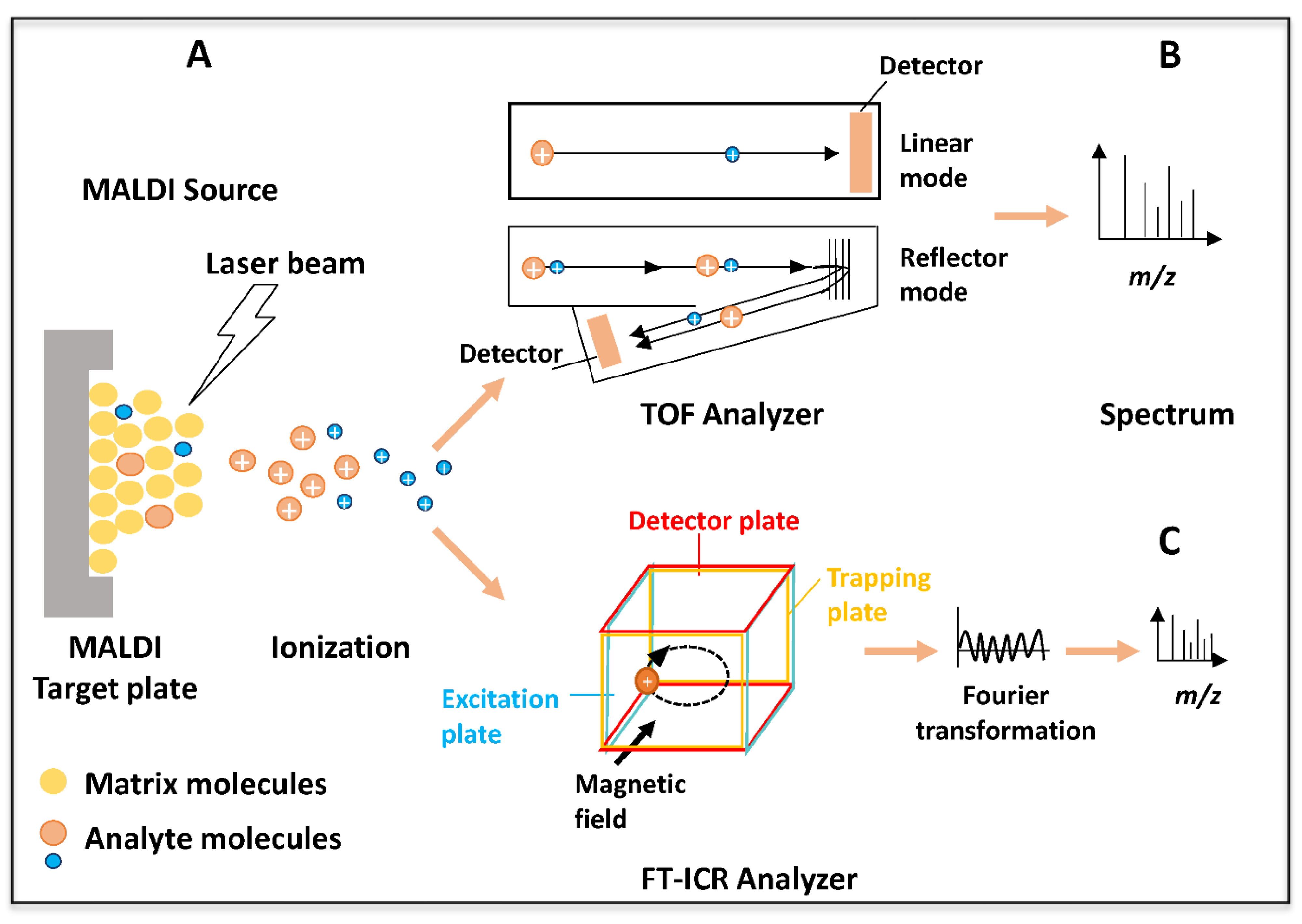
2. MALDI-TOF and MALDI-FT-ICR Mass Spectrometry: A Brief Presentation

The discovery of “soft ionization” methods—MALDI and electrospray ionization (ESI)—in the mid-1980s represented a milestone in the history of MS [39,40]. In fact, before the invention of “soft ionization” techniques, some biomolecules decomposed or were destroyed when analyzed by traditional MS instruments. MALDI is a soft ionization technique used in MS that can preserve the structure of large, non-volatile, and labile molecules, allowing for the sensitive detection of many kinds of nonvolatile biomolecules, including proteins, nucleic acids, and carbohydrates [41]. A typical MALDI preparation relies on embedding these biomolecules in an energy-absorbing crystalline matrix (typically, a small aromatic acid). The use of a large excess of matrix over the biomolecules that are of interest for the analysis is at the core of the MALDI principle [41]. More specifically, in a typical experimental procedure (as shown in Figure 1A), the laser beam hits the co-crystallized mixture of the matrix with the analyte. After the matrix absorbs the laser energy, it desorbs from the sample surface, carrying the embedded analytes into the gas phase. In this process, analyte molecules become ions by a proton transfer from the matrix to the analyte. Then, the charged particles, predominantly singly charged ions, are accelerated with a constant amount of kinetic energy to a mass analyzer such as the Time-of-Flight (TOF) analyzer (Figure 1B). While flying through the TOF, the ions are separated according to their mass-to-charge ratio (m/z).
As also illustrated in Figure 1B, in modern MALDI-TOF instruments, sample analysis can be performed using linear and/or reflector MS modes. The linear MS mode is well-suited for analytes with molecular weights above 4000 Da and enables very sensitive analyses. Analytes below 4000 Da are analyzed in reflector MS mode, with improved mass accuracy and resolution. Specifically, when operating in reflector MS mode, a reflector mirror which reflects ions back toward the reflector detector is used. Therefore, a longer flight ion path is provided, which increases ion separation through an enhanced resolution (Figure 1B). The reflector (also used in the MS/MS mode) is a two-stage electrostatic mirror consisting of a series of plates. The reflector also improves mass accuracy and resolution by correcting the dispersion time caused by variations of initial kinetic energy that generate slight differences in ion velocity and by filtering out neutral molecules. In the presence of a second TOF analyzer (MALDI-TOF/TOF), samples analyzed in the reflector MS mode may be further analyzed by the MS/MS mode to obtain a fragmentation pattern used to determine structural information. This platform is especially used in peptide/protein identification and amino acid sequencing. MALDI-TOF MS allows for the analysis of hundreds of proteins or other biomolecules in a single spectrum and has been used in several diagnostic applications in recent years, including: (i) the study of the distributions of particular biochemical compounds on tissues (MALDI-based imaging MS) [42,43]; (ii) the analysis of nucleic acids for genotyping single nucleotide polymorphisms (SNPs) [44]; (iii) the profiling of biological samples to aid in the discovery of new of disease biomarkers in exploratory studies with diagnostic applicability. Such biomarkers might be used as indicators of progress or the severity of diseases/disorders [45,46,47,48,49,50,51], and for (iv) the identification, characterization, and typing of bacteria, yeasts, and viruses [52,53,54,55,56].
Although less used than TOF because of its high costs, another mass analyzer coupled with a MALDI source is the FT-ICR, which provides better performance than a MALDI-TOF instrument in terms of resolution, mass accuracy, and dynamic range. The FT-ICR analyzer measures the mass-to-charge ratio (m/z) of ions according to the cyclotron frequency of the ions (Figure 1C). After ion generation in the MALDI source, the ions are transferred to the Penning trap consisting of a cell under a high vacuum positioned in a superconducting magnet. The ions are excited by a radiofrequency and generate a signal. The frequency of the signal from each ion is equal to its orbital frequency, which is inversely related to its m/z value. The signal intensity of each frequency is proportional to the number of ions with that m/z value. The signal is amplified and all the frequency components are determined. The mass spectrum is generated using Fourier analysis (Figure 1C). The frequencies match the m/z, and the amplitudes are correlated with the abundance of the analytes [57]. In comparison to any other mass analyzer, FT-ICR provides the highest mass accuracy and mass resolving power, reaching up to ppb mass accuracy, high dynamic range, and mass resolving power greater than 1,000,000 in routine analyses [58]. Very recently, molecular structures with m/z differences of 1.79 mDa were resolved and identified with high mass accuracy and sensitivity [37]. For these reasons, MALDI-FT-ICR is very suitable for the analysis of complex biological samples in proteomic, metabolomic, and lipidomic research in different fields of application, including microbiology, imaging, and biomarker discovery [59,60,61].

3. MALDI-MS for Pathogen Detection: A General Overview

Over the past decade, MALDI-MS has become a routine laboratory technique for the rapid, accurate, and cost-effective identification of cultured bacteria and fungi in clinical microbiology, which requires minimal consumables or reagents [62,63,64]. MALDI-TOF technology has been used for the study, and analyses of several viruses such as human herpesviruses, influenza viruses, and diseases that are related to severe enterovirus infections such as echovirus, coxsackievirus A and B, and poliovirus [53,54,65,66,67,68]. Moreover, the advantage of this approach over the low-throughput LC-MS-based methods is the wide availability of this instrumentation in clinical laboratories, including those in developing countries; consequently, the implementation of the developed methodology would not require a high economic cost [62].
MALDI-MS approaches used for SARS-CoV-2 detection are illustrated in Figure 2. As exemplified in Figure 2, current MALDI-MS approaches aimed at the detection of SARS-CoV-2 are based on “biotyping” [69,70] and “genotyping” [71,72,73] strategies. One study is based on “proteotyping” with high resolution MALDI-FTICR at the peptide level [74], while several studies used the “biomolecular host profiling” strategy to uncover biomarkers of diagnostic utility generated after SARS-CoV-2 infection [75,76,77,78,79]. In particular, the investigations by Iles et al. and Chivte et al. used both biotyping and biomolecular host profiling strategies [69,70].
The identification of microorganisms by MS is a relatively recent concept that is different from the traditional typing based on DNA restriction or amplification methods.
Specifically, proteins are extracted and profiled by MALDI-TOF MS to generate the fingerprint that is used to identify the microorganisms. However, it is difficult and very challenging to perform the MS-biotyping analysis with viruses as they have low protein content in general. The MS-biotyping procedure is mainly used for bacteria, yeasts, and molds. This “biotyping” procedure allows for the identification of a strain by matching its fingerprint obtained from a mass spectrum with a database containing spectra from many well-characterized and verified pathogens. The success of MALDI biotyping mostly resides in the shorter time it requires to produce results in comparison to classical genotypic or phenotypic identification. While accurate identification is most reliably accomplished at the species level, limitations of the method may arise when differentiating closely related species. However, to overcome these limitations, tandem MS approaches at the peptide level have been employed [80,81,82]. The tryptic digestion of the extracted proteins from the classical biotyping preparation in combination with nano-LC and the subsequent identification of the peptides by tandem MS increases the discrimination power at the subspecies level [83]. The term “proteotyping” refers predominantly to proteomic bottom-up approaches. “Proteotyping” strategies mainly use single signature peptides detected in mass maps to identify, type, and subtype strains, using high-resolution MS. More specifically, MS proteotyping facilitates the detection of signature peptides in mass maps that are unique and specific to that particular microorganism. This approach enables the unambiguous identification of these unique and specific peptides (proteotypic peptides), exploiting the high mass accuracy provided by high resolution mass spectrometers [84]. Proteotyping approaches have been used in the identification of different viruses such as the influenza virus [84], the parainfluenza virus [85], and the oncovirus [86], and for discerning different biopathogen species as well as strains within the same species. High-resolution MALDI-based approaches are the MS instruments most suited to proteotyping; in addition to the simplicity of their sample preparation, these approaches employ high-throughput processes and automation.
MS-based genotyping methods are based on the detection by MS (especially MALDI-TOF MS) of the amplicons from the PCR amplification of the genetic material. The genotyping approaches exploiting the MS detection of PCR amplicons are very sensitive; consequently, enrichment methods such as cell culturing can be avoided. These approaches have been used successfully for the identification of several viruses and bacteria [71,72,73,87,88,89].
Very recently, low-cost panels enabling the accurate, high-throughput, and low-cost detection of SARS-CoV-2, dominant SARS-CoV-2 variants, and influenza RNA have been developed by Agena Bioscience® (San Diego, CA, USA). In particular, the MassARRAY SARS-CoV-2 Panel (EUA, CE-IVD) [90] will be briefly described in the next sections.
In contrast to the previously described approaches, which detect viral proteins or viral genes directly and unambiguously, the biomolecular host profiling approaches instead measure changes in host biomolecular MS profiling. These studies are very challenging as a large and stratified cohort population is required. Moreover, disease assessments to verify the presence of other comorbidities are mandatory in order to better elucidate MS profiling readouts and avoid confounding the results [91,92].
Table 1 mainly summarizes all the MALDI-MS-based SARS-CoV-2 detection studies: the specimen, experimental strategies, MS-targeted molecules, and other critical details such as the time necessary to complete the whole assay and the diagnostic performances such as sensitivity and specificity.
The general workflow of all these investigations is illustrated in Figure 3. Control subjects and (PCR-diagnosed) SARS-CoV-2 patients were enrolled; different sampling procedures were used to collect samples. Subsequently, depending on the MALDI-MS approaches used (proteotyping, biotyping, genotyping, and biomolecular host profiling), the extraction of biomolecules (RNA or proteins) was performed. Samples were subsequently subjected to MALDI-MS analysis, and finally, the resulting data were analyzed using online or in-house database searching or machine learning algorithms, thus allowing for the detection of SARS-CoV-2 infection.

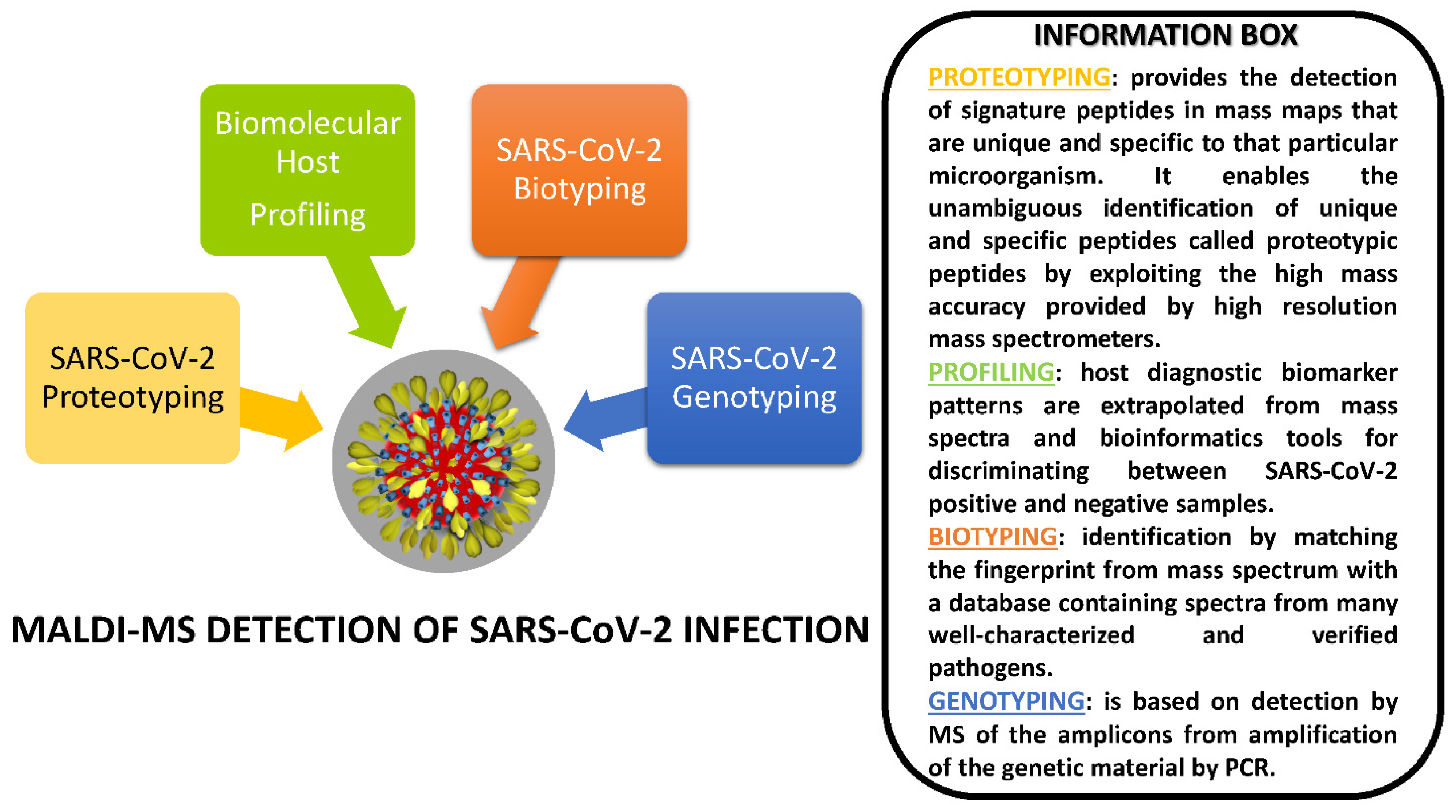
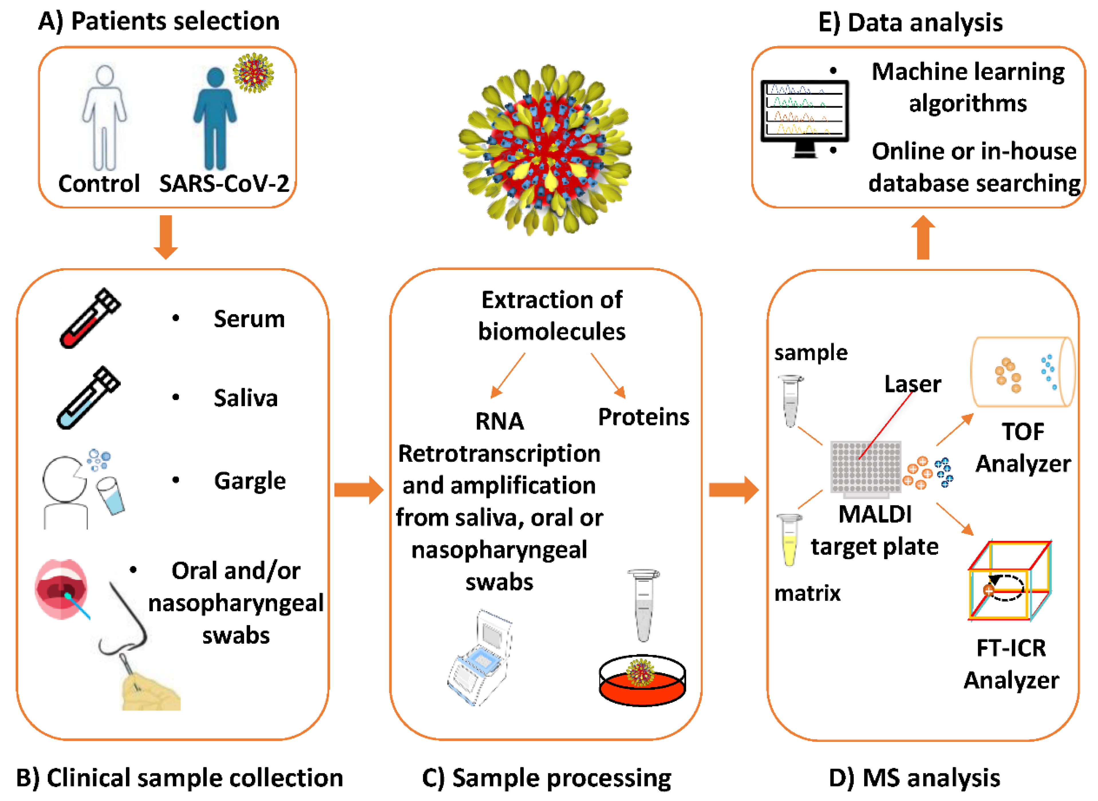
4. MALDI-MS Investigations Targeting SARS-CoV-2

In this section, we report the MALDI-MS-based investigations which aimed to detect SARS-CoV-2 infection with the use of “proteotyping” [74], “biotyping” [69,70], “genotyping” [71,72,73], and biomolecular host profiling [75,76,77,78,79] approaches. Interestingly, two of these investigations used both biotyping and biomolecular host profiling strategies [69,70].

4.1. Proteotyping Approach for SARS-CoV-2 Detection by MALDI-MS

Dollman and colleagues applied a proteotyping strategy for the detection of peptides unique to the SARS-CoV-2 by mass alone; they used High Resolution MALDI-FT-ICR Mass Spectrometry [74]. In particular, nasopharyngeal (NP) swabs from infected patients were cultured in Vero E6 cells or directly prepared before the MALDI-MS analysis. The use of a virus culture provided a more reliable approach to overcome potential false negatives deriving from specimens with low viral loads. The authors performed the MALDI-FT-ICR-MS analyses on the tryptic whole virus digest peptide products. In the case of cell-cultured SARS-CoV-2 samples with higher virus titers in comparison to other samples, twenty of the most prominent signals were confidently matched to fragments of the N, M, and S proteins with a high mass accuracy (<3 ppm) (Table 1). Detected peptides covered a large range of identified N- and C-terminal, fusion peptide receptor, and RNA-binding domains.
The acquired mass spectra from the non-cell-cultured NP samples showed peptide ions with reduced intensity and lower signal-to-noise ratio (S/N) as compared to those from experiments using cell-cultured samples; in this case, additional matrix cluster ions were also observed. However, despite the lower virus titers, the tryptic peptides of M, N, and S proteins were detected, including a new peptide at m/z 843.4255. In particular, six peptides were always detected in ten different NP samples, and five represent reliable signature peptides for SARS-CoV-2 proteotyping. In fact, due to the shared clinical symptoms presented by the SARS-CoV-2 with other circulating influenza viruses, the authors, by means of a specifically constructed algorithm and database, showed that these peptides were detected in all SARS-CoV-2 strains and exhibited a difference in mass greater than 3 ppm in comparison to those fragments detected in more than 95% of the cases from all influenza virus proteins of the circulating strains.
Table 2 reports the limit of detection (LOD) of the various analytical assays (including LOD related to conventional RT-PCR [93]) calculated based on the information from each of the authors, in order to make the comparison more straightforward for the readers. To determine the LOD of this MS platform, serial dilution experiments on the cell-cultured sample were performed, and the most abundant viral peptide signal at m/z 1635.8238 was monitored by MS. The peak was detected down to 0.75 ng, matching 7.5 × 10⁵ copies (Table 2).
This represents an excellent result, considering that a recent study reported maximum estimated levels of virus collected by NP swabs varying from 106 to 109 copies [94].
Interestingly, the authors stressed that this copy number is comparable with the 105–106 copies of virus necessary for high-quality PCR sequencing; however, as they additionally claimed, it is also higher than the 103–104 copies usually required for virus detection by PCR (Table 2). As underlined by the authors, the detection limits of 105 copies could be significantly improved when operating in an automated selected ion monitoring (SIM) strategy for the detection of only selected peptides for a proteotyping assay (Table 2). Furthermore, this MS approach does not require the separation of viral components, thus allowing for mass spectra acquisition from hundreds of digested virus samples in only a few minutes. The use of immobilized enzymes for proteolytic digestion as well as the implementation of an automated or semi-automated spectral acquisition method could drastically reduce processing and acquisition time analysis. Other advantages include better speed of the analysis as compared to LC-MS approaches and the possibility to confidently assign viral peptides by mass alone, without the use of MS/MS sequencing, which further reduces the time required for the analysis. For all these reasons, MS virus proteotyping should play a key role in molecular detection, not only for SARS-CoV-2 but also for other respiratory viruses and bio-pathogens. The high costs of the MS equipment, due to the high-resolution FT-ICR analyzer, make this MS-based proteotyping assay too expensive, although their costs are comparable to sequencers and PCR tools, as the same authors observed.

4.2. Biotyping Approach for SARS-CoV-2 Detection by MALDI-MS

With the aim of carefully and systematically analyzing all the challenging issues of an MS-based diagnostic process, Iles and colleagues developed a clinical MALDI-TOF MS assay for SARS-CoV-2 detection in a saliva or gargle solution [69]. The samples needed to be deactivated by UV-C irradiation, which preserves the viral membrane and proteins necessary for MS analysis. On the contrary, the use of heat for inactivating SARS-CoV-2 in NP samples as well as the use of SDS or Triton media for storage and transport could strongly interfere with MS analysis. Moreover, in comparison to NP or oropharyngeal swabs (OP), the saliva or gargle solution could be collected without causing as much discomfort to individuals. The authors performed several experiments in order to optimize the MALDI-TOF detection of viral protein and its proteolytic peptide detection. They finally developed a clinical diagnostic protocol based on (1) the collection of gargle/saliva samples; (2) the rapid processing of the gargle samples by filtration, acetone precipitation with subsequent pellet resuspension, and protein solubilization; (3) MALDI TOF analysis; (4) output data processing by an appropriate software (see Table 1).
The authors found that the use of ice-cold acetone for viral particle enrichment by precipitation was more advantageous over other organic solvents. The precipitation, followed by centrifugation, allowed for the enrichment of the pellet, with the enrichment of large virus particles and the removal of unwanted background proteins contained in the supernatant. Moreover, the use of ice-cold acetone also strongly contributed to virus inactivation.
Sinapinic acid (SA) was found to be more sensitive for the detection of virus-specific glycoproteins in comparison to alpha-cyano-4-hydroxycinnamic acid (CHCA), which is used for the routine detection of microbial intracellular proteins. Low resolution MALDI-TOF spectra were acquired in which S-protein subunits with their fragments, S1 (at ~m/z 79,000) and S2 (m/z from ~62,000 to ~72,000), were detected together with other putatively identified viral envelope protein fragments (m/z from ~26,000 to ~47,000). S1 peaks were higher in the SARS-CoV-2 virus culture spiked samples than in the volunteer samples, demonstrating that this marker alone could be an indicator of the identification of the coronavirus, with nearly 100% detection and specificity at 103–104 PFU SARS-CoV-2 virions in a gargle/saliva sample, which correspond to 10–102 copies (Table 2).
Interestingly, through the clinical information extrapolated by MALDI-TOF analysis, the authors demonstrated not only the possibility to detect S protein fragments, but also other peaks matching with Immunoglobulin light chains indicative of an oral upper respiratory immune response, and elevated levels of gargle/saliva IgA heavy chain peak indicative of a viral immune response.
These features reveal the diagnostic utility of MALDI-TOF MS as a powerful and economically ideal solution, with its ease of sampling and speed of analysis. However, other studies and validation tests on saliva/gargle spiked with cultured virus, or a direct comparison of the MALDI-TOF MS analysis with an RT-PCR detection of COVID-19 in clinical samples is required.
Starting from the results obtained by the study of Iles and coworkers, Chivte and colleagues analyzed gargle samples with MALDI-TOF MS to assess the presence of SARS-CoV-2 infection and compared the resulting spectra with the corresponding results of an RT-qPCR from NP swabs [70]. In particular, they analyzed 60 gargle samples (see Table 1) from volunteer student athletes, including 30 PCR positive and 30 PCR negative samples. Spectral clear differences were observed in the m/z range from 20,000 to 200,000. This extended m/z range allowed them to analyze the area under the curve (AUC) of putative viral and host proteins in order to assess the presence of SARS-CoV-2 infection. The receiver operating characteristic (ROC) analysis identified five peaks which showed highly sensitive and specific discrimination between COVID-19 positive and negative samples, including peaks which probably corresponded to the S1 and S2 fragments of the SARS-CoV-2 S protein, a potential biomarker near m/z 112,000, and peaks putatively assigned as human immunoglobulins or human α-amylase (Table 1 and Table 3). The putative identification of these peaks was assessed through a comparison with the UNIPROT database and from the work of Iles et al.; however, these results need to be further validated. For these biomarkers, an elevated concordance was achieved (90%) with the RT-PCR results, demonstrating that this methodology is a promising tool for a rapid and inexpensive COVID-19 assay. To evaluate the LOD of the MALDI-TOF protocol, they analyzed a saliva sample with a very low SARS-CoV-2 viral load by quantitative RT-qPCR. In particular, the Ct value of this sample was 36.09, which is less than the quantifiable limit of the assay (~30 copies) (Table 2). Interestingly, the authors observed a signal in the mass spectrum for the potential biomarker peak found between 78,600 and 80,500 m/z, demonstrating that the MALDI-TOF protocol could detect SARS-CoV-2 in samples containing very low viral loads.

4.3. Genotyping Approach for SARS-CoV-2 Detection by MALDI-MS

Wandernoth and colleagues evaluated the ability of MALDI-TOF MS to detect nucleic acid from SARS-CoV-2 in biological samples and compared the results to those of rRT-PCR [71]. In particular, they analyzed oral and NP swab samples from 22 patients who tested positive and 22 patients who tested negative by RT-PCR for SARS-CoV-2 infection (see Table 1). They used the new commercially available MS-based assay MassARRAY® (Agena Bioscience®, San Diego, CA, USA), a genotyping panel for the detection of SARS-CoV-2 developed by Agena Bioscience, which received the CE-IVD mark in Europe for the detection of nucleic acid from SARS-CoV-2 in respiratory specimens. This platform provides a robust route for the detection of the SARS-CoV-2 virus in human samples. It consists in a four-step process represented by a one-step RT-PCR reaction to reverse transcribe viral RNA into cDNA, followed by the amplification of the obtained nucleic acid material, primer extension, and the MALDI-TOF analysis of the products on a matrix-loaded silicon chip array. It allowed for the detection of five SARS-CoV-2 specific targets: three in the nucleocapsid region and two in the ORF1ab region. The results of the rRT-PCR and MALDI-TOF MS analyses were comparable in all samples. The LOD was estimated at 10 genome copies (Table 2). Interestingly, time-to-results was faster for rRT-PCR, while hands-on time was comparable between the rRT-PCR and MALDI-TOF techniques. Very recently, Rybicka and colleagues [72] also tested the new commercially available MS-based assay MassARRAY® SARS-CoV-2 Panel (Agena Bioscience) and compared it to the RT-PCR diagnostic test. Oral and NP swabs from 168 suspected COVID-19 patients with symptoms of respiratory infection were simultaneously processed with both assays (Table 1). Among the 168 analyzed samples, different results were obtained for 10.12% (17 samples). In particular, in four samples considered negative by RT-PCR, viral genes were detected by the MS-based method. Furthermore, 87% of patients previously considered as suspected cases by RT-PCR resulted unambiguously positive in all MS assays. An estimated LOD of the assay was 10 copies (Table 2). Using the MassARRAY® SARS-CoV-2 Panel, the authors were able to detect SARS-CoV-2 in low viral load specimens and with very few microliters of viral RNA. The authors emphasized that their data analysis is in agreement with other studies, reporting false-negative results from RT-PCR of about 30%, and that MALDI-TOF MS seems to be an ideal tool for the detection and discrimination of mutations. They pointed out that the MassARRAY® System provides automation, minimal hands-on time, and onboard data analysis, delivering easy-to-interpret data with a simple and fast workflow.
Study limitations may arise from the lack of information about isolated RNA concentration as well as from the inability to reanalyze suspected COVID-19 patients in order to evaluate the progress of infection.
Hernandez and colleagues evaluated the ability of two different technologies to detect SARS-CoV-2 in saliva samples. In particular, they compared the already mentioned RT-PCR/MALDI-TOF MS-based assay (AGENA MASS ARRAY) with the cobas® 6800/8800 SARS-CoV-2 real-time RT-PCR (Roche, Basel, Switzerland) for conventional real time RT-PCR [73]. They collected saliva samples from 60 patients, which were previously subjected to molecular testing for SARS-CoV-2 in NP or anterior nare specimens. The Agena MassARRAY® platform allowed for the detection of viral targets: three in the nucleocapsid (N) gene (N1, N2, N3) and two in the Orf1ab gene (ORF1, Orf1ab) (Table 1). However, only two targets were detected by using the cobas® 6800/8800 SARS-CoV-2 real-time RT-PCR (Roche): the SARS-CoV-2-specific Orf1ab gene (T1) and the pan-Sarbecovirus envelope E gene (T2).
Interestingly, SARS-CoV-2 detection in saliva samples by the AGENA system showed high sensitivity and specificity (Table 1) when compared to RT-PCR results from NP or anterior nare specimens. Analogue sensitivity and specificity were obtained by cobas® 6800/8800 SARS-CoV-2 real-time RT-PCR (Roche). Among the Agena targets, the most sensitive were the N2 target (103 copies) (Table 2), followed by the N1 target.
The Roche platform showed a higher sensitivity in comparison to the Agena platform, having achieved a lower LOD (390.6 copies/mL).
These data demonstrate that saliva constitutes an appropriate matrix for SARS-CoV-2 detection on the novel Agena system, with a comparable performance to the more ubiquitous real-time RT-PCR technology. As stated by the same authors, one limitation of this study is the lack of a standardized collection method, because saliva samples were collected randomly within two days of initial NP and anterior nare collection, and not at one time point.

4.4. Biomolecular Host Profiling Approach for SARS-CoV-2 Detection by MALDI-MS

In this section, we report on MALDI-MS studies performed on a variety of biological specimens in which host diagnostic biomarker patterns had been extrapolated rather than direct SARS-CoV-2 detection [75,76,77,78,79]. The investigations by Iles and colleagues [69] and by Chivte and colleagues [70], which assessed the presence of both viral and host proteins, were already reported in the previous section (see Table 1 and Table 3). Many of these investigations used oral or respiratory samples [75,76,77,78], which are the commonly accepted specimens for SARS-CoV-2 detection; interestingly, one such study was conducted using serum samples to decipher the molecular changes induced by the systemic effects after infection [79]. In these investigations, MALDI-TOF readouts were processed by machine learning (ML) techniques (Table 1) to extrapolate novel or unknown diagnostic information from a mass spectra dataset. Additionally, the robustness and clinical significance of the ML models employed were assessed.
Nachtigall et al. described a method of detecting SARS-CoV-2 in NP swabs using the MALDI-TOF MS and ML analyses [75]. They proposed this approach starting from the rationale that several clinical laboratories use MALDI-MS as a high-throughput, cost-effective, and robust technology for the conventional diagnosis of pathogen infections.
They analyzed a total of 362 NP swab samples, 211 RT-PCR positives and 151 RT-PCR negatives, from subjects coming from three different countries (Argentina, Chile, and Peru) (Table 1). The biological samples were placed on the MALDI target plate without prior sample purification.
They established a mass analysis range between 3 and 15.5 kDa and applied a two-tailed Wilcoxon rank sum test, allowing for the identification of 31 m/z peaks that could differentiate SARS-CoV-2 positive from negative samples (none of these were identified). Seven of these were the most relevant peaks at m/z 3095, 3152, 3337, 3358, 4532, 7612, and 10,444 (in particular, the peak at m/z 7612 was common to all the samples from the different laboratories) (see also Table 3).
Extracted m/z features that discriminate between SARS-CoV-2 positive and negative samples were then used for principal component analysis (PCA). The PCA provided only partial data separation when samples from the three different countries were analyzed all together; on the contrary, complete data separation was achieved on samples from each country independently. Therefore, more advanced methods are needed to better discriminate SARS-CoV-2-positive and negative samples.
Next, the authors tested different feature selection methods and ML approaches to determine the top performing analysis strategies. Feature selection methods can be used in data pre-processing to obtain efficient data reduction, a useful step in order to identify accurate data models [95]. In particular, they applied feature selection methods with six different ML algorithms in a smaller set of 80 SARS-CoV-2-positive and negative samples. The resulting data demonstrated that although the support vector machine model (SVM-R) with no feature selection was the best method for SARS-CoV-2 detection, model accuracy did not vary substantially among the ML methods. Finally, Nachtigall and colleagues tested the SVM-R ML algorithm, with and without the feature selection methods, on all 362 samples from all three countries. This ML algorithm with no feature selection method allowed for the discrimination of the control group from patients with COVID-19 and reached high sensitivity and specificity levels (Table 1). Through a comparison of the results obtained from RT–PCR and MALDI-TOF coupled with ML, the authors found a concordance rate that was acceptable as a clinical diagnostic approach (>80%), confirming that the MALDI-MS and ML analyses represent promising alternatives as fast screening assays for SARS-CoV-2.
Tran and colleagues described as proof-of-concept a novel, automated, ML-enhanced MALDI-TOF MS approach for analyzing nasal swabs from patients with suspected COVID-19 [76].
The authors evaluated the performance of the MALDI-TOF MS method using SARS-CoV-2 RNA PCR positive and negative samples in order to determine the accuracy, the positive percent agreement (PPA), and the negative percent agreement (NPA) of the MALDI-TOF MS method as compared to the PCR method. They collected 226 nasal swab samples that were analyzed both by RT-PCR and by MALDI-TOF MS. Normalized mass spectra were subjected to ML analysis by the Auto-ML MILO (Machine Intelligence Learning Optimizer) platform [96]. The MILO as ML platform consists in a series of algorithms, scalers, scorers, and feature selectors/transformers, which are used to generate models that are then statistically assessed to ultimately identify the best performing model for a specific purpose. Briefly, its infrastructure consists of two datasets: the balanced dataset used for training and validation, and an unbalanced dataset for generalization/secondary testing. Finally, the selected model is deployed and used to test new data and make predictions. Among the 226 nasal swab samples, 27 were eliminated due to polymer contamination of the sample. Overall, 199 samples were analyzed by both MALDI-TOF MS and RT-PCR; out of these, 107 samples were COVID-19 positive (28 asymptomatic and 79 symptomatic) while 92 were COVID-19 negative (Table 1). The data related to MALDI-TOF spectra obtained from these 199 samples were divided into Datasets A and B, with dataset A used as a training and initial validation dataset (which included 42 COVID-19 negative cases and 40 COVID-19 positive ones). Optimized models obtained from Dataset A were then tested with dataset B, used as generalization test set to assess their true performance (constituted by 50 negative cases and 67 positive cases).
MILO produced a total of 379,269 models, two of which—DNN and GBM—showed high performance characteristics (Table 1). The authors described their ML-enhanced MALDI-TOF approach as an attempt to address both throughput and speed limitations observed in molecular platforms; interestingly, the authors demonstrated the ability of both PCA and ML models to classify MALDI-TOF mass spectra for discriminating positive and negative COVID-19 cases.
The high-throughput performance of this platform was also demonstrated by the short time required for the analysis (total turnaround time was less than 1 h, with the potential of performing up to 1104 analyses per day, per instrument) as compared to commercial RT-PCR platforms, which need, for example, batch testing for optimal reagent use.
Another investigation that described the analysis of NP samples by MALDI-TOF MS coupled with ML was performed by Deulofeu and colleagues [77]. In particular, they used different ML approaches for the analysis of the MALDI-TOF mass spectra from 237 NP samples in order to identify a signature characterizing negative and positive samples for SARS-CoV-2 infection (Table 1). These samples were analyzed by both RT-PCR and MALDI-TOF MS. The mass spectra obtained were used to build different ML models in order to identify the best model that would be able to distinguish the positive COVID-19 cases from negative ones. All the parameters of a MALDI-TOF MS approach, including the matrix, sample dilution, and mass range, were optimized. Interestingly, the effect of the use of different viral transport media on the end results was also investigated. MS data analyzed by the ML models were able to identify the more appropriate viral transport media for the detection of SARS-CoV-2 infected samples. In particular, the best results were achieved using the SVM model, which obtained elevated levels of accuracy, sensitivity, and specificity (Table 1). These data demonstrated the utility of the developed method for the detection of SARS-CoV-2-positive samples and the simplicity, safety, rapidity, and cheapness of MALDI-TOF MS as a diagnostic test. The authors estimated the cost of MALDI-TOF MS to be 25% more than the cost of the RT-PCR analyses. They also stated that the time required to receive results is around 6 h for a conventional RT-PCR, whereas the analyses of the NP samples by MALDI-TOF MS take less than one-third of this (Table 1).
Finally, Rocca and colleagues also investigated the potential role of MALDI-TOF MS combined with ML methods in the identification and the discrimination between COVID-19 positive samples and COVID-19 negative ones [78].
They used NP swab samples from 311 patients that were analyzed by MALDI-TOF MS analysis without processing (Table 1). They used 20 main spectrum profiles obtained from nine COVID-19 positive samples, eight COVID-19 negative samples, and three positive samples for other respiratory viruses to create a new “in-house” database (named BE COVID-19). Using this database, they searched for potential discriminatory peaks that could differentiate positive from negative samples, using two different software (Flex analysis v3.4 and ClinPro tools, Bruker Daltonik GmbH, Bremen, Germany). From the evaluation of parameters and statistical analysis, six peaks at m/z 3372, 3442, 3465, 3488, 6347, and 10,836 demonstrated a decrease or an absence of COVID-19 positive samples and were considered as potential biomarkers (Table 3).
The authors applied ML algorithms and used 432 spectra (obtained from 55 COVID-19 positive samples, 57 COVID-19 negative samples, 24 samples positive for the influenza virus, and 8 other respiratory virus samples) as a training set for the generation of three classification models; to analyze the performance of this approach based on ML algorithms with the combination of potential biomarkers, a validation test was performed on 501 spectra (167 clinical samples), obtaining an accuracy of 67.66%, a sensitivity of 61.76%, and a specificity of 71.72%.
These preliminary results demonstrated that the method still needs to be improved, and the authors stated that further studies on a larger cohort of patients, evaluating different extraction methods and improving ML algorithms, will be performed. An important weakness of this study might be the limited number of samples used to build the “in-house” database. In fact, a larger number of clinical samples are required to obtain a reliable spectrum profile library, in order to reduce the inter-individual variability effects and to determine specific discriminatory peaks with more confidence.
Yan et al. utilized a high-throughput serum peptidome profiling method based on MALDI-TOF MS for the detection of COVID-19 [79]. They analyzed serum samples from a total of 298 individuals: 146 COVID-19 patients (classified into mild, typical, severe, and critical) and 152 control patients (including 73 non-COVID-19 patients with similar clinical symptoms such as fever/cough, 33 tuberculosis patients, and 46 healthy individuals) (Table 1).
After sterilization, the serum samples were profiled by MALDI-TOF MS. The MALDI-TOF data of COVID-19 patients and control samples were randomly split into training cohorts, with 198 samples (101 controls and 97 patients), and validation cohorts, with 100 samples (51 controls and 49 patients). Using ML methods, twenty-five peaks were identified as the distinctive features between COVID-19 patients and control participants, with statistically significant differences (Table 3). Most of the peaks were located in the range of m/z 5000–30,000. Distinctive features were observed: e.g., m/z = 6357, 6654, 6639, 13,886, and 28,232 were significantly down-regulated in the COVID-19 group, while m/z = 7614, 15,123, 15,867, and 28,091 were significantly up-regulated in the COVID-19 group (Table 3). The classification model for the detection of COVID 19 was constructed with these 25 feature peaks in the training cohort by using eight ML methods.
The ROC curves and AUC were evaluated for each of the ML models. Later, the efficiency of these models was assessed in the validation cohort, which was independent from the samples included in the training cohort. The Principal Component Analysis (PCA) of the 25 features demonstrated that the COVID-19 patients and control cases in the test group were well-separated. Among all the models, the LR model demonstrated the best classification performance and it was considered the most recommended by the authors for future applications in the detection of COVID-19, with the highest accuracy (99%), sensitivity (98%), precision (100%), and specificity (100%) (Table 1).
Starting from the observation that the symptoms of SARS-CoV-2 infected patients are frequently shared by patients affected by other respiratory infections, the authors highlighted that this could lead to the misinterpretation of results; so they emphasized the ability of this platform to discriminate COVID-19 patients from non-COVID-19 subjects with similar clinical symptoms. The authors concluded that these results also demonstrated that MALDI-TOF-based serum profiling could be a powerful tool for screening, routine surveillance, and diagnosis of COVID-19 in populations. However, it is important to note that the sampling time of the COVID-19 serum samples analyzed ranged from 3 to 28 days from the onset of symptoms, which consists in a relatively long period of disease progression. This condition could represent one potential limitation of this study, as a screening method should be able to detect the virus infection as early as possible in order to avoid an uncontrolled viral spread and to stop the pandemic.

5. Discussion

5.1. Comparison of MALDI-MS vs. RT-PCR Techniques for SARS-CoV-2 Detection: Advantages and Limitations

As a resource for new, alternative and/or complementary, rapid COVID-19 diagnostic tests, this review explored the recent developments and applications of MALDI MS-based technologies for the accurate and unambiguous SARS-CoV-2 clinical diagnosis, which may help to control the COVID-19 pandemic.
From the perspective of a high-throughput, routine, diagnostic clinical setting, a viral infection diagnostic tool should consist of an integrated analytical platform, which needs to be fast, sensitive, highly specific, reproducible, and cost-effective. MS techniques have rapidly complemented or replaced conventional methods in clinical diagnostic laboratories and have been successfully implemented in the investigation of viruses and their pathogenesis [17,21,22,23,24,25,26,27,97,98,99]. In particular, MALDI-MS has become an indispensable and versatile method for biochemical and clinical investigations [100,101]. MALDI-MS represents one such technology, characterized by its ease of use, high-throughput capabilities, and cost effectiveness, thus becoming a routine tool in clinical microbiology laboratories and also proving to be capable of supporting clinical decision-making [36,64].
However, unlike what happens with bacteria, neither MS nor MALDI-MS currently have a low impact on virus detection in the routine diagnostics of clinical samples [102]. In fact, the approach used for biotyping is not well-suited for the identification of viruses for some reasons. First of all, viruses cannot be isolated by simple methods, unlike bacteria. As a matter of fact, bacteria are routinely grown and isolated from colonies, while viruses can be found in a complex cellular background, making their detection more difficult. Hence, the sensitivity is severely limited by the dynamic range of the MS instrument used. A second reason is that bacteria contain significantly more proteins than viruses, with poor signals in the m/z below 12,000, the range covered by whole-cell MALDI-TOF MS. Hence, the interpretation of low-resolution mass spectra made up of few viral peaks in a complex human background is more challenging.
As a matter of choice, the clinical sensitivity and specificity of each diagnostic tool should be evaluated in various clinically relevant real-life situations (for example, the viral load, the site and the quality of sample collection, timing, and illness severity). The current gold standard technique for the molecular diagnosis of SARS-CoV-2 infection is the RT-qPCR, which allows for the analysis of thousands of samples in a single day and shows a high testing sensitivity of 95% [103] and a low limit of detection of <10 copies/reaction [104] under ideal circumstances. Nevertheless, several investigations have reported that clinically evident COVID-19 infections often go undetected by SARS-CoV-2 RT-PCR testing [10,105,106,107,108]; the evidence that virus shedding can occur at undetectable levels during the very early and late stages of SARS-CoV-2 infection demonstrates that RT-PCR results should always be interpreted in a wider context. Indeed, the most efficient strategy for the diagnosis of SARS-CoV-2 infection in suspected patients should encompass a combination of SARS-CoV-2 detection by RT-PCR with clinical and epidemiologic observations (symptoms, previous exposure to virus, negative diagnostic tests for other respiratory diseases), whereas additional follow-up testing with RT-PCR should be required from patients with initially negative results and high suspicion of COVID-19.
The time required for conventional PCR can vary from about 4–6 h for sample processing (including sample preparation, RNA extraction, reverse transcription PCR, and readout of amplified DNA products), up to a few days, considering the time needed for the eventual transport of the specimen to the laboratory.
Most of the reports described here that are related to Biotyping, Proteotyping, and Genotyping approaches are proof-of-principle studies showing the feasibility of viral protein or viral nucleic acid detection by MALDI-MS, which often do not involve stress testing in low viral load samples. Nevertheless, as reported in Table 1 and Table 2, the specificity, sensitivity, and the detection limit of the SARS-CoV-2 virus by MALDI-MS diagnostic methods were high and comparable to reference methods such as PCR.
The MALDI-MS detection of a virus has a fast turnaround time from sample collection to diagnosis (Table 1). The time required is mainly encumbered by the procedures needed for sample processing, considering that spectral acquisition could be performed in an automated or semiautomated manner.
Furthermore, MALDI-MS analysis is cost-effective despite the initial investment in expensive equipment. In fact, especially in the case of ultra-high-resolution MS, the purchase of the mass spectrometer instrument accounts for the major cost; however, it is a one-time cost and not inconsistent with the costs required for multiple PCR sequencers and associated specialized reagents and equipment [74].

5.2. Pre-Analytical and Analytical Issues for Molecular Detection of SARS-CoV-2

As a rule, the reliability of each diagnostic tool is influenced by several factors which could reduce the sensitivity and specificity of the obtained results, and which have to be estimated to avoid erroneous interpretations by the clinical laboratories [16,109]. There are several critical issues and challenges affecting the diagnosis of SARS-CoV-2 infections, including sampling operations, specimen source, and sampling time, which could have an impact on the end result. It is essential to handle bodily fluids according to standardized procedures in order to distinguish reliable molecular biomarkers and to assess the influence of pre-analytical parameters on the final result in order to avoid artifacts. As revealed by the literature reviewed in the previous sections, there is high variability in all of the abovementioned critical technical factors; for example, most studies do not clarify if the tested samples were analyzed by MALDI-MS immediately after collection or if they had been deep-frozen until analysis. This represents an important limitation, considering that several pre-analytical and analytical variables, including storage time and temperature, could alter the analysis of the MS profile of biological specimens, and that several studies demonstrated that the proteins could undergo degradation processes [51,110,111,112,113]. Thus, especially for the biomolecular host profiling studies, it is not possible to exclude the fact that the observed differences in the molecular fingerprints obtained by MALDI-MS could derive from the degradation of the molecular species due to the action of endo-proteases, including those of microbial origin in the nasal environment, or from the inherent biological variability among subjects, also due to genetic factors affecting the phenotype. Worthy of note is the fact that most studies do not clarify if the tested samples were collected at the time of symptom onset rather than time of exposure, disregarding another important issue which could lead to high variance in the detection of SARS-CoV-2 infection in the first few days after virus exposure. The success of an accurate diagnostic procedure mainly depends on the collection of proper specimens at the right time of infection, and from the appropriate anatomic location/organ.

5.3. Detection of SARS-CoV-2 in Different Types of Clinical Specimens

Respiratory sampling is the preferred and widely approved method for SARS-CoV-2 detection by RT-qPCR. The choice of optimal clinical specimens takes into consideration several factors such as ease of accessibility, non-invasive collection procedure, a lower risk to health care professionals during sample collection, and good viral loads for higher possibilities of detection. NP and/or OP swabs are best recommended for the screening or diagnosis of an early infection, when low viral loads are known to occur and false-negative results can derive from differences in analytical sensitivity among methods [16,114]. For these reasons, most of the studies reviewed here are based on the use of NP or OP samples [71,72,74,75,76,77,78]. Interestingly, nasal swab samples were directly applied to MALDI-MS, and spectral readouts were used to identify infected or non-infected subjects; consequently, non-purified viral samples were used and any virus would be markedly diluted [71,72,74,75,76,77,78]. These conditions have allowed for a higher viral load that not only facilitates the detection by MS but also reduces the time needed for the experimental procedure and analysis. On the other hand, NP or OP swabs show limitations in sample collection and present a risk to medical staff through sneezing or coughing, which can lead to aerosol exposure of viral particles [115].
An alternative option for collecting upper respiratory tract specimens in order to detect SARS-CoV-2 is self-collected saliva or gargle specimen [93]. Saliva is a reliable biological fluid that can easily be provided by the patient; it does not require healthcare personnel in a screening program [93,116]. In fact, a mass screening program should rely on devices that can be used by non-medical staff to promptly assess whether an individual is positive to COVID-19 [117]. Moreover, a gargle sample is obtained from an easy-to-perform procedure and is comfortable for the patient. Reliable and rapid diagnostic techniques based on the use of saliva or gargle specimens might represent a cost-saving procedure considering that their collection does not require the need to develop and maintain specific infrastructure for swab collection as well as the need for dedicated healthcare personnel. The studies reviewed here, which reported the use of saliva/gargle specimens, analyzed the feasibility of these biological fluids as a diagnostic sample for detecting SARS-CoV-2 infection [69,70,73]. It is worth noting that compared to swab-based diagnostics, several studies majorly revealed a higher viral load in NP than in saliva specimens [118], probably due to dilution effects. However, it should be noted that the possibilities to detect SARS-CoV-2 infection from saliva, gargle, NP, or OP specimens could decrease with the time since the onset of symptoms, leading to a higher risk of false-negative results [16]. In fact, in later infection, the main site of replication could shift to the low respiratory tract, and repeated testing or lower respiratory tract specimens may represent the alternative choice [115,119].
Serum samples could be used to understand the molecular mechanism for the diagnosis of COVID-19. As a consequence of viral infection, serum may contain mediators produced by the systemic effects or released to the lung, and this could reflect the physiological or pathological state [30].
Serum samples, and more generally, serological methods, could play a key role in the epidemiology of COVID-19 and in defining the immune status of asymptomatic patients, but they are not recommended for the screening or diagnosis of early infections [120]. However, serology could contribute to the confirmation of the diagnosis of COVID-19 by providing information about the type and the concentration of various immunoglobulins generated after SARS-CoV-2 infection [121].

5.4. Asymptomatic Infection and the Spread of New SARS-CoV-2 Variants

Another critical issue is the limited resources employed to facilitate and enable the widespread rapid screening of asymptomatic individuals necessary to control the potential of silent chains of virus transmission. Asymptomatic patients are the carriers of the live, replicating virus and contribute to the unknown viral transmission and spread. In some cases, the methods used for SARS-CoV-2 detection in these subjects can be less sensitive than in the symptomatic ones [109,122,123]. It is worth noting that only a limited number of studies reviewed here have considered the asymptomatic population [70,76]. However, they demonstrated that the MALDI-TOF MS platform was able to detect SARS-CoV-2 infection in asymptomatic individuals, and with elevated accuracy [76] (see Table 1), which could be of paramount importance for the reopening of businesses, recreational facilities, schools, and all non-hospital settings.
In addition to asymptomatic infection, SARS-CoV-2 variants are another challenge for virus detection [123,124]. In fact, new variants caused by mutations in the genome of the SARS-CoV-2 are now emerging as the virus spreads throughout the world, and this is of great relevance considering that such mutations may alter various aspects of virus biology such as pathogenicity, infectivity, transmissibility, and/or antigenicity [125]. Nowadays, the mutations and evolution of SARS-CoV-2 are mainly traced by next generation sequencing, but routine genomic testing is expensive and difficult to perform in real time due to the lack of resources and expertise in many areas [126,127]. Additionally, emerging variants raise the risk of target dropouts and false-negative results due to primer/probe binding site mismatches. Therefore, alternative and/or complementary methods of rapidly detecting and monitoring the evolution of virus strains are of vital importance.
Very recently, some studies employed MALDI-MS to study and distinguish strains of the major variants of concern using mass signatures [124,128,129]. These studies also demonstrate how such MALDI-MS data can be used to chart viral evolution and to construct mass-based phylogenetic trees, similar to those obtained using conventional gene sequence phylogenetics. The use of MS datasets to track the evolution of SARS-CoV-2 in order to identify such mutations that may limit detection by PCR or those that allow immune escape or reduce the efficacy of existing vaccines and/or therapies have great future potential.

6. Conclusions

Laboratory SARS-CoV-2 testing assays are the primary mitigation measure for the prompt identification and isolation of infected individuals. In fact, the rapid and accurate detection of SARS-CoV-2 infection and the close monitoring of persons in contact with cases are key steps in managing the COVID-19 pandemic, until vaccines can be extensively administered or until antiviral drugs become widely available. In this context, MS-based technologies may represent an alternative resource or may support PCR-based methods as a diagnostic test in the fight against the novel coronavirus. To date, MALDI-MS is routinely used in clinical practice for the rapid, unambiguous, and cost-effective identification of pathogens in biological samples. It is worth noting that several MS-laboratories around the world have been working to deliver guidelines and protocols on sample collection, processing, and data analysis in order to develop rapid and robust methods for the high-throughput screening of SARS-CoV-2 infection. Furthermore, clinical trials using MALDI-MS are currently ongoing for the management of the COVID-19 pandemic [130,131]. In particular, one of these studies is based on MALDI-TOF MS profiling and combined with ML methods to detect individuals infected with SARS-CoV-2 from saliva samples, while the other one is based on the MALDI-TOF MS analysis of both saliva and blood samples to search for discriminating profiles between COVID-19 and non-COVID-19 individuals [130,131]. Nevertheless, some limitations still exist for the applications of MALDI-MS technology in routine virus diagnostics. In fact, unlike what happens for bacteria, in the case of biotyping approaches, the major limitations could arise from the relatively low protein content of viruses, the higher molecular weight of viral proteins (>20,000 Da), and a probable carryover of debris from the cell substrate in which the viruses are cultured in vitro.
On the other hand, despite the high costs of the instrument, the high-resolution MALDI MS with proteotyping strategies, based on SARS-CoV-2 signature peptides, has demonstrated excellent performance characteristics especially for the high-accuracy detection of SARS-CoV-2. Considering high accuracy, high throughput, and speed of analysis, the use of more innovative MS-based tools and assays represents an essential and feasible complement to PCR.
MALDI-MS-based genotyping approaches also enable the high-throughput and low-cost detection of SARS-CoV-2. Nevertheless, the level of accuracy of these approaches is not yet comparable to those of proteotyping strategies. In any case, conventional tests such as PCR or immunoassay have become insufficient, also considering the shortages of medium, reagents, collection devices, and consumables that hinder day-to-day laboratory operations and the ability to improve testing capacity. In this scenario, MALDI-MS-based genotyping strategies could represent reliable diagnostic techniques that require alternative consumables. Moreover, recent investigations have demonstrated the ability of these platforms to assess unfolding genomic variation in a timely manner, and have highlighted the potential of diagnostic results to serve as a reliable system for the detection of emergent SARS-CoV-2 variants of concern [128,129].
Finally, regarding the proof-of-principle studies, the biomolecular host profiling approaches that assess host response to predict infection are a further intriguing alternative to the current testing methods for the identification of SARS-CoV-2 infected individuals. However, viral load differs among infected individuals, in particular between symptomatic and asymptomatic subjects, which leads to a broad variation in host response. Therefore, these approaches warrant further investigations that would test their threshold for the detection of viral infection and their ability to differentiate types of infectious or non-infectious causes of host modulation, in order to avoid confounding results. Additionally, these strategies also have multiple desirable features for a clinical test during the COVID-19 pandemic, which includes minimal sample preparation, few reagents, and rapid and high-throughput data acquisition.
Arguably, as part of a pandemic preparedness plan, a massive amount of novel scientific knowledge as well as the use of innovative MS-based tools and assays will support the development of diagnostics facilitating a reliable response to emerging infectious diseases.
Certainly, the expanded application of MS-based technologies and related multi-omics strategies could become one of the reference methodologies for other future epidemics/pandemics that may arise.

Author Contributions

Conceptualization, R.T.; writing—original draft preparation, M.P. and R.T.; writing—reviewing and editing, M.P., R.S., C.P. and R.T.; preparation of figures, R.T. and S.C.; preparation of tables, M.P., R.S., R.T. and S.C.; supervision, R.S. and R.T. All authors have read and agreed to the published version of the manuscript.

Funding

S.C. is supported by a fellowship from the Ph.D. Program in Life Sciences (XXXVI cycle).

Informed Consent Statement

Not applicable.

Data Availability Statement

Not applicable.

Conflicts of Interest

The authors declare no conflict of interest.

References

  1. Coronavirus Update (Live): 92,895,303 Cases and 1,989,457 Deaths from COVID-19 Virus Pandemic—Worldometer. Available online: https://www.worldometers.info/coronavirus/ (accessed on 21 October 2021).
  2. Peiris, J.S.; Yuen, K.Y.; Osterhaus, A.D.; Stöhr, K. The severe acute respiratory syndrome. N. Engl. J. Med. 2003, 349, 2431–2441. [Google Scholar] [CrossRef] [Green Version]
  3. Muller, C.P. Do asymptomatic carriers of SARS-COV-2 transmit the virus? Lancet Reg. Health Eur. 2021, 4, 100082. [Google Scholar] [CrossRef]
  4. Johansson, M.A.; Quandelacy, T.M.; Kada, S.; Prasad, P.V.; Steele, M.; Brooks, J.T.; Slayton, R.B.; Biggerstaff, M.; Butler, J.C. SARS-CoV-2 Transmission From People Without COVID-19 Symptoms. JAMA Netw. Open 2021, 4, e2035057. [Google Scholar] [CrossRef] [PubMed]
  5. Atripaldi, L.; Sale, S.; Capone, M.; Montesarchio, V.; Parrella, R.; Botti, G.; Ascierto, P.A.; Madonna, G. Could asymptomatic carriers spread the SARS-CoV-2 infection? Experience from the Italian second wave. J. Transl. Med. 2021, 19, 93. [Google Scholar] [CrossRef]
  6. SeyedAlinaghi, S.; Mirzapour, P.; Dadras, O.; Pashaei, Z.; Karimi, A.; MohsseniPour, M.; Soleymanzadeh, M.; Barzegary, A.; Afsahi, A.M.; Vahedi, F.; et al. Characterization of SARS-CoV-2 diferent variants and related morbidity and mortality: A systematic review. Eur. J. Med. Res. 2021, 26, 51. [Google Scholar] [CrossRef]
  7. Becker, M.; Dulovic, A.; Junker, D.; Ruetalo, N.; Kaiser, P.D.; Pinilla, Y.T.; Heinzel, C.; Haering, J.; Traenkle, B.; Wagner, T.R.; et al. Immune response to SARS-CoV-2 variants of concern in vaccinated individuals. Nat. Commun. 2021, 12, 3109. [Google Scholar] [CrossRef]
  8. D’Cruz, R.J.; Currier, A.W.; Sampson, V.B. Laboratory Testing Methods for Novel Severe Acute Respiratory Syndrome-Coronavirus-2 (SARS-CoV-2). Front. Cell Dev. Biol. 2020, 8, 468. [Google Scholar] [CrossRef] [PubMed]
  9. Kucirka, L.M.; Lauer, S.A.; Laeyendecker, O.; Boon, D.; Lessler, J. Variation in False-Negative Rate of Reverse Transcriptase Polymerase Chain Reaction–Based SARS-CoV-2 Tests by Time Since Exposure. Ann. Intern. Med. 2020, 173, 262–267. [Google Scholar] [CrossRef] [PubMed]
  10. Arevalo-Rodriguez, I.; Buitrago-Garcia, D.; Simancas-Racines, D.; Zambrano-Achig, P.; Del Campo, R.; Ciapponi, A.; Sued, O.; Martinez-García, L.; Rutjes, A.W.; Low, N.; et al. False-negative results of initial RT-PCR assays for COVID-19: A systematic review. PLoS ONE 2020, 15, e0242958. [Google Scholar] [CrossRef]
  11. Tahamtan, A.; Ardebili, A. Real-time RT-PCR in COVID-19 detection: Issues affecting the results. Expert. Rev. Mol. Diagn. 2020, 20, 453–454. [Google Scholar] [CrossRef] [Green Version]
  12. Rosebrock, A.P. DNA Cross-Reactivity of the CDC-Specified SARS-CoV-2 Specimen Control Leads to Potential for False Negatives and Underreporting of Viral Infection. Clin. Chem. 2020, 67, 435–437. [Google Scholar] [CrossRef]
  13. Kanji, J.N.; Zelyas, N.; MacDonald, C.; Pabbaraju, K.; Khan, M.N.; Prasad, A.; Hu, J.; Diggle, M.; Berenger, B.M.; Tipples, G. False negative rate of COVID-19 PCR testing: A discordant testing analysis. Virol. J. 2021, 18, 1–6. [Google Scholar] [CrossRef]
  14. Huggett, J.F.; Benes, V.; Bustin, S.A.; Garson, J.A.; Harris, K.; Kammel, M.; Kubista, M.; McHugh, T.D.; Moran-Gilad, J.; Nolan, T.; et al. Cautionary Note on Contamination of Reagents Used for Molecular Detection of SARS-CoV-2. Clin. Chem. 2020, 66, 1369–1372. [Google Scholar] [CrossRef]
  15. Wan, Z.; Zhao, Y.; Lu, R.; Dong, Y.; Zhang, C. Rapid antigen detection alone may not be sufficient for early diagnosis and/or mass screening of COVID-19. J. Med. Virol. 2021, 93, 6462–6464. [Google Scholar] [CrossRef]
  16. Tang, Y.-W.; Schmitz, J.E.; Persing, D.H.; Stratton, C.W. Laboratory Diagnosis of COVID-19 Infection: Current Issues and Challenges. J. Clin. Microbiol. 2020, 58, e00512–e00520. [Google Scholar] [CrossRef] [PubMed] [Green Version]
  17. Terracciano, R.; Preianò, M.; Fregola, A.; Pelaia, C.; Montalcini, T.; Savino, R. Mapping the SARS-CoV-2-Host Protein-Protein Interactome by Affinity Purification Mass Spectrometry and Proximity-Dependent Biotin Labeling: A Rational and Straightforward Route to Discover Host-Directed Anti-SARS-CoV-2 Therapeutics. Int. J. Mol. Sci. 2021, 22, 532. [Google Scholar] [CrossRef] [PubMed]
  18. Banerjee, S. Empowering Clinical Diagnostics with Mass Spectrometry. ACS Omega 2020, 5, 2041–2048. [Google Scholar] [CrossRef] [PubMed] [Green Version]
  19. Fung, A.W.S.; Sugumar, V.; Ren, A.H.; Kulasingam, V. Emerging role of clinical mass spectrometry in pathology. J. Clin. Pathol. 2020, 73, 61–69. [Google Scholar] [CrossRef] [Green Version]
  20. Aggarwal, S.; Acharjee, A.; Mukherjee, A.; Baker, M.S.; Srivastava, S. Role of Multiomics Data to Understand Host–Pathogen Interactions in COVID-19 Pathogenesis. J. Proteome Res. 2021, 20, 1107–1132. [Google Scholar] [CrossRef]
  21. Trauger, S.A.; Junker, T.; Siuzdak, G. Investigating viral proteins and intact viruses with mass spectrometry. In Modern Mass Spectrometry, 1st ed.; Schalley, C.A., Ed.; Springer: Berlin/Heidelberg, Germany, 2003; Volume 225, pp. 265–282. [Google Scholar]
  22. Downard, K.M.; Morrissey, B.; Schwahn, A.B. Mass spectrometry analysis of the influenza virus. Mass Spectrom. Rev. 2009, 28, 35–49. [Google Scholar] [CrossRef]
  23. Ganova-Raeva, L.M.; Khudyakov, Y.E. Application of mass spectrometry to molecular diagnostics of viral infections. Expert Rev. Mol. Diagn. 2013, 13, 377–388. [Google Scholar] [CrossRef]
  24. Milewska, A.; Ner-Kluza, J.; Dabrowska, A.; Bodzon-Kulakowska, A.; Pyrc, K.; Suder, P. Mass spectrometry in virological sciences. Mass Spectrom. Rev. 2020, 39, 499–522. [Google Scholar] [CrossRef] [PubMed] [Green Version]
  25. Majchrzykiewicz-Koehorst, J.A.; Heikens, E.; Trip, H.; Hulst, A.G.; de Jong, A.L.; Viveen, M.C.; Sedee, N.J.; van der Plas, J.; Coenjaerts, F.E.; Paauw, A. Rapid and generic identification of influenza A and other respiratory viruses with mass spectrometry. J. Virol. Methods 2015, 213, 75–83. [Google Scholar] [CrossRef] [PubMed] [Green Version]
  26. Foster, M.W.; Gerhardt, G.; Robitaille, L.; Plante, P.L.; Boivin, G.; Corbeil, J.; Moseley, M.A. Targeted Proteomics of Human Metapneumovirus in Clinical Samples and Viral Cultures. Anal. Chem. 2015, 87, 10247–10254. [Google Scholar] [CrossRef]
  27. Santana, W.I.; Williams, T.L.; Winne, E.K.; Pirkle, J.L.; Barr, J.R. Quantification of viral proteins of the avian H7 subtype of influenza virus: An isotope dilution mass spectrometry method applicable for producing more rapid vaccines in the case of an influenza pandemic. Anal. Chem. 2014, 86, 4088–4095. [Google Scholar] [CrossRef] [PubMed] [Green Version]
  28. Yuan, Z.-C.; Hu, B. Mass Spectrometry-Based Human Breath Analysis: Towards COVID-19 Diagnosis and Research. J. Anal. Test. 2021, 5, 287–297. [Google Scholar] [CrossRef]
  29. Griffin, J.H.; Downard, K.M. Mass spectrometry analytical responses to the SARS-CoV2 coronavirus in review. Trends Analyt. Chem. 2021, 142, 116328. [Google Scholar] [CrossRef]
  30. Mahmud, I.; Garrett, T.J. Mass Spectrometry Techniques in Emerging Pathogens Studies: COVID-19 Perspectives. J. Am. Soc. Mass Spectrom. 2020, 31, 2013–2024. [Google Scholar] [CrossRef]
  31. Rais, Y.; Fu, Z.; Drabovich, A.P. Mass spectrometry-based proteomics in basic and translational research of SARS-CoV-2 coronavirus and its emerging mutants. Clin. Proteom. 2021, 18, 19. [Google Scholar] [CrossRef]
  32. De Silva, I.W.; Nayek, S.; Singh, V.; Reddy, J.; Granger, J.K.; Verbeck, G.F. Paper Spray Mass Spectrometry Utilizing Teslin® Substrate for Rapid Detection of Lipid Metabolite Changes during COVID-19 Infection. Analyst 2020, 145, 5725–5732. [Google Scholar] [CrossRef]
  33. Singh, P.; Chakraborty, R.; Marwal, R.; Radhakrishan, V.S.; Bhaskar, A.K.; Vashisht, H.; Dhar, M.S.; Pradhan, S.; Ranjan, G.; Imran, M. A Rapid and Sensitive Method to Detect SARS-CoV-2 virus using Targeted-Mass Spectrometry. J. Proteins Proteom. 2020, 11, 159–165. [Google Scholar] [CrossRef] [PubMed]
  34. Cardozo, K.H.M.; Lebkuchen, A.; Okai, G.G.; Schuch, R.A.; Viana, L.G.; Olive, A.N.; dos Santos Lazari, C.; Fraga, A.M.; Granato, C.F.H.; Pintão, M.C.T.; et al. Establishing a mass spectrometry-based system for rapid detection of SARS-CoV-2 in large clinical sample cohorts. Nat. Commun. 2020, 11, 6201. [Google Scholar] [CrossRef] [PubMed]
  35. Kipping, M.; Tänzler, D.; Sinz, A. A rapid and reliable liquid chromatography/mass spectrometry method for SARS-CoV-2 analysis from gargle solutions and saliva. Anal. Bioanal. Chem. 2021, 413, 6503–6511. [Google Scholar] [CrossRef]
  36. Greco, V.; Piras, C.; Pieroni, L.; Ronci, M.; Putignani, L.; Roncada, P.; Urbani, A. Applications of MALDI-TOF mass spectrometry in clinical proteomics. Expert Rev. Proteom. 2018, 15, 683–696. [Google Scholar] [CrossRef] [PubMed]
  37. Bowman, A.P.; Blakney, G.T.; Hendrickson, C.L.; Ellis, S.R.; Heeren, R.M.A.; Smith, D.F. Ultra-High Mass Resolving Power, Mass Accuracy, and Dynamic Range MALDI Mass Spectrometry Imaging by 21-T FT-ICR MS. Anal. Chem. 2020, 92, 3133–3142. [Google Scholar] [CrossRef] [Green Version]
  38. Buchan, B.W.; Ledeboer, N.A. Emerging Technologies for the Clinical Microbiology Laboratory. Clin. Microbiol. Rev. 2014, 27, 783–822. [Google Scholar] [CrossRef] [Green Version]
  39. Karas, M.; Bachmann, D.; Bahr, U.; Hillenkamp, F. Matrix-assisted ultraviolet laser desorption of non-volatile compounds. Int. J. Mass Spectrom. Ion Process. 1987, 78, 53–68. [Google Scholar] [CrossRef]
  40. Whitehouse, C.M.; Dreyer, R.N.; Yamashita, M.; Fenn, J.B. Electrospray interface for liquid chromatographs and mass spectrometers. Anal. Chem. 1985, 57, 675–679. [Google Scholar] [CrossRef]
  41. Hillenkamp, F.; Peter-Katalinic, J. The MALDI Process and Method. In MALDI MS: A Practical Guide to Instrumentation, Methods and Application, 1st ed.; Hillenkam, F., Karas, M., Eds.; Wiley-VCH Verlag GmbH & Co. KgaA: Weinheim, Germany, 2007; pp. 1–28. [Google Scholar]
  42. Baker, T.C.; Han, J.; Borchers, C.H. Recent advancements in matrix-assisted laser desorption/ionization mass spectrometry imaging. Curr. Opin. Biotechnol. 2017, 43, 62–69. [Google Scholar] [CrossRef]
  43. Ivanova, M.; Dyadyk, O.; Ivanov, D.; Clerici, F.; Smith, A.; Magni, F. Matrix-assisted laser desorption/ionization mass spectrometry imaging to uncover protein alterations associated with the progression of IgA nephropathy. Virchows Arch. 2020, 476, 903–914. [Google Scholar] [CrossRef]
  44. Zhao, F.; Zhang, J.; Wang, X.; Liu, L.; Gong, J.; Zhai, Z.; He, L.; Meng, F.; Xiao, D. A multisite SNP genotyping and macrolide susceptibility gene method for Mycoplasma pneumoniae based on MALDI-TOF MS. iScience 2021, 24, 102447. [Google Scholar] [CrossRef]
  45. Chinello, C.; Cazzaniga, M.; De Sio, G.; Smith, A.J.; Gianazza, E.; Grasso, A.; Rocco, F.; Signorini, S.; Grasso, M.; Bosari, S.; et al. Urinary signatures of Renal Cell Carcinoma investigated by peptidomic approaches. PLoS ONE 2014, 9, e106684. [Google Scholar] [CrossRef]
  46. Terracciano, R.; Preianò, M.; Maggisano, G.; Pelaia, C.; Savino, R. Hexagonal Mesoporous Silica as a Rapid, Efficient and Versatile Tool for MALDI-TOF MS Sample Preparation in Clinical Peptidomics Analysis: A Pilot Study. Molecules 2019, 24, 2311. [Google Scholar] [CrossRef] [Green Version]
  47. Terracciano, R.; Preianò, M.; Palladino, G.P.; Carpagnano, G.E.; Foschino Barbaro, M.P.; Pelaia, G.; Savino, R.; Maselli, R. Peptidome profiling of induced sputum by mesoporous silica beads and MALDI-TOF MS for non-invasive biomarker discovery of chronic inflammatory lung diseases. Proteomics 2011, 11, 3402–3414. [Google Scholar] [CrossRef] [PubMed]
  48. Corigliano, A.; Preianò, M.; Terracciano, R.; Savino, R.; De Gori, M.; Galasso, O.; Gasparini, G. C3f is a potential tool for the staging of osteoarthritis. J. Biol. Regul. Homeost. Agents 2017, 31, 29–35. [Google Scholar] [PubMed]
  49. Preianò, M.; Maggisano, G.; Murfuni, M.; Villella, C.; Colica, C.; Fregola, A.; Pelaia, C.; Lombardo, N.; Pelaia, G.; Savino, R.; et al. Rapid Detection and Identification of Antimicrobial Peptide Fingerprints of Nasal Fluid by Mesoporous Silica Particles and MALDI-TOF/TOF Mass Spectrometry: From the Analytical Approach to the Diagnostic Applicability in Precision Medicine. Int. J. Mol. Sci. 2018, 19, 4005. [Google Scholar] [CrossRef] [Green Version]
  50. Lombardo, N.; Preianò, M.; Maggisano, G.; Murfuni, M.S.; Messina, L.; Pelaia, G.; Savino, R.; Terracciano, R. A rapid differential display analysis of nasal swab fingerprints to distinguish allergic from non-allergic rhinitis subjects by mesoporous silica particles and MALDI-TOF mass spectrometry. Proteomics 2017, 17, 1600215. [Google Scholar] [CrossRef]
  51. Preianò, M.; Maggisano, G.; Murfuni, M.S.; Villella, C.; Pelaia, C.; Montalcini, T.; Lombardo, N.; Pelaia, G.; Savino, R.; Terracciano, R. An Analytical Method for Assessing Optimal Storage Conditions of Gingival Crevicular Fluid and Disclosing a Peptide Biomarker Signature of Gingivitis by MALDI-TOF MS. Proteom. Clin. Appl. 2018, 12, e1800005. [Google Scholar] [CrossRef]
  52. Angeletti, S. Matrix assisted laser desorption time of flight mass spectrometry (MALDI-TOF MS) in clinical microbiology. J. Microbiol. Methods 2017, 138, 20–29. [Google Scholar] [CrossRef] [PubMed]
  53. Calderaro, A.; Arcangeletti, M.-C.; Rodighiero, I.; Buttrini, M.; Gorrini, C.; Motta, F.; Germini, D.; Medici, M.-C.; Chezzi, C.; De conto, F. Matrix-assisted laser desorption/ionization time-of-flight (MALDI-TOF) mass spectrometry applied to virus identification. Sci. Rep. 2014, 4, 6803. [Google Scholar] [CrossRef] [PubMed] [Green Version]
  54. Cobo, F. Application of maldi-tof mass spectrometry in clinical virology: A review. Open Virol. J. 2013, 7, 84–90. [Google Scholar] [CrossRef]
  55. Buchan, B.W.; Ledeboer, N.A. Advances in identification of clinical yeast isolates by use of matrix-assisted laser desorption ionization-time of flight mass spectrometry. J. Clin. Microbiol. 2013, 51, 1359–1366. [Google Scholar] [CrossRef] [PubMed] [Green Version]
  56. Normand, A.C.; Gabriel, F.; Riat, A.; Cassagne, C.; Bourgeois, N.; Huguenin, A.; Chauvin, P.; De Geyter, D.; Bexkens, M.; Rubio, E.; et al. Optimization of MALDI-ToF mass spectrometry for yeast identification: A multicenter study. Med. Mycol. 2020, 58, 639–649. [Google Scholar] [CrossRef] [PubMed]
  57. Cho, Y.; Ahmed, A.; Islam, A.; Kim, S. Developments in FT-ICR MS instrumentation, ionization techniques, and data interpretation methods for petroleomics. Mass Spectrom. Rev. 2015, 34, 248–263. [Google Scholar] [CrossRef]
  58. Shaw, J.B.; Lin, T.Y.; Leach, F.E., 3rd; Tolmachev, A.V.; Tolić, N.; Robinson, E.W.; Koppenaal, D.W.; Paša-Tolić, L. 21 Tesla Fourier Transform Ion Cyclotron Resonance Mass Spectrometer Greatly Expands Mass Spectrometry Toolbox. J. Am. Soc. Mass Spectrom. 2016, 27, 1929–1936. [Google Scholar] [CrossRef]
  59. Sun, X.; Wu, P.; Zhao, C.; Zheng, F.; Hu, C.; Lu, X.; Xu, G. Protein profiling analysis based on matrix-assisted laser desorption/ionization-Fourier transform ion cyclotron resonance mass spectrometry and its application in typing Streptomyces isolates. Talanta 2020, 208, 120439. [Google Scholar] [CrossRef]
  60. Dilillo, M.; Ait-Belkacem, R.; Esteve, C.; Pellegrini, D.; Nicolardi, S.; Costa, M.; Vannini, E.; de Graaf, E.L.; Caleo, M.; McDonnell, L.A. Ultra-High Mass Resolution MALDI Imaging Mass Spectrometry of Proteins and Metabolites in a Mouse Model of Glioblastoma. Sci. Rep. 2017, 7, 603. [Google Scholar] [CrossRef] [Green Version]
  61. Piga, I.; Heijs, B.; Nicolardi, S.; Giusti, L.; Marselli, L.; Marchetti, P.; Mazzoni, M.R.; Lucacchini, A.; McDonnell, L.A. Ultra-high resolution MALDI-FTICR-MSI analysis of intact proteins in mouse and human pancreas tissue. Int. J. Mass Spectrom. 2017, 437, 10–16. [Google Scholar] [CrossRef]
  62. Patel, R. MALDI-TOF-MS for the diagnosis of infectious diseases. Clin. Chem. 2015, 61, 100–111. [Google Scholar] [CrossRef] [Green Version]
  63. Croxatto, A.; Prod’hom, G.; Greub, G. Application of MALDI-TOF mass spectrometry in clinical diagnostic microbiology. FEMS Microbiol. Rev. 2021, 36, 380–407. [Google Scholar] [CrossRef]
  64. Welker, M.; van Belkum, A.; Girard, V.; Charrier, J.P.; Pincus, D. An update on the routine application of MALDI-TOF MS in clinical microbiology. Expert Rev. Proteom. 2019, 16, 695–710. [Google Scholar] [CrossRef]
  65. Sjöholm, M.I.L.; Dillner, J.; Carlson, J. Multiplex detection of human herpesviruses from archival specimens by using matrix-assisted laser desorption ionization-time of flight mass spectrometry. J. Clin. Microbiol. 2008, 46, 540–545. [Google Scholar] [CrossRef] [PubMed] [Green Version]
  66. Chou, T.-C.; Hsu, W.; Wang, C.-H.; Chen, Y.-J.; Fang, J.-M. Rapid and specific influenza virus detection by functionalized magnetic nanoparticles and mass spectrometry. J. Nanobiotechnol 2011, 9, 52. [Google Scholar] [CrossRef] [Green Version]
  67. Piao, J.; Jiang, J.; Xu, B.; Wang, X.; Guan, Y.; Wu, W.; Liu, L.; Zhang, Y.; Huang, X.; Wang, P.; et al. Simultaneous detection and identification of enteric viruses by PCR-mass assay. PLoS ONE 2012, 7, e42251. [Google Scholar] [CrossRef] [PubMed]
  68. Peng, J.; Yang, F.; Xiong, Z.; Guo, J.; Du, J.; Hu, Y.; Jin, Q. Sensitive and rapid detection of viruses associated with hand foot and mouth disease using multiplexed MALDI-TOF analysis. J. Clin. Virol. 2013, 56, 170–174. [Google Scholar] [CrossRef]
  69. Iles, R.K.; Zmuidinaite, R.; Iles, J.K.; Carnell, G.; Sampson, A.; Heeney, J.L. Development of a Clinical MALDI-ToF Mass Spectrometry Assay for SARS-CoV-2: Rational Design and Multi-Disciplinary Team Work. Diagnostics 2020, 10, 746. [Google Scholar] [CrossRef]
  70. Chivte, P.; LaCasse, Z.; Seethi, V.D.R.; Bharti, P.; Bland, J.; Kadkol, S.S.; Gaillard, E.R. MALDI-ToF Protein Profiling as Potential Rapid Diagnostic Platform for COVID-19. J. Mass Spectrom. Adv. Clin. Lab. 2021, 21, 31–41. [Google Scholar]
  71. Wandernoth, P.; Kriegsmann, K.; Groh-Mohanu, C.; Daeumer, M.; Gohl, P.; Harzer, O.; Kriegsmann, M.; Kriegsmann, J. Detection of Severe Acute Respiratory Syndrome Coronavirus 2 (SARS-CoV-2) by Mass Spectrometry. Viruses 2020, 12, 849. [Google Scholar] [CrossRef]
  72. Rybicka, M.; Miłosz, E.; Bielawski, K.P. Superiority of MALDI-TOF Mass Spectrometry over Real-Time PCR for SARS-CoV-2 RNA Detection. Viruses 2021, 13, 730. [Google Scholar] [CrossRef]
  73. Hernandez, M.M.; Banu, R.; Shrestha, P.; Pate, A.; Chen, F.; Cao, L.; Fabre, S.; Tan, J.; Lopez, H.; Chiu, N.; et al. RT-PCR/MALDI-TOF mass spectrometry-based detection of SARS-CoV-2 in saliva specimens. J. Med. Virol. 2021, 93, 5481–5486. [Google Scholar] [CrossRef]
  74. Dollman, N.L.; Griffin, J.H.; Downard, K.M. Detection, Mapping, and Proteotyping of SARS-CoV-2 Coronavirus with High Resolution Mass Spectrometry. ACS Infect. Dis. 2020, 6, 3269–3276. [Google Scholar] [CrossRef]
  75. Nachtigall, F.M.; Pereira, A.; Trofymchuk, O.S.; Santos, L.S. Detection of SARS-CoV-2 in nasal swabs using MALDI-MS. Nat. Biotechnol. 2020, 38, 1168–1173. [Google Scholar] [CrossRef]
  76. Tran, N.K.; Howard, T.; Walsh, R.; Pepper, J.; Loegering, J.; Phinney, B.; Salemi, M.R.; Rashidi, H.H. Novel application of automated machine learning with MALDI-TOF-MS for rapid high-throughput screening of COVID-19: A proof of concept. Sci. Rep. 2021, 11, 8219. [Google Scholar] [CrossRef] [PubMed]
  77. Deulofeu, M.; García-Cuesta, E.; Peña-Méndez, E.M.; Conde, J.E.; Jiménez-Romero, O.; Verdú, E.; Serrando, M.T.; Salvadó, V.; Boadas-Vaello, P. Detection of SARS-CoV-2 Infection in Human Nasopharyngeal Samples by Combining MALDI-TOF MS and Artificial Intelligence. Front. Med. (Lausanne) 2021, 8, 661358. [Google Scholar] [CrossRef]
  78. Rocca, M.F.; Zintgraff, J.C.; Dattero, M.E.; Santos, L.S.; Ledesma, M.; Vay, C.; Prieto, M.; Benedetti, E.; Avaro, M.; Russo, M.; et al. A combined approach of MALDI-TOF mass spectrometry and multivariate analysis as a potential tool for the detection of SARS-CoV-2 virus in nasopharyngeal swabs. J. Virol. Methods 2020, 286, 113991. [Google Scholar] [CrossRef]
  79. Yan, L.; Yi, J.; Huang, C.; Zhang, J.; Fu, S.; Li, Z.; Lyu, Q.; Xu, Y.; Wang, K.; Yang, H. Rapid Detection of COVID-19 Using MALDI-TOF-Based Serum Peptidome Profiling. Anal. Chem. 2021, 93, 4782–4787. [Google Scholar] [CrossRef] [PubMed]
  80. Chenau, J.; Fenaille, F.; Caro, V.; Haustant, M.; Diancourt, L.; Klee, S.R.; Junot, C.; Ezan, E.; Goossens, P.L.; Becher, F. Identification and validation of specific markers of Bacillus anthracis spores by proteomics and genomics approaches. Mol. Cell. Proteom. 2014, 13, 716–732. [Google Scholar] [CrossRef] [PubMed] [Green Version]
  81. Chen, S.H.; Parker, C.H.; Croley, T.R.; McFarland, M.A. Identification of Salmonella Taxon-Specific Peptide Markers to the Serovar Level by Mass Spectrometry. Anal. Chem. 2019, 91, 4388–4395. [Google Scholar] [CrossRef]
  82. Karlsson, R.; Gonzales-Siles, L.; Gomila, M.; Busquets, A.; Salvà-Serra, F.; Jaén-Luchoro, D.; Jakobsson, H.E.; Karlsson, A.; Boulund, F.; Kristiansson, E.; et al. Proteotyping bacteria: Characterization, differentiation and identification of pneumococcus and other species within the Mitis Group of the genus Streptococcus by tandem mass spectrometry proteomics. PLoS ONE 2018, 13, e0208804. [Google Scholar] [CrossRef] [Green Version]
  83. Gekenidis, M.-T.; Studer, P.; Wuthrich, S.; Brunisholz, R.; Drissner, D. Beyond the Matrix-Assisted Laser Desorption Ionization (MALDI) Biotyping Workflow: In Search of Microorganism-Specific Tryptic Peptides Enabling Discrimination of Subspecies. Appl. Environ. Microbiol. 2014, 80, 4234–4241. [Google Scholar] [CrossRef] [Green Version]
  84. Downard, K.M. Proteotyping for the Rapid Identification of Pandemic Influenza Virus and other Biopathogens. Chem. Soc. Rev. 2013, 42, 8584–8595. [Google Scholar] [CrossRef]
  85. Nguyen, A.P.; Downard, K.M. Proteotyping of the Parainfluenza Virus with High Resolution Mass Spectrometry. Anal. Chem. 2013, 85, 1097–1105. [Google Scholar] [CrossRef] [PubMed]
  86. Uddin, R.; Downard, K.M. Subtyping of Hepatitis C Virus with High Resolution Mass Spectrometry. Clin. Mass Spectrom. 2017, 4−5, 19–24. [Google Scholar] [CrossRef]
  87. Yi, X.; Li, J.; Yu, S.; Zhang, A.; Xu, J.; Yi, J.; Zou, J.; Nie, X.; Huang, J.; Wang, J. A new PCR-based mass spectrometry system for high-risk HPV, part I: Methods. Am. J. Clin. Pathol. 2011, 136, 913–919. [Google Scholar] [CrossRef] [PubMed] [Green Version]
  88. von Wintzingerode, F.; Böcker, S.; Schlötelburg, C.; Chiu, N.H.L.; Storm, N.; Jurinke, C.; Cantor, C.R.; Göbel, U.B.; van den Boom, D. Base-specific fragmentation of amplified 16S rRNA genes analyzed by mass spectrometry: A tool for rapid bacterial identification. Proc. Natl. Acad. Sci. USA 2002, 99, 7039–7044. [Google Scholar] [CrossRef] [Green Version]
  89. Lefmann, M.; Honisch, C.; Böcker, S.; Storm, N.; von Wintzingerode, F.; Schlötelburg, C.; Moter, A.; van den Boom, D.; Göbel, U.B. Novel mass spectrometry-based tool for genotypic identification of mycobacteria. J. Clin. Microbiol. 2004, 42, 339–346. [Google Scholar] [CrossRef] [Green Version]
  90. MassARRAY® SARS-CoV-2 Panel-Agena Bioscience. Available online: https://agenabio.com/wp-content/uploads/2020/04/GEN0027-02-CoV2-EUA-Product-Sheet-WEB.pdf (accessed on 5 October 2021).
  91. Bittremieux, W.; Adams, C.; Laukens, K.; Dorrestein, P.C.; Bandeira, N. Open Science Resources for the Mass Spectrometry-Based Analysis of SARS-CoV-2. J. Proteome Res. 2021, 20, 1464–1475. [Google Scholar] [CrossRef]
  92. SoRelle, J.A.; Patel, K.; Filkins, L.; Park, J.Y. Mass Spectrometry for COVID-19. Clin. Chem. 2020, 66, 1367–1368. [Google Scholar] [CrossRef]
  93. Kevadiya, B.D.; Machhi, J.; Herskovitz, J.; Oleynikov, M.D.; Blomberg, W.R.; Bajwa, N.; Soni, D.; Das, S.; Hasan, M.; Patel, M.; et al. Diagnostics for SARS-CoV-2 infections. Nat. Mater. 2021, 20, 593–605. [Google Scholar] [CrossRef]
  94. Bar-On, Y.M.; Flamholz, A.; Phillips, R.; Milo, R. SARS-CoV-2 (COVID-19) by the numbers. eLife 2020, 9, e57309. [Google Scholar] [CrossRef]
  95. Chandrashekar, G.; Sahin, F. A survey on feature selection methods. Comput. Electr. Eng. 2014, 40, 16–28. [Google Scholar] [CrossRef]
  96. Tran, N.K.; Albahra, S.; Pham, T.N.; Holmes IV, J.H.; Greenhalgh, D.; Palmieri, T.L.; Wajda, J.; Rashidi, H.H. Novel application of an automated-machine learning development tool for predicting burn sepsis: A proof of concept. Sci. Rep. 2020, 10, 12354. [Google Scholar] [CrossRef] [PubMed]
  97. Ahsan, N.; Rao, R.; Wilson, R.S.; Punyamurtula, U.; Salvato, F.; Petersen, M.; Ahmed, M.K.; Abid, M.R.; Verburgt, J.C.; Kihara, D.; et al. Mass spectrometry-based proteomic platforms for better understanding of SARS-CoV-2 induced pathogenesis and potential diagnostic approaches. Proteomics 2021, 21, e2000279. [Google Scholar] [CrossRef]
  98. Haas, P.; Muralidharan, M.; Krogan, N.J.; Kaake, R.M.; Hüttenhain, R. Proteomic Approaches to Study SARS-CoV-2 Biology and COVID-19 Pathology. J. Proteome Res. 2021, 20, 1133–1152. [Google Scholar] [CrossRef] [PubMed]
  99. Barzelighi, H.M.; Daraei, B. The Review of SARS-CoV-2: Recent Perspective and Advances in Detection. Infect. Epidemiol. Microbiol. 2020, 6, 229–249. [Google Scholar]
  100. Singhal, N.; Kumar, M.; Kanaujia, P.K.; Virdi, J.S. MALDITOF Mass Spectrometry: An Emerging Technology for Microbial Identification and Diagnosis. Front. Microbiol. 2015, 6, 791. [Google Scholar] [CrossRef] [Green Version]
  101. Torres-Sangiao, E.; Rodriguez, C.L.; García-Riestra, C. Application and Perspectives of MALDI–TOF Mass Spectrometry in Clinical Microbiology Laboratories. Microorganisms 2021, 9, 1539. [Google Scholar] [CrossRef]
  102. Grossegesse, M.; Hartkopf, F.; Nitsche, A.; Schaade, L.; Doellinger, J.; Muth, T. Perspective on Proteomics for Virus Detection in Clinical Samples. J. Proteome Res. 2020, 19, 4380–4388. [Google Scholar] [CrossRef]
  103. Corman, V.M.; Landt, O.; Kaiser, M.; Molenkamp, R.; Meijer, A.; Chu, D.K.; Bleicker, T.; Brünink, S.; Schneider, J.; Schmidt, M.L.; et al. Detection of 2019 novel coronavirus (2019-nCoV) by real-time RT-PCR. Eurosurveillance 2020, 25, 2000045. [Google Scholar] [CrossRef] [Green Version]
  104. Chu, D.K.W.; Pan, Y.; Cheng, S.M.S.; Hui, K.P.Y.; Krishnan, P.; Liu, Y.; Ng, D.Y.M.; Wan, C.K.C.; Yang, P.; Wang, Q.; et al. Molecular Diagnosis of a Novel Coronavirus (2019-nCoV) Causing an Outbreak of Pneumonia. Clin. Chem. 2020, 66, 549–555. [Google Scholar] [CrossRef] [Green Version]
  105. Li, R.; Pei, S.; Chen, B.; Song, Y.; Zhang, T.; Yang, W.; Shaman, J. Substantial undocumented infection facilitates the rapid dissemination of novel coronavirus (SARS-CoV-2). Science 2020, 368, 489–493. [Google Scholar] [CrossRef] [PubMed] [Green Version]
  106. Lippi, G.; Simundic, A.M.; Plebani, M. Potential preanalytical and analytical vulnerabilities in the laboratory diagnosis of coronavirus disease 2019 (COVID-19). Clin. Chem. Lab. Med. 2020, 58, 1070–1076. [Google Scholar] [CrossRef] [PubMed] [Green Version]
  107. Woloshin, S.; Patel, N.; Kesselheim, A.S. False Negative Tests for SARS-CoV-2 Infection-Challenges and Implications. N. Engl. J. Med. 2020, 383, e38. [Google Scholar] [CrossRef]
  108. Green, D.A.; Zucker, J.; Westblade, L.F.; Whittier, S.; Rennert, H.; Velu, P.; Craney, A.; Cushing, M.; Liu, D.; Sobieszczyk, M.E.; et al. Clinical Performance of SARS-CoV-2 Molecular Tests. J. Clin. Microbiol. 2020, 58, e00995-20. [Google Scholar] [CrossRef] [PubMed]
  109. Keaney, D.; Whelan, S.; Finn, K.; Lucey, B. Misdiagnosis of SARS-CoV-2: A Critical Review of the Influence of Sampling and Clinical Detection Methods. Med. Sci. 2021, 9, 36. [Google Scholar] [CrossRef]
  110. Preianò, M.; Falcone, D.; Maggisano, G.; Montalcini, T.; Navarra, M.; Paduano, S.; Savino, R.; Terracciano, R. Assessment of pre-analytical and analytical variables affecting peptidome profiling of gingival crevicular fluid by MALDI-TOF mass spectrometry. Clin. Chim. Acta 2014, 437, 120–128. [Google Scholar] [CrossRef]
  111. Preianò, M.; Maggisano, G.; Lombardo, N.; Montalcini, T.; Paduano, S.; Pelaia, G.; Savino, R.; Terracciano, R. Influence of storage conditions on MALDI-TOF MS profiling of gingival crevicular fluid: Implications on the role of S100A8 and S100A9 for clinical and proteomic based diagnostic investigations. Proteomics 2016, 16, 1033–1045. [Google Scholar] [CrossRef]
  112. Manconi, B.; Castagnola, M.; Cabras, T.; Olianas, A.; Vitali, A.; Desiderio, C.; Sanna, M.T.; Messana, I. The intriguing heterogeneity of human salivary proline-rich proteins: Short title: Salivary proline-rich protein species. J. Proteom. 2016, 134, 47–56. [Google Scholar] [CrossRef]
  113. Esser, D.; Alvarez-Llamas, G.; de Vries, M.P.; Weening, D.; Vonk, R.J.; Roelofsen, H. Sample Stability and Protein Composition of Saliva: Implications for Its Use as a Diagnostic Fluid. Biomark. Insights 2008, 3, 25–27. [Google Scholar] [CrossRef] [PubMed]
  114. Zou, L.; Ruan, F.; Huang, M.; Liang, L.; Huang, H.; Hong, Z.; Yu, J.; Kang, M.; Song, Y.; Xia, J.; et al. SARS-CoV-2 viral load in upper respiratory specimens of infected patients. N. Engl. J. Med. 2020, 382, 1177–1179. [Google Scholar] [CrossRef]
  115. Loefelholz, M.J.; Tang, Y.W. Laboratory diagnosis of emerging human coronavirus infections—the state of the art. Emerg. Microbes Infect. 2020, 9, 747–756. [Google Scholar] [CrossRef]
  116. Azzi, L.; Carcano, G.; Gianfagna, F.; Grossi, P.; Gasperina, D.D.; Genoni, A.; Fasano, M.; Sessa, F.; Tettamanti, L.; Carinci, F.; et al. Saliva is a reliable tool to detect SARS-CoV-2. J. Infect. 2020, 81, e45–e50. [Google Scholar] [CrossRef]
  117. Azzi, L.; Maurino, V.; Baj, A.; Dani, M.; d’Aiuto, A.; Fasano, M.; Lualdi, M.; Sessa, F.; Alberio, T. Diagnostic Salivary Tests for SARS-CoV-2. J. Dent. Res. 2021, 100, 115–123. [Google Scholar] [CrossRef] [PubMed]
  118. Kapoor, P.; Chowdhry, A.; Kharbanda, O.P.; Popli, D.B.; Gautam, K.; Saini, V. Exploring salivary diagnostics in COVID-19: A scoping review and research suggestions. BDJ Open 2021, 7, 8. [Google Scholar] [CrossRef] [PubMed]
  119. Mathuria, J.P.; Yadav, R.; Rajkumar. Laboratory diagnosis of SARS-CoV-2-A review of current methods. J. Infect. Public Health 2020, 13, 901–905. [Google Scholar] [CrossRef] [PubMed]
  120. Wolfel, R.; Corman, V.M.; Guggemos, W.; Seilmaier, M.; Zange, S.; Müller, M.A.; Niemeyer, D.; Jones, T.; Vollmar, P.; Rothe, C.; et al. Virological assessment of hospitalized patients with COVID-2019. Nature 2020, 581, 465–469. [Google Scholar] [CrossRef] [Green Version]
  121. Zhang, W.; Du, R.H.; Li, B.; Zheng, X.-S.; Yang, X.-L.; Hu, B.; Wang, Y.-Y.; Xiao, G.-F.; Yan, B.; Shi, Z.-L.; et al. Molecular and serological investigation of 2019-nCoV infected patients: Implication of multiple shedding routes. Emerg. Microbes Infect. 2020, 9, 386–389. [Google Scholar] [CrossRef] [Green Version]
  122. Griffin, S. Covid-19: Lateral flow tests are better at identifying people with symptoms, finds Cochrane review. BMJ 2021, 372, n823. [Google Scholar] [CrossRef]
  123. Jiang, C.; Li, X.; Ge, C.; Ding, Y.; Zhang, T.; Cao, S.; Meng, L.; Lu, S. Molecular detection of SARS-CoV-2 being challenged by virus variation and asymptomatic infection. J. Pharm. Anal. 2021, 11, 257–264. [Google Scholar] [CrossRef]
  124. Mann, C.; Griffin, J.H.; Downard, K.M. Detection and evolution of SARS-CoV-2 coronavirus variants of concern with mass spectrometry. Anal. Bioanal. Chem. 2021, 413, 7241–7249. [Google Scholar] [CrossRef]
  125. Harvey, W.T.; Carabelli, A.M.; Jackson, B.; Gupta, R.K.; Thomson, E.C.; Harrison, E.M.; Ludden, C.; Reeve, R.; Rambaut, A. COVID-19 Genomics UK (COG-UK) Consortium SARS-CoV-2 variants, spike mutations and immune escape. Nat. Rev. Microbiol. 2021, 19, 409–424. [Google Scholar] [CrossRef] [PubMed]
  126. Volz, E.; Hill, V.; McCrone, J.T.; Price, A.; Jorgensen, D.; O’Toole, Á.; Southgate, J.; Johnson, R.; Jackson, B.; Nascimento, F.F.; et al. Evaluating the Effects of SARS-CoV-2 Spike Mutation D614G on Transmissibility and Pathogenicity. Cell 2021, 184, 64–75. [Google Scholar] [CrossRef] [PubMed]
  127. Bull, R.A.; Adikari, T.N.; Ferguson, J.M.; Hammond, J.M.; Stevanovski, I.; Beukers, A.G.; Naing, Z.; Yeang, M.; Verich, A.; Gamaarachchiet, H.; et al. Analytical validity of nanopore sequencing for rapid SARS-CoV-2 genome analysis. Nat. Commun. 2020, 11, 6272. [Google Scholar] [CrossRef] [PubMed]
  128. Hernandez, M.M.; Banu, R.; Gonzalez-Reiche, A.S.; van de Guchte, A.; Khan, Z.; Shrestha, P.; Cao, L.; Chen, F.; Shi, H.; Hanna, A.; et al. Robust clinical detection of SARS-CoV-2 variants by RT-PCR/MALDI-TOF multi-target approach. medRxiv 2021. Available online: https://www.medrxiv.org/content/10.1101/2021.09.09.21263348v1.full (accessed on 21 October 2021).
  129. Zhao, F.; Zhang, J.; Wang, X.; Hou, X.; Qin, T.; Meng, F.; Xu, X.; Li, T.; Zhou, H.; Kan, B.; et al. A novel strategy for the detection of SARS-CoV-2 variants based on multiplex PCR-MALDI-TOF MS. medRxiv 2021, 17, e0126721. [Google Scholar]
  130. National Library of Medicine (U.S.). Exploration of Feasability of Blood and Saliva Proteomic Analysis through Harvesting by Silica Matrix (NanoDx-CoV-19) (NanoDxCoV19). Identifier: NCT04597216. October 2020. Available online: https://clinicaltrials.gov/ct2/show/NCT04597216 (accessed on 21 October 2021).
  131. National Library of Medicine (U.S.). SARS-CoV2 (COVID-19) Diagnosis in Human Saliva by MALDI-TOF MS Profiling (CoviDiagMS). Identifier: NCT0446063. March 2021. Available online: https://clinicaltrials.gov/ct2/show/NCT04460638 (accessed on 21 October 2021).
Figure 1. Schematics of the MALDI-MS preparation and the equipment used for SARS-CoV-2 detection. In the sample preparation step, analytes are mixed with a large excess of matrix and are spotted onto a stainless steel target plate, where they co-crystallize. In the MALDI source, (A) the co-crystals are irradiated with a laser beam, inducing the desorption and ionization of analytes. The generated ions are accelerated in an electric field, which directs them to the analyzer. In the TOF analyzer, (B) ions are separated according to their m/z in a flight tube by two different modes: linear and reflectron. The ions are accelerated toward a detector, which amplifies ion signals and converts them into a mass spectrum. In the FT-ICR analyzer, (C) the m/z are measured according to the cyclotron frequency of the ions, trapped in a circular orbit, and subjected to a magnetic field, subsequently generating a signal. Using a Fourier transform, the signal is converted into a mass spectrum. The MALDI mass spectrum represents the mass-to-charge ratios of each ionized molecular species in a sample.
Biochem 01 00018 g001
Figure 2. Overview of the MALDI-MS approaches for SARS-CoV-2 detection. MALDI-MS biotyping, proteotyping, and genotyping provide the direct detection of SARS-CoV-2. MALDI-MS biomolecular host profiling approaches uncover biomarkers associated with COVID-19.
Biochem 01 00018 g002
Figure 3. Workflow of main MALDI MS-based investigations targeting SARS-CoV-2. (A) Control subjects and (PCR-diagnosed) SARS-CoV-2 patient enrollment. (B) Clinical sample (serum, saliva, gargle, oral, and/or nasopharyngeal swab) collection from control subjects and SARS-CoV-2 patients. (C) Sample processing based on the extraction of RNA or proteins. RNAs are retro-transcripted and amplified from saliva, oral, and/or nasopharyngeal swabs in genotyping methods; proteins are extracted from body fluid samples or from cell-cultured SARS-CoV-2 samples in the proteotyping and biotyping methods; in the case of biomolecular host profiling methods, proteins are extracted from bodily fluids. (D) MS analysis based on the ionization in a MALDI source and the separation of the ions into two different types of analyzers: TOF and FT-ICR. (E) Data analysis using online or in-house database searching or machine learning algorithms for the detection of SARS-CoV-2 infection.
Biochem 01 00018 g003
Table 1. MALDI-MS-based SARS-CoV-2 detection studies: specimen, experimental strategy, target molecules, and diagnostic performances.
Specimen Sample SizeMS Instrumentation Identification MethodMolecular TargetUse of Database or Algorithms for IdentificationTimeDiagnostic PerformancesReferences
Nasopharyngeal samplesNot specifiedMALDI-FT-ICRProteotypingViral proteins:
-
N (1);
-
S (2);
-
M (3).
Online database searchingSimilar to RT-PCR time frame (few minutes for mass spectra acquisition) Only analytical performancesDollman et al. [74]
Saliva or gargle samples35 samplesMALDI-TOF MSBiotyping and Biomolecular Host ProfilingHost proteins;
viral proteins:
-
S;
-
Veps (4).
Output data processed with appropriate software (not specified)45 min for sample preparation, 3 min per sample for MALDI-TOF analysis, a few seconds for data results analysisSensitivity of ~100% (5)Iles et al. [69]
Gargle samples
-
30 COVID-19 positive samples (89% asymptomatic)
-
30 COVID-19 negative samples
MALDI-TOF MSBiotyping and Biomolecular Host ProfilingHost proteins;
viral proteins:
-
S.
Online database searchingNot mentioned
-
Sensitivity of 93.33–100%
-
Specificity of 90–93.33% (6)
Chivte et al. [70]
Oral or nasopharyngeal samples
-
22 COVID-19 positive samples
-
22 COVID-19 negative samples
MALDI-TOF MSGenotypingViral genes:
-
N1;
-
N2;
-
N3;
-
ORF1 (7);
-
ORF1ab.
Not specified8 h for the entire processNot mentionedWandernoth et al. [71]
Oral or nasopharyngeal samples168 suspected COVID-19 samplesMALDI-TOF MSGenotypingViral genes:
-
N1;
-
N2;
-
N3;
-
ORF1;
-
ORF1ab.
Online database searching8 h for the entire processNot mentionedRybicka et al. [72]
Saliva samples
-
34 COVID-19 positive samples
-
26 COVID-19 negative samples
MALDI-TOF MSGenotypingViral genes:
-
N1;
-
N2;
-
N3;
-
ORF1;
-
Orf1ab.
Not specifiedNot mentioned
-
Sensitivity of 97.14%
-
Specificity of 100% (8)
Hernandez et al. [73]
Nasopharyngeal samples
-
211 COVID-19 positive samples
-
151 COVID-19 negative samples
MALDI-TOF MSBiomolecular Host ProfilingHost proteinsMachine learning algorithmsNot mentioned
-
Sensitivity of 94.7%
-
Specificity of 92.6% (9)
Nachtigall et al. [75]
Nasal samples
-
107 COVID-19 positive samples (28 asymptomatic and 79 symptomatic)
-
92 COVID-19 negative samples
MALDI-TOF MSBiomolecular Host ProfilingHost proteinsMachine learning algorithmsTotal turnaround time < 1 h
-
Accuracy of 98.3%, PPA (10) of 100%, NPA (11) of 96% for DNN (12) model
-
Accuracy of 96.6%, PPA of 98.5%, and NPA of 94% for GBM (13) model (9)
Tran et al. [76]
Nasopharyngeal samples237 samplesMALDI-TOF MSBiomolecular Host ProfilingHost proteinsMachine learning algorithmsTurnaround time < 2 h
-
Sensitivity of 100%
-
Specificity of 92%
-
Accuracy of 97% (9)
Deulofeu et al. [77]
Nasopharyngeal samples311 samplesMALDI-TOF MSBiomolecular Host ProfilingHost proteinsIn-house database searching and machine learning algorithmsNot mentioned
-
Sensitivity of 61.76%
-
Specificity of 71.72%
-
Accuracy of 67.66% (9)
Rocca et al. [78]
Serum samples
-
146 COVID-19 positive samples
-
152 COVID-19 negative samples
MALDI-TOF MSBiomolecular Host ProfilingHost proteinsMachine learning algorithmsLess than 1 min per sample for MALDI-TOF analysis
-
Sensitivity of 98%
-
Specificity of 100%
-
Accuracy of 99% (9)
Yan et al. [79]
(1) N: nucleocapsid protein; (2) S: spike protein; (3) M: membrane protein; (4) Veps: viral envelope proteins; (5) Diagnostic sensitivity of the method is reported; (6) Sensitivity and specificity are referred to AUC and ROC curve analyses; (7) ORF: open reading frame; (8) Percent agreement and Cohen’s kappa were calculated to assess sensitivity and specificity; (9) Diagnostic performances are referred to the Machine learning model used. (10) PPA: positive percent agreement; (11) NPA: negative percent agreement; (12) DNN: deep neural network; (13) GBM: XGBoost gradient boosting machine.
Table 2. Diagnostic performances and estimated costs of the different tests used for SARS-CoV-2 detection.
SpecimenTest CategoryLOD (1)Analysis TimeEstimated Cost per SampleKey PointsReferences
Respiratory tract specimensRT-PCR<10 copies/reactions
(103–104 copies) (2)
4–6 hUSD 10–15Current gold standard for COVID-19 diagnosis.Kevadiya et al. [93]
Nasopharyngeal samplesMALDI-FT-ICR105 copiesSimilar to RT-PCR time frameUSD 100LOD could be improved in automated selected ion monitoring (SIM) strategy (reaching 103–104 copies).Dollman et al. [74]
Saliva or gargle samplesMALDI-TOF MS~10–102 copies~50 min Less than USD 1Specificity at 10–102 copies was reached only for the S1 protein peak.Iles et al. [69]
Gargle samplesMALDI-TOF MS~30 copiesNot mentionedNot mentionedMS protocol was sensitive and comparable with RT-PCR forlow viral loads.Chivte et al. [70]
Oral or nasopharyngeal samplesRT-PCR/MALDI-TOF MS~10 copies8 h Not mentionedTime-to-results was faster for RT-PCR, while hands-on time was comparable between RT-PCR and MS assay techniques.Wandernoth et al. [71]
Oral or nasopharyngeal samplesRT-PCR/MALDI-TOF MS~10 copies8 h ~ EUR 10 The MS assay was able to detect SARS-CoV-2 in low viral load specimens.Rybicka et al. [72]
Saliva samplesRT-PCR/MALDI-TOF MS~103 copiesNot mentionedNot mentionedThe LOD of 103 copies was obtained for the N2 target.Hernandez et al. [73]
(1) LOD: limit of detection; (2) 103–104 copies are typically required for PCR detection [74].
Table 3. Sample size, protein patterns, and their expression trend in COVID-19 vs. non-COVID-19 samples in Biomolecular Host Profiling studies.
SpecimenSample SizePatient ClassificationProtein/Peptide Identitym/zExpression (Downregulated ↓, Upregulated ↑) Against ControlBioinformatic ToolSensitivity/
Specificity of ML Diagnostic
References
Gargle60 samples
-
30 COVID-19 positive samples (89% asymptomatic)
-
30 COVID-19 negative samples
Immunoglobulin heavy chain or amylase55,500–59,000
-
AUC (1)
-
ROC (2)
ML (3) not applied. Chivte et al. [70]
Immunoglobulin heavy chain doubly charged 27,900–29,400
Not identified~112,000
Nasopharyngeal 362 samples
-
211 COVID-19 positive samples
-
151 COVID-19 negative samples
Not identified3358
-
ML (SVM-R) (4)
-
PCA (5)
-
ROC
-
Sensitivity of 94.7%
-
Specificity of 92.6%
Nachtigall et al. [75]
3095
4532
3337
3152
10,444
7612
Nasal 199 samples
-
107 COVID-19 positive samples (28 asymptomatic and 79 symptomatic)
-
92 COVID-19 negative samples
Not identifiedNot specifiedNot specified
-
ML (DNN (6) and GBM (7))
-
PCA
-
AUC
-
ROC
-
Accuracy of 98.3%, PPA (8) of 100%, NPA (9) of 96% for DNN model
-
Accuracy of 96.6%, PPA of 98.5%, NPA of 94% for GBM model
Tran et al. [76]
Nasopharyngeal 237 samplesNot mentionedNot identifiedNot specifiedNot specified
-
ML (SVM)
-
PCA
-
Sensitivity of 100%
-
Specificity of 92%
Deulofeu et al. [77]
Nasopharyngeal 311 samplesNot mentionedNot identified3372
-
ML
-
AUC
-
ROC
-
Sensitivity of 61.76%
-
Specificity of 71.72%
Rocca et al. [78]
3442
3465
3488
6347
10,836
Serum 298 samples
-
146 COVID-19 positive samples
-
152 COVID-19 negative samples
Not identified6357
-
ML (LR) (10)
-
AUC
-
ROC
-
PCA
-
Sensitivity of 98%
-
Specificity of 100%
Yan et al. [79]
6654
6639
28,232
Platelet basic protein13,886
Platelet factor 4 variant 7614
Hemoglobin subunit alpha15,123
Hemoglobin subunit beta15,867
WD repeat-containing protein28,091
(1) Area under the receiver operating characteristic (ROC) curve; (2) Receiver operating characteristic; (3) Machine learning; (4) Support vector machine with a radial kernel; (5) Principal component analysis; (6) DNN: deep neural network; (7) GBM: XGBoost gradient boosting machine; (8) PPA: positive percent agreement; (9) NPA: negative percent agreement; (10) Logistic regression.
Publisher’s Note: MDPI stays neutral with regard to jurisdictional claims in published maps and institutional affiliations.

