Review

IgM-Associated Cryoglobulinaemia

1
Department of Haematology, University College London Hospital, London NW1 2BU, UK
2
Health Services Laboratories, London NW1 2BU, UK
*
Author to whom correspondence should be addressed.
Hemato 2023, 4(3), 240-249; https://doi.org/10.3390/hemato4030019
Submission received: 18 May 2023 / Revised: 3 July 2023 / Accepted: 4 July 2023 / Published: 21 July 2023
(This article belongs to the Section Plasma Cell Disorders)

Abstract

:
Cryoglobulinaemia is characterised by serum immunoglobulins that precipitate at temperatures below 37 °C and redissolve on warming. Monoclonal IgM immunoglobulin can be associated with type I and II cryoglobulinaemia with underlying Waldenström macroglobulinemia, monoclonal gammopathy of undetermined significance, or another non-Hodgkin lymphoma. In this research, we review the clinical characteristics of monoclonal IgM-associated cryoglobulinaemia and suggest a management approach for addressing them. Laboratory testing is critical as even a minimal amount of measurable cryoglobulin may result in symptoms. Accurate detection of cryoglobulins may be challenging, care must be taken with preanalytical variables, and repeated testing of monoclonal protein and cryoglobulins is indicated if clinical suspicion is high. Presentations range from asymptomatic to showing multisystem involvement, meaning that careful evaluation of the features and a thorough interrogation of organ systems and the underlying clone are critical. Immediate management is required for clinical red-flag features. Due to their rarity, data to inform treatment decisions are scant and collaborative research is imperative must be conducted to aid researchers in efforts to define optimal treatment strategies.

1. Introduction

Cryoglobulinaemia is defined by serum immunoglobulins that precipitate at temperatures below 37 °C and redissolve on warming. The first description originated in 1933 in a patient with myeloma, which was previously diagnosed as Raynaud’s disease [1]. By 1974, Brouet and colleagues identified three distinct immunological types of the diseased based on a series of 86 patients and this classification remains in use today [2]. Type I cryoglobulinaemia consists of monoclonal immunoglobulins only. Type II consists of monoclonal immunoglobulin components possessing avidity for the polyclonal component of a different isotype. Type III cryoglobulinaemia is defined by mixed polyclonal immunoglobulins of any isotype that mostly form complexes. In the original series, type III was the most common (50%), with similar proportions of type I and type II. As a polyclonal disorder, type III cryoglobulins are most commonly found in viral infections (mainly hepatitis) and autoimmune disorders.
IgM-related disorders can be associated with type I and II cryoglobulins. In type II cryoglobulinaemia, this is most frequently an IgM immunoglobulin with the ability to bind polyclonal IgG. This, the ability to bind to the Fc portion of IgG, is defined as rheumatoid factor activity. The rheumatoid factor (RF) detected in type II cryoglobulinaemia is a monoclonal IgM-kappa in over 85% of cases [3]. In this review, we discuss clinical characteristics and laboratory considerations, suggesting a management approach for monoclonal IgM-associated cryoglobulinaemia.

2. Clonal IgM Disorders

IgM monoclonal (M) protein is observed in premalignant and malignant clonal disorders. IgM monoclonal gammopathy of undetermined significance (MGUS) is defined by asymptomatic circulating IgM M-proteins below 30 g/L, with lymphoplasmacytic bone marrow infiltration of less than 10% in the absence of an overt neoplasm [4]. IgM MGUS may progress to non-Hodgkin lymphomas (NHL), mainly Waldenström macroglobulinemia (WM), chronic lymphocytic lymphoma or rarely, plasma cell neoplasms, including IgM myeloma. Type I and II cryoglobulinaemia may be associated with any of these disorders. The term IgM monoclonal gammopathy of clinical significance (MGCS) has been used to describe disorders with typically low marrow infiltrate (<10% marrow infiltration), but with clinically significant organ manifestations related to the IgM [5]. Type I and II cryoglobulinaemia are included in IgM MGCS alongside cold agglutinin disease, IgM-associated neuropathies, systemic light-chain (AL) amyloidosis, Schnitzler syndrome, and others. Most recently, IgM cryoglobulins have been included in a subset of monoclonal gammopathies of thrombotic significance due to their potential thrombotic manifestations [6]. Type II cryoglobulins are most commonly associated with chronic hepatitis C infection and autoimmune conditions, such as Sjögren’s syndrome. This is a risk factor (as well as a low-complement C3, C4, CD4/CD8 T-cell ratio) in developing lymphoproliferative disorders, including marginal zone lymphoma [7,8,9]. An increase in the usage of immunoglobulin heavy-chain variable-region (IGHV) gene IGHV 1-69 is observed in B-cell clones of hepatitis C-associated mixed cryoglobulins and NHL [10]. This suggests that chronic antigen stimulation and B-cell clonal expansion can serve as a model of lymphomagenesis, although exact biological methods are only incompletely understood.

3. Clinical Characteristics

A wide spectrum of symptoms may be present depending on the pattern of organ involvement, and severity may range from asymptomatic to symptomatic disease (Figure 1). The symptoms of type I disease occur due to vascular occlusion due to monoclonal immunoglobulin. Type II symptoms are typically due to small- and medium-sized vessel vasculitis from immune complexes of monoclonal IgM that binds polyclonal IgG. Cutaneous involvement is the most frequently described manifestation triggered by the cold and is most common in the extremities. Symptoms may include purpura, livedo reticularis, cold intolerance, digital ischaemic ulcers, and necrosis. Other symptoms are outlined in Figure 1. In a single-centre report of 45 patients with type I cryoglobulinaemia, 26 patients had IgM isotype and 19 were disagnosed with IgG. Of those with IgM-associated cryoglobulinaemia, 46% had skin involvement, while less than 10% had peripheral neuropathy (8%), arthralgia (8%) or renal involvement (4%) [11]. In this cohort, around a third of IgM patients had underlying WM, a third MGUS and a third other NHL, whereas with those with IgG two-thirds had MGUS. Peripheral neuropathy was seen in 42% of IgG cases and only 8% of IgM instances, with similar proportions of other symptoms reported. Elsewhere, peripheral neuropathy has been noted in around 50% of IgM cases. These cases are mainly instances of sensory neuropathy (70%). However, sensorimotor polyneuropathy and mononeuritis multiplex were also seen [12]. Central nervous system involvement is rare unless this occurs due to hyperviscosity [13]. No studies report specific presenting features of type II cryoglobulinaemia in those with circulating monoclonal IgM. In a mixed cohort of 203 type II patients with an underlying haematological disorder in 23%, skin manifestations predominated (85%). Compared to type I cryoglobulinaemia, there was a greater proportion of peripheral neuropathy (56%), joint (41%), renal (38%), gastrointestinal (6%) and pulmonary (2%) involvement. Hyperviscosity is almost never seen in type II [14]. This is unlike the case in type I iterations due to vascular occlusion. The most commonest description of peripheral neuropathy in patients with mixed cryoglobulinaemia is of a smallfibre sensory neuropathy [15].

4. Diagnostic Evaluation

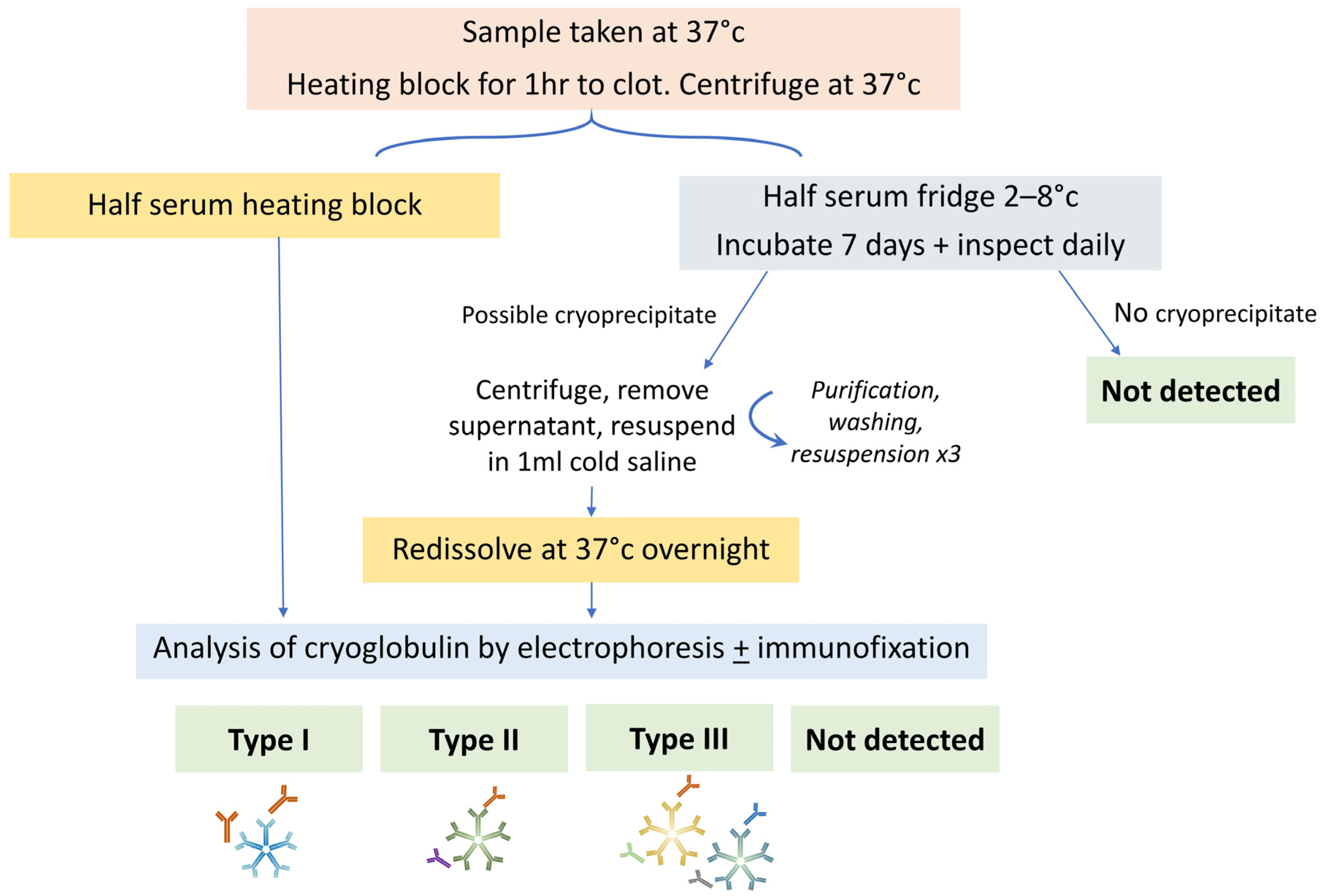
4.1. Cryoglobulin Detection

A schema of sample testing is shown in Figure 2. Samples should be taken in prewarmed tubes and transported at 37 °C, separated by centrifugation for 10 min at 2500 rpm at 37 °C, and the sample should be split into two aliquots (one at 4 °C and one at 37 °C). Half of the serum should be incubated at 37 °C and analysed via electrophoresis to identify the presence of an M-protein. Half of the serum is incubated for 7 days at 4 °C and inspected daily for the formation of a cryoprecipitate. If a precipitate is observed that is not present in the 37 °C sample, it is washed three times in saline solution at 4 °C, resuspended in saline and warmed to 37 °C in order to redissolve it. An analysis of the cryoprecipitate is then performed by electrophoresis and immunofixation. The cryoglobulin may be quantified as a cryocrit (%) by the relative volume of precipitate as a proportion of the total serum volume, performed visually, meaning that accuracy and reliability is poor, particularly at low concentrations. Alternatively, it may be monitored by the concentration of the M-protein in the cryoprecipitate. Typically, those with type I have higher cryocrit than type II and therefore may cause intravascular occlusion [16]. Figure 3 shows the appearance of a cryoprecipitate in the laboratory.
The detection of cryoglobulins may be challenging due to a number of potential pre-analytical errors, such as samples being collected at too low a temperature and in incompletely filled tubes. Ideally, 10 mL of blood sample should be provided. The accurate detection of cryoglobulins requires samples to be taken into pre-warmed tubes which must not be allowed to cool below 37 °C until the serum is separated as the cryoglobulin may precipitate and not be detected. This can be challenging, particularly in the setting of laboratory testing performed some distance from where samples have been taken as the temperature may fall by the time the sample reaches the laboratory. Additionally, a false-negative or underestimation of M-protein may result from the same process. Figure 4 shows an example of this from samples taken from the same patient. Similarly, a raised plasma viscosity in the absence of a raised IgM M-protein may trigger clinicians to assess for cryoglobulins [17]. In a large French study, 9% of cases with negative cryoglobulin detection were positive on a follow-up test [3]. Care must be taken with preanalytical variables and repeat testing of M-protein and cryoglobulins should be indicated if clinical suspicion is present.
Laboratory testing is critical as even a minimal amount of measurable cryoglobulin may result in symptoms. In one study where two-thirds of the cohort were symptomatic, 58% of those with IgM type I cryoglobulinaemia demonstrated a cryocrit of <1%, which was a significantly greater proportion than those with IgG cryoglobulins [11]. Cryoglobulin concentration did not differ in MGUS compared to other lymphoproliferative-associated type I cryoglobulins in a mixed cohort of 64 patients with IgG and IgM M-protein [18]. Symptoms do not directly correlate with cryocrit and may depend instead on the temperature at which precipitation occurs [13]. It should be noted that cryofibrinogens (cryoproteins that precipitate in plasma only, and not serum) may be associated with clinical vascultic symptoms and that testing should therefore be requested. However, these do not relate to an underlying IgM gammopathy [6] and so are not discussed further here.

4.2. Organ Involvement
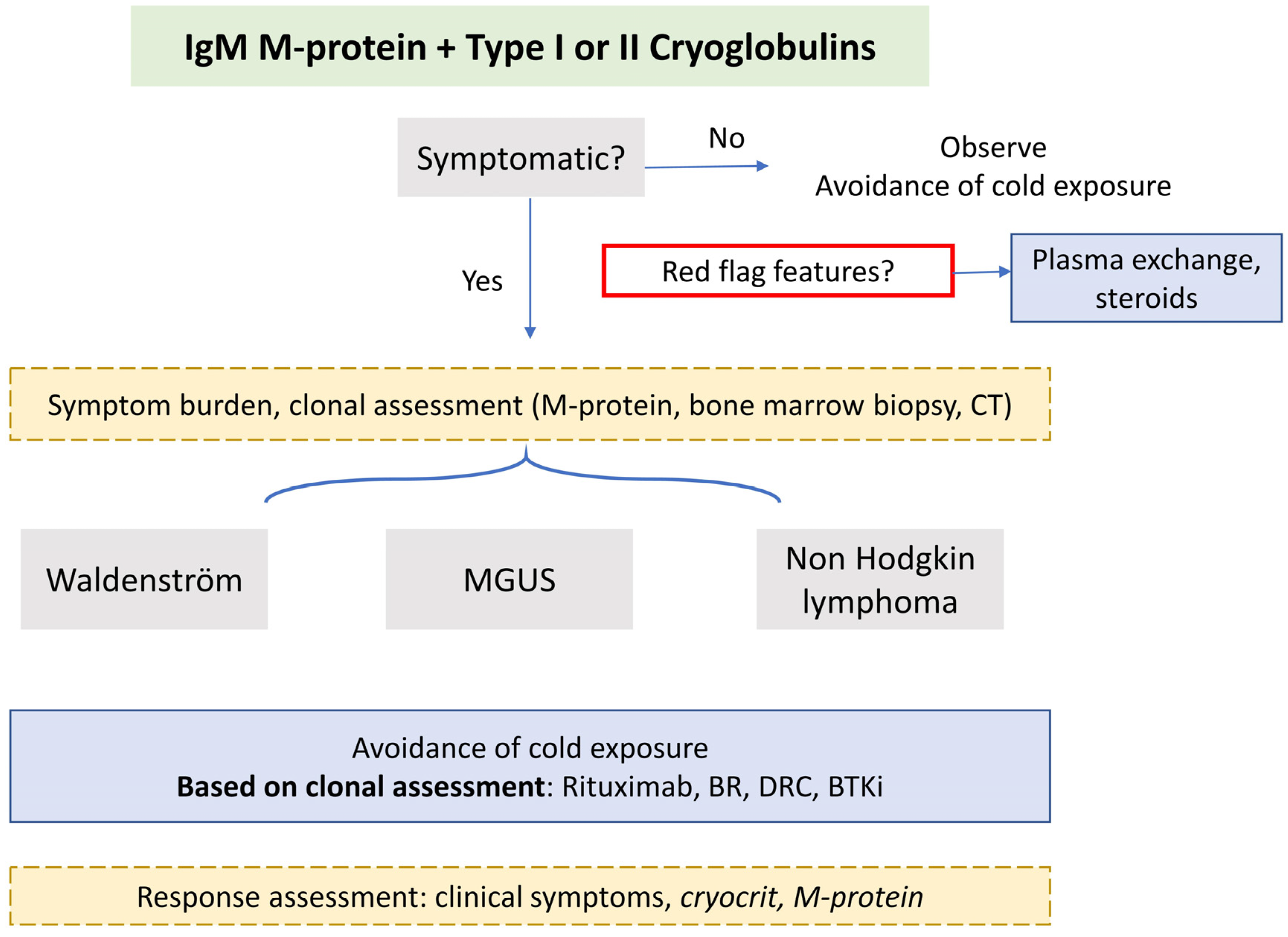
Symptoms of disease should be corroborated by laboratory tests and investigations which include assessments of organ involvement and the underlying clonal disorder. Investigations including urinalysis to screen for renal involvement (proteinuria, haematuria, renal insufficiency) [19], nerve conduction studies, autoimmune screening, and virology (HIV, hepatitis B and C serology) are useful. In those presenting with peripheral neuropathy, close collaboration with a neurologist with expertise in peripheral nerves is recommended as there is a wide differential of IgM-associated neuropathies (including anti-MAG peripheral neuropathy, non-MAG neuropathy, CANOMAD [chronic ataxic neuropathy ophthalmoplegia M-protein agglutination disialosyl antibodies syndrome], AL amyloidosis, central nervous system involvement of WM (Bing Neel syndrome), neurolymphomatosis or treatment-emergent neuropathy from drugs such as bortezomib, vinca alkaloids). In those with IgM monoclonal protein, alongside rapidly progressing axonal and demyelinating peripheral neuropathy, cryoglobulinaemia should be considered after AL amyloidosis is excluded [20]. As such, screening tests for neuropathy, including renal and liver function, HbA1c, serum B12 and folate, N-terminal pro B-type natriuretic peptide and cardiac troponin, should be undertaken. Rheumatoid factor, complement factors and viral studies are essential, particularly where type II cryoglobulins are detected, as immune complex deposition may cause the activation of proinflammatory complement proteins and raise the subsequent consumption of complement factors. Rheumatoid factor is associated with decreased C4 and CH50 (screening assay for the activation of the classical complement pathway) [3]. The consumption of complement due to activation of the classical pathway may be observed in type I cryoglobulinaemia. Decreased levels of complement and increased rheumatoid factor activity have been variably reported in up to a third in some series [11,18]. This has also been observed in cases of monoclonal type I IgG cryoglobulin (predominantly IgG1 subclass) [21]. Virology screening is necessary as most cases of type II cryoglobulins are associated with hepatitis. In a routine laboratory, hepatitis-related mixed cryoglobulins constitute the majority of all positive cryoglobulins detected. If indicated, a biopsy may be required to distinguish from other causes, particularly in establishing renal or nerve involvement. However, there are no standardised diagnostic criteria. Intravascular precipitation of IgM triggered by cold exposure results in thrombotic obstruction and ischaemia in small vessels, as evidenced via biopsy in type I cryoglobulinaemia. Leucocytoclastic vasculitis, a small-vessel vasculitis characterised by immune complex-mediated vasculitis of the dermal capillaries and venules, may be evident in type II. Membranoproliferative glomerulonephritis is characteristic and nephrotic-range proteinuria, intraluminal thrombi and extracapillary proliferation are associated with end-stage renal disease in mixed cryoglobulinaemia [19]. Nerve biopsy may demonstrate large-fibre axonal degeneration without regeneration, accompanied by vasculitis [22]. Intravascular cryoglobulin deposition without vasculitis may be present in the vasa nervorum or myelin sheaths and electron microscopy may identify monoclonal immunoglobulin deposition [23,24].
Prior to any treatment, clonal assessment should be undertaken in all patients to assess the burden of the underlying haematological disease. Standard staging includes bone marrow biopsy and aspirate (immunohistochemistry, flow cytometry, FISH, and Congo red staining—to exclude amyloidosis) and molecular testing for MYD88 and CXCR4 mutations, as well as CT imaging of chest, abdomen, and pelvis.

5. Management

There are a paucity of data to guide optimal management. A suggested management approach is outlined in Figure 5. Symptomatic cryoglobulinaemia is an indication for treatment and the treatment choice should be guided by the severity of symptoms. Mild symptoms may be abated with cold prevention alone and asymptomatic findings of cryoglobulins may require observation alone to be identified. However, rapidly progressive nephropathy and neuropathy have been reported at various stages of the disease course, meaning that careful monitoring is recommended [12]. Treatment for cryoglobulin symptoms is reported in up to 80% of cohorts comprising patients with symptomatic cryoglobulinaemia [25]. Response assessment is not standardised and most focus on symptomatic improvement [11]. Clinical symptom resolution is a priority, but disease activity markers (cryocrit for type I; complement and rheumatoid factor activity in type II) have been suggested [26]. Cryocrit at treatment initiation, change in cryocrit and the time taken to the nadir were predictive of symptom improvement in a mixed cohort of IgG and IgM type I cases. The underlying diagnosis of MGUS or lymphoma did not affect symptom improvement in one series [25]. Treatment regimens are heterogenous with small sample sizes.

5.1. Red Flag Symptoms

In those with red-flag features or life-threatening manifestations (as outlined in Figure 1), plasma exchange may ameliorate critical symptoms and is utilised in up to a third of all cryoglobulinaemia in mixed cohorts; warming procedures should be in place with all priming fluids to pre-warm the circuit apparatus and the replacement products should be run through a warmer [14,18,25]. Steroids are used in up to 90% of all cases (1 mg/kg), normally together with immunosuppression [14,18]. Plasma exchange should be also be considered first-line treatment in those with severe refractory symptoms who fail to respond to (or who are ineligible for) other treatments [27].

5.2. Symptomatic Disease

In the absence of robust evidence, definitive treatment should be directed at the underlying clone: in the majority of cases, this will be WM, and in rare cases another kind of NHL (chronic lymphocytic leukaemia or marginal zone lymphoma) [26]. Colchicine has been described as efficacious in patients with limited cutaneous manifestations (purpura) and mixed cryoglobulinaemia. However, these cohorts did not include patients with underlying haematological disorders, and so treatments derived on its basis are unlikely to be solely effective [28]. Rituximab combinations or bortezomib-based treatment are the most commonly employed treatment varieties [11], with symptom response rates of approximately 80% [11,13,18]. The disappearance of cryoglobulin may be seen in half of patients [25]. A pilot clinical trial of anti-CD38 therapy, isatuximab, is open for patients with IgG type I cryoglobulinaemia (NCT05114109), but no trial data are available for IgM type I cryoglobulinaemia. No studies have explored therapy exclusively for mixed cryoglobulinaemic vasculitis in the context of IgM disorders. However, a randomised controlled trial compared rituximab monotherapy with azathioprine or cyclophosphamide for the treatment of severe cryoglobulinemic vasculitis in 59 patients (skin ulcers, active glomerulonephritis, peripheral neuropathy) with or without hepatitis C virus (HCV). Survival at 12 months was higher in the rituximab-treated group (64.3 vs. 3.5%, p <  0.0001), and the improved reduction in vasculitis score was well tolerated [29].
As is standard, we consider delaying rituximab if IgM M-protein is significantly raised (>40g/L) to prevent IgM flare and hyperviscosity. Transient disease exacerbation due to the IgM flare has been described following the use of rituximab in patients with type I cryoglobulins and low burden of disease (<10% infiltrate) [30] and in patients of type II cryoglobulinaemia [31]. A study examining plasma exchange prior to rituximab to prevent IgM flare is ongoing (NCT04692363). Historically, there has been concern to treat emergent neurotoxicity with the use of bortezomib, which may be a particular concern in those with cryoglobulinaemic peripheral neuropathy. However weakly, subcutaneous administration and close monitoring may ameliorate the risk and have been shown to be efficacious in WM [32]. Effective depletion of clonal disease by bortezomib is likely to supersede its potential neurotoxicty. Autologous stem cell transplantation has been employed in cryoglobulinaemia associated with underlying myeloma, but has not used exclusively for those with WM, MGUS or NHL. Bruton’s tyrosine kinase inhibitors are a mainstay treatment in WM but their use in IgM cryoglobulinaemia has not been reported; the speed and depth of clonal depletion could be keys to heightening the effectiveness of this class of treatment.
The treatment of mixed cryoglobulinaemia with active cryoglobulinaemic vasculitis depends on the underlying cause of the illness. Studies examining mixed cryoglobuinaemia (including monoclonal IgG or IgM) demonstrate that the overwhelming majority of sicknesses of this kind are due to HCV (60 to 90%) [33]. Type II cryoglobulinaemia associated with HCV are most frequently unrelated to an underlying overt NHL [3]. Monoclonal B-lymphocytosis is a common finding in HCV-positive with small clones in the peripheral blood, bone marrow or liver. Therefore, treatment has focused on interferon-based antiviral therapy [34] and rituximab to deplete B-cell clonal expansion [35,36].
In a retrospective review of 67 patients with HCV and mixed cryoglobulinaemia (type II cryoglobulinaemia in 83%) treated with antivirals alone [37], 9% of patients had an overt NHL, 30% had a small B-cell clone, with a 20% rate of MYD88 mutation detected in the peripheral blood. Clonality by immunoglobulin gene rearrangement was demonstrated in peripheral blood mononuclear cells in all cases with type II cryoglobulins and in a quarter of type III cryoglobulins. Antiviral treatment resulted in a negative HCV viraemia in week 12 and a complete clinical response of vasculitis in 60% of cases. Clonal eradication in the peripheral blood was demonstrated in only 22%. Overall, six patients had overt NHL, with only one achieving a complete haematological response. Four (80%) required chemoimmunotherapy after antiviral failure to trear progressive disease.
In cases of HCV-associated indolent lymphoproliferative disorders (predominantly marginal zone lymphoma), a 1-year progression-free survival rate of 75% has been reported with the use of direct antivirals alone [38]. Therefore, we support the use of this approach. Disseminated lymphoproliferative disease may require chemoimmunotherapy (Rituximab-bendamustine or rituximab- cyclophosphamide-vincristine-prednisolone) [39,40,41]. Serum levels of C4 and CH50 are reliable markers of mixed cryoglobulin activity and normalisation may define an immunological response [42].

6. Conclusions

Type I and II cryoglobulins may be present in patients with IgM-associated disorders and are part of a distinctive entity of IgM MGCS. Presentations range from asymptomatic disease to multisystem involvement, meaning that a careful evaluation of the features and thorough interrogation of organ systems and the underlying clone is critical. Patients may present with WM, IgM MGUS or NHL. Treatment approaches and the formal assessment of severity and response criteria in cryoglobulinaemia are not standardised. Immediate management is required for clinical red-flag features. Due to their rarity, data with the capacity to inform treatment decisions are scant and collaborative research is imperative in order to define optimal treatment strategies.

Author Contributions

J.K. wrote the manuscript. S.J.S. and S.D. reviewed the manuscript. All authors have read and agreed to the published version of the manuscript.

Funding

This research received no external funding.

Institutional Review Board Statement

Not applicable.

Informed Consent Statement

Not applicable.

Data Availability Statement

Not applicable.

Conflicts of Interest

The authors declare no conflict of interest.

References

  1. Wintrobe, N.; Buell, M. Hypreproteinemia Associated with Multiple Myeloma with Report of Case in Which Extraordinary Hyperproteinemia Was Associated with Thrombosis of Retinal Veins and Symptom Suggesting Raynauds Disease; Bull Johns Hopkins Hospital: Baltimore, MD, USA, 1933. [Google Scholar]
  2. Brouet, J.C.; Clauvel, J.P.; Danon, F.; Klein, M.; Seligmann, M. Biologic and clinical significance of cryoglobulins. A report of 86 cases. Am. J. Med. 1974, 57, 775–788. [Google Scholar] [CrossRef] [PubMed]
  3. Kolopp-Sarda, M.N.; Nombel, A.; Miossec, P. Cryoglobulins Today: Detection and Immunologic Characteristics of 1675 Positive Samples from 13,439 Patients Obtained Over Six Years. Arthritis Rheumatol. 2019, 71, 1904–1912. [Google Scholar] [CrossRef] [PubMed]
  4. Swerdlow, S.H.; Campo, E.; Pileri, S.A.; Harris, N.L.; Stein, H.; Siebert, R.; Advani, R.; Ghielmini, M.; Salles, G.A.; Zelenetz, A.D.; et al. The 2016 revision of the World Health Organization classification of lymphoid neoplasms. Blood 2016, 127, 2375–2390. [Google Scholar] [CrossRef] [Green Version]
  5. Khwaja, J.; D’Sa, S.; Minnema, M.C.; Kersten, M.J.; Wechalekar, A.; Vos, J.M. IgM monoclonal gammopathies of clinical significance: Diagnosis and management. Haematologica 2022, 107, 2037–2050. [Google Scholar] [CrossRef] [PubMed]
  6. Gkalea, V.; Fotiou, D.; Dimopoulos, M.A.; Kastritis, E. Monoclonal Gammopathy of Thrombotic Significance. Cancers 2023, 15, 480. [Google Scholar] [CrossRef] [PubMed]
  7. Tzioufas, A.G.; Boumba, D.S.; Skopouli, F.N.; Moutsopoulos, H.M. Mixed monoclonal cryoglobulinemia and monoclonal rheumatoid factor cross-reactive idiotypes as predictive factors for the development of lymphoma in primary Sjögren’s syndrome. Arthritis Rheum. 1996, 39, 767–772. [Google Scholar] [CrossRef]
  8. Theander, E.; Henriksson, G.; Ljungberg, O.; Mandl, T.; Manthorpe, R.; Jacobsson, L.T. Lymphoma and other malignancies in primary Sjögren’s syndrome: A cohort study on cancer incidence and lymphoma predictors. Ann. Rheum. Dis. 2006, 65, 796–803. [Google Scholar] [CrossRef]
  9. Monti, G.; Pioltelli, P.; Saccardo, F.; Campanini, M.; Candela, M.; Cavallero, G.; De Vita, S.; Ferri, C.; Mazzaro, C.; Migliaresi, S.; et al. Incidence and characteristics of non-Hodgkin lymphomas in a multicenter case file of patients with hepatitis C virus-related symptomatic mixed cryoglobulinemias. Arch. Intern. Med. 2005, 165, 101–105. [Google Scholar] [CrossRef] [Green Version]
  10. Marasca, R.; Vaccari, P.; Luppi, M.; Zucchini, P.; Castelli, I.; Barozzi, P.; Cuoghi, A.; Torelli, G. Immunoglobulin gene mutations and frequent use of VH1-69 and VH4-34 segments in hepatitis C virus-positive and hepatitis C virus-negative nodal marginal zone B-cell lymphoma. Am. J. Pathol. 2001, 159, 253–261. [Google Scholar] [CrossRef] [Green Version]
  11. Zhang, L.-L.; Cao, X.-X.; Shen, K.-N.; Han, H.-X.; Zhang, C.-L.; Qiu, Y.; Zhao, H.; Gao, X.-M.; Feng, J.; Zhang, L.; et al. Clinical characteristics and treatment outcome of type I cryoglobulinemia in Chinese patients: A single-center study of 45 patients. Ann. Hematol. 2020, 99, 1735–1740. [Google Scholar] [CrossRef]
  12. Néel, A.; Perrin, F.; Decaux, O.; Dejoie, T.; Tessoulin, B.; Halliez, M.; Mahé, B.; Lamy, T.; Fakhouri, F.; Jego, P.; et al. Long-term outcome of monoclonal (type 1) cryoglobulinemia. Am. J. Hematol. 2014, 89, 156–161. [Google Scholar] [CrossRef]
  13. Harel, S.; Mohr, M.; Jahn, I.; Aucouturier, F.; Galicier, L.; Asli, B.; Malphettes, M.; Szalat, R.; Brouet, J.-C.; Lipsker, D.; et al. Clinico-biological characteristics and treatment of type I monoclonal cryoglobulinaemia: A study of 64 cases. Br. J. Haematol. 2015, 168, 671–678. [Google Scholar] [CrossRef]
  14. Terrier, B.; Krastinova, E.; Marie, I.; Launay, D.; Lacraz, A.; Belenotti, P.; de Saint-Martin, L.; Quemeneur, T.; Huart, A.; Bonnet, F.; et al. Management of noninfectious mixed cryoglobulinemia vasculitis: Data from 242 cases included in the CryoVas survey. Blood 2012, 119, 5996–6004. [Google Scholar] [CrossRef] [PubMed] [Green Version]
  15. Gemignani, F.; Brindani, F.; Alfieri, S.; Giuberti, T.; Allegri, I.; Ferrari, C.; Marbini, A. Clinical spectrum of cryoglobulinaemic neuropathy. J. Neurol. Neurosurg. Psychiatry 2005, 76, 1410–1414. [Google Scholar] [CrossRef] [Green Version]
  16. Monti, G.; Galli, M.; Invernizzi, F.; Pioltelli, P.; Saccardo, F.; Monteverde, A.; Pietrogrande, M.; Renoldi, P.; Bombardieri, S.; Bordin, G. Cryoglobulinaemias: A multi-centre study of the early clinical and laboratory manifestations of primary and secondary disease. GISC. Italian Group for the Study of Cryoglobulinaemias. QJM Int. J. Med. 1995, 88, 115–126. [Google Scholar]
  17. Hira-Kazal, R.; Sayar, Z.; Kothari, J.; Ayrton, P.; Berney, S.; Maher, J. Cryoglobulinaemia identified by repeated analytical failure of laboratory tests. Lancet 2014, 383, 382. [Google Scholar] [CrossRef]
  18. Terrier, B.; Karras, A.; Kahn, J.E.; Le Guenno, G.; Marie, I.; Benarous, L.; Lacraz, A.; Diot, E.; Hermine, O.; de Saint-Martin, L.; et al. The spectrum of type I cryoglobulinemia vasculitis: New insights based on 64 cases. Medicine 2013, 92, 61–68. [Google Scholar] [CrossRef] [PubMed] [Green Version]
  19. Zaidan, M.; Terrier, B.; Pozdzik, A.; Frouget, T.; Rioux-Leclercq, N.; Combe, C.; Lepreux, S.; Hummel, A.; Noël, L.-H.; Marie, I.; et al. Spectrum and Prognosis of Noninfectious Renal Mixed Cryoglobulinemic GN. J. Am. Soc. Nephrol. 2016, 27, 1213–1224. [Google Scholar] [CrossRef] [PubMed] [Green Version]
  20. D’Sa, S.; Kersten, M.J.; Castillo, J.J.; Dimopoulos, M.; Kastritis, E.; Laane, E.; Leblond, V.; Merlini, G.; Treon, S.P.; Vos, J.M.; et al. Investigation and management of IgM and Waldenström-associated peripheral neuropathies: Recommendations from the IWWM-8 consensus panel. Br. J. Haematol. 2017, 176, 728–742. [Google Scholar] [CrossRef]
  21. Kolopp-Sarda, M.N.; Azevedo, P.M.; Miossec, P. IgG subclasses in cryoglobulins: Link to composition and clinical manifestations. Arthritis Res. Ther. 2020, 22, 267. [Google Scholar] [CrossRef]
  22. Nemni, R.; Corbo, M.; Fazio, R.; Quattrini, A.; Comi, G.; Canal, N. Cryoglobulinaemic neuropathy. A clinical, morphological and immunocytochemical study of 8 cases. Brain 1988, 111 Pt 3, 541–552. [Google Scholar] [CrossRef] [PubMed]
  23. Vallat, J.M.; Desproges-Gotteron, R.; Leboutet, M.J.; Loubet, A.; Gualde, N.; Treves, R. Cryoglobulinemic neuropathy: A pathological study. Ann. Neurol. 1980, 8, 179–185. [Google Scholar] [CrossRef] [PubMed]
  24. Ciompi, M.; Marini, D.; Siciliano, G.; Melchiorre, D.; Bazzichi, L.; Sartucci, F.; Murri, L. Cryoglobulinemic peripheral neuropathy: Neurophysiologic evaluation in twenty-two patients. Biomed. Pharmacother. 1996, 50, 329–336. [Google Scholar] [CrossRef] [PubMed]
  25. Sidana, S.; Rajkumar, S.V.; Dispenzieri, A.; Lacy, M.Q.; Gertz, M.A.; Buadi, F.K.; Hayman, S.R.; Dingli, D.; Kapoor, P.; Gonsalves, W.I.; et al. Clinical presentation and outcomes of patients with type 1 monoclonal cryoglobulinemia. Am. J. Hematol. 2017, 92, 668–673. [Google Scholar] [CrossRef]
  26. Motyckova, G.; Murali, M. Laboratory testing for cryoglobulins. Am. J. Hematol. 2011, 86, 500–502. [Google Scholar] [CrossRef]
  27. Pratt, G.; El-Sharkawi, D.; Kothari, J.; D’Sa, S.; Auer, R.; McCarthy, H.; Krishna, R.; Miles, O.; Kyriakou, C.; Owen, R. Diagnosis and management of Waldenström macroglobulinaemia-A British Society for Haematology guideline. Br. J. Haematol. 2022, 197, 171–187. [Google Scholar] [CrossRef] [PubMed]
  28. Invernizzi, F.; Monti, G. Colchicine and mixed cryoglobulinemia. Arthritis Rheum. 1993, 36, 722–723. [Google Scholar] [CrossRef]
  29. De Vita, S.; Quartuccio, L.; Isola, M.; Mazzaro, C.; Scaini, P.; Lenzi, M.; Campanini, M.; Naclerio, C.; Tavoni, A.; Pietrogrande, M.; et al. A randomized controlled trial of rituximab for the treatment of severe cryoglobulinemic vasculitis. Arthritis Rheum. 2012, 64, 843–853. [Google Scholar] [CrossRef] [Green Version]
  30. Nehme-Schuster, H.; Korganow, A.S.; Pasquali, J.L.; Martin, T. Rituximab Inefficiency during Type I Cryoglobulinaemia; Rheumatology: Oxford, UK, 2005; Voluem 44, pp. 410–411. [Google Scholar]
  31. Sène, D.; Ghillani-Dalbin, P.; Amoura, Z.; Musset, L.; Cacoub, P. Rituximab may form a complex with IgMkappa mixed cryoglobulin and induce severe systemic reactions in patients with hepatitis C virus-induced vasculitis. Arthritis Rheum. 2009, 60, 3848–3855. [Google Scholar] [CrossRef]
  32. Khwaja, J.; Uppal, E.; Baker, R.; Trivedi, K.; Rismani, A.; Gupta, R.; Proctor, I.; Kyriakou, C.; D’Sa, S. Bortezomib-based therapy is effective and well tolerated in frontline and multiply pre-treated Waldenström macroglobulinaemia including BTKi failures: A real-world analysis. EJHaem 2022, 3, 1330–1334. [Google Scholar] [CrossRef]
  33. Ferri, C.; Sebastiani, M.; Giuggioli, D.; Cazzato, M.; Longombardo, G.; Antonelli, A.; Puccini, R.; Michelassi, C.; Zignego, A.L. Mixed cryoglobulinemia: Demographic, clinical, and serologic features and survival in 231 patients. Semin. Arthritis Rheum. 2004, 33, 355–374. [Google Scholar] [CrossRef] [PubMed]
  34. Hermine, O.; Lefrère, F.; Bronowicki, J.-P.; Mariette, X.; Jondeau, K.; Eclache-Saudreau, V.; Delmas, B.; Valensi, F.; Cacoub, P.; Brechot, C.; et al. Regression of splenic lymphoma with villous lymphocytes after treatment of hepatitis C virus infection. N. Engl. J. Med. 2002, 347, 89–94. [Google Scholar] [CrossRef]
  35. Saadoun, D.; Resche Rigon, M.; Sene, D.; Terrier, B.; Karras, A.; Perard, L.; Schoindre, Y.; Coppéré, B.; Blanc, F.; Musset, L.; et al. Rituximab plus Peg-interferon-alpha/ribavirin compared with Peg-interferon-alpha/ribavirin in hepatitis C-related mixed cryoglobulinemia. Blood 2010, 116, 326–334. [Google Scholar] [CrossRef]
  36. Dammacco, F.; Tucci, F.A.; Lauletta, G.; Gatti, P.; De Re, V.; Conteduca, V.; Sansonno, S.; Russi, S.; Mariggiò, M.A.; Chironna, M.; et al. Pegylated interferon-alpha, ribavirin, and rituximab combined therapy of hepatitis C virus-related mixed cryoglobulinemia: A long-term study. Blood 2010, 116, 343–353. [Google Scholar] [CrossRef] [Green Version]
  37. Pozzato, G.; Mazzaro, C.; Artemova, M.; Abdurakhmanov, D.; Grassi, G.; Crosato, I.; Mauro, E.; Ghersetti, M.; Zorat, F.; Bomben, R.; et al. Direct-acting antiviral agents for hepatitis C virus-mixed cryoglobulinaemia: Dissociated virological and haematological responses. Br. J. Haematol. 2020, 191, 775–783. [Google Scholar] [CrossRef] [PubMed]
  38. Arcaini, L.; Besson, C.; Frigeni, M.; Fontaine, H.; Goldaniga, M.; Casato, M.; Visentini, M.; Torres, H.A.; Loustaud-Ratti, V.; Peveling-Oberhag, J.; et al. Interferon-free antiviral treatment in B-cell lymphoproliferative disorders associated with hepatitis C virus infection. Blood 2016, 128, 2527–2532. [Google Scholar] [CrossRef] [Green Version]
  39. Kang, H.J.; Kim, W.S.; Kim, S.J.; Lee, J.J.; Yang, D.H.; Kim, J.S.; Lee, S.R.; Lee, G.W.; Kim, H.J.; Kim, H.Y.; et al. Phase II trial of rituximab plus CVP combination chemotherapy for advanced stage marginal zone lymphoma as a first-line therapy: Consortium for Improving Survival of Lymphoma (CISL) study. Ann. Hematol. 2012, 91, 543–551. [Google Scholar] [CrossRef]
  40. Rummel, M.J.; Niederle, N.; Maschmeyer, G.; Banat, G.A.; von Grünhagen, U.; Losem, C.; Kofahl-Krause, D.; Heil, G.; Welslau, M.; Balser, C.; et al. Bendamustine plus rituximab versus CHOP plus rituximab as first-line treatment for patients with indolent and mantle-cell lymphomas: An open-label, multicentre, randomised, phase 3 non-inferiority trial. Lancet 2013, 381, 1203–1210. [Google Scholar] [CrossRef] [PubMed]
  41. Laribi, K.; Tempescul, A.; Ghnaya, H.; Denizon, N.; Besançon, A.; Anghel, A.; Farhi, J.; Truong, C.; Lemaire, P.; Poulain, S.; et al. The bendamustine plus rituximab regimen is active against primary nodal marginal zone B-cell lymphoma. Hematol. Oncol. 2017, 35, 536–541. [Google Scholar] [CrossRef]
  42. Lauletta, G.; Russi, S.; Pavone, F.; Vacca, A.; Dammacco, F. Direct-acting antiviral agents in the therapy of hepatitis C virus-related mixed cryoglobulinaemia: A single-centre experience. Arthritis Res. Ther. 2017, 19, 74. [Google Scholar] [CrossRef] [Green Version]
Figure 1. Cryoglobulin symptoms and organ assessment. Key: NCS, nerve conduction studies; EMG, electromyography.
Hemato 04 00019 g001
Figure 2. Laboratory cryoglobulin detection.
Hemato 04 00019 g002
Figure 3. Appearances of a cryoprecipitate in the laboratory.
Hemato 04 00019 g003
Figure 4. IgM M-protein quantification in the same patient with concurrent cryoglobulin (a) sample taken to the laboratory at room temperature, (b) sample kept at 37 °C from blood puncture until serum separation.
Hemato 04 00019 g004
Figure 5. Management approach for IgM type I and II cryoglobulins.
Hemato 04 00019 g005
Disclaimer/Publisher’s Note: The statements, opinions and data contained in all publications are solely those of the individual author(s) and contributor(s) and not of MDPI and/or the editor(s). MDPI and/or the editor(s) disclaim responsibility for any injury to people or property resulting from any ideas, methods, instructions or products referred to in the content.

