Review

The Clinical Impact of Precisely Defining Mantle Cell Lymphoma: Contributions of Elaine Jaffe

1
Lymphoid Malignancies Branch, Center for Cancer Research, National Cancer Institute, National Institutes of Health, Bethesda, MD 20892, USA
2
Hematology Division, Department of Medicine, Brigham and Women’s Hospital, Boston, MA 02115, USA
*
Author to whom correspondence should be addressed.
Hemato 2022, 3(3), 508-517; https://doi.org/10.3390/hemato3030035
Submission received: 18 July 2022 / Revised: 11 August 2022 / Accepted: 12 August 2022 / Published: 16 August 2022

Abstract

:
Mantle cell lymphoma (MCL) is an aggressive yet incurable B-cell lymphoma that was only first recognized as a distinct subtype in 1992, with early reports suggesting a poor median survival. Elaine Jaffe is a renowned hematopathologist and scientist from the National Cancer Institute who was instrumental in many of the early descriptions of MCL that distinguished it from other B-cell lymphomas. Further, she has led multiple international collaborations that have harmonized the lymphoma classification systems that are currently in use today. The early morphologic descriptions of MCL along with the contributions of immunologic and genetic techniques have confirmed MCL as a distinct entity with unique biology and clinical behavior. Importantly, these scientific discoveries laid the foundation for unprecedented therapeutic breakthroughs that have led to significant improvements in overall survival.

1. Introduction

Non-Hodgkin lymphoma (NHL) is a generic term applied to a broad range of malignant lymphoid neoplasms with striking underlying clinical and biologic heterogeneity. Lymphomas represent malignant transformation of lymphocytes at various stages of differentiation and have acquired hallmark cancer capabilities, including the ability to proliferate, resist cellular apoptosis, and evade the host immune response. Yet, important biologic differences exist across NHL subtypes that manifest as differences in disease behavior and therapeutic vulnerabilities.
Over the last 7 decades, the classification of lymphoid malignancies has been a complex and iterative process that evolved with the emergence of novel biologic insights and advances in analytic methods. New subtypes are often first introduced as provisional entities and subsequently validated to be sufficiently distinct to merit a unique therapeutic approach. The National Cancer Institute (NCI) has contributed substantially to the body of knowledge on lymphoma biology, classification, and management through the years. The bedrock upon which its lymphoma studies have been built is accurate and reproducible diagnosis, initially based entirely on histologic examination under a microscope and, over time, with a remarkably sophisticated battery of assays for the expression of specific genes and proteins. Indeed, the lymphoma classification systems purport to make scientifically and clinically meaningful distinctions between lymphoma subtypes by defining relatively homogeneous entities from a clinical, morphologic, immunologic, and genetic perspective with the goal of improving clinical outcomes.
For the past nearly 50 years, the precision of the diagnosis of lymphoma at the NCI has been established and maintained by Elaine Jaffe, where she currently serves as the head of the Hematopathology Section of the Laboratory of Pathology. At NCI, the pathologists have participated as full partners in the clinical studies and used their scientific expertise to establish new insights into lymphoma biology that have direct influence on patient management. The function of a clinical research team requires expertise in several domains: diagnostic imaging, patient care, surgery, pharmacology, and knowledge of the disease being treated. Excellence in each area is critical to obtaining the best outcomes, while variability in a domain can undermine the results. The work at the NCI has been able to rely on the accuracy of lymphoma diagnosis because of the excellence of Dr. Jaffe and her team. If Dr. Jaffe says it, you can count on it. And everyone knows it. A comprehensive recounting of the insights emerging from her work would take volumes to cover. Beyond her role within the NCI, she is a renowned physician scientist who has made numerous seminal contributions to our understanding of the biology and classification of lymphoma subtypes over her illustrious career. In her career, Dr. Jaffe has championed the critical importance of accurate diagnosis in making therapeutic progress. Early on, Dr. Jaffe and her NCI colleagues described important biologic differences between lymphomas arising from B-cell origin compared with those with T-cell or monocytic origin. Indeed, she was the first to demonstrate that nodular lymphomas originated from follicular B lymphocytes [1]. She and her colleagues had the foresight to understand the importance of incorporating the immunologic aspects of lymphoma, including the nature and function of the malignant cell along with the surrounding immune cell infiltrates [2,3]. Throughout her career, she has pioneered scientific discoveries within and across lymphoma subtypes, and she has led international collaborations that modernize lymphoma classification by integrating traditional pathology with the emergence of novel immunologic and genomic approaches. No more illustrative example exists than her scientific contribution to our understanding of the entity mantle cell lymphoma (MCL). She originally described key morphologic and immunologic features of “intermediate lymphocytic lymphoma (ILL)” that suggested it was a distinct B-cell lymphoma and first proposed the term “mantle cell lymphoma” in 1991 [4]. In this article, we highlight the seminal contributions of Dr. Jaffe along a discovery timeline that led to our modern conceptualization of MCL as a B-cell lymphoma with at least two distinct clinical subtypes with divergent clinical behavior (Figure 1). What is striking about this timeline is that numerous therapeutic advances followed from seminal observations in pathobiology and the prognosis has improved. The clinical impact of having a universally recognized set of diagnostic criteria establishing a defined and reproducible clinical entity has allowed clinical research to focus on developing new therapeutic approaches. The Nebraska Lymphoma Study Group has documented therapeutic progress through the last 30 years made possible by defining MCL as a distinct disease entity [5]. Indeed, one can make a cogent argument that the improved clinical outcomes in MCL have outpaced improvements in any other lymphoma subtype and the contributions of Dr. Jaffe and other pioneering scientists enabled that success [5].

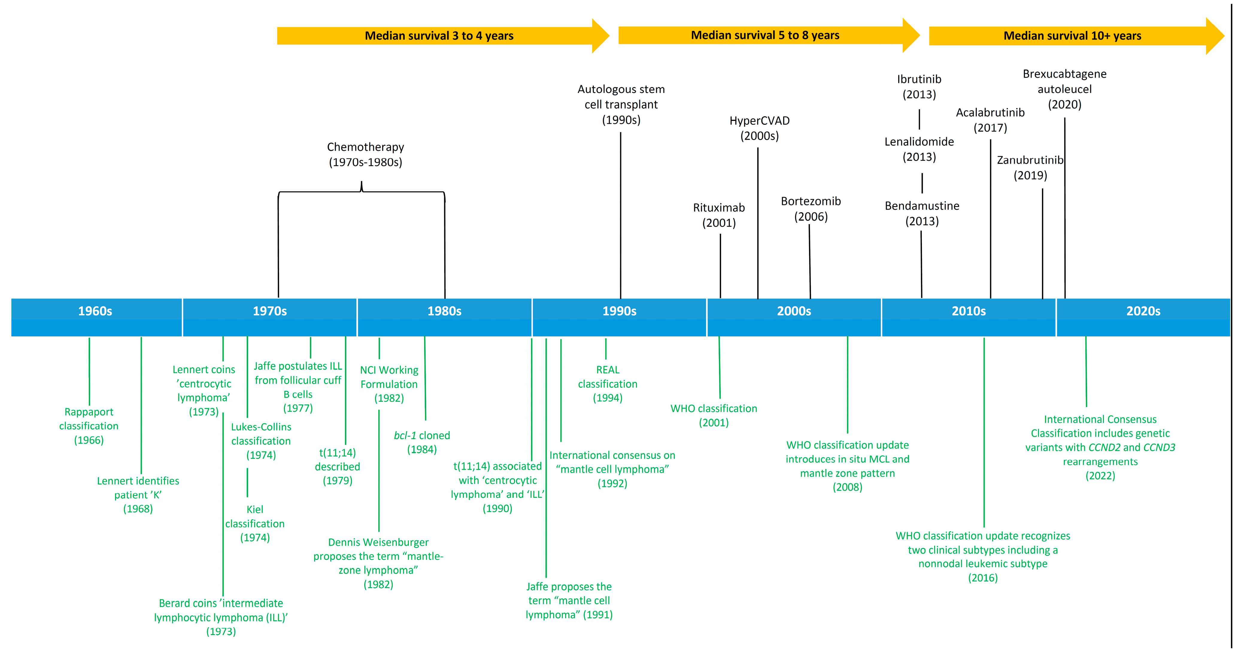
2. History and Evolution of Lymphoid Malignancies Classification

In 1966, Henry Rappaport of the United States Armed Forces Institute of Pathology proposed a simple and reproducible lymphoma classification system that relied exclusively on morphologic criteria [6]. Lymphomas were subdivided based on their underlying growth pattern as either nodular or diffuse. Further, the appearance of the malignant cell and its differentiation state was used to classify tumors as either well-differentiated, poorly differentiated, undifferentiated, or histiocytic [7]. In the Rappaport system, nodular lymphomas were typically composed of small lymphocytes and were comprised mostly of indolent disorders, while the “histiocytic” and poorly differentiated lymphomas were more aggressive and required chemotherapy. Notably, this system predated our modern understanding of cellular immunology and did not classify tumors based on B-cell or T-cell lineage.
In the 1970s, both the German pathologist Karl Lennert and the American pathologists L.J. Lukes and R.D. Collins proposed functional approaches to lymphoma classification that incorporated lymphocytic lineage based on cell surface immune markers and enzyme histochemical features along with morphology [8,9]. During this time period, separate classification schemes were used in different parts of the world and, since no broad international consensus existed, it was difficult to compare pathological and clinical results. Further, all systems lacked extensive clinical correlations and did not consider clinical features when classifying tumors.
In this context, the National Cancer Institute sponsored a panel of expert hematopathologists known as the international Working Formulation (WF) with a goal of providing a reproducible method to translate the various classification systems into clinical trial results and reports of clinical outcomes [10]. The WF system applied many concepts of preceding systems and incorporated clinical data. It defined subtypes based on their general clinical prognosis, and entities were grouped as low-grade, intermediate-grade, or high-grade. Notably, the IWF did not include the immunologic orientation to subclassify, rendering it less reproducible and limiting the discovery of new entities. Indeed, the panel of expert hematopathologists asked to review and classify the cases in the study disagreed with one another and with their own initial reading a substantial and alarming fraction of the time.
In 1994, the International Lymphoma Study Group (ILSG) convened a panel of 19 international expert hematopathologists to develop a consensus list of distinct clinical entities called the Revised European–American Classification of Lymphoid Neoplasms (REAL) classification [11]. The concept behind the REAL classification was to describe disease entities according to all available information (morphology, immunophenotype, genetic, and clinical features), with varying degrees of relative importance for each entity. To further validate the REAL classification, the ILSG conducted a study in which five expert pathologists reviewed over 1300 cases of NHL at various international centers [12,13]. This effort confirmed that the REAL classification was easily used by expert hematopathologists and had greater inter-observer reproducibility than other classification systems [14]. The REAL classification laid the foundation for the World Health Organization (WHO) classification system that was the first true international consensus on the classification of lymphoid malignancies in 2001 [15]. A cardinal feature of the WHO classification system was to periodically review new data and periodically incorporate them into updated classification systems that occurred in both 2008 [16] and 2016 [17].

3. Mantle Cell Lymphoma as a Distinct Entity

A primary objective of all lymphoma classification systems is to build upon previous iterations by describing novel entities that were previously difficult to classify; they are often predicated on the emergence of new technology. For example, in 1968 Karl Lennert described a lymph node biopsy from “patient K” that was comprised of small lymphocytes with a diffuse growth pattern that had completely effaced the lymph node architecture [18]. Lennert recognized that this lymphoma was unclassifiable with existing systems and he labeled this and other similar cases as ‘type K’. After analyzing subsequent biopsies from the same patient, he coined the term “centrocytic lymphoma” (CC) to describe lymphomas with a diffuse growth pattern of small cells that resembled cleaved follicular center cells [8,19]. Similarly, the American pathologist Costan Berard had recognized that some lymphomas were not easily classified as either well-differentiated or poorly differentiated by the Rappaport classification system and he proposed the term “malignant lymphoma, lymphocytic type intermediate grade of differentiation” or “intermediate lymphocytic lymphoma” (ILL) [20]. Both CC and ILL shared pathologic features between nodular lymphomas (i.e., follicular lymphoma) and well-differentiated lymphocytic lymphoma (i.e., chronic lymphocytic leukemia). They exhibited a mostly diffuse growth pattern but could also have areas with a vaguely nodular pattern. The cells were small and monotonous with clumped chromatin and scant cytoplasm with nuclei that varied in shape, including round, slightly clefted, and irregular [3]. Immunologically, these tumors also exhibited features intermediate between well-differentiated and poorly differentiated tumors and expressed surface immunoglobulin. In 1977, Dr. Jaffe postulated on the possible cell of origin based on these morphologic and immunologic criteria:
“The cells of nodular lymphomas are neoplastic counterparts of follicular B lymphocytes whereas well-differentiated lymphocytic lymphoma (WDL) cells are more closely related to medullary-cord B cells. Lymphoma of intermediate differentiation type may derive from B cells of the lymphoid cuff at the margins of follicles and thus exhibit features at the interface between nodular lymphomas and WDL”. Jaffe et al., Cancer Treat Rep. 1977;61(6):953–962.
The American pathologist Dennis Weisenburger recognized that there was a distinctive B-cell lymphoma that appeared to originate from the mantle zones of secondary lymphoid follicles [21]. He described 12 cases of lymphoma that morphologically resembled ILL, but with a growth pattern of atypical lymphocytes proliferating as wide mantles around normal appearing germinal centers and proposed the term “mantle-zone lymphoma”. Importantly, he reported that the clinical course was often aggressive, which further solidified the need to distinguish them from well-differentiated lymphomas [22]. In 1987, Jaffe and colleagues reviewed the histologic, immunologic, and clinical features of ILL that supported its consideration as a distinct clinicopathologic entity that was virtually identical to CC. The authors concluded that this entity was a tumor derived from lymphocytes of the mantle cuff based on the growth pattern and the expression of alkaline phosphatase on the neoplastic cells, which resembled that seen in mantle cuffs [23,24].
The emergence of molecular biology provided additional methods to sub-classify lymphomas. In 1979, the recurrent rearrangement t(11;14) (q14;q32) was first described in four cases of lymphoid neoplasms [25] and it was soon appreciated that recurrent translocations were often characteristic of specific lymphoma subtypes and this could be a powerful new technology to advance our understanding of distinct pathobiology of specific lymphomas [26]. In 1984, NCI researchers determined that the breakpoint of t(11;14) was within the joining segment of immunoglobulin heavy chain (IGH) typically located on chromosome 14 band q32 and characterized a new gene, named bcl-1 (B-cell lymphoma/leukemia 1), located on chromosome 11 band q13 [27]. It was shortly after that studies linked this recurrent translocation with specific lymphoma subtypes. Rearrangements involving bcl-1 were enriched in cases of ILL [28], but virtually never observed in follicular lymphoma, Burkitt lymphoma, or diffuse large B-cell lymphomas [29]. In 1990, Mike Williams and colleagues used Southern blotting and probes for immunoglobulin heavy and light chains, bcl-1, bcl-2, and c-myc in 14 patients [30]. They described rearrangements of bcl-1 in four (29%) cases and none had bcl-2 rearrangements. At the same time, NCI researchers used a genomic probe of the major breakpoint region in bcl-1 and showed that 10 (53%) ILL cases were associated with rearrangements [31]. When using multiple probes in 12 cases of CC, Williams and colleagues described that 11 (92%) were associated with bcl-1 rearrangements [32]. In 1991, it was discovered that the candidate oncogene PRAD1 located on chromosome 11q13 encoded a protein structurally similar to the cyclins (named cyclin D1) [33,34,35,36]. Taken together, we now know that the bcl-1 rearrangement involves the PRAD1/CCND1 gene located downstream of its major breakpoint region that encodes cyclin D1 and places it under the transcriptional control of IGH. The cyclin D1 translocation leads to unregulated cell cycle control underpinning MCL biology.
In 1991, Raffeld and Jaffe reviewed the morphologic, immunophenotypic, and genetic data supporting the notion that ILL, CC, and mantle zone lymphoma were identical neoplasms that should be unified as a distinct lymphoma subtype [4]. These entities were comprised of small to medium sized lymphocytes with scant neoplasm that expressed pan B-cell markers including CD20, CD19, and CD22 along with the pan T-cell marker CD5 and exhibited characteristically strong surface immunoglobulin expression while lacking CD10, Bcl-6, and CD23. Further, frequent expression of alkaline phosphatase suggested that these tumors were derived from follicular mantle zone cells [4]. They proposed that these entities be unified under the term “mantle cell lymphoma” (MCL) and recognized that diffuse, vaguely nodular, and expanded mantle zone growth patterns could be observed. In 1992, the term MCL was universally accepted to describe ILL, CC, and mantle zone lymphoma based on characteristic clinical features, immunophenotype, and a hallmark translocation [37].
Importantly, the recognition of MCL as a unique B-cell lymphoma did not signify the end of scientific discovery within this entity but represented the beginning. Since the original descriptions, important scientific discoveries have been made in MCL that distinguish it from related B-cell lymphomas that highlight the clinical and biologic heterogeneity within this entity. It is appreciated that tumor proliferation can vary widely in MCL and proliferation signatures by gene expression profiling are closely associated with response to chemotherapy and survival [38]. Further, it is now recognized that not all cases of MCL have demonstrable cyclin D1 expression, and cases of cyclin D1-negative MCL exhibit the classic morphologic and immunologic features of MCL but are often associated with rearrangements involving CCND2 or CCND3 [39]. Lastly, it has been noted that select cases of MCL exhibit very indolent behavior and that cyclin D1 positive B cells can occasionally be identified within the inner mantle zones of follicles, a process first described as in situ MCL and later updated to in situ mantle cell neoplasia (ISMN) [16,17,40]. Current classification systems recognize two distinct clinical variants of MCL, including a classical variant that typically expresses SOX11, often involving lymph nodes along with frequent extra-nodal involvement, and can behave aggressively [41,42]. An indolent form of MCL also exists that is frequently SOX11 negative and presents with non-nodal disease involving the spleen, peripheral blood, and bone marrow [17].

4. Therapeutic Advances in Mantle Cell Lymphoma

This article is primarily focused on the contribution of pathologic observations that led to the recognition of MCL as a distinct biologic entity, but it is notable that, since 1991, the prognosis for patients with MCL has improved significantly (Figure 1). Indeed, since the time of original description, research has focused on identifying pathogenetic mechanisms and oncogenic signaling pathways that drive MCL and the translation of these observations into novel therapies. It is beyond the scope of this article to comprehensively review the evolution of therapy for MCL, but we choose to highlight specific therapeutic advances and their effect on outcomes.
A feature of MCL that was initially quite puzzling was the wide range of natural histories associated with the diagnosis. Some patients had a very indolent course and did not require therapeutic intervention for years. Others had an extremely aggressive disease that spread rapidly and killed them in a few months. The initial reports of survival for MCL patients treated with standard lymphoma combination regimens such as cyclophosphamide, vincristine, prednisone (CVP) or cyclophosphamide, doxorubicin, vincristine, and prednisone CHOP showed a very poor outcome compared with other B-cell lymphomas with a median survival of only 3 to 4 years [13,43]. Further, in distinction to aggressive B-cell lymphomas, durable remission was rare and MCL is considered largely incurable with combination chemotherapy. For this reason, the standard approach starting in the 1990s and early 2000s was to intensify chemotherapy to include either autologous stem cell transplantation (ASCT) as part of frontline therapy [44,45] or to treat with highly dose-intensive regimens developed from acute lymphocytic leukemia (ALL) such as fractionated cyclophosphamide, vincristine, doxorubicin, and dexamethasone (hyper-CVAD), often followed by ASCT as consolidation [46]. Although this intensification of therapy was only applicable to patients able to tolerate the myelosuppressive nature of these approaches, the rates of complete response and durable remissions appeared to improve.
In the early 2000s, the monoclonal anti-CD20 antibody rituximab emerged and was tested in combination chemotherapy regimens for NHL, including MCL. Early studies of rituximab added to CHOP demonstrated improvement in rates of complete response, but these data did not translate into a significant improvement in progression-free or overall survival [47,48]. Nonetheless, rituximab was incorporated into virtually all frontline regimens for MCL [49,50,51] and improved rates of complete response and overall survival when delivered as maintenance therapy after ASCT [52]. Further, for older patients who are not deemed suitable candidates for consolidation with ASCT, rituximab maintenance improved overall survival [53]. Although multiple regimens remain in use for MCL and the approach varies considerably based on practice setting and the age of the patient, both epidemiologic and long term data from clinical trials suggest that rituximab has improved survival in all patients with MCL when added to chemotherapy, including older patients [54,55,56,57]. Overall, rituximab along with intensification of chemotherapy in younger patients has improved the median survival of MCL to 5 to 8 years, although this approach is associated with a continuous incidence of relapse and does not cure most patients [56].
In the 2010s, targeted agents emerged that appeared to have unique activity in MCL, including proteasome inhibitors, immunomodulatory agents, and inhibitors of the Bruton tyrosine kinase (BTK) pathway [58,59,60,61]. At the same time, bendamustine with rituximab (BR) emerged as a safe and effective induction regimen for MCL that was tolerable for patients of all ages [62]. Indeed, induction therapy with BR is as effective as more intensive induction regimens prior to ASCT and has become the most commonly used chemotherapy regimen in community practice [57,63].
Most recently, combination regimens are being tested in MCL that do not use traditional chemotherapy at all, with the hope of more broad tolerability. Lenalidomide with rituximab has been shown to be safe and highly effective as a frontline regimen with frequent durable remissions [60,64]. The BTK inhibitor ibrutinib has been studied with rituximab in patients with indolent forms of MCL as well as prior to intensive chemotherapy and has shown to induce very high rates of complete response [65]. Ibrutinib also improved the complete response rate and progression free survival when added to BR in a recent randomized study [66]. Finally, the emergence of chimeric antigen receptor T-cell therapy is associated with very high rates of complete response in MCL and is associated with durable remissions [67,68]. Taken together, these data suggest that future studies in MCL will test combinations of targeted agents with and without chemotherapy or immunotherapy. Current estimates suggest that following these therapeutic advances, the overall survival for MCL treated in the modern era may be longer than a decade and should continue to improve [69].

5. Trainees and Mentees

Beyond personal accomplishments as a scientist and researcher, Elaine Jaffe also impacted the field of hematopathology by her mentorship and teaching of close colleagues and trainings for nearly 4 decades. Nearly 75 pathologists have trained under her direct or indirect mentorship since the late 1970s, many of whom also made seminal contributions to our understanding of lymphoma biology and classification. Notable former mentees and/or close collaborators during their formative years include Stefania Pittaluga, Mark Raffeld, Elias Campo, Leticia Quintanilla-Martinez, and Falko Fend. In this way, her approach of identifying novel and biologically relevant associations between pathologic observations and clinical outcomes has been handed down and continues to impact the field.

6. Conclusions

Lymphoma treatment relies on accurate and reproducible diagnosis, while the lymphoma classification systems aim to make scientifically and clinically meaningful distinctions between lymphoma subtypes by defining relatively homogeneous entities from a clinical, morphologic, immunologic, and genetic perspective with the goal of improving clinical outcomes. Throughout her illustrious career, Dr. Elaine Jaffe has made important contributions to the management of individual patients at the NCI and has been instrumental in identifying new lymphoma subtypes, such as MCL. She has championed international efforts to harmonize the classification of lymphoma and these efforts have formed the foundation for therapeutic success.

Funding

This research was funded by the Intramural Research Program of the National Institutes of Health.

Institutional Review Board Statement

Not applicable.

Informed Consent Statement

Not applicable.

Data Availability Statement

Not applicable.

Conflicts of Interest

The authors report no financial conflict of interest.

References

  1. Jaffe, E.S.; Shevach, E.M.; Frank, M.M.; Berard, C.W.; Green, I. Nodular lymphoma—Evidence for origin from follicular B lymphocytes. N. Engl. J. Med. 1974, 290, 813–819. [Google Scholar] [CrossRef] [PubMed]
  2. Jaffe, E.S.; Braylan, R.C.; Nanba, K.; Frank, M.M.; Berard, C.W. Functional markers: A new perspective on malignant lymphomas. Cancer Treat. Rep. 1977, 61, 953–962. [Google Scholar] [PubMed]
  3. Berard, C.W.; Jaffe, E.S.; Braylan, R.C.; Mann, R.B.; Nanba, K. Immunologic aspects and pathology of the malignant lymphomas. Cancer 1978, 42, 911–921. [Google Scholar] [CrossRef]
  4. Raffeld, M.; Jaffe, E.S. bcl-1, t(11;14), and mantle cell-derived lymphomas. Blood 1991, 78, 259–263. [Google Scholar] [CrossRef] [PubMed]
  5. Armitage, J.O.; Longo, D.L. Mantle-Cell Lymphoma. N. Engl. J. Med. 2022, 386, 2495–2506. [Google Scholar] [CrossRef] [PubMed]
  6. Rappaport, H. Tumors of the Hematopoietic System; Armed Forces Institute of Pathology: Washington, DC, USA, 1966. [Google Scholar]
  7. Hicks, E.B.; Rappaport, H.; Winter, W.J. Follicular lymphoma; A re-evaluation of its position in the scheme of malignant lymphoma, based on a survey of 253 cases. Cancer 1956, 9, 792–821. [Google Scholar]
  8. Lennert, K. Malignant Lymphomas Other than Hodgkin’s Disease: Histology, Cytology, Ultrastructure, Immunology; Springer: Berlin/Heidelberg, Germany, 1978. [Google Scholar]
  9. Lukes, R.J.; Collins, R.D. Immunologic characterization of human malignant lymphomas. Cancer 1974, 34 (Suppl. 4), 1488–1503. [Google Scholar] [CrossRef]
  10. National Cancer Institute sponsored study of classifications of non-Hodgkin’s lymphomas: Summary and description of a working formulation for clinical usage. The Non-Hodgkin’s Lymphoma Pathologic Classification Project. Cancer 1982, 49, 2112–2135. [CrossRef]
  11. Harris, N.L.; Jaffe, E.S.; Stein, H.; Banks, P.M.; Chan, J.K.; Cleary, M.L.; Delsol, G.; De Wolf-Peeters, C.; Falini, B.; Gatter, K.C.; et al. A revised European-American classification of lymphoid neoplasms: A proposal from the International Lymphoma Study Group. Blood 1994, 84, 1361–1392. [Google Scholar] [CrossRef]
  12. A clinical evaluation of the International Lymphoma Study Group classification of non-Hodgkin’s lymphoma. The Non-Hodgkin’s Lymphoma Classification Project. Blood 1997, 89, 3909–3918. [CrossRef]
  13. Armitage, J.O.; Weisenburger, D.D. New approach to classifying non-Hodgkin’s lymphomas: Clinical features of the major histologic subtypes. Non-Hodgkin’s Lymphoma Classification Project. J. Clin. Oncol. 1998, 16, 2780–2795. [Google Scholar] [CrossRef] [PubMed]
  14. Harris, N.L.; Jaffe, E.S.; Diebold, J.; Flandrin, G.; Muller-Hermelink, H.K.; Vardiman, J. Lymphoma classification—From controversy to consensus: The R.E.A.L. and WHO Classification of lymphoid neoplasms. Ann. Oncol. 2000, 11, S3–S10. [Google Scholar] [CrossRef]
  15. Jaffe, E.S.; Harris, N.L.; Stein, H.; Vardiman, J. Pathology and Genetics of Tumours of Haematopoietic and Lymphoid Tissues; IARC Press: Lyon, France, 2001; Volume 3. [Google Scholar]
  16. Campo, E.; Swerdlow, S.H.; Harris, N.L.; Pileri, S.; Stein, H.; Jaffe, E.S. The 2008 WHO classification of lymphoid neoplasms and beyond: Evolving concepts and practical applications. Blood 2011, 117, 5019–5032. [Google Scholar] [CrossRef] [PubMed]
  17. Swerdlow, S.H.; Campo, E.; Pileri, S.A.; Harris, N.L.; Stein, H.; Advani, R.; Ghielmini, M.; Salles, G.A.; Zelenetz, A.D.; Jaffe, E.S. The 2016 revision of the World Health Organization classification of lymphoid neoplasms. Blood 2016, 127, 2375–2390. [Google Scholar] [CrossRef]
  18. Klapper, W.; Koch, K.; Mechler, U.; Borck, C.; Fuhry, E.; Siebert, R. Lymphoma ‘type K.’-in memory of Karl Lennert (1921–2012). Leukemia 2013, 27, 519–521. [Google Scholar] [CrossRef] [PubMed]
  19. Tolksdorf, G.; Stein, H.; Lennert, K. Morphological and immunological definition of a malignant lymphoma derived from germinal-centre cells with cleaved nuclei (centrocytes). Br. J. Cancer 1980, 41, 168–182. [Google Scholar] [CrossRef]
  20. Berard, C.W.; Dorfman, R.F. Histopathology of malignant lymphomas. Clin. Haematol. 1974, 3, 39–76. [Google Scholar] [CrossRef]
  21. Weisenburger, D.D.; Kim, H.; Rappaport, H. Mantle-zone lymphoma: A follicular variant of intermediate lymphocytic lymphoma. Cancer 1982, 49, 1429–1438. [Google Scholar] [CrossRef]
  22. Weisenburger, D.D.; Nathwani, B.N.; Diamond, L.W.; Winberg, C.D.; Rappaport, H. Malignant lymphoma, intermediate lymphocytic type: A clinicopathologic study of 42 cases. Cancer 1981, 48, 1415–1425. [Google Scholar] [CrossRef]
  23. Jaffe, E.S.; Bookman, M.A.; Longo, D.L. Lymphocytic lymphoma of intermediate differentiation--mantle zone lymphoma: A distinct subtype of B-cell lymphoma. Hum. Pathol. 1987, 18, 877–880. [Google Scholar] [CrossRef]
  24. Nanba, K.; Jaffe, E.S.; Braylan, R.C.; Soban, E.J.; Berard, C.W. Alkaline phosphatase-positive malignant lymphoma. A subtype of B-cell lymphomas. Am. J. Clin. Pathol. 1977, 68, 535–542. [Google Scholar] [CrossRef] [PubMed]
  25. Van Den Berghe, H.; Parloir, C.; David, G.; Michaux, J.L.; Sokal, G. A new characteristic karyotypic anomaly in lymphoproliferative disorders. Cancer 1979, 44, 188–195. [Google Scholar] [CrossRef]
  26. Yunis, J.J.; Oken, M.M.; Kaplan, M.E.; Ensrud, K.M.; Howe, R.R.; Theologides, A. Distinctive chromosomal abnormalities in histologic subtypes of non-Hodgkin’s lymphoma. N. Engl. J. Med. 1982, 307, 1231–1236. [Google Scholar] [CrossRef] [PubMed]
  27. Tsujimoto, Y.; Yunis, J.; Onorato-Showe, L.; Erikson, J.; Nowell, P.C.; Croce, C.M. Molecular cloning of the chromosomal breakpoint of B-cell lymphomas and leukemias with the t(11;14) chromosome translocation. Science 1984, 224, 1403–1406. [Google Scholar] [CrossRef]
  28. Weisenburger, D.D.; Sanger, W.G.; Armitage, J.O.; Purtilo, D.T. Intermediate lymphocytic lymphoma: Immunophenotypic and cytogenetic findings. Blood 1987, 69, 1617–1621. [Google Scholar] [CrossRef]
  29. Rimokh, R.; Berger, F.; Cornillet, P.; Wahbi, K.; Rouault, J.P.; Ffrench, M.; Bryon, P.A.; Gadoux, M.; Gentilhomme, O.; Germain, D.; et al. Break in the BCL1 locus is closely associated with intermediate lymphocytic lymphoma subtype. Genes Chromosomes Cancer 1990, 2, 223–226. [Google Scholar] [CrossRef]
  30. Williams, M.E.; Westermann, C.D.; Swerdlow, S.H. Genotypic characterization of centrocytic lymphoma: Frequent rearrangement of the chromosome 11 bcl-1 locus. Blood 1990, 76, 1387–1391. [Google Scholar] [CrossRef]
  31. Medeiros, L.J.; Van Krieken, J.H.; Jaffe, E.S.; Raffeld, M. Association of bcl-1 rearrangements with lymphocytic lymphoma of intermediate differentiation. Blood 1990, 76, 2086–2090. [Google Scholar] [CrossRef]
  32. Williams, M.E.; Meeker, T.C.; Swerdlow, S.H. Rearrangement of the chromosome 11 bcl-1 locus in centrocytic lymphoma: Analysis with multiple breakpoint probes. Blood 1991, 78, 493–498. [Google Scholar] [CrossRef]
  33. Motokura, T.; Bloom, T.; Kim, H.G.; Juppner, H.; Ruderman, J.V.; Kronenberg, H.M.; Arnold, A. A novel cyclin encoded by a bcl1-linked candidate oncogene. Nature 1991, 350, 512–515. [Google Scholar] [CrossRef]
  34. Bosch, F.; Jares, P.; Campo, E.; Lopez-Guillermo, A.; Piris, M.A.; Villamor, N.; Tassies, D.; Jaffe, E.S.; Monteserrat, E.; Rozman, C.; et al. PRAD-1/cyclin D1 gene overexpression in chronic lymphoproliferative disorders: A highly specific marker of mantle cell lymphoma. Blood 1994, 84, 2726–2732. [Google Scholar] [CrossRef] [PubMed]
  35. Rosenberg, C.L.; Wong, E.; Petty, E.M.; Bale, A.; Tsujimoto, Y.; Harris, N.L.; Arnold, A. PRAD1, a candidate BCL1 oncogene: Mapping and expression in centrocytic lymphoma. Proc. Natl. Acad. Sci. USA 1991, 88, 9638–9642. [Google Scholar] [CrossRef] [PubMed]
  36. Rimokh, R.; Berger, F.; Delsol, G.; Charrin, C.; Bertheas, M.F.; Ffrench, M.; Garoscio, M.; Felman, P.; Coiffier, B.; Bryon, P.A.; et al. Rearrangement and overexpression of the BCL-1/PRAD-1 gene in intermediate lymphocytic lymphomas and in t(11q13)-bearing leukemias. Blood 1993, 81, 3063–3067. [Google Scholar] [CrossRef] [PubMed]
  37. Banks, P.M.; Chan, J.; Cleary, M.L.; Delsol, G.; De Wolf-Peeters, C.; Gatter, K.; Grogan, T.M.; Harris, N.L.; Isaacson, P.G.; Jaffe, E.S.; et al. Mantle cell lymphoma. A proposal for unification of morphologic, immunologic, and molecular data. Am. J. Surg. Pathol. 1992, 16, 637–640. [Google Scholar] [CrossRef]
  38. Rosenwald, A.; Wright, G.; Wiestner, A.; Chan, W.C.; Connors, J.M.; Campo, E.; Gascoyne, R.D.; Grogan, T.M.; Muller-Hermelink, H.K.; Smeland, E.B.; et al. The proliferation gene expression signature is a quantitative integrator of oncogenic events that predicts survival in mantle cell lymphoma. Cancer Cell. 2003, 3, 185–197. [Google Scholar] [CrossRef]
  39. Fu, K.; Weisenburger, D.D.; Greiner, T.C.; Dave, S.; Wright, G.; Rosenwald, A.; Chiorazzi, M.; Iqbal, J.; Gesk, S.; Siebert, R.; et al. Cyclin D1-negative mantle cell lymphoma: A clinicopathologic study based on gene expression profiling. Blood 2005, 106, 4315–4321. [Google Scholar] [CrossRef]
  40. Carvajal-Cuenca, A.; Sua, L.F.; Silva, N.M.; Pittaluga, S.; Royo, C.; Song, J.Y.; Sargent, R.L.; Espinet, B.; Climent, F.; Jacobs, S.A.; et al. In situ mantle cell lymphoma: Clinical implications of an incidental finding with indolent clinical behavior. Haematologica 2012, 97, 270–278. [Google Scholar] [CrossRef]
  41. Navarro, A.; Clot, G.; Royo, C.; Jares, P.; Hadzidimitriou, A.; Agathangelidis, A.; Bikos, V.; Darzentas, N.; Papadaki, T.; Salaverria, I.; et al. Molecular subsets of mantle cell lymphoma defined by the IGHV mutational status and SOX11 expression have distinct biologic and clinical features. Cancer Res. 2012, 72, 5307–5316. [Google Scholar] [CrossRef]
  42. Fernandez, V.; Salamero, O.; Espinet, B.; Sole, F.; Royo, C.; Navarro, A.; Carnacho, F.; Bea, S.; Hartmann, E.; Amador, V.; et al. Genomic and gene expression profiling defines indolent forms of mantle cell lymphoma. Cancer Res. 2010, 70, 1408–1418. [Google Scholar] [CrossRef]
  43. Bosch, F.; Lopez-Guillermo, A.; Campo, E.; Ribera, J.M.; Conde, E.; Piris, M.A.; Vallespi, T.; Woessner, S.; Montserrat, E. Mantle cell lymphoma: Presenting features, response to therapy, and prognostic factors. Cancer 1998, 82, 567–575. [Google Scholar] [CrossRef]
  44. Vose, J.M.; Bierman, P.J.; Weisenburger, D.D.; Lynch, J.C.; Bociek, Y.; Chan, W.C.; Greiner, T.C.; Armitage, J.O. Autologous hematopoietic stem cell transplantation for mantle cell lymphoma. Biol. Blood Marrow Transpl. 2000, 6, 640–645. [Google Scholar] [CrossRef]
  45. Vandenberghe, E.; Ruiz de Elvira, C.; Loberiza, F.R.; Conde, E.; Lopez-Guillermo, A.; Gisselbrecht, C.; Guihot, F.; Vose, J.M.; van Biesen, K.; Rizzo, J.D.; et al. Outcome of autologous transplantation for mantle cell lymphoma: A study by the European Blood and Bone Marrow Transplant and Autologous Blood and Marrow Transplant Registries. Br. J. Haematol. 2003, 120, 793–800. [Google Scholar] [CrossRef] [PubMed]
  46. Khouri, I.F.; Romaguera, J.; Kantarjian, H.; Palmer, J.L.; Pugh, W.C.; Korbling, M.; Hagemeister, F.; Samuels, B.; Rodrigeuz, A.; Giralt, S.; et al. Hyper-CVAD and high-dose methotrexate/cytarabine followed by stem-cell transplantation: An active regimen for aggressive mantle-cell lymphoma. J. Clin. Oncol. 1998, 16, 3803–3809. [Google Scholar] [CrossRef] [PubMed]
  47. Howard, O.M.; Gribben, J.G.; Neuberg, D.S.; Grossbard, M.; Poor, C.; Janicek, M.J.; Shipp, M.A. Rituximab and CHOP induction therapy for newly diagnosed mantle-cell lymphoma: Molecular complete responses are not predictive of progression-free survival. J. Clin. Oncol. 2002, 20, 1288–1294. [Google Scholar] [CrossRef] [PubMed]
  48. LaCasce, A.S.; Vandergrift, J.L.; Rodriguez, M.A.; Abel, G.A.; Crosby, A.L.; Czuczman, M.S.; Nademanee, A.P.; Blayney, D.W.; Gordon, L.I.; Millenson, M.; et al. Comparative outcome of initial therapy for younger patients with mantle cell lymphoma: An analysis from the NCCN NHL Database. Blood 2012, 119, 2093–2099. [Google Scholar] [CrossRef] [PubMed]
  49. Geisler, C.H.; Kolstad, A.; Laurell, A.; Anderson, N.S.; Pederson, L.B.; Jerkeman, M.; Eriksson, M.; Nordstrom, M.; Kimby, E.; Boesen, A.M.; et al. Long-term progression-free survival of mantle cell lymphoma after intensive front-line immunochemotherapy with in vivo-purged stem cell rescue: A nonrandomized phase 2 multicenter study by the Nordic Lymphoma Group. Blood 2008, 112, 2687–2693. [Google Scholar] [CrossRef]
  50. Romaguera, J.E.; Fayad, L.; Rodriguez, M.A.; Broglio, K.R.; Hagermeister, F.B.; Pro, B.; McLaughlin, P.; Younes, A.; Samaniego, F.; Goy, A.; et al. High rate of durable remissions after treatment of newly diagnosed aggressive mantle-cell lymphoma with rituximab plus hyper-CVAD alternating with rituximab plus high-dose methotrexate and cytarabine. J. Clin. Oncol. 2005, 23, 7013–7023. [Google Scholar] [CrossRef]
  51. Hermine, O.; Hoster, E.; Walewski, J.; Bosly, A.; Stilgenbauer, S.; Thieblemont, C.; Szymczyk, M.; Bouabdallah, R.; Kneba, M.; Hallek, M.; et al. Addition of high-dose cytarabine to immunochemotherapy before autologous stem-cell transplantation in patients aged 65 years or younger with mantle cell lymphoma (MCL Younger): A randomised, open-label, phase 3 trial of the European Mantle Cell Lymphoma Network. Lancet 2016, 388, 565–575. [Google Scholar]
  52. Le Gouill, S.; Thieblemont, C.; Oberic, L.; Moreau, A.; Bouabdallah, K.; Dartigeas, C.; Damaj, G.; Gastinne, T.; Ribrag, V.; Feugier, P.; et al. Rituximab after Autologous Stem-Cell Transplantation in Mantle-Cell Lymphoma. N. Engl. J. Med. 2017, 377, 1250–1260. [Google Scholar] [CrossRef]
  53. Kluin-Nelemans, H.C.; Hoster, E.; Hermine, O.; Walewski, J.; Trney, M.; Geisler, C.H.; Stilgenbauer, S.; Thieblemont, C.; Vehling-Kaiser, U.; Doorduijn, J.K.; et al. Treatment of older patients with mantle-cell lymphoma. N. Engl. J. Med. 2012, 367, 520–531. [Google Scholar] [CrossRef]
  54. Romaguera, J.E.; Fayad, L.E.; Feng, L.; Hartig, K.; Weaver, P.; Rodriguez, M.A.; Hagemeister, F.B.; Pro, B.; McLaughlin, P.; Younes, A.; et al. Ten-year follow-up after intense chemoimmunotherapy with Rituximab-HyperCVAD alternating with Rituximab-high dose methotrexate/cytarabine (R-MA) and without stem cell transplantation in patients with untreated aggressive mantle cell lymphoma. Br. J. Haematol. 2010, 150, 200–208. [Google Scholar] [CrossRef]
  55. Griffiths, R.; Mikhael, J.; Gleeson, M.; Danese, M.; Dreyling, M. Addition of rituximab to chemotherapy alone as first-line therapy improves overall survival in elderly patients with mantle cell lymphoma. Blood 2011, 118, 4808–4816. [Google Scholar] [CrossRef]
  56. Eskelund, C.W.; Kolstad, A.; Jerkeman, M.; Raty, R.; Laurell, A.; Eloranta, S.; Smedby, K.E.; Husby, S.; Pedersen, L.B.; Andersen, N.S.; et al. 15-year follow-up of the Second Nordic Mantle Cell Lymphoma trial (MCL2): Prolonged remissions without survival plateau. Br. J. Haematol. 2016, 175, 410–418. [Google Scholar] [CrossRef] [PubMed]
  57. Martin, P.; Cohen, J.B.; Wang, M.; Kumar, A.; Hill, B.; Villa, D.; Switchenko, J.M.; Kahl, B.; Maddocks, K.; Grover, N.S.; et al. Treatment Outcomes and Roles of Transplantation and Maintenance Rituximab in Patients With Previously Untreated Mantle Cell Lymphoma: Results From Large Real-World Cohorts. J. Clin. Oncol 2022, JCO2102698. [Google Scholar] [CrossRef] [PubMed]
  58. Goy, A.; Younes, A.; McLaughlin, P.; Pro, B.; Romaguera, J.E.; Hagermeister, F.; Fayad, L.; Dang, N.H.; Samaniego, F.; Wang, M.; et al. Phase II study of proteasome inhibitor bortezomib in relapsed or refractory B-cell non-Hodgkin’s lymphoma. J. Clin. Oncol. 2005, 23, 667–675. [Google Scholar] [CrossRef] [PubMed]
  59. Robak, T.; Huang, H.; Jin, J.; Zhu, J.; Liu, T.; Samoilova, O.; Pylypenko, H.; Verhoef, G.; Siritanaratkul, N.; Osmanov, E.; et al. Bortezomib-based therapy for newly diagnosed mantle-cell lymphoma. N. Engl. J. Med. 2015, 372, 944–953. [Google Scholar] [CrossRef]
  60. Ruan, J.; Martin, P.; Shah, B.; Schuster, S.J.; Smith, S.M.; Furman, R.R.; Christos, P.; Rodriguez, A.; Svoboda, J.; Lewis, J.; et al. Lenalidomide plus Rituximab as Initial Treatment for Mantle-Cell Lymphoma. N. Engl. J. Med. 2015, 373, 1835–1844. [Google Scholar] [CrossRef]
  61. Ladetto, M.; Cortelazzo, S.; Ferrero, S.; Evangelista, A.; Mian, M.; Tavarozzi, R.; Zanni, M.; Cavallo, F.; Di Rocco, A.; Stefoni, V.; et al. Lenalidomide maintenance after autologous haematopoietic stem-cell transplantation in mantle cell lymphoma: Results of a Fondazione Italiana Linfomi (FIL) multicentre, randomised, phase 3 trial. Lancet Haematol. 2021, 8, e34–e44. [Google Scholar] [CrossRef]
  62. Rummel, M.J.; Niederle, N.; Maschmeyer, G.; Banat, G.A.; von Grunhagen, U.; Losem, C.; Kofahl-Krause, D.; Heil, G.; Welslau, M.; Balser, C.; et al. Bendamustine plus rituximab versus CHOP plus rituximab as first-line treatment for patients with indolent and mantle-cell lymphomas: An open-label, multicentre, randomised, phase 3 non-inferiority trial. Lancet 2013, 381, 1203–1210. [Google Scholar] [CrossRef]
  63. Chen, R.W.; Li, H.; Bernstein, S.H.; Kahwash, S.; Rimsza, L.M.; Forman, S.J.; Constine, L.; Shea, T.C.; Cashen, A.F.; Blum, K.A.; et al. RB but not R-HCVAD is a feasible induction regimen prior to auto-HCT in frontline MCL: Results of SWOG Study S1106. Br. J. Haematol. 2017, 176, 759–769. [Google Scholar] [CrossRef]
  64. Ruan, J.; Martin, P.; Christos, P.; Cerchietti, L.; Tam, W.; Shah, B.; Schuster, S.J.; Rodriguez, A.; Hyman, D.; Calvo-Vidal, M.N.; et al. Five-year follow-up of lenalidomide plus rituximab as initial treatment for mantle cell lymphoma. Blood 2018, 132, 2016–2025. [Google Scholar] [CrossRef] [PubMed]
  65. Gine, E.; de la Cruz, F.; Jimenez Ubieto, A.; Lopez Jimenez, J.; Martin Garcia-Sancho, A.; Terol, M.J.; Gonzalez Barca, E.; Casanova, M.; de la Fuente, A.; Marin-Niebla, A.; et al. Ibrutinib in Combination With Rituximab for Indolent Clinical Forms of Mantle Cell Lymphoma (IMCL-2015): A Multicenter, Open-Label, Single-Arm, Phase II Trial. J. Clin. Oncol. 2022, 40, 1196–1205. [Google Scholar] [CrossRef] [PubMed]
  66. Wang, M.L.; Jurczak, W.; Jerkeman, M.; Trotman, J.; Zinzani, P.; Belada, D.; Boccomini, C.; Flinn, I.W.; Giri, P.; Goy, A.; et al. Ibrutinib plus Bendamustine and Rituximab in Untreated Mantle-Cell Lymphoma. N. Engl. J. Med. 2022, 386, 2482–2494. [Google Scholar] [CrossRef]
  67. Wang, M.; Munoz, J.; Goy, A.; Locke, F.L.; Jacobson, C.A.; Hill, B.T.; Timmerman, J.M.; Holmes, H.; Jaglowski, S.; Flinn, I.W.; et al. KTE-X19 CAR T-Cell Therapy in Relapsed or Refractory Mantle-Cell Lymphoma. N. Engl. J. Med. 2020, 382, 1331–1342. [Google Scholar] [CrossRef] [PubMed]
  68. Wang, M.; Munoz, J.; Goy, A.; Locke, F.L.; Jacobson, C.A.; Hill, B.T.; Timmerman, J.M.; Holmes, H.; Jaglowski, S.; Flinn, I.W.; et al. Three-Year Follow-Up of KTE-X19 in Patients With Relapsed/Refractory Mantle Cell Lymphoma, Including High-Risk Subgroups, in the ZUMA-2 Study. J. Clin. Oncol. 2022, JCO2102370. [Google Scholar] [CrossRef]
  69. Castellino, A.; Wang, Y.; Larson, M.C.; Maurer, M.J.; Link, B.K.; Farooq, U.; Feldman, A.L.; Syrbu, S.I.; Habermann, T.M.; Paludo, J.; et al. Evolving frontline immunochemotherapy for mantle cell lymphoma and the impact on survival outcomes. Blood Adv. 2022, 6, 1350–1360. [Google Scholar] [CrossRef]
Figure 1. The timeline from the 1960s to the modern day depicting the scientific discoveries that ultimately led to recognition of mantle cell lymphoma (MCL) as a distinct biologic entity and therapeutic advances. Below the timeline are the classification systems of lymphoma and early pathologic descriptions of entities now classified as MCL. Above the timeline are the standard therapies for the period and the corresponding median survival for patients with MCL.
Hemato 03 00035 g001
Publisher’s Note: MDPI stays neutral with regard to jurisdictional claims in published maps and institutional affiliations.

