Review

Evolution in the Definition of Follicular Lymphoma and Diffuse Large B-Cell Lymphoma: A Model for the Future of Personalized Medicine

by
Elaine S. Jaffe
1,* and
Antonino Carbone
2
1
Laboratory of Pathology, Center for Cancer Research, National Cancer Institute, National Institutes of Health, Bethesda, MD 20892, USA
2
Department of Pathology, Centro di Riferimento Oncologico di Aviano (CRO), Istituto di Ricovero e Cura a Carattere Scientifico (IRCCS), Via F. Gallini 2, I-33081 Aviano, Italy
*
Author to whom correspondence should be addressed.
Hemato 2022, 3(3), 466-474; https://doi.org/10.3390/hemato3030032
Submission received: 13 June 2022 / Revised: 2 July 2022 / Accepted: 13 July 2022 / Published: 21 July 2022

Abstract

:
The definitions of follicular lymphoma (FL) and diffuse large B-cell lymphoma (DLBCL) are evolving in the era of personalized medicine. Early stages of the evolution of FL have been recognized. Two histological manifestations of early lesions are in situ follicular neoplasia and duodenal type FL. Additionally, FL frequently undergoes histological transformation, the most common form being DLBCL. High-grade B-cell lymphoma with double hit, with translocations involving BCL2 and MYC are important clinically. Rarer forms of transformation include classic Hodgkin lymphoma (CHL) and histiocytic sarcoma. In addition to conventional FL associated with the BCL2 translocation, alternative forms of BCL2-negative FL have been observed. These are heterogenous clinically and genetically. A distinctive group of B-cell lymphomas of follicle cell derivation arise in young patients and include pediatric type FL, testicular FL and a large B-cell lymphoma with IRF4 rearrangement. Historically DLBCL was separated into only two histological variants, centroblastic and immunoblastic. In 2017 the WHO classification recommended (1) the segregation of activated B cell and germinal center B cell derived DLBCL, (2) the identification of high-grade B-cell lymphoma with double hit, and (3) the recognition of an aggressive lymphoma that may resemble Burkitt lymphoma, currently designated in the International Consensus Classification as Large B-cell lymphoma with 11q aberration. Today we appreciate greater genomic complexity among aggressive B-cell lymphomas. Recent studies with NGS and mutational profiling have identified clinically significant genetic subgroups. It is hoped that these data ultimately will lead to targeted therapy based on the genetic profile.

1. Introduction

The diagnosis of lymphoma is evolving in the era of personalized medicine. Indicative of these changes is the evolution that has occurred in the criteria and definition of follicular lymphoma (FL) and diffuse large B-cell lymphoma (DLBCL), two of the most common subtypes of B-cell lymphoma.
FL is a B cell malignancy that mimics normal follicles histologically and phenotypically. FL B-cells maintain the same state of differentiation as that of germinal centre (GC) B cells within the secondary lymphoid follicle [1,2]. Morphologically FL is a follicular proliferation of centrocytes and centroblasts associated with follicular dendritic cells (FDCs). The immunophenotypic profile of FL is CD20+, CD19+, CD79a+, CD10+, CD5, Bcl-2+, Bcl-6+. The genetic alterations include JH/BCL2 rearrangement, t(14;18) and somatic mutations of VH. Most patients present with advanced stage disease, (IIIA–IV A), and with indolent, but generally incurable disease [1,2].
Diffuse large B-cell lymphoma NOS includes morphologic variants, i.e., centroblastic, immunoblastic, anaplastic and other rare variants. More recently, the subtypes of DLBCL have been related to the cell of origin, i.e., the germinal center B-cell (GCB) and activated B-cell (ABC) subtypes [3]. However, aggressive B-cell lymphomas are diverse and include many other specific clinico-pathologic entities; among these are T-cell histiocyte rich large B-cell lymphoma, EBV positive DLBCL NOS, primary mediastinal large B-cell lymphoma [4].
FL and DLBCL NOS are the most common forms of malignant lymphoma, with recent studies identifying much greater diversity than originally thought.

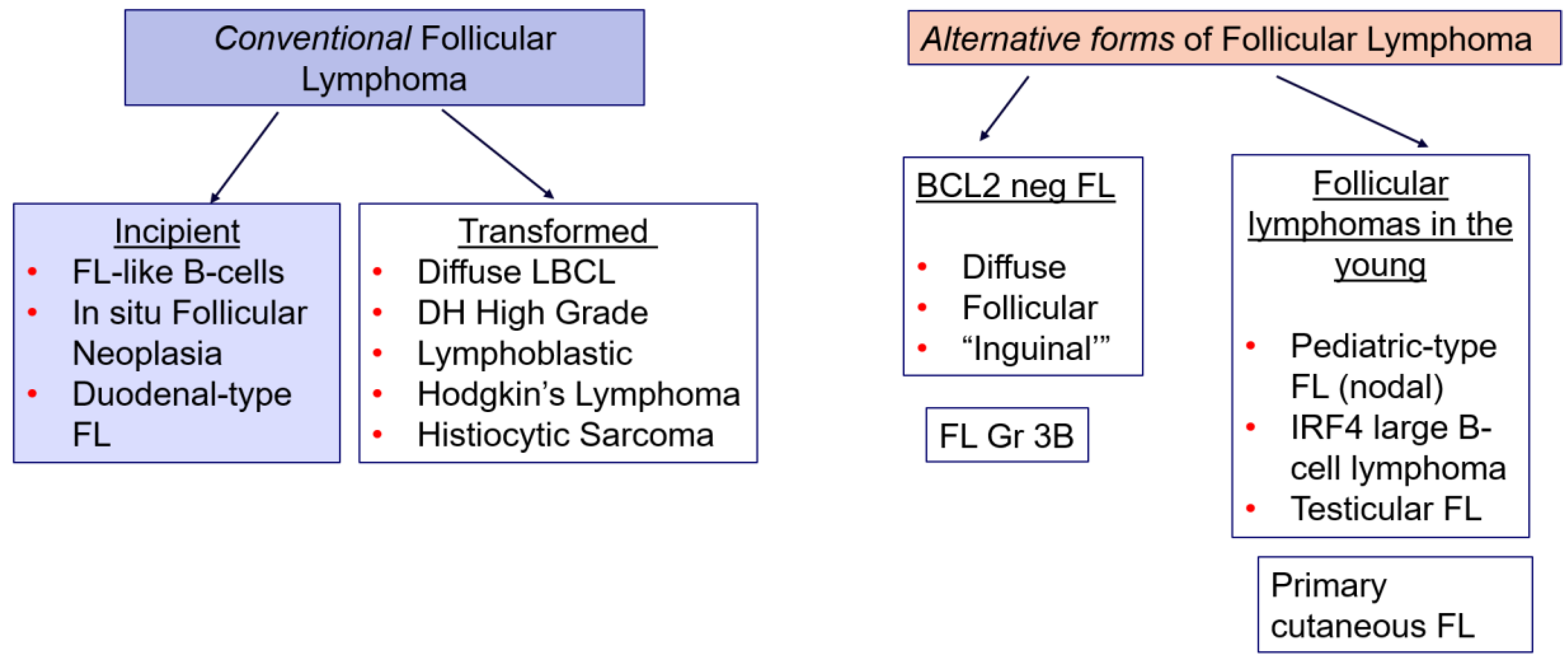
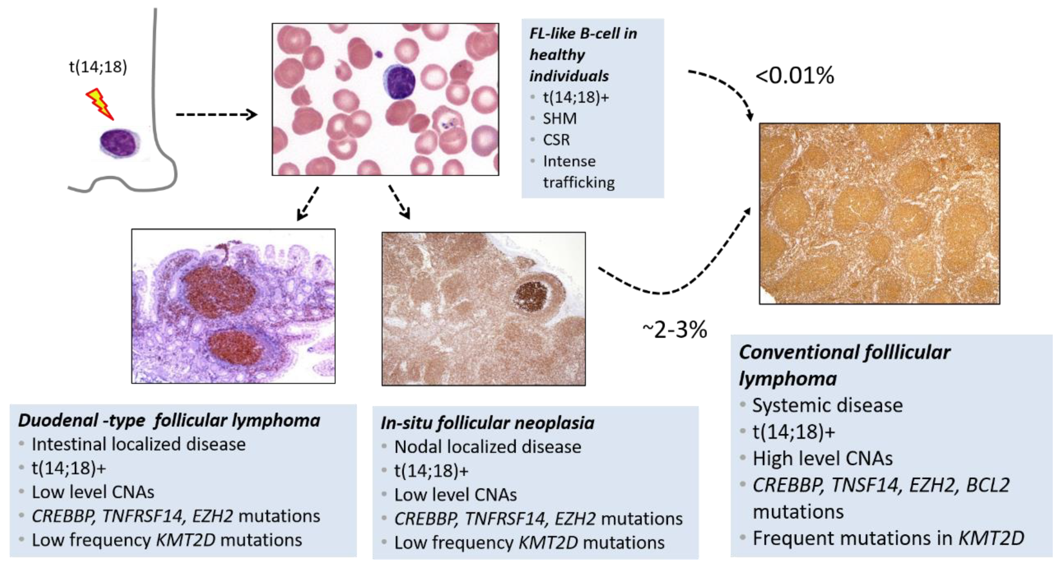
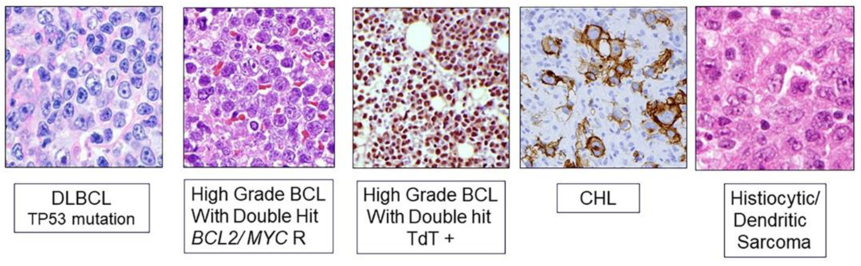
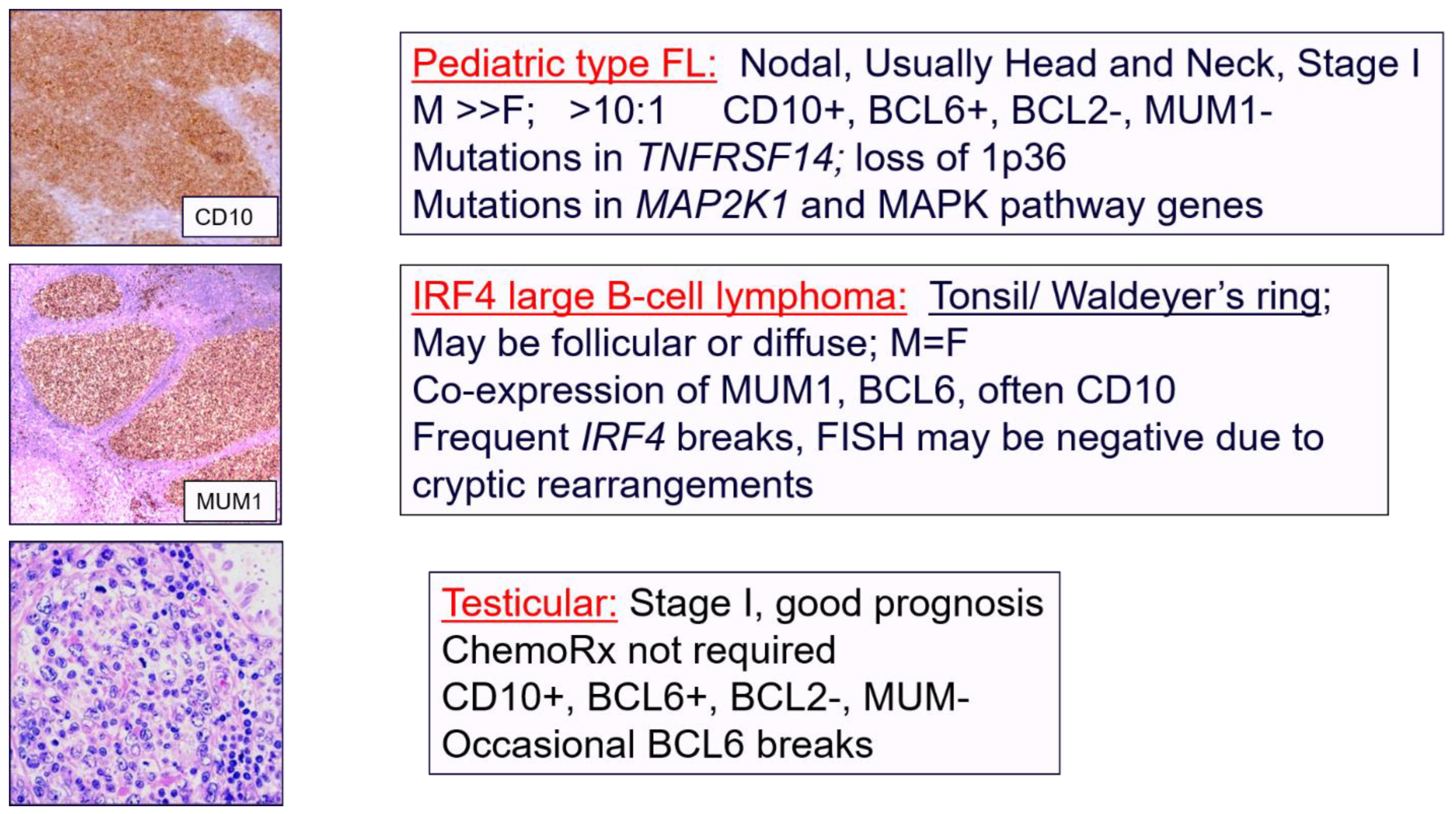
2. Evolving Spectrum of Follicular Lymphoma

The earliest stages in the evolution of FL have been explored (Figure 1). The classical model of FL lymphomagenesis is a multistage and progressive process, whereby t(14;18)(q32;q21) represents the founder event, and clinically significant disease results from a successive accumulation of genetic and epigenetic alterations. The discovery that non-neoplastic B cells that carry the t(14;18) in healthy individuals, later referred to as FL-like B-cells, influenced many aspects of FL research. FL like B cells are found in about 70% of individuals over the age of 50 [2].
In addition, the histological counterpart of this early lesion, is termed in situ follicular neoplasia (ISFN). It can be seen as an incidental finding in routine lymph node biopsies. ISFN is detected in 2–3% of routine lymph node biopsies. However, fewer than 5% of these patients will ever develop clinically significant FL. By array comparative genomic hybridization (CGH), ISFN has a very low level of genomic aberrations beyond the BCL2 translocation [5]. However, these early lesions may show mutations in CREBBP, EZH2, and TNFRSF14 [6]. Mutations in KMT2D appear to be a later event, associated with increased risk of progression.
Another early form of the disease is duodenal-type FL in which the BCL2 positive cells expand within the intestinal lymphoid tissue (Figure 2). All of these early lesions have a relatively low incidence of progression to clinically significant FL and this is especially rare in circulating FL-like B cells as found by sensitive PCR testing of normal peripheral blood [1,7].
Similarly, knowledge of the diverse forms of histological transformation that may occur in FL has been expanded. The two most common forms of transformation are DLBCL and high-grade B-cell lymphoma with double hit (Figure 3) but other rarer forms of histologic transformation include classic Hodgkin lymphoma, and histiocytic sarcoma. Some cases of high grade B-cell lymphoma may be TdT-positive and resemble B lymphoblastic lymphoma leukemia (B-ALL/LBL) [8]. Most cases expressing TdT carry both MYC and BCL2 rearrangement. However, their mutational profile is distinct from B-ALL/LBL [9]. Thus, they are best considered a variant of double hit lymphoma.
Transformation to classic Hodgkin lymphoma and histiocytic sarcoma indicates the plasticity of the hematopoietic system. Histiocytic or dendritic cell sarcomas show loss of the B-cell program, most likely related to loss of expression of PAX5 [10]. Secondary histiocytic sarcomas share some of the molecular alterations of FL but also show evidence of evolution with acquisition of new mutations that are characteristic of primary histiocytic sarcoma with mutations in the RAS/RAF/MAPK pathway (Figure 3) [11,12].
Alternative forms of FL, so-called BCL2 negative FL, have been recognized (Figure 1). BCL2 negative FL are both clinically and pathologically heterogeneous and probably do not constitute a single disease entity. However, BCL2 negative FL should be segregated from BCL2 rearranged FL, as both clinical and biological differences exist [13]. A distinctive subgroup includes those cases presenting often with inguinal disease that tend to be low stage. These cases have a high frequency of STAT6 mutations, are frequently positive for CD23, a helpful feature in diagnosis. These cases are negative for BCL2R still carry some of the genetic alterations of BCL2 positive FL with frequent mutations in CREBBP [13]. The recent proposal from the International Consensus Classification (ICC) of lymphoid neoplasms proposed the terminology of BCL2-R negative, CD23-positive follicle center lymphoma for this lesion [14]. It is likely that this subtype of follicle center lymphoma is suitable for different management approaches [15].
B-cell lymphomas of follicle cell derivation occurring in young patients differ from those in adults. These include pediatric type FL, testicular FL and large B-cell lymphoma with IRF4 rearrangement [16]. Pediatric type FL (PTFL) has a low level of genomic complexity. Aberrations in MAP2K1 and 1p36/TNFRSF14 are the most common genetic changes in PTFL, each observed in 30–70% of the cases [17,18]. A recurrent loss-of-function mutation in IRF8, a tumor suppressor gene, was also reported more recently [19]. Recent work has shown that PTFL and the pediatric variant of nodal marginal zone lymphoma (PMZL) are morphological variants with a common molecular profile [19]. These cases typically present with localized disease, Stage I, and recurrence following simple surgical excision is rare. It is important to distinguish these cases from more aggressive B-cell lymphomas in young patients, as the management is entirely different [20].
IRF4 large B-cell lymphoma most commonly presents in young patients (Figure 1 and Figure 4) with involvement of tonsil and Waldeyer’s ring. In contrast to PTFL, which is mainly seen in young boys, IRF4 large B-cell lymphoma is seen equally in males and females. These cases can be follicular or diffuse and show co-expression of MUM1 associated with IRF4 rearrangement, are BCL6 positive and often CD10 positive. They are of germinal center B-cell derivation; and have a relatively good prognosis in contrast to other forms of diffuse large B-cell lymphoma seen in young patients [1,16,21].
Finally, there is testicular follicular lymphoma, which is a rare condition seen in young boys; these patients have a very good prognosis and in the vast majority of cases appear to be cured by simple orchiectomy [22,23]. Systemic chemotherapy is not required.
Another rare form of FL is primary cutaneous follicular lymphoma (Figure 1). It is negative for BCL2 rearrangement and negative for CD10 and has some of the genetic alterations that are observed in pediatric type FL with 1p36 deletions and mutations in TNFSR14. However, it is negative for most of the genetic alterations seen in classic nodal FL. This is an indolent disease that should be managed conservatively. However, if there is evidence of BCL2-R or BCL2 protein expressed, clinical evaluation is suggested to rule out secondary cutaneous involvement from a systemic FL [24,25,26].
In conclusion, FL is not a single disease but is a family of tumors derived from follicle center B cells. The therapeutic options vary widely from aggressive therapy to a minimal intervention.

3. Diffuse Large B-Cell Lymphoma NOS and Aggressive B-Cell Lymphomas

Historically DLBCL was separated based on the cytological appearance in routine H&E–stained sections. The two most common variants were centroblastic and immunoblastic. We now recognize that DLBCLs are a complex group of aggressive B-cell lymphomas [4]. Figure 5 shows a chart that outlines the major subtypes of aggressive B-cell lymphomas at the time of the 2016 WHO classification [3], which recommended the segregation of ABC and GCB derived diffuse large B-cell lymphoma, and the recognition of high-grade B-cell lymphomas with double hit. The most common and well characterized form of “double hit” lymphoma, is a tumor that has translocations involving BCL2 and c-MYC. Some of these tumors represent progression from follicular lymphoma. In addition, not all cases with a double hit involving MYC and BCL2 are detected by FISH. By gene expression profiling the identification of a double hit signature can uncover double hit high grade B cell lymphomas with genetic events that are cryptic to FISH analysis [27].
In 2017 the WHO bluebook also identified a rare variant of aggressive lymphoma, seen mainly in young patients, and most often presenting with nodal disease. This tumor was included as a provisional entity and the term “Burkitt-like lymphoma with 11q aberration” was proposed [28]. More recent studies have shown that these tumors lack the common mutational findings of Burkitt lymphoma, are more correctly considered a variant of GCB derived diffuse large B-cell lymphoma [29,30]. In the recently published report from the International Consensus Committee, the term large B-cell lymphoma with 11q aberration is offered as the preferred nomenclature for this lesion [14]. While clinically aggressive, it is not included among the “high-grade” B-cell lymphomas.
Figure 6 shows a flow chart for the diagnosis of aggressive B-cell lymphomas that provides a model for current clinical practice. The first step is biopsy of lymph node or extranodal lesion. At this stage one should consider any one of a group of specific entities, such as such as primary mediastinal large B-cell lymphoma, EBV positive large B-cell lymphoma and HHV8/KSHV associated lymphomas. FISH is still important to recognize high grade B-cell lymphomas with double hit and represents a valuable tool in the clinical setting.
It is controversial as to whether aggressive B-cell lymphomas with dual translocations involving MYC and BCL6 should be retained in the “double hit” category [31,32]. These cases are heterogeneous at the gene expression level, and do not appear to have the very aggressive clinical behavior of the classic double hit lymphomas with MYC and BCL2. Further study of such cases is warranted to determine their proper place in the classification of aggressive B-cell lesions [14].
A new era is emerging with NGS and mutational profiling. This technology is on the horizon for clinical practice, although not currently required for the routine diagnosis of diffuse large B-cell lymphoma. However, there are many clinical settings in which NGS is commonly used in clinical practice.
Two major studies in the last few years showed that there is genetic heterogeneity in diffuse large B-cell lymphoma and that this heterogeneity can identify clinically significant genetic subgroups [33,34]. Variations in prognosis based on the mutational profile could be shown. The subtype termed MCD is based on the co-occurrence of the L265P mutation in MYD88 and mutations in CD79B. This subtype is highly enriched in primary central nervous system lymphoma, testicular diffuse large B-cell lymphoma, and other aggressive extranodal diffuse large B-cell lymphomas. The ICC group discussed the option of creating a separate category for extranodal DLBCL but deferred taking this step for the present time [14]. The subtype designated as BN2 is based on fusions involving BCL6 and mutations in NOTCH2. This subtype is heterogeneous in its gene expression profile and may represent an aggressive variant of marginal zone lymphoma. The prognosis of the BN2 group seems to be somewhat better than many of the other forms of diffuse large B-cell lymphoma [34]. Thus, it might represent marginal zone lymphoma with increased transformed cells.
In more recent work, even greater heterogeneity in the molecular classification of diffuse large B-cell lymphoma was observed [35]. New molecular variants were uncovered including a subset of aggressive lymphomas with a high degree of aneuploidy and a high frequency of p53 mutation and deletion [35]. The term A53 refers to these key features: p53 mutation and aneuploidy.
The historical cell of origin approach (ABC or activated B-cell; GCB or germinal center B-cell) for the classification of DLBCL can be integrated in part to the new data derived from genomic profiling based on NGS studies [36]. For example, the EZB and C4 subgroups are related to germinal center B cells. Mutations in EZB are common in germinal center derived neoplasms, including FL. The CD4 cluster, as designated by Chapuy et al. [33] includes the ST2 subset, as recognized by Wright et al. [35]. These cases are characterized by mutations in SGK1 and TET2 (hence ST2) and includes tumors with features of T-cell/histiocyte-rich large B-cell lymphoma related to nodular lymphocyte predominant B-cell lymphoma [14]. Tumors related to activated B-cells (ABC) are more heterogeneous, with the MCD group being a major subset. MCD tumors as noted above are almost always extranodal. N1, with mutations in NOTCH1 is ABC as well. The hope is that these data will be clinically relevant and lead to targeted therapy in the future based on the genetic profile [33,35,36,37,38]. A challenge will be to make this technology accessible for routine practice and diagnosis.

4. Pathology Provides a Roadmap for Disease Discovery and Treatment

Disease discovery and disease definition using routine diagnostic tools are critical first steps in elucidating the pathogenesis of lymphomas. Discovery of recurrent genetic alterations have usually followed on the heels of a precise description of the lymphoma entity based on clinical, morphological, or immunophenotypic grounds [39,40]. In other words, it starts with the microscope but insights from genetics, epigenetics, and knowledge of the cellular microenvironment, lead to refinement of diagnostic criteria, and ultimately appropriate therapy.

Author Contributions

E.S.J. designed the work, wrote the manuscript; A.C. wrote the manuscript. All authors have read and agreed to the published version of the manuscript.

Funding

This work was supported by the intramural research budget of the Center for Cancer Research, National Cancer Institute: ZIA SC 000550.

Institutional Review Board Statement

Not applicable.

Informed Consent Statement

Not applicable.

Data Availability Statement

Not applicable.

Conflicts of Interest

The authors declare no conflict of interest.

References

  1. Jaffe, E.S.; Harris, N.L.; Swerdlow, S.H.; Ott, G.; Nathwani, B.N.; de Jong, D.; Yoshino, T.; Spagnolo, D.; Gascoyne, R.D. Follicular lymphoma. In WHO Classification of Tumours of Haematopoietic and Lymphoid Tissues, 4th ed.; Swerdlow, S.H., Campo, E., Harris, N.L., Jaffe, E.S., Pileri, S.A., Stein, H., Thiele, J., Eds.; IARC Press: Lyon, France, 2017; pp. 266–277. [Google Scholar]
  2. Carbone, A.; Roulland, S.; Gloghini, A.; Younes, A.; von Keudell, G.; López-Guillermo, A.; Fitzgibbon, J. Follicular lymphoma. Nat. Rev. Dis. Primers 2019, 5, 83. (In English) [Google Scholar] [CrossRef]
  3. Swerdlow, S.H.; Campo, E.; Pileri, S.A.; Harris, N.L.; Stein, H.; Siebert, R.; Advani, R.; Ghielmini, M.; Salles, G.A.; Zelenetz, A.D.; et al. The 2016 revision of the World Health Organization classification of lymphoid neoplasms. Blood 2016, 127, 2375–2390. (In English) [Google Scholar] [CrossRef] [PubMed] [Green Version]
  4. Gascoyne, R.D.; Chan, J.K.C.; Campo, E.; Rosenwald, A.; Jaffe, E.S.; Stein, H.; Chan, W.C.; Swerdlow, S.H. Diffuse large B-cell lymphoma, NOS. In WHO Classification of Tumours of Haematopoietic and Lymphoid Tissues, 4th ed.; IARC, Ed.; IARC Press: Lyon, France, 2017; pp. 291–297. [Google Scholar]
  5. Mamessier, E.; Song, J.Y.; Eberle, F.C.; Pack, S.; Drevet, C.; Chetaille, B.; Abdullaev, Z.; Adelaide, J.; Birnbaum, D.; Chaffanet, M.; et al. Early lesions of follicular lymphoma: A genetic perspective. Haematologica 2014, 99, 481–488. [Google Scholar] [CrossRef] [PubMed] [Green Version]
  6. Schmidt, J.; Ramis-Zaldivar, J.E.; Bonzheim, I.; Steinhilber, J.; Muller, I.; Haake, A.; Yu, S.C.; Raffeld, M.; Fend, F.; Salaverria, I.; et al. CREBBP gene mutations are frequently detected in in situ follicular neoplasia. Blood 2018, 132, 2687–2690. (In English) [Google Scholar] [CrossRef] [Green Version]
  7. Jaffe, E.S.; Quintanilla-Martinez, L. t(14;18)-positive B cells: Is it seed or soil? Blood 2018, 132, 1631–1632. (In English) [Google Scholar] [CrossRef]
  8. Geyer, J.T.; Subramaniyam, S.; Jiang, Y.; Elemento, O.; Ferry, J.A.; de Leval, L.; Nakashima, M.O.; Liu, Y.C.; Martin, P.; Mathew, S.; et al. Lymphoblastic transformation of follicular lymphoma: A clinicopathologic and molecular analysis of 7 patients. Hum. Pathol. 2015, 46, 260–271. (In English) [Google Scholar] [CrossRef]
  9. Bhavsar, S.; Liu, Y.C.; Gibson, S.E.; Moore, E.M.; Swerdlow, S.H. Mutational Landscape of TdT+ Large B-cell Lymphomas Supports Their Distinction From B-lymphoblastic Neoplasms: A Multiparameter Study of a Rare and Aggressive Entity. Am. J. Surg. Pathol. 2022, 46, 71–82. (In English) [Google Scholar] [CrossRef]
  10. Feldman, A.L.; Arber, D.A.; Pittaluga, S.; Martinez, A.; Burke, J.S.; Raffeld, M.; Camos, M.; Warnke, R.; Jaffe, E.S. Clonally related follicular lymphomas and histiocytic/dendritic cell sarcomas: Evidence for transdifferentiation of the follicular lymphoma clone. Blood 2008, 111, 5433–5439. (In English) [Google Scholar] [CrossRef] [Green Version]
  11. Egan, C.; Lack, J.; Skarshaug, S.; Pham, T.A.; Abdullaev, Z.; Xi, L.; Pack, S.; Pittaluga, S.; Jaffe, E.S.; Raffeld, M. The mutational landscape of histiocytic sarcoma associated with lymphoid malignancy. Mod. Pathol. 2021, 34, 336–347. (In English) [Google Scholar] [CrossRef]
  12. Shanmugam, V.; Griffin, G.K.; Jacobsen, E.D.; Fletcher, C.D.M.; Sholl, L.M.; Hornick, J.L. Identification of diverse activating mutations of the RAS-MAPK pathway in histiocytic sarcoma. Mod. Pathol. 2019, 32, 830–843. (In English) [Google Scholar] [CrossRef]
  13. Nann, D.; Ramis-Zaldivar, J.E.; Müller, I.; Gonzalez-Farre, B.; Schmidt, J.; Egan, C.; Salmeron-Villalobos, J.; Clot, G.; Mattern, S.; Otto, F.; et al. Follicular lymphoma t(14;18)-negative is genetically a heterogeneous disease. Blood Adv. 2020, 4, 5652–5665. (In English) [Google Scholar] [CrossRef] [PubMed]
  14. Campo, E.; Jaffe, E.S.; Cook, J.R.; Quintanilla-Martinez, L.; Swerdlow, S.H.; Anderson, K.C.; Brousset, P.; Cerroni, L.; de Leval, L.; Dirnhofer, S.; et al. The International Consensus Classification of Mature Lymphoid Neoplasms: A Report from the Clinical Advisory Committee. Blood, 2022; in press. (In English). [Google Scholar] [CrossRef]
  15. Katzenberger, T.; Kalla, J.; Leich, E.; Stocklein, H.; Hartmann, E.; Barnickel, S.; Wessendorf, S.; Ott, M.M.; Muller-Hermelink, H.K.; Rosenwald, A.; et al. A distinctive subtype of t(14;18)-negative nodal follicular non-Hodgkin lymphoma characterized by a predominantly diffuse growth pattern and deletions in the chromosomal region 1p36. Blood 2009, 113, 1053–1061. Available online: http://www.ncbi.nlm.nih.gov/entrez/query.fcgi?cmd=Retrieve&db=PubMed&dopt=Citation&list_uids=18978208 (accessed on 1 July 2022). (In English). [PubMed] [Green Version]
  16. Liu, Q.; Salaverria, I.; Pittaluga, S.; Jegalian, A.G.; Xi, L.; Siebert, R.; Raffeld, M.; Hewitt, S.M.; Jaffe, E.S. Follicular lymphomas in children and young adults: A comparison of the pediatric variant with usual follicular lymphoma. Am. J. Surg. Pathol. 2013, 37, 333–343. (In English) [Google Scholar] [CrossRef] [Green Version]
  17. Louissaint, A., Jr.; Schafernak, K.T.; Geyer, J.T.; Kovach, A.E.; Ghandi, M.; Gratzinger, D.; Roth, C.G.; Paxton, C.N.; Kim, S.; Namgyal, C.; et al. Pediatric-type nodal follicular lymphoma: A biologically distinct lymphoma with frequent MAPK pathway mutations. Blood 2016, 128, 1093–1100. (In English) [Google Scholar] [CrossRef] [PubMed] [Green Version]
  18. Schmidt, J.; Ramis-Zaldivar, J.E.; Nadeu, F.; Gonzalez-Farre, B.; Navarro, A.; Egan, C.; Montes-Mojarro, I.A.; Marafioti, T.; Cabecadas, J.; van der Walt, J.; et al. Mutations of MAP2K1 are frequent in pediatric-type follicular lymphoma and result in ERK pathway activation. Blood 2017, 130, 323–327. (In English) [Google Scholar] [CrossRef] [PubMed] [Green Version]
  19. Salmeron-Villalobos, J.; Egan, C.; Borgmann, V.; Müller, I.; Gonzalez-Farre, B.; Ramis-Zaldivar, J.E.; Nann, D.; Balagué, O.; Lopez-Guerra, M.; Colomer, D.; et al. PNMZL and PTFL: Morphological variants with a common molecular profile—A unifying hypothesis. Blood Adv. 2022; in press. (In English). [Google Scholar] [CrossRef]
  20. Louissaint, A., Jr.; Ackerman, A.M.; Dias-Santagata, D.; Ferry, J.A.; Hochberg, E.P.; Huang, M.S.; Iafrate, A.J.; Lara, D.O.; Pinkus, G.S.; Salaverria, I.; et al. Pediatric-type nodal follicular lymphoma: An indolent clonal proliferation in children and adults with high proliferation index and no BCL2 rearrangement. Blood 2012, 120, 2395–2404. (In English) [Google Scholar] [CrossRef] [Green Version]
  21. Ramis-Zaldivar, J.E.; Gonzalez-Farré, B.; Balagué, O.; Celis, V.; Nadeu, F.; Salmerón-Villalobos, J.; Andrés, M.; Martin-Guerrero, I.; Garrido-Pontnou, M.; Gaafar, A.; et al. Distinct molecular profile of IRF4-rearranged large B-cell lymphoma. Blood 2020, 135, 274–286. (In English) [Google Scholar] [CrossRef]
  22. Finn, L.S.; Viswanatha, D.S.; Belasco, J.B.; Snyder, H.; Huebner, D.; Sorbara, L.; Raffeld, M.; Jaffe, E.S.; Salhany, K.E. Primary follicular lymphoma of the testis in childhood. Cancer 1999, 85, 1626–1635. Available online: http://www.ncbi.nlm.nih.gov/pubmed/10193956 (accessed on 1 July 2022).
  23. Lones, M.A.; Raphael, M.; McCarthy, K.; Wotherspoon, A.; Terrier-Lacombe, M.J.; Ramsay, A.D.; Maclennan, K.; Cairo, M.S.; Gerrard, M.; Michon, J.; et al. Primary follicular lymphoma of the testis in children and adolescents. J. Pediatr. Hematol. Oncol. 2012, 34, 68–71. (In English) [Google Scholar] [CrossRef] [Green Version]
  24. Barasch, N.J.K.; Liu, Y.C.; Ho, J.; Bailey, N.; Aggarwal, N.; Cook, J.R.; Swerdlow, S.H. The molecular landscape and other distinctive features of primary cutaneous follicle center lymphoma. Hum. Pathol. 2020, 106, 93–105. (In English) [Google Scholar] [CrossRef]
  25. Gángó, A.; Bátai, B.; Varga, M.; Kapczár, D.; Papp, G.; Marschalkó, M.; Kuroli, E.; Schneider, T.; Csomor, J.; Matolcsy, A.; et al. Concomitant 1p36 deletion and TNFRSF14 mutations in primary cutaneous follicle center lymphoma frequently expressing high levels of EZH2 protein. Virchows Arch. 2018, 473, 453–462. (In English) [Google Scholar] [CrossRef] [PubMed]
  26. Lucioni, M.; Fraticelli, S.; Neri, G.; Feltri, M.; Ferrario, G.; Riboni, R.; Paulli, M. Primary Cutaneous B-Cell Lymphoma: An Update on Pathologic and Molecular Features. Hemato 2022, 3, 318–340. Available online: https://www.mdpi.com/2673-6357/3/2/23 (accessed on 1 July 2022).
  27. Hilton, L.K.; Tang, J.; Ben-Neriah, S.; Alcaide, M.; Jiang, A.; Grande, B.M.; Rushton, C.K.; Boyle, M.; Meissner, B.; Scott, D.W.; et al. The double-hit signature identifies double-hit diffuse large B-cell lymphoma with genetic events cryptic to FISH. Blood 2019, 134, 1528–1532. (In English) [Google Scholar] [CrossRef] [PubMed] [Green Version]
  28. Salaverria, I.; Martin-Guerrero, I.; Wagener, R.; Kreuz, M.; Kohler, C.W.; Richter, J.; Pienkowska-Grela, B.; Adam, P.; Burkhardt, B.; Claviez, A.; et al. A recurrent 11q aberration pattern characterizes a subset of MYC-negative high-grade B-cell lymphomas resembling Burkitt lymphoma. Blood 2014, 123, 1187–1198. [Google Scholar] [CrossRef] [Green Version]
  29. Gonzalez-Farre, B.; Ramis-Zaldivar, J.E.; Salmeron-Villalobos, J.; Balagué, O.; Celis, V.; Verdu-Amoros, J.; Nadeu, F.; Sábado, C.; Ferrández, A.; Garrido, M.; et al. Burkitt-like lymphoma with 11q aberration: A germinal center-derived lymphoma genetically unrelated to Burkitt lymphoma. Haematologica 2019, 104, 1822–1829. (In English) [Google Scholar] [CrossRef]
  30. Wagener, R.; Seufert, J.; Raimondi, F.; Bens, S.; Kleinheinz, K.; Nagel, I.; Altmuller, J.; Thiele, H.; Hubschmann, D.; Kohler, C.W.; et al. The mutational landscape of Burkitt-like lymphoma with 11q aberration is distinct from that of Burkitt lymphoma. Blood 2019, 133, 962–966. (In English) [Google Scholar] [CrossRef] [Green Version]
  31. Moore, E.M.; Aggarwal, N.; Surti, U.; Swerdlow, S.H. Further Exploration of the Complexities of Large B-Cell Lymphomas with MYC Abnormalities and the Importance of a Blastoid Morphology. Am. J. Surg. Pathol. 2017, 41, 1155–1166. (In English) [Google Scholar] [CrossRef]
  32. Pillai, R.K.; Sathanoori, M.; Van Oss, S.B.; Swerdlow, S.H. Double-hit B-cell lymphomas with BCL6 and MYC translocations are aggressive, frequently extranodal lymphomas distinct from BCL2 double-hit B-cell lymphomas. Am. J. Surg. Pathol. 2013, 37, 323–332. (In English) [Google Scholar] [CrossRef]
  33. Chapuy, B.; Stewart, C.; Dunford, A.J.; Kim, J.; Kamburov, A.; Redd, R.A.; Lawrence, M.S.; Roemer, M.G.M.; Li, A.J.; Ziepert, M.; et al. Molecular subtypes of diffuse large B cell lymphoma are associated with distinct pathogenic mechanisms and outcomes. Nat. Med. 2018, 24, 679–690. (In English) [Google Scholar] [CrossRef]
  34. Schmitz, R.; Wright, G.W.; Huang, D.W.; Johnson, C.A.; Phelan, J.D.; Wang, J.Q.; Roulland, S.; Kasbekar, M.; Young, R.M.; Shaffer, A.L.; et al. Genetics and Pathogenesis of Diffuse Large B-Cell Lymphoma. N. Engl. J. Med. 2018, 378, 1396–1407. (In English) [Google Scholar] [CrossRef]
  35. Wright, G.W.; Huang, D.W.; Phelan, J.D.; Coulibaly, Z.A.; Roulland, S.; Young, R.M.; Wang, J.Q.; Schmitz, R.; Morin, R.D.; Tang, J.; et al. A Probabilistic Classification Tool for Genetic Subtypes of Diffuse Large B Cell Lymphoma with Therapeutic Implications. Cancer Cell 2020, 37, 551–568.e514. (In English) [Google Scholar] [CrossRef] [PubMed]
  36. Ennishi, D.; Hsi, E.D.; Steidl, C.; Scott, D.W. Toward a New Molecular Taxonomy of Diffuse Large B-cell Lymphoma. Cancer Discov. 2020, 10, 1267–1281. [Google Scholar] [CrossRef] [PubMed]
  37. Lacy, S.E.; Barrans, S.L.; Beer, P.A.; Painter, D.; Smith, A.G.; Roman, E.; Cooke, S.L.; Ruiz, C.; Glover, P.; Van Hoppe, S.J.L.; et al. Targeted sequencing in DLBCL, molecular subtypes, and outcomes: A Haematological Malignancy Research Network report. Blood 2020, 135, 1759–1771. (In English) [Google Scholar] [CrossRef] [PubMed]
  38. Sha, C.; Barrans, S.; Cucco, F.; Bentley, M.A.; Care, M.A.; Cummin, T.; Kennedy, H.; Thompson, J.S.; Uddin, R.; Worrillow, L.; et al. Molecular High-Grade B-Cell Lymphoma: Defining a Poor-Risk Group That Requires Different Approaches to Therapy. J. Clin. Oncol. 2019, 37, 202–212. (In English) [Google Scholar] [CrossRef]
  39. Jaffe, E.S.; Harris, N.L.; Stein, H.; Isaacson, P.G. Classification of lymphoid neoplasms: The microscope as a tool for disease discovery. Blood 2008, 112, 4384–4399. (In English) [Google Scholar] [CrossRef] [Green Version]
  40. Carbone, A. Classification of Tumors of the Hematopoietic and Lymphoid Tissues. Discovering Diseases—Defining Their Features. Hemato 2020, 1, 7–9. Available online: https://www.mdpi.com/2673-6357/1/1/4 (accessed on 1 July 2022).
Figure 1. Evolving spectrum of follicular lymphoma.
Hemato 03 00032 g001
Figure 2. Incipient follicular lymphoma (FL) includes FL-like B cells found in the peripheral blood at low levels in normal individuals, most commonly after age 50. Tissue manifestations of incipient FL include in situ follicular neoplasia and duodenal-type FL. Abbreviations. CNAs, chromosomal numeric aberrations; SHM, somatic hypermutations; CSR: Class switch recombination. Reproduced with permission from Jaffe and Quintanilla-Martinez [7].
Hemato 03 00032 g002
Figure 3. Transformed Follicular Lymphoma. All show clonal identity and retain the BCL2 rearrangement.
Hemato 03 00032 g003
Figure 4. Follicular Lymphomas (FL) in young patients (<30 years) include pediatric type FL, IRF4 large B-cell lymphoma, testicular FL. Modified and adapted from Liu Q et al. [16].
Hemato 03 00032 g004
Figure 5. Aggressive B-cell lymphomas. Modified and adapted from Swerdlow et al. [3].
Hemato 03 00032 g005
Figure 6. Flow Chart for the Diagnosis of Aggressive B-cell Lymphomas.
Hemato 03 00032 g006
Publisher’s Note: MDPI stays neutral with regard to jurisdictional claims in published maps and institutional affiliations.

