Review

The Evolution of Circulating Biomarkers for Use in Acetaminophen/Paracetamol-Induced Liver Injury in Humans: A Scoping Review

by
Mitchell R. McGill
1,2,3,* and
Steven C. Curry
4,5
1
Department of Environmental Health Sciences, Fay W. Boozman College of Public Health, University of Arkansas for Medical Sciences, Little Rock, AR 72205, USA
2
Department of Pharmacology and Toxicology, College of Medicine, University of Arkansas for Medical Sciences, Little Rock, AR 72205, USA
3
Department of Pathology, College of Medicine, University of Arkansas for Medical Sciences, Little Rock, AR 72205, USA
4
Division of Clinical Data Analytics and Decision Support, University of Arizona College of Medicine—Phoenix, Phoenix, AZ 85006, USA
5
Department of Medical Toxicology, Banner—University Medical Center Phoenix, Phoenix, AZ 85006, USA
*
Author to whom correspondence should be addressed.
Livers 2023, 3(4), 569-596; https://doi.org/10.3390/livers3040039
Submission received: 12 September 2023 / Revised: 13 October 2023 / Accepted: 18 October 2023 / Published: 27 October 2023
(This article belongs to the Special Issue Recent Advances in Acetaminophen Hepatotoxicity)

Abstract

:
Acetaminophen (APAP) is a widely used drug, but overdose can cause severe acute liver injury. The first reports of APAP hepatotoxicity in humans were published in 1966, shortly after the development of alanine aminotransferase (ALT) and aspartate aminotransferase (AST) as the first biomarkers of liver injury as opposed to liver function. Thus, the field of liver injury biomarkers has evolved alongside the growth in APAP hepatotoxicity incidence. Numerous biomarkers have been proposed for use in the management of APAP overdose patients in the intervening years. Here, we comprehensively review the development of these markers from the 1960s to the present day and briefly discuss possible future directions.

1. Introduction

Acetaminophen (APAP; also known as paracetamol) toxicity was recognized as a clinical problem around 1966, when the first two cases of APAP overdose in patients were described by D.G. Davidson—then associated with the storied Ward 3, Regional Poisoning Treatment Center at the Royal Infirmary in Edinburgh [1]—and W. Noel Eastham [2]. APAP-induced liver and renal damage had been observed in cats [3] several years before and in rodents earlier in the same year [4], but APAP had not been thought of as a significant risk to humans. Indeed, many considered it safer than other analgesics available at that time (for example, see [5]). However, more reports of APAP toxicity soon followed [6,7,8,9,10,11,12,13,14] as its incidence rapidly increased throughout the UK and Europe [15]. Within a decade, APAP overdose arrived in North America as well, with the first reports appearing in 1971 [8]. Today, APAP overdose is a major cause of acute liver failure (ALF) in the US and UK, and a lesser cause in other countries [15]. In the US, for example, approximately 50,000 to 80,000 emergency department visits per year are attributable to APAP overdose [16,17,18], and APAP hepatotoxicity accounts for roughly half of all ALF cases [19].
The first reports by Davidson and Eastham are as notable for what they did not include as for what they did. In addition to presentation and symptomology, the authors provided detailed descriptions of the gross and microscopic histopathology of the liver and kidney that laid the foundation for later mechanistic investigations into modes of cell death and inflammation in APAP hepatotoxicity. They also provided results from 11 (Case 1) and 18 (Case 2) clinical chemistry, hematology, and coagulation tests, and suggested ways in which these data might be used to differentiate APAP toxicity from other causes of liver damage. Conspicuously lacking for the modern reader, however, was any mention of the serum transaminases, alanine aminotransferase (ALT; also known as SGPT, for serum glutamate-pyruvate transaminase) and aspartate aminotransferase (AST; also known as SGOT, for serum glutamate-oxaloacetate transaminase). These two enzymes were described as liver injury biomarkers only a decade earlier [20,21,22]. Although AST was routinely measured in serum in many clinical laboratories by 1960, ALT evidently took longer to catch on. Though ALT reagent kits [23] were becoming available around that time and were even available for the newly developed automated analyzers of the time [24,25], they were not as ubiquitous as they are today. Thus, the history of APAP toxicity has run parallel with the evolution of liver injury biomarkers. Furthermore, because APAP overdose is common, it is relatively easy to obtain serum and plasma specimens from APAP toxicity patients. As a result, many recent advances in liver injury biomarker development have come from studies on APAP overdose.
Here, we review APAP toxicity biomarkers in more-or-less chronological order. We begin by briefly discussing the broad categories of use of biomarkers in APAP overdose patients; then, we describe the earliest tests for liver injury, like ALT and AST, in the context of APAP overdose; continue with development of APAP toxicity diagnostics based on APAP and APAP metabolite levels; describe advances in both protein and microRNA (miRNA)-based markers; and conclude with a discussion of the lessons we can learn from these data and possible future directions in APAP biomarker research. Note that some prior knowledge of APAP metabolism, mechanisms of toxicity, and treatments is assumed. The reader is directed to other reviews for more information on these topics [26,27,28].

2. Uses of Biomarkers in APAP Overdose

Biomarkers in APAP overdose can be broadly grouped into (1) biomarkers for diagnosis, (2) biomarkers for prognosis, and (3) mechanistic biomarkers. Biomarkers for diagnosis should be elevated early enough after overdose to permit detection in early-presenting patients and should be specific enough for APAP that they can distinguish between liver injury caused by the drug and liver injury due to other etiologies in a broad set of liver injury patients. The latter is especially difficult as we will see in our discussion of APAP-protein adducts, which one might reasonably expect to have high specificity for APAP overdose. More biomarkers of prognosis are available, but the evaluation of those markers is complicated, due, in part, to variations in endpoints. For example, one study may be designed to identify biomarkers that predict elevated peak prothrombin time (PT), while another study is designed to look for biomarkers that predict death. While there is an association between peak PT and death, the correlation is imperfect, and so these different designs can yield different results. In another example, studies may be designed to discover biomarkers that identify patients who are at risk of developing high ALT elevations (and therefore in need of treatment) despite presenting with ALT values in the normal range. This may seem straightforward until one considers that ALT elevation is defined in some studies as anything >50 U/L (in other words, any release of ALT from the liver above roughly the upper limit of normal), while in others it is defined as only values >1000 U/L (which are more likely to lead to poor outcomes). In other words, the clinical utility of a biomarker depends on the particular outcome one wants to predict, and many different outcomes have been used in the literature. We can simplify things by sub-dividing prognostic biomarkers into (A) biomarkers intended to predict hepatotoxicity in patients who initially present without evidence of liver injury and (B) biomarkers intended to predict poor clinical outcomes, like encephalopathy or death. We will use this convention roughly throughout the manuscript. However, the reader should consider exactly what outcome the authors of a study are trying to predict and whether or not that outcome is clinically meaningful or optimal.

3. ALT, AST, and Other Enzymes

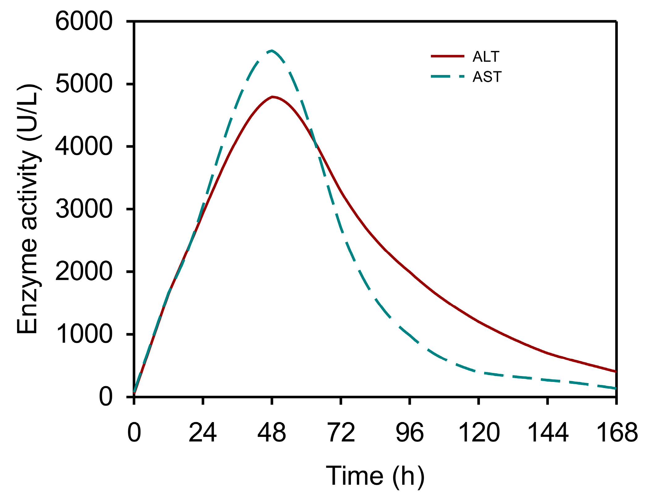
3.1. The Kinetics of Serum Aminotransferases after APAP Overdose

ALT and AST are transaminases present in both the cytosol (ALT1 and AST1) and mitochondria (ALT2 and AST2) of hepatocytes. Both ALT1 and AST1 activity are present in circulation at baseline (with lesser contributions from ALT2 and AST2) [29,30]—generally believed to be due to normal hepatocyte turnover, though other mechanisms may be involved. Although the hepatic AST/ALT ratio is approximately 2.5:1, AST1 has a shorter half-life in blood, resulting in a roughly 1:1 ratio in circulation under normal conditions [31]. Karmen et al. and De Ritis et al. independently reported elevations of ALT and AST activity in serum from patients with liver injury in 1955 [20,22,32,33]. By the mid-to-late 1960s, ALT (measured using the coupled enzyme reaction approach developed by Karmen for AST [20] and later applied to ALT [21]) had become the standard blood biomarker of hepatocellular damage for both clinical and experimental use, and has remained so ever since [33]. Thomson and Prescott briefly mentioned ALT and AST values in the range of 100–200 “units” in one APAP overdose patient in 1966 [6], just after the reports from Davidson and Eastham, but the first complete time courses of these enzymes in an APAP patient were published by a group in Australia two years later [34]. These investigators observed peak values on day 4 of hospitalization followed by a sharp decline in AST the next day and a more gradual recess of ALT over the next several days. Similar kinetics have been reported by other groups since then [35,36,37,38,39,40], and it is now generally agreed that both transaminases begin to rise with a 1:1 AST/ALT ratio within 24 h of overdose, peak between 48 and 72 h with a 1:1 to 3:1 ratio, and fall thereafter, with a faster drop in AST in the absence of preexisting liver disease. The serum half-lives for AST and ALT during the latter phase are around 15–20 h and 40–50 h, respectively [37]. A typical ALT time course in humans is shown in Figure 1.

3.2. Uses and Limitations of Serum Aminotransferases in APAP Toxicity

Elevated aminotransferases can occur for a number of reasons, so they cannot be used to diagnose APAP overdose specifically. However, they are useful for the detection of potential liver injury that should elicit further investigations. It has been reported that ALT values also have some prognostic value to predict hepatotoxicity and therefore guide N-acetyl-l-cysteine (NAC) treatment. James Dear and colleagues reviewed the charts of 410 APAP overdose patients from their institution to determine if an initial ALT above 50 U/L could predict later injury, as indicated primarily by peak ALT ≥ 1000 U/L or prothrombin time >2 [41]. While the positive predictive value (PPV) of ALT > 50 was low, the negative predictive value (NPV) was around 100%. Of course, this was likely because the patients with ALT < 50 presented before the development of liver injury and received prompt NAC treatment (even if the patients’ clinical histories may have indicated later presentation). Curry and others reported that among 160 APAP overdose patients in whom NAC was commenced within 4 h of having an ALT ≤ 50 U/L, just 10 (6%) developed a peak ALT > 1000 U/L. Of 127 patients with an initial ALT > 100 U/L near the time of beginning NAC therapy, 29 developed hepatic encephalopathy, and 12 died. No patient died if NAC therapy was commenced when ALT was <200 U/L [42].
As for patients who present after the onset of injury, a decline in either ALT or AST in serial measurements along with improvements in hepatic synthetic function and a lack of, or improving, encephalopathy likely indicate that the patient has passed the peak of injury and may no longer benefit from NAC, which targets only the early induction of APAP toxicity. AST may be especially useful for this purpose because it falls faster than ALT and could theoretically allow treatment discontinuation and patient discharge earlier [37]. Taking this idea a step further, it has been suggested that a AST/ALT ratio < 0.4 can identify patients who are beyond the window for NAC treatment without the need for serial measurements [32]. Ideas such as these have been criticized, however, based on evidence that NAC improves survival even when administered very late post-overdose. Two papers [43,44] by the late Roger Williams are often cited to support this view [45]. In one of these studies [43], the investigators included patients treated with NAC within 10–36 h post-overdose, with a median time of 17 h—well within the early phase of APAP toxicity in humans, when the transaminases are still rising, as discussed above. The second study [44], however, is more compelling. In that experiment, the investigators randomized patients with liver failure into two groups, NAC and no NAC, and looked at patient survival over the next 21 days. Importantly, the patients who received NAC were treated ≥30 h after overdose, though the authors did not describe how the exact time of ingestion was determined. Approximately 60% of the NAC patients survived compared to 20% of the controls [44]. While the investigators relied on small sample sizes, with only 25 patients per group, the results are supported by data from mice and primary human hepatocytes. It has been demonstrated that NAC protects against APAP-induced liver injury in mice even up to 2 h after overdose [46] and as late as 6 h after initial APAP exposure in primary human hepatocytes [47]. Both time points are after APAP metabolism is complete [46,48] and when there is already evidence of oxidative stress and mitochondrial damage [46,47]. However, all of these time points—30 h in humans, 2 h in mice, and 6 h in human hepatocytes—are still before the typical peak of transaminases, around 48–72 h in humans and human hepatocytes and 12 h in mice. By the time transaminases have peaked and begun to fall at even later time points, it seems likely correct that NAC is not useful.
In addition to guiding NAC treatment, ALT and AST may have some prognostic utility to predict poor outcomes. Although large ALT and AST elevations, by themselves, do not predict death in APAP hepatotoxicity or any other forms of liver injury [38,39,49], combining ALT and AST together or with other markers may improve their value for this purpose. For example, patients who died after APAP overdose tended to have a greater AST/ALT ratio at the time of NAC treatment in one cohort [37]. However, that study included only six patients who died. Larger studies are needed to further explore and validate this use of the AST/ALT ratio. In another example, it has been noted that patients with shorter doubling times for ALT and AST from the approximate time of APAP ingestion to peak injury usually have higher peak INR values [40], with the latter having an association with liver failure and death in the most severe cases. Based on the latter study and on the observation that slower declines in serum APAP concentration are associated with faster onset of toxicity, Sivilotti et al. [50] proposed the parameter “APAP × AT” to predict liver injury that is both rapid and severe after APAP overdose (defined as reaching ALT values ≥1000 U/L within 24 h of ingestion and high peak INR, respectively) despite NAC treatment. APAP × AT is the multiplication product of the earliest paired serum APAP and aminotransferase values available—in other words, the product of APAP and either ALT or AST nearest to the time of patient presentation or treatment. Because serum APAP declines due to metabolism while aminotransferases proportionally increase due to liver damage, this product is more-or-less stable over time until about the peak of injury [50], making it somewhat independent of the time elapsed since APAP ingestion. However, INR is only a surrogate for death, and the ability of APAP × AT to actually predict death after APAP overdose has not yet been tested. Overall, then, the utility of ALT and AST to predict poor clinical outcomes remains limited.

3.3. Other Early Enzymes

About the same time that serum ALT and AST were found to increase in patients with liver injury, some of the same investigators noted that high lactate dehydrogenase (LDH) activities “were observed in patients with myocardial infarction, diabetic acidosis, acute stem cell leukemia, chronic myelogenous leukemia and hepatitis” [51]. These results were quickly confirmed by others [52]. However, LDH is expressed in other organs at or near the level in the liver [51,53,54], so serum values are not specific for liver damage. Indeed, serum LDH is elevated in numerous diseases [55]. As a result, it is not widely viewed as an important liver injury marker and has no diagnostic value for APAP overdose beyond what ALT and AST provide. But, recent data have demonstrated that it does have prognostic value. We used an untargeted proteomics approach to analyze serum samples from 58 transplant-free survivors and non-survivors of APAP-induced ALF [49]. Much to our surprise, LDH displayed the greatest ability to predict death out of >1600 proteins that we were able to measure [49]. We then confirmed the prognostic value of LDH in this context through a retrospective review of laboratory data from 238 patients hospitalized with ALF at our institution over a 12-year period [39]. Importantly, LDH performed about as well as, or better than, the current prognostic tools, the Model for End-Stage Liver Disease (MELD) score and the King’s College Criteria, in both studies [39,49], with a preliminary cutoff of 2000 U/L. Furthermore, only a single laboratory value is needed to use LDH, whereas the Model for End-stage Liver Disease (MELD) score, the King’s College Criteria (KCC), the Acute Liver Failure Study Group Prognostic Index (ALFSGPI), and other prognostic scores include multiple laboratory results and/or demographic factors. Interestingly, the isoform of LDH that is dominant in the liver, LDH-M, was elevated in these patients, while the non-liver form, LDH-H, decreased [49]. The latter may have offset some of the total LDH elevation caused by liver injury, so specific measurements of LDH-M may have had an even greater prognostic value than total LDH activity. Although LDH can be increased by hemolysis, which may be common in ischemic hepatitis, the effect is negligible at serum LDH values > 1000 U/L [56], so interference due to hemolysis is generally not a concern in ALF patients—especially when the 2000 U/L LDH cutoff is used. Finally, while serum LDH elevations occur in many conditions, the increases seen in acute liver injury are much greater, so these high values provide some specificity. Thus, LDH appears to be a promising prognostic biomarker to predict the need for a liver transplant in ALF, including APAP-induced ALF. In addition, the combination of LDH with the MELD score (our so-called MELD-LDH score)—or with other parameters still to be tested—may improve utility further [39,49].
It was also noted early on that malate dehydrogenase increases in serum after liver injury [57], and recent work revealed that it correlates with serum ALT and other markers of liver injury [58]. In addition, we found that MDH1 was elevated in non-survivors of APAP-induced ALF compared to survivors [49], indicating that it may have prognostic value for poor outcomes. However, additional studies are needed to fully characterize the clinical or regulatory utility of MDH.

4. Serum APAP and the Rumack–Matthew Nomogram

Arguably, the first biomarker to be measured in APAP hepatotoxicity after or around the same time as the aminotransferases was serum APAP. The earliest methods to estimate APAP in biological specimens predate the initial cases of APAP toxicity in humans [59,60], and even the first reports of APAP overdose by Davidson and Eastham included values for serum total p-aminophenol, a product of the hydrolysis of APAP and APAP metabolites [2]. Today, clinical assays use a variety of methods to measure APAP, including conversion to p-aminophenol followed by a reaction with the chromogen o-cresol and the enzyme multiplied immunoassay technique (EMIT). However, it was quickly realized that serum p-aminophenol and even parent APAP values alone have limited value for the diagnosis or prediction of APAP hepatotoxicity, overall, and other parameters like serum APAP half-life were suggested instead [61]. This issue was addressed to some degree by the Rumack–Matthew nomogram, which introduced a time dimension to help interpret serum APAP. In 1973, Henry Matthew, the medical director of the Regional Poisoning Treatment Center in Edinburgh, and Barry Rumack, a clinician visiting from the US, began working on a nomogram for APAP overdose inspired by the Done nomogram for salicylate poisoning [62] (Rumack, personal communication). Rumack and Matthew plotted the logarithm of serum APAP levels from 64 Ward 3 patients with acute APAP ingestions (30 of whom were included in a publication by Laurie Prescott [61]) on the y-axis against time-after-ingestion on the x-axis, and then drew a straight line dividing those who developed ALT >1000 U/L from those with lower peak ALT values. The final graph was published in 1975 [63]. Patients above the line are likely to develop clinically significant hepatotoxicity and should therefore be treated with NAC, while those below the line generally do not require treatment (Figure 2). The Rumack–Matthew line began at 200 µg/mL APAP at four hours post-ingestion, but when clinical trials of NAC began, the US FDA required a line 25% lower, beginning at 150 µg/mL. The latter is now referred to sometimes as the “treatment line” in the US. Other modifications of the Rumack–Matthew nomogram have been introduced in the intervening years, but the basic premise is the same and the graph is still useful in emergency departments today. In addition, new ways to use the nomogram have been introduced, such as the APAP ratio, which is the first APAP value measured between 4 and 24 h post-ingestion divided by the APAP concentration on the standard nomogram line at the same time point [64,65,66]. Unfortunately, it is difficult to use the nomogram when the time of APAP ingestion cannot be determined or in cases of cumulative overdose from multiple supratherapeutic ingestions over time. Curry found that in 335 subjects with definite APAP poisoning (both acute and chronic), the history of the time of ingestion was thought to be reliable only 12% of the time [67]. Furthermore, the x-axis of the nomogram ends at 24 h, so it cannot be used for later-presenting patients who may still benefit somewhat from NAC. Some APAP assays used in clinical laboratories are also susceptible to false results due to bilirubin [68,69,70,71,72] and even NAC [72,73] interference, which would both be expected to be an issue in hospitalized patients with very high serum bilirubin or NAC levels after APAP-induced liver injury or treatment (though this is not an issue with all such assays). Thus, some serum APAP results may be misleading.
Another challenge with the nomogram is that it does not explicitly take into account the lag time from APAP ingestion to NAC treatment, which is a major factor in the development of hepatotoxicity and adverse outcomes [74,75,76], presumably because greater delays in treatment allow more time for the formation of the reactive metabolite of APAP, N-acetyl-p-benzoquinone imine (NAPQI). To address this, Sivilotti et al. introduced the psi parameter [77]. Psi is essentially the area under the serum APAP concentration curve from an approximate time of hepatic glutathione depletion post-ingestion (usually set as 4 or 6 h) to the time of NAC initiation, and so theoretically reflects the window of time in which NAPQI formation exceeds detoxification by glutathione before glutathione can be restored by NAC [77]. This parameter was found to be a strong predictor of hepatotoxicity development in early-presenting patients with ALT <100 U/L at admission [78,79,80]. However, like the Rumack–Matthew nomogram, it is difficult to use when the time of ingestion is unknown. Extrapolation of the curve is necessary and may not be accurate.

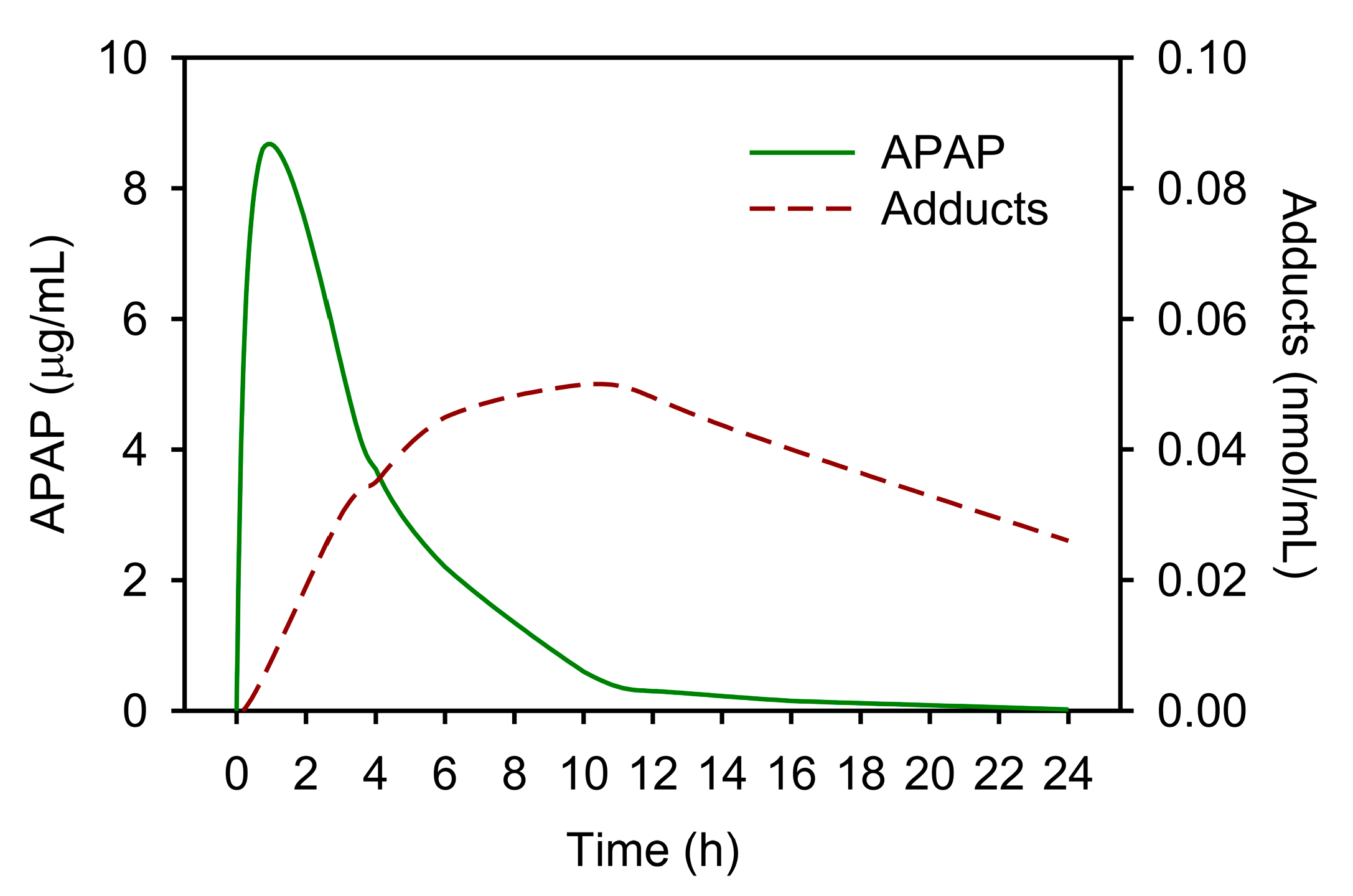
5. APAP-Protein Adducts

5.1. Diagnostic Utility of APAP-Protein Adducts as a Biomarker

APAP-protein adducts, measured as protein-derived APAP-cysteine (APAP-CYS) after the removal of free cysteine and proteolysis of the specimen, overcome some major shortcomings of serum APAP, APAP × AT, psi, and similar endpoints that are based on, or incorporate, serum APAP levels. Like APAP, adducts can become detectable in circulation as early as 1 h after a therapeutic dose, with a peak between 3 and 12 h after therapeutic or sub-hepatotoxic doses [81,82] and between 24 and 72 h after overdose [65] (similar to ALT), but they are more stable than APAP and decline slowly, with a half-life of 1–2 days [82,83,84,85] (Figure 3). Thus, serum adducts provide a longer diagnostic window than APAP alone. Furthermore, bilirubin and NAC have no effect on the HPLC-electrochemical detection (HPLC-EC) method of adduct measurement that is currently used clinically. Generally, values >1 nmol/mL are associated with overdose and serum ALT > 1000 U/L [83,85,86]. Note, however, that some critical considerations are outlined in Section 5.3 of this paper.

5.2. Evolution of APAP-Protein Adduct Methods

The first studies to explore the fundamental mechanisms of APAP hepatotoxicity were undertaken by Bernard Brodie, David Jollow, Jerry Mitchell, and others at the US National Institutes of Health and published in 1973–1974 [27,87,88,89,90,91]. In those papers, the authors discovered that APAP is converted to a reactive metabolite that can deplete hepatic glutathione and bind to proteins. The original methods to measure APAP-protein adducts in these early studies required the administration of radiolabeled APAP followed by multiple sample extraction steps to remove non-protein-bound drug and metabolites, and, finally, the measurement of radioactivity [88]. These methods were laborious and could not be easily applied to serum. The first convenient methods to measure adducts were antibody-based [92]. The development of the original antibodies against APAP-protein adducts came about through a collaboration between Jack Hinson and Dean Roberts while they were working at the National Center for Toxicological Research (NCTR) of the US Food and Drug Administration in the mid-1980s [92]. The two met carpooling to work due to the remote location of the NCTR in a rural area of central Arkansas in the US. During a drive, Hinson described his work on APAP metabolism and protein alkylation to Roberts, who then offered to make an antibody to recognize APAP-cysteine adducts in proteins. According to Hinson:
“It resulted from a conversation we had while we were commuting to NCTR. I described the APAP toxicity mechanism. I clearly remember [Dean] saying, ‘I can make an antibody to that’. Following the development of the anti-APAP antibody, we studied the presence of APAP-protein adducts in mice. As expected, we found adducts in liver. An unexpected finding was adducts in serum. The appearance of serum adducts correlated with the appearance of liver enzymes in the serum (ALT) and were determined to be of hepatic origin. We immediately recognized that serum of APAP overdose victims would have APAP-protein adducts too. We collaborated with Dr. Henrik Poulsen from the University of Copenhagen, Denmark, to assay clinical samples from APAP overdose patients that he had stockpiled in his freezer. This resulted in a publication in the Lancet.”
Hinson later discovered the more sensitive and specific HPLC-EC method [93]:
“The HPLC-EC assay came about by our attempts to assay for 3-nitrotyrosine in APAP samples. We were working with Phil Mayeux and a medical student who had taken a year off to perform research, Ken Muldrew. We had observed nitrotyrosine in livers of APAP-treated mice by immunohistochemical methods. We had also observed it in homogenates from APAP-treated livers using Western blot. Following a published procedure for 3-nitrotyrosine, Ken did not observe any nitrotyrosine but did observe a peak coming out of the HPLC column very late. It was observed only in the liver homogenates from the APAP-treated livers. I postulated that it may be the APAP-cysteine adduct. I then synthesized the 3-cysteinyl-acetaminophen conjugate and confirmed the synthesis by NMR spectral analysis of the product. The peak [in the HPLC-EC assay] was subsequently confirmed to be APAP-cysteine”.
The physician-scientist Laura James joined Hinson and Roberts shortly before the development of the HPLC-EC method and helped to collect the first human samples for HPLC-EC analyses. In her words:
“In subsequent studies, serum samples obtained from patients with APAP overdose were assayed using the HPLC assay. APAP adducts were detected in patients with a documented history of APAP overdose and resulting liver injury. Collectively, these findings were published in the 2002 Muldrew manuscript. Publication of this manuscript caught the attention of hepatologists funded by the National Institute of Diabetes and Digestive and Kidney Diseases (NIDDK) of the US National Institutes of Health through the Acute Liver Failure Study Group. Subsequent research collaborations with this NIDDK-funded network defined the range and magnitude of APAP adducts in multiple clinical scenarios, including ‘low-dose’ exposure, acute overdose with early NAC treatment and acute overdose with delayed or no NAC treatment. Ultimately, the collaborations demonstrated that measurement of adducts in human serum samples could accurately identify patients with APAP-induced ALF and found that approximately 20% of patients with ALF of unknown etiology were secondary to APAP-induced injury. This understanding prompted the development of AcetaSTAT, a rapid, lateral flow diagnostic assay for the semi-qualitative detection of APAP-protein adducts in human sera”.
The development of the HPLC-EC method for adducts was followed by mass spectrometry-based methods [94,95], but HPLC-EC is currently the only method used clinically in the US. As alluded to in the quote above, a lateral flow immunoassay called AcetaSTAT has also been developed and is pending the completion of various studies before FDA review. A prototype of this device reportedly had 100% sensitivity and 96% specificity for APAP-induced liver injury in one small clinical study [86].

5.3. Limitations of APAP-Protein Adducts for Diagnosis and Proposed Solutions

APAP-protein adducts can be detected in circulation during sub-toxic and therapeutic use. Mice treated with 75 mg/kg, which is 5× greater than the equivalent therapeutic dose in humans of approximately 14–15 mg/kg (1 g divided by 70 kg for an average healthy adult, for example) but still sub-toxic, have detectable serum adducts around 0.2 nmol/mL [48]. More importantly, some human volunteers taking therapeutic doses have had adduct values as high as 0.9 to 1.0 nmol/mL [96]—around the proposed diagnostic threshold. Although the diagnosis of APAP overdose may require both an adduct level >1.0 and elevated ALT, there is evidence that co-incidental liver injury leading to ALT elevations can increase APAP-protein adduct concentrations in blood during therapeutic APAP use. For example, we treated mice with the sub-toxic 75 mg/kg dose of APAP, induced co-incidental liver injury by ischemia-reperfusion (IR), or performed sham surgery, and then compared plasma adducts between the IR and sham groups [48]. IR increased plasma adducts by approximately 9-fold, from far below the 1.0 nmol/mL threshold in sham animals to just under it [48]. More significantly, for clinical use, Curry et al. found that some patients classified as having liver injury “definitely not” due to APAP had adduct levels > 1.0 nmol/mL and even as high as 2.86 nmol/mL, which is consistent with elevated adducts due to co-incidental liver damage in patients using sub-toxic APAP doses [67]. To address this issue, Curry proposed the use of 95% probability intervals. That is, the areas on a graph in which 95% of paired serum APAP and serum APAP-protein adduct values from patients with liver injury due to APAP overdose or due to another cause are predicted to fall when adducts are plotted on the y-axis against ALT on the x-axis [67] (Figure 4). Using this approach, the authors noted, for example, that “finding a serum APAP-CYS [APAP-protein] concentration of 1.4 μM [nmol/mL] when corresponding ALT activity was 12,000 IU/L would be unexpected in APAP-induced hepatic necrosis”. (See example case in Figure 4.) In other words, even though adducts would be >1.0 nmol/mL and ALT > 1000 U/L, the clinician could still discern that APAP is not a likely cause of the liver injury in such a case because the elevated adduct levels are still low compared to the very high ALT. Importantly, Curry’s data also indicate that there is a zone of overlap between APAP overdose and non-APAP overdose patients. In cases that fall within this zone, as always, the clinician must look at the complete clinical picture to determine a likely diagnosis or etiology. The 95% prediction interval requires further testing and validation, but moving forward 1.0 nmol/mL may serve as the generally recommended diagnostic cutoff in straightforward APAP overdose cases while the 95% prediction intervals may be helpful when considering overdose in patients with more complicated or uncertain presentations or histories. Finally, it is notable that kidney failure, which is often present in APAP-induced ALF [97], is associated with a more prolonged elimination half-life of serum APAP-CYS, probably from a larger apparent volume of distribution [98]. The latter may also affect the clinical interpretation of adduct values.

5.4. Factors Affecting Serum Adduct Levels at Therapeutic Doses of Acetaminophen

Data from clinical trials indicate that APAP-protein adducts are detectable in serum in most people during therapeutic APAP use [82,96,99]. While the adduct concentrations are generally low, a few subjects in these studies have had values that reached or exceeded the proposed diagnostic threshold for APAP overdose of 1 nmol/mL, as mentioned above. Andrew Monte and colleagues have explored possible mechanisms for this inter-individual variation in adduct levels using ‘omics approaches. For example, in a manuscript describing a small, exploratory study in which they performed genomics analysis on samples from eight subjects administered therapeutic doses of APAP, they reported that there were 194 variants in 65 genes that were associated with adduct concentration [100]. They further reported that patients with higher adduct levels were more likely to have damaging variants in genes involved in signal transduction, and singled out ARHGAP11A and MUC20 as examples. Although these observations are interesting, their causal significance is currently unclear. Nevertheless, they point to potential genetic or otherwise personal factors affecting APAP-protein adduct levels.
Finally, postmortem blood APAP-protein adduct concentrations do not reflect antemortem levels. They can be dramatically elevated in patients who have taken APAP, but have no evidence of APAP-induced hepatotoxicity [101]. This should be kept in mind when investigating causes of liver injury or death in deceased subjects.

6. The Rise of Mechanism-Based Biomarkers

6.1. Mitotoxicity Biomarkers

Mitochondrial protein alkylation was noted by Jollow et al. in the early mouse studies published in 1973 [88], and mitochondrial swelling was observed in liver sections from APAP overdose patients as early as 1979 [102]. However, the importance of mitochondria in APAP toxicity was not recognized at that time. In the late 1980s and early 1990s, research groups led by Steve Cohen at the University of Connecticut, Hartmut Jaeschke at Baylor University, and others, demonstrated that mitochondria are damaged during APAP hepatotoxicity, leading to reduced respiration and increased mitochondrial oxidative stress [103,104]. Additional studies in the 2000s, mostly from Dr. Jaeschke’s group, further demonstrated that these are critical events in the toxicity, driving much of the liver damage [36,105,106,107,108,109,110,111]. Also, in the 2000s, Neil Kaplowitz and colleagues revealed that the c-Jun N-terminal kinases 1/2 are important in the pathogenesis of APAP-induced liver injury [112,113,114,115].
We became interested in translating these mechanisms from rodent models to humans while working together with Dr. Jaeschke around 2009. It is difficult to obtain liver tissue from APAP overdose patients, but blood samples are drawn from those patients daily, so we took up the challenge of studying these mechanisms in patients using blood as a window to peer into the liver. We initially focused on the mitochondrial damage and reasoned that because one can see mitochondrial content spilling into the cytosol after APAP overdose [116], the same mitochondrial macromolecules must also spill into blood when plasma membrane integrity is lost and other large molecules like ALT are released. Consistent with that, when we measured mitochondrial matrix components like glutamate dehydrogenase (GLDH) (by activity) and mitochondrial DNA (mtDNA; by PCR) in plasma from APAP overdose patients, we found that they were elevated [36]. However, it was not enough to simply show that these mitochondrial markers increased during APAP toxicity. We needed a model of liver injury without mitochondrial damage to demonstrate that the molecules are only present, or at least present at higher levels, in blood when mitochondrial damage has occurred. Our solution came when we found a paper in which the authors compared the mechanisms of furosemide and bromobenzene hepatotoxicity and found that furosemide, specifically, caused liver damage without altering mitochondrial respiration [117]. When we then compared plasma or serum GLDH and mtDNA in mice with furosemide and APAP toxicity, they were much higher in the APAP mice [36], indicating that these markers do indeed have some specificity for mitochondrial injury. The GLDH data were recently reproduced by another group [118]. Although they framed their results as a challenge to our work, their data confirmed our fundamental observation that mitochondrial markers like GLDH are higher in blood from mice with APAP toxicity compared to furosemide toxicity [118,119]. We later published similar results for long-chain acylcarnitines in mice [120]. While we were unable to detect an increase in acylcarnitines in adult humans after APAP overdose, another group reported elevations in a pediatric overdose population [121], further supporting the hypothesis that mitochondrial damage occurs in humans as it does in mice. Importantly, data from both studies indicated that early NAC treatment can explain why we did not observe elevated acylcarnitines in our patient population. NAC mitigated the acylcarnitine increase in mice, and acylcarnitine elevations were more apparent in pediatric patients with delayed NAC treatment. We also found that GLDH and mtDNA are higher in serum from non-survivors of APAP-induced ALF than in survivors [38], indicating that mitochondrial damage not only occurs but is a driver of APAP hepatotoxicity in humans. We supported these conclusions with additional data from human HepaRG cells and primary human hepatocytes, which further demonstrated the central role of mitochondrial damage in APAP-induced liver injury in humans [47,94,122].
Another marker that may reflect mitotoxicity in the context of APAP overdose is nuclear DNA fragments. In mice, nuclear DNA fragmentation in the liver occurs partly as a result of the translocation of endonucleases from damaged mitochondria to the nucleus [108], so the presence of high levels of fragmented DNA (or nucleosomes) in the circulation may reflect mitochondrial destruction. We and others were able to measure nuclear DNA fragments in the circulation of APAP overdose patients with liver injury using an anti-histone ELISA [36,38,123], again lending credence to the idea that mitochondrial damage occurs in humans as in mice.
Unfortunately, the clinical value of GLDH, mtDNA, nuclear DNA fragments, and acylcarnitines to predict poor outcomes in APAP overdose patients appears limited at best [38,123]. However, another mitochondrial marker, carbamoyl phosphate synthetase 1 (CPS1), is also elevated in APAP hepatotoxicity [124,125] and may have both mechanistic and prognostic value. A study from 2006 demonstrated that circulating CPS1 values increased at 8 and 24 h after surgery in the cecal ligation and puncture model of sepsis-induced liver damage [126]. Importantly, this increase was co-incident with the loss of mitochondria in the liver and changes in mitochondrial morphology, while ALT release occurred much later. The authors interpreted this to mean that CPS1 appeared in serum as a result of mitochondrial destruction specifically, rather than simply necrosis [126]. Bishr Omary and coworkers later reported that CPS1 is elevated in the culture medium and circulation from primary mouse hepatocytes and mice, respectively, during APAP-induced injury, and in serum from APAP overdose patients [124]. They also found that serum CPS1 levels were greater in non-survivors of APAP-induced ALF and in transplant recipients than in spontaneous survivors [125], indicating that these levels can predict poor outcomes. Notably, CPS1 levels modestly improved the MELD score for prognosis [125].

6.2. Cell Death Mode Markers

While we were working on mitochondrial biomarkers, other groups were exploring the mode of cell death in human APAP hepatotoxicity by measuring keratin 18 (K18) and high mobility group box 1 (HMGB1) protein in blood. An antibody (“M30”) against a caspase-cleaved form of K18 (ccK18) had been developed in the 1990s and was used to label apoptotic cells in sections from colonic carcinoma tissue and cancer cell lines [127]. It was later applied to serum from cancer patients using an ELISA [128,129]. Rutherford et al. [130] then measured ccK18 in serum from patients with ALF of various etiologies and noted that median levels were higher in APAP overdose patients than in healthy controls. However, they did not measure total K18 for comparison, so it was not possible to determine the relative contributions of apoptosis and necrosis. At about the same time, Bechmann et al. [131] measured ccK18 in the circulation of one patient with APAP-induced ALF and reported only minor elevations in comparison to total K18 (“M65”), indicating “a low rate of apoptosis”. Since then, most studies have demonstrated that only about 10–20% of total K18 is in the caspase-cleaved form. For example, Volkmann et al. [132] reported ccK18 values around 8–9000 and total K18 values around 50–55,000 U/L (~15–16%) in APAP patients while Craig et al. reported median values of 9646 and 45,615, respectively (21%) [123]. Importantly, even these percentages likely overestimate the amount of apoptotic vs. necrotic cell death in the human liver after APAP overdose. Very few apoptotic cells appear in the liver after APAP overdose in mice, and caspase inhibitors do not protect against the injury at all [47,94,133,134,135]. Furthermore, we could not detect any caspase 3 activity in plasma from APAP overdose patients with liver injury, even though we observed very high levels in plasma from mice with hepatocyte apoptosis due to galactosamine/LPS treatment [36]. Finally, histological studies have revealed that only ~0.6% of hepatocytes display characteristic morphological features of apoptosis during APAP hepatotoxicity in humans compared to >60% with necrotic appearance [135], though the authors of that study insisted on an interpretation of the data that likely exaggerates the importance of apoptosis. In any case, even when considering only the ccK18 and K18 data, necrosis is the dominant mode of cell death during APAP hepatotoxicity.
There are conflicting data regarding the prognostic value of K18 and ccK18 to predict death. On the one hand, Rutherford et al. measured serum ccK18 in the earliest available samples from a subset of 52 patients with ALF of various etiologies and reported that the median levels were significantly greater in non-survivors and transplant recipients compared to spontaneous survivors [130]. Consistent with these data, Church et al. [136] measured K18 and ccK18 in samples from patients with non-APAP DILI from the DILI Network (DILIN) and reported that K18 levels were significantly greater in patients who died or required a transplant. On the other hand, Craig et al. measured K18 and ccK18 in serum from APAP overdose patients specifically and reported that there were no significant differences in either biomarker between those who spontaneously survived (n = 14) and those who died or received a liver transplant (n = 12), at the time of admission [117]. Results from ROC analyses, sensitivity and specificity, and the diagnostic odds ratio were unimpressive (AUCs ≤ 0.67 and 95% CIs for odds ratios overlapping one, for example) [123]. Regarding the prediction of hepatotoxicity in early-presenting APAP overdose patients, Dear and colleagues measured K18, ccK18, microRNA-122 (miR-122), and GLDH in admission samples from APAP overdose patients who presented with normal ALT values < 50 U/L and reported that K18 was one of the best markers to predict later injury in these early presenters [137]. GLDH was also associated with later injury, but not as strongly [137].
Like K18, HMGB1 has also been interpreted as a marker of necrotic cell death. Scaffidi et al. [138] stained for HMGB1 in HeLa cells undergoing TNF-α/cyclohexamide-stimulated apoptosis and observed that it localized to and remained within the nucleus throughout the apoptotic process. Additionally, there was little release of HMGB1 into the cell culture medium during apoptosis compared to necrosis induced by CCCP and other compounds [138]. Finally, they reported that an anti-HMGB1 antibody decreased inflammatory cell recruitment in the liver (as assessed by myeloperoxidase) after APAP-induced hepatocyte necrosis in mice, implying that HMGB1 was released into serum by necrosis and had a pro-inflammatory effect [138]. Importantly, HMGB1 release has been reported in APAP overdose patients as well, further supporting necrosis as the primary mode of cell death in APAP hepatotoxicity in humans [123,137]. Additionally, like K18 and GLDH, HMGB1 levels can forecast APAP hepatotoxicity in early-presenting APAP overdose patients with normal ALT values [137], though they likely cannot predict death [123,139].

6.3. Other Mechanistic Biomarkers

The results from an intriguing recent study indicate that bile acids leak out of bile canaliculi early in APAP hepatotoxicity and are taken up by hepatocytes, where they accumulate and contribute to hepatocyte death [140]. While this hypothesis requires confirmation, especially in light of prior data demonstrating that bile acid depletion worsens APAP-induced injury [141], we and others previously reported that bile acids increase in the serum in APAP hepatotoxicity in both mice and humans [142,143]. Furthermore, we found that glycodeoxycholic acid, in particular, can predict death in APAP overdose patients [142]. Thus, bile acids may also be considered mechanistic biomarkers in APAP patients, pending independent replication of the mechanistic studies mentioned. In addition, some bile acids appear to increase earlier than ALT, indicating that they may have value as prognostic markers to predict later injury in early-presenting patients with normal ALT.
Additional studies have addressed the utility of these and other biomarkers for the diagnosis of APAP-induced liver injury [144]. While such studies may be useful when extrapolating the results to DILI more generally, for which diagnosis is a greater challenge [145], in most cases, their kinetics mirror the kinetics of ALT (see [36] for example). It is not clear what value they might add to diagnosis or prognosis if they cannot detect injury earlier.

7. Inflammation Biomarkers

The role of inflammation in APAP hepatotoxicity and liver repair is complex, multiphasic, and controversial [27,146,147,148]. Early studies in the 1980s found that Kupffer cells are activated in rats during APAP toxicity [149,150], and that inactivation with gadolinium chloride protects against APAP in mice [151]. However, later work revealed that total Kupffer cell ablation, instead of simply inactivation, with a single dose of liposomal clodronate either worsens injury in mice [152] or has no effect [153]. More recently, we demonstrated that two doses of liposomal clodronate actually protect against APAP, in contrast to the single dose regimen, but also that the protective effect was likely due to Nrf2 activation rather than anything directly having to do with Kupffer cells [153]. Furthermore, NADPH oxidase and C-C chemokine receptor 2 (CCR2) KO mice are not protected against APAP [154,155]. Thus, overall, it appears that Kupffer cells do not contribute significantly to APAP hepatotoxicity. Similar conflicting data exist for neutrophils [27,156,157,158], though recent data indicate that the role of neutrophils may depend upon the APAP dose or extent of injury [159], which may explain some discrepancies in the literature. Studies are currently ongoing to investigate other inflammatory cell types in the context of APAP overdose.
Regardless, a number of cytokines and other pro- and anti-inflammatory markers have been measured in APAP overdose patients. In 2001, James et al. reported increased interleukin-8 (IL-8; aka CXCL8) in the circulation of pediatric overdose patients, but no elevations in IL-6 or IL-10, compared to healthy controls [160]. They also reported that patients with high peak IL-8 had higher peak prothrombin times, suggesting some prognostic utility, despite the possible confounding effects of time-to-NAC treatment in their analysis (delayed NAC treatment may result in both higher IL-8 and worse outcomes independent of each other, and a novel marker like IL-8 is probably not needed to determine if presentation was delayed) [160]. A 2003 study by another group reproduced the observation that IL-8 is elevated in APAP-induced liver injury in humans and revealed that IL-8 and hyaluronic acid serum levels were higher in non-survivors of APAP overdose compared to survivors [161]. Similarly, we observed a trend toward increased IL-8 in non-survivors, though it did not achieve statistical significance [162].
In contrast to their 2001 study, James et al. later observed that IL-6 is significantly elevated after APAP overdose, but only in patients with more severe liver injury than those included in their initial work [163]. They also found in that study that monocyte chemoattractant protein 1 (MPC1; aka CCL2) was elevated in APAP toxicity patients, and, like IL-8, was associated with peak prothrombin time [163]. Consistent with those observations, we recently reported that both IL-6 and MCP1 are significantly higher in serum from non-survivors of APAP-induced ALF compared to spontaneous survivors at some early time points [162]. However, in contrast to James et al., we also observed elevated IL-10, and it too was associated with poor outcomes [162]. Elevations in IL-6, IL-8, and IL-10 have also been reported in other DILI patients [163,164]. Overall, it appears that IL-6, IL-8, and MCP1 are consistently elevated in serum from APAP overdose patients with liver injury and may have prognostic value to predict poor outcomes. IL-10 may also be prognostic but requires further investigation. Other inflammation markers including pentraxin 3 [165], Lect2 [166], and neopterin [167] also seem to be elevated in APAP-induced liver injury and associated with patient outcome, but have not been explored as thoroughly as the cytokines and chemokines already mentioned.

8. miRNA-Based Biomarkers

Wang et al. 2009 [168] first reported that serum levels of multiple miRNAs are altered in mice with APAP-induced liver injury, and these data were extended to APAP overdose patients by the research group of Kevin Park at the University of Liverpool two years later [169]. Since then, many miRNAs have been shown to increase or decrease in circulation in APAP hepatotoxicity [169,170,171,172,173,174], but miR-122 seems to display the largest and most consistent changes [175].
The most promising application of miR-122 at this point still appears to be in the prediction of hepatotoxicity in APAP overdose patients presenting with normal ALT and liver function test results. It is rare for patients presenting with ALT < 50 U/L to later develop injury (as described in Section 3.2, above), and those who do rarely develop severe hepatotoxicity. This is even clear from the studies supporting miR-122 use themselves, as pointed out in a letter to the editor [176]. However, others argue that if the potential for hepatotoxicity, despite NAC, can be ruled out in these early presenters using a marker specifically for that purpose, then NAC treatment can be tailored, unnecessary hospitalizations can be avoided, and patients who require care can still be identified and promptly treated. K18, HMGB1, and—to a lesser extent—GLDH may have some value for this purpose, as mentioned in Section 6 [137], but miR-122 and perhaps K18 appear to be uniquely positioned for this because they seem to be more sensitive to hepatocellular stress. For example, we observed that K18 was elevated in patients with compensated cirrhosis, despite normal ALT [82]. Furthermore, serum miR-122 was significantly elevated in 18 human subjects after a moderate alcohol binge that had no effect on ALT or GGT [177]. Dear et al. demonstrated that miR-122 at admission has high sensitivity and specificity to predict peak ALT >100 U/L in patients who present with an initial ALT value < 50 [137]. While there are concerns about the biological variation in miR-122 [136] as well as technical challenges with current measurement methods (e.g., PCR), new analytical approaches are in development that may help to overcome some of the relevant problems [178,179,180]. In addition, an alternative approach may be to measure the levels of proteins encoded by genes that are miR-122 targets. miR-122 decreases in the liver during APAP-induced injury in mice [168], so it is theoretically possible that miR-122 targets are upregulated in the liver and could be released into the circulation. Finally, despite the likely utility of miR-122 as a prognostic marker for later injury in early-presenters, it appears that it is only modestly associated with death in APAP-induced ALF [181,182], if at all. Overall, the necessity of new markers to predict hepatotoxicity in early presenters has been questioned, but if one were to be used clinically then miR-122 appears to be a reasonable candidate.
Specific miRNA profiles have also been linked to liver regeneration and survival [182,183]. For example, Salehi et al. [183] described miRNA profiles in patients who underwent auxiliary liver transplantation. In this procedure, the patient’s damaged liver is left in place and allowed to regenerate, while the transplanted liver takes over the normal liver functions. After successful regeneration, immunosuppression is withdrawn and the transplanted liver is allowed to atrophy, leaving the patient’s recovered liver. This unique scenario allowed the researchers to monitor miRNA changes in biopsy samples from the native liver at multiple time points during the regeneration period. Of the 11 total patients enrolled in the study, 7 experienced successful regeneration leading to the complete withdrawal of immunosuppression, while 4 failed to regenerate. The investigators isolated RNA from their biopsies and performed microarray analysis to measure numerous miRNAs, followed by pathway analysis to determine what signaling pathways would be expected to be altered due to the detected changes in miRNA. The authors reported decreased levels of miRNAs that can suppress the expression of pro-proliferative genes (e.g., CCND2)—miR-23a, miR-150, miR-503, and miR-663—and increased levels of miRNAs that can activate expression of pro-proliferative genes (e.g., VEGF)—miR-20a, miR-126, miR-130a, and miR-520e—over time from the day of transplantation to later phases of regeneration in patients whose native livers successfully regenerated. They later measured some of these miRNAs in the serum from patients with APAP-induced ALF and found that miR-23a, miR-150, and miR-503a (the miRNAs associated with suppressed proliferation) were lower in patients who survived compared to those who died or received a liver transplant, consistent with the idea that these miRNAs are biomarkers of liver regeneration [184]. Finally, they measured these same miRNAs plus additional regeneration-linked species in the serum from 192 patients with APAP-induced ALF and explored their abilities to differentiate between survivors and non-survivors individually, in combination, and when combined with conventional clinical endpoints [181]. While they again observed an association between patient outcome and miR-150, as well as several other miRNAs, the discriminatory powers of the individual miRNA species were modest. However, the prognostic value improved when some of the miRNAs were combined, and further improved when these miRNA species were considered with the MELD score and vasopressor use [181]. Unfortunately, the authors of the latter study also discovered that there was considerable within- and between-person variation in miRNA levels over time. Some species that were elevated in non-survivors compared to survivors at an early time point, for example, were lower compared to survivors at the later time point, and vice versa. Furthermore, the inclusion of multiple miRNAs increases test complexity and cost. These are significant barriers to clinical use that will need to be overcome.

9. Liver Regeneration and Repair Biomarkers

Circulating miRNA profiles are not the only regeneration-associated biomarkers. Arguably, the oldest markers of liver function that are still in use today, namely bilirubin and prothrombin time, are really markers of liver regeneration. Bilirubin undergoes conjugation in the liver to facilitate its excretion, so the accumulation of bilirubin in serum after injury indicates impaired metabolic or excretory liver function. Prothrombin time, on the other hand, is a measure of blood clotting. As most clotting factors are synthesized in the liver, long prothrombin times after injury indicate impaired synthetic function. As the liver regenerates and liver function recovers, values for these tests return to normal. It is not surprising then that these markers correlate well with patient outcome in liver failure. Indeed, the MELD score, the KCC, and the ALF Study Group Prognostic Index (ALFSG PI) include both [185,186,187]. However, these markers often cannot be used to forecast regeneration because their kinetics are more-or-less parallel to the kinetics of regeneration. That is, liver function improves as regeneration occurs, not before, though there is evidence that remnant hepatocytes induce basal function to partially offset the liver damage [188]. Because function markers mostly parallel regeneration, changes in these markers may not be useful to predict recovery vs. death. Schmidt and Dalhoff [189] also reported that circulating α-fetoprotein (AFP), a marker of hepatocyte proliferation, is increased in the serum from survivors of APAP-induced ALF compared to non-survivors. AFP is already measured clinically as a tumor marker and as part of the screening tests for chromosomal abnormalities during gestation, so it could have immediate clinical application. Like bilirubin and prothrombin time, however, AFP values do not increase until after the peak of liver injury. Several other biomarkers of regeneration have been proposed but require further validation. Serum phosphate is elevated in non-survivors of APAP-induced liver injury and in those who received a liver transplant compared to survivors [190]. α-NH-butyric acid increases in serum after PHx in mice and can discriminate between survivors and non-survivors of ALF (including APAP-induced ALF) in pediatric patients as well as bilirubin and prothrombin time can [191]. Lect2 is also elevated in non-survivors of ALF (again, including APAP-induced ALF) compared to survivors [166]. Similarly, CD133/CD39 double-positive microparticles are elevated in patients with APAP-induced acute-on-chronic liver failure (ACLF), and data from rodent studies indicate that CD39 promotes liver recovery [192]. It was also recently reported that some hepatocytes adopt a senescent phenotype after injury, leading to the secretion of CXCL14, which then inhibits liver regeneration [193]. Interestingly, the authors also reported that serum CXCL14 was elevated in patients with APAP-induced ALF and could predict non-survivors. Finally, we recently discovered that some species of phosphatidic acid (PA) may be involved in liver regeneration after APAP hepatotoxicity in mice and are also elevated in the serum from APAP overdose patients with liver injury [194,195], though we have not yet tested the prognostic value of serum PA.

10. Miscellaneous Other Biomarkers

Various other biomarkers that do not fit well into the categories above have also been explored in APAP hepatotoxicity. We have repeatedly observed elevated agininosuccinate synthetase 1 (ASS1) in circulation after APAP hepatotoxicity in both mice and humans [196,197]. Furthermore, our data from mice indicate that it may be a more sensitive and specific biomarker of liver injury than ALT [196,197]. In our experiments, plasma ASS1 increased as early as 2 h post-APAP overdose, before an increase in ALT [196]. It was also elevated in models of mild liver injury but not in a model of benign ALT elevation [197]. In addition, a dose of APAP (100 mg/kg) that did not cause an increase in ALT did increase ASS1 [196]. However, more research is needed to fully evaluate the utility of ASS1. We have also shown that aldehyde dehydrogenase 1a1 (ALDH1A1), alcohol dehydrogenase 1 (ADH1), fructose-1,6-bisphophate (FBP1), and a number of other serum markers are also more specific for liver injury than ALT using the APAP model [197]. Based on that, we proposed that such markers may be useful as part of a screen-and-confirm approach to identify and diagnose idiosyncratic drug-induced liver injury in clinical trials or even clinically in which one would screen for possible injury using ALT and then confirm the injury using a more specific marker [145]. Procalcitonin, often used as a marker of bacterial infection, is also elevated in APAP overdose patients with liver injury and appears to be a result of the injury and resulting inflammation rather than an indication of infection [198,199]. Interestingly, procalcitonin elevation preceded ALT elevation by 33 h in one study [200], indicating that it may be useful as an early biomarker of liver damage to predict later ALT elevations. Unfortunately, while the latter study included a logistic regression analysis of the ability of admission procalcitonin levels to predict injury, the authors did not limit the analysis to patients who presented with normal ALT, so the prognostic value in that scenario remains unclear. Circulating fatty acid binding protein 1 (FABP1) is also elevated in liver injury [201]. It is also prognostic for death in APAP-induced ALF, especially in combination with the KCC and the ALSGPI. The serum or plasma levels of several other potential biomarkers, including C-reactive protein (CRP) [165], taurine [202], and sphingolipids [203], also change during APAP hepatotoxicity, but the clinical significance of these changes has yet to be explored.
The overall findings for all of the major biomarkers discussed above are summarized in Table 1.

11. Conclusions and Future Directions in Biomarker Development

11.1. New Models for Biomarker Discovery

A challenge in identifying and validating biomarkers for APAP overdose in humans is the difficulty in obtaining serum or plasma samples from a sufficient number of APAP overdose patients, especially from non-survivors, to obtain meaningful results. Developing cell culture models may present an alternative approach. A challenge with cell culture models is that most hepatocyte cell lines, such as HepG2 cells, do not express the cytochrome P450 enzymes required to initiate APAP hepatotoxicity [204,205]. Furthermore, most hepatocyte culture systems cannot replicate the human pathophysiology to any significant degree because they lack the other cell types in the liver (Kupffer cells, stellate cells, cholangiocytes, and sinusoidal epithelial cells, for example) as well as the normal hepatic architecture. They also typically lack extrahepatic cells that may have a role in APAP-induced liver injury or repair, like neutrophils and monocytes. However, new models are emerging to overcome these challenges. HepaRG is a convenient cell line that expresses many P450s and develops APAP hepatotoxicity that mostly resembles what we see in humans and mice [94]. Meanwhile, model systems such as organ-on-chip are increasingly popular and permit the co-culture of primary hepatocytes or HepaRG cells with other hepatic cell types in structures that somewhat resemble the hepatic sinusoid [206]. Finally, multi-organ chips are also being developed that incorporate similar liver systems [207,208,209,210]. As these systems mature and become more affordable and accessible, they may be useful for the routine study of novel liver injury biomarkers which can then be validated in later clinical studies.

11.2. Understanding What Makes a Good Biomarker

Whatever methods are used to identify and validate new biomarkers, it is imperative that we come to understand what makes a good biomarker of liver injury. Aside from the usual considerations of technical and biological variations, organ specificity and specificity for liver injury are critical considerations. A challenge with ALT, for example, is that it can be elevated in the absence of liver injury [197]. Furthermore, it is not entirely specific for the liver, as it can be elevated with muscle damage, too [211]. Clinical feasibility is another key issue. For example, because fatal APAP overdose and ALF, more generally, are relatively uncommon, it may be difficult to obtain commercial interest in biomarkers to predict survival. This is an advantage of markers like LDH, which are already widely used clinically for other reasons. Another factor may be abundance within an organ. Abundance within the liver may also be an important consideration. One may ask why LDH can predict poor outcomes in APAP overdose patients while ALT cannot. It has been proposed that the molecular weight and subcellular location of a protein may be factors, the idea being that large proteins or those buried deeper into the cells—within organelles, for example—are less likely to be released. However, LDH forms a large tetramer of approximately 140 kD, and both LDH and ALT1—the dominant isoform released into serum [212,213,214]—are primarily cytosolic. Another possible explanation is that LDH is only released with membrane damage and leakage, reflecting true hepatocyte injury, while ALT can be released via other mechanisms that are less specific for cell death. This is plausible, but there are little data to support it at this moment. Finally, a third explanation is that LDH is either induced by APAP or is simply more abundant in the liver than ALT in the first place, such that LDH continues to increase with increased injury (and therefore with a greater probability of poor outcomes) while ALT plateaus. Indeed, our prior data indicate that LDH continues to increase for about 24 h after ALT peaks [39], though further investigation is needed to confirm that. Furthermore, we have observed far greater LDH activity than ALT activity in liver tissue homogenates, though LDH did not appear to be induced (McGill et al., unpublished observations). In any case, additional research is clearly needed to understand what physiological and biochemical properties of a biomarker are most desirable for either diagnosis or prognosis in liver injury.

11.3. Conclusions

A great deal of research has been conducted to identify circulating biomarkers for use in APAP overdose patients. ALT and the Rumack–Matthew nomogram continue to dominate clinical use, while the measurement of APAP-protein adducts is emerging as a novel diagnostic tool that may be useful when carefully interpreted. Emerging prognostic biomarkers also show promise for use in the near future. However, additional research is recommended to validate these latter markers.

Author Contributions

Conceptualization, M.R.M.; methodology, M.R.M.; software, M.R.M.; investigation, M.R.M. and S.C.C.; resources, M.R.M.; writing—original draft preparation, M.R.M.; writing—review and editing, M.R.M. and S.C.C.; visualization, M.R.M.; project administration, M.R.M.; funding acquisition, M.R.M. All authors have read and agreed to the published version of the manuscript.

Funding

M.M. was funded in part by National Institutes of Health grants UL1TR003107 and by a research grant from Haleon. The funding agencies had no role in the conceptualization, design, writing, editing or submission of this manuscript.

Institutional Review Board Statement

Not applicable.

Informed Consent Statement

Not applicable.

Data Availability Statement

Not applicable.

Acknowledgments

We thank Jack Hinson, Dean Roberts, Laura James, and Barry Rumack for their helpful discussions and correspondence. The laboratory of MRM was supported in part by the National Institutes of Health grant UL1 TR003107.

Conflicts of Interest

M.R.M. has consulted for Acetaminophen Toxicity Diagnostics, LLC, within the last ten years. He has also received research funding from GlaxoSmithKline/Haleon within the last ten years. S.C.C. has received research funding from Johnson & Johnson within the last ten years. None of these companies had a role in the conception, writing, editing, or submission of this manuscript.

References

  1. Proudfoot, A.T.; Good, A.M.; Bateman, D.N. Clinical toxicology in Edinburgh, two centuries of progress. Clin. Toxicol. 2013, 51, 509–514. [Google Scholar] [CrossRef] [PubMed]
  2. Davidson, D.G.; Eastham, W.N. Acute liver necrosis following overdose of paracetamol. Br. Med. J. 1966, 2, 497–499. [Google Scholar] [CrossRef] [PubMed]
  3. Eder, H. Chronic Toxicity Studies on Phenacetin, N-Acetyl-p-Aminophenol (NAPA) and Acetylsalicylic Acid on Cats. Acta Pharmacol. Toxicol. 1964, 21, 197–204. [Google Scholar] [CrossRef] [PubMed]
  4. Boyd, E.M.; Bereczky, G.M. Liver Necrosis from Paracetamol. Br. J. Pharmacol. Chemother. 1966, 26, 606–614. [Google Scholar] [CrossRef] [PubMed]
  5. Bone, J.M. Aspirin and Gastric Bleeding. Br. Med. J. 1967, 3, 810–811. [Google Scholar] [CrossRef] [PubMed]
  6. Thomson, J.S.; Prescott, L.F. Liver damage and impaired glucose tolerance after paracetamol overdosage. Br. Med. J. 1966, 2, 506–507. [Google Scholar] [CrossRef] [PubMed]
  7. Toghill, P.; Williams, R.; Stephens, J.; Carroll, J. Acute Hepatic Necrosis Following an Overdose of Paracetamol. Gastroenterology 1969, 56, 773–776. [Google Scholar] [CrossRef]
  8. Boyer, T.D.; Rouff, S.L. Acetaminophen-Induced Hepatic Necrosis and Renal Failure. JAMA J. Am. Med. Assoc. 1971, 218, 440–441. [Google Scholar] [CrossRef]
  9. James, O.; Roberts, S.; Douglas, A.; Lesna, M.; Pulman, L.; Smith, P.; Watson, A. Liver Damage after Paracetamol Overdose. Comparison of Liver-Function Tests, Fasting Serum Bile Acids, and Liver Histology. Lancet 1975, 306, 579–581. [Google Scholar] [CrossRef]
  10. McJunkin, B.; Barwick, K.W.; Little, W.C.; Winfield, J.B. Fatal Massive Hepatic Necrosis Following Acetaminophen Overdose. JAMA 1976, 236, 1874–1875. [Google Scholar] [CrossRef]
  11. Barker, J.D.; de Carle, D.J.; Anuras, S. Chronic Excessive Acetaminophen Use and Liver Damage. Ann. Intern. Med. 1977, 87, 299–301. [Google Scholar] [CrossRef] [PubMed]
  12. Krenzelok, E.P.; Best, L.; Manoguerra, A.S. Acetaminophen Toxity. Am. J. Hosp. Pharm. 1977, 34, 391–394. [Google Scholar] [CrossRef] [PubMed]
  13. Ferguson, D.R.; Snyder, S.K.; Cameron, A.J. Hepatotoxicity in Acetaminophen Poisoning. Mayo Clin. Proc. 1977, 52, 246–248. [Google Scholar] [CrossRef] [PubMed]
  14. Groarke, J.F.; Averett, J.M.; Hirschowitz, B.I. Acetaminophen and Hepatic Necrosis. N. Engl. J. Med. 1977, 296, 233. [Google Scholar] [CrossRef] [PubMed]
  15. Lee, W.M. Acetaminophen Toxicity: A History of Serendipity and Unintended Consequences. Clin. Liver Dis. 2020, 16, 34–44. [Google Scholar] [CrossRef] [PubMed]
  16. Manthripragada, A.D.; Zhou, E.H.; Budnitz, D.S.; Lovegrove, M.C.; Willy, M.E. Characterization of acetaminophen overdose-related emergency department visits and hospitalizations in the United States. Pharmacoepidemiol. Drug Saf. 2011, 20, 819–826. [Google Scholar] [CrossRef] [PubMed]
  17. Li, C.; Martin, B.C. Trends in emergency department visits attributable to acetaminophen overdoses in the United States: 1993–2007. Pharmacoepidemiol. Drug Saf. 2011, 20, 810–818. [Google Scholar] [CrossRef] [PubMed]
  18. Budnitz, D.S.; Lovegrove, M.C.; Crosby, A.E. Emergency Department Visits for Overdoses of Acetaminophen-Containing Products. Am. J. Prev. Med. 2011, 40, 585–592. [Google Scholar] [CrossRef]
  19. Stravitz, R.T.; Fontana, R.J.; Karvellas, C.; Durkalski, V.; McGuire, B.; Rule, J.A.; Tujios, S.; Lee, W.M.; Acute Liver Failure Study Group. Future directions in acute liver failure. Hepatology 2023, 78, 1266–1289. [Google Scholar] [CrossRef]
  20. Karmen, A.; Wróblewski, F.; LaDue, J.S. Transaminase Activity in Human Blood. J. Clin. Investig. 1955, 34, 126–133. [Google Scholar] [CrossRef]
  21. Wróblewski, F.; Ladue, J.S. Serum Glutamic Pyruvic Transaminase SGP-T in Hepatic Disease: A Preliminary Report. Ann. Intern. Med. 1956, 45, 801–811. [Google Scholar] [CrossRef]
  22. De Ritis, F.; Coltori, M.; Giusti, G. Transaminase Activity of Human Serum in Viral Hepatitis; Preventive Note. Minerva Medica 1955, 46, 1207–1209. [Google Scholar] [PubMed]
  23. Alfred, D. Reagents and Method for Assaying Biological Samples. U.S. Patent 3,413,198, 26 November 1968. [Google Scholar]
  24. Kessler, G.; Rush, R.; Leon, L.; Delea, A.; Cupiola, R. Automated 340 Nm Measurement of SGOT, SGPT and LDH. Clin. Chem. 1970, 16, 530–531. [Google Scholar]
  25. Smith, A.F.; Taylor, R.H. Comparison of two-point (AutoAnalyzer II) with kinetic methods for transaminase assay. J. Clin. Pathol. 1973, 26, 42–47. [Google Scholar] [CrossRef] [PubMed]
  26. McGill, M.R.; Jaeschke, H. Metabolism and Disposition of Acetaminophen: Recent Advances in Relation to Hepatotoxicity and Diagnosis. Pharm. Res. 2013, 30, 2174–2187. [Google Scholar] [CrossRef] [PubMed]
  27. McGill, M.R.; Hinson, J.A. The development and hepatotoxicity of acetaminophen: Reviewing over a century of progress. Drug Metab. Rev. 2020, 52, 472–500. [Google Scholar] [CrossRef] [PubMed]
  28. Jaeschke, H.; Ramachandran, A. Central mechanisms of acetaminophen hepatotoxicity: Mitochondrial dysfunction by protein adducts and oxidant stress. Drug Metab. Dispos. 2023, 51. [Google Scholar] [CrossRef]
  29. Rej, R.; Keese, C.R.; Giaever, I. Direct immunological determination of aspartate aminotransferase isoenzymes. Clin. Chem. 1981, 27, 1597–1601. [Google Scholar] [CrossRef]
  30. Panteghini, M.; Pagani, F.; Cuccia, C. Activity of serum aspartate aminotransferase isoenzymes in patients with acute myocardial infarction. Clin. Chem. 1987, 33, 67–71. [Google Scholar] [CrossRef]
  31. Zoppini, G.; Cacciatori, V.; Negri, C.; Stoico, V.; Lippi, G.; Targher, G.; Bonora, E. The aspartate aminotransferase-to-alanine aminotransferase ratio predicts all-cause and cardiovascular mortality in patients with type 2 diabetes. Medicine 2016, 95, e4821. [Google Scholar] [CrossRef]
  32. Mcgovern, A.J.; Vitkovitsky, I.V.; Jones, D.L.; Mullins, M.E. Can AST/ALT Ratio Indicate Recovery after Acute Paracetamol Poisoning? Clin. Toxicol. 2015, 53, 164–167. [Google Scholar] [CrossRef]
  33. McGill, M.R. The past and present of serum aminotransferases and the future of liver injury biomarkers. EXCLI J. 2016, 15, 817–828. [Google Scholar] [CrossRef] [PubMed]
  34. Maclean, D.; Peters, T.J.; Brown, R.A.; McCathie, M.; Baines, G.F.; Robertson, P.G. Treatment of Acute Paracetamol Poisoning. Lancet 1968, 292, 849–852. [Google Scholar] [CrossRef] [PubMed]
  35. Singer, A.J.; Carracio, T.R.; Mofenson, H.C. The Temporal Profile of Increased Transaminase Levels in Patients with Acetaminophen-Induced Liver Dysfunction. Ann. Emerg. Med. 1995, 26, 49–53. [Google Scholar] [CrossRef] [PubMed]
  36. McGill, M.R.; Sharpe, M.R.; Williams, C.D.; Taha, M.; Curry, S.C.; Jaeschke, H. The mechanism underlying acetaminophen-induced hepatotoxicity in humans and mice involves mitochondrial damage and nuclear DNA fragmentation. J. Clin. Investig. 2012, 122, 1574–1583. [Google Scholar] [CrossRef] [PubMed]
  37. Curtis, R.M.; Sivilotti, M.L.A. A descriptive analysis of aspartate and alanine aminotransferase rise and fall following acetaminophen overdose. Clin. Toxicol. 2015, 53, 849–855. [Google Scholar] [CrossRef] [PubMed]
  38. McGill, M.R.; Staggs, V.S.; Sharpe, M.R.; Lee, W.M.; Jaeschke, H. Serum Mitochondrial Biomarkers and Damage-Associated Molecular Patterns Are Higher in Acetaminophen Overdose Patients with Poor Outcome. Hepatology 2014, 60, 1336–1345. [Google Scholar] [CrossRef]
  39. Price, J.R.; Hagrass, H.; Filip, A.B.; McGill, M.R. LDH and the MELD-LDH in Severe Acute Liver Injury and Acute Liver Failure: Preliminary Confirmation of a Novel Prognostic Score for Risk Stratification. J. Appl. Lab. Med. 2023, 8, 504–513. [Google Scholar] [CrossRef]
  40. Green, T.J.; Sivilotti, M.L.; Langmann, C.; Yarema, M.; Juurlink, D.; Burns, M.J.; Johnson, D.W. When Do the Aminotransferases Rise after Acute Acetaminophen Overdose. Clin. Toxicol. 2010, 48, 787–792. [Google Scholar] [CrossRef]
  41. Al-Hourani, K.; Mansi, R.; Pettie, J.; Dow, M.; Bateman, D.N.; Dear, J.W. The predictive value of hospital admission serum alanine transaminase activity in patients treated for paracetamol overdose. QJM 2013, 106, 541–546. [Google Scholar] [CrossRef]
  42. Curry, S.C.; Padilla-Jones, A.; Kang, A.; Snow, J.; Heise, C. Serum ALT Activity at Commencement of N-Acetylecysteine Therapy as a Predictor of Hepatotoxocity, Hepatic Encephalopathy, and Mortality in Patients with Acetaminophen Toxicity. J. Med. Toxicol. 2019, 15, 71. [Google Scholar] [CrossRef]
  43. Harrison, P.; Keays, R.; Bray, G.; Alexander, G.; Williams, R. Improved outcome of paracetamol-induced fulminant hepatic failure by late administration of acetylcysteine. Lancet 1990, 335, 1572–1573. [Google Scholar] [CrossRef] [PubMed]
  44. Keays, R.; Harrison, P.M.; Wendon, J.A.; Forbes, A.; Gove, C.; Alexander, G.J.; Williams, R. Intravenous acetylcysteine in paracetamol induced fulminant hepatic failure: A prospective controlled trial. BMJ 1991, 303, 1026–1029. [Google Scholar] [CrossRef] [PubMed]
  45. Mullins, M.E.; Schwarz, E. Re: Remien Etal.: Mathematical Modeling of Liver Injury and Dysfunction after Acetaminophen Overdose. Hepatology 2012, 56, 2427–2428. [Google Scholar] [CrossRef] [PubMed]
  46. James, L.P.; McCullough, S.S.; Lamps, L.W.; Hinson, J.A. Effect of N-Acetylcysteine on Acetaminophen Toxicity in Mice: Relationship to Reactive Nitrogen and Cytokine Formation. Toxicol. Sci. 2003, 75, 458–467. [Google Scholar] [CrossRef] [PubMed]
  47. Xie, Y.; McGill, M.R.; Dorko, K.; Kumer, S.C.; Schmitt, T.M.; Forster, J.; Jaeschke, H. Mechanisms of acetaminophen-induced cell death in primary human hepatocytes. Toxicol. Appl. Pharmacol. 2014, 279, 266–274. [Google Scholar] [CrossRef] [PubMed]
  48. McGill, M.R.; Lebofsky, M.; Norris, H.-R.; Slawson, M.H.; Bajt, M.L.; Xie, Y.; Williams, C.D.; Wilkins, D.G.; Rollins, D.E.; Jaeschke, H. Plasma and liver acetaminophen-protein adduct levels in mice after acetaminophen treatment: Dose–response, mechanisms, and clinical implications. Toxicol. Appl. Pharmacol. 2013, 269, 240–249. [Google Scholar] [CrossRef]
  49. Vazquez, J.H.; Kennon-McGill, S.; Byrum, S.D.; Mackintosh, S.G.; Jaeschke, H.; Williams, D.K.; Lee, W.M.; Dranoff, J.A.; McGill, M.R. Proteomics Indicates Lactate Dehydrogenase Is Prognostic in Acetaminophen-Induced Acute Liver Failure Patients and Reveals Altered Signaling Pathways. Toxicol. Sci. 2022, 187, 25–34. [Google Scholar] [CrossRef]
  50. Sivilotti, M.L.A.; Green, T.J.; Langmann, C.; Yarema, M.; Juurlink, D.; Johnson, D. Multiplying the Serum Aminotransferase by the Acetaminophen Concentration to Predict Toxicity Following Overdose. Clin. Toxicol. 2010, 48, 793–799. [Google Scholar] [CrossRef]
  51. Wróblewski, F.; Ladue, J.S. Lactic Dehydrogenase Activity in Blood. Proc. Soc. Exp. Biol. Med. 1955, 90, 210–213. [Google Scholar] [CrossRef]
  52. Hsieh, K.M.; Blumenthal, H.T. Serum Lactic Dehydrogenase Levels in Various Disease States. Proc. Soc. Exp. Biol. Med. 1956, 91, 626–630. [Google Scholar] [CrossRef] [PubMed]
  53. Hammond, K.D.; Balinsky, D. Isozyme Studies of Several Enzymes of Carbohydrate Metabolism in Human Adult and Fetal Tissues, Tumor Tissues, and Cell Cultures. Cancer Res. 1978, 38, 1323–1328. [Google Scholar] [PubMed]
  54. Milne, E.; Doxey, D. Lactate dehydrogenase and its isoenzymes in the tissues and sera of clinically normal dogs. Res. Vet. Sci. 1987, 43, 222–224. [Google Scholar] [CrossRef] [PubMed]
  55. Farhana, A.; Lappin, S.L. Biochemistry, Lactate Dehydrogenase (LDH); StatPearls Publishing: Treasure Island, FL, USA, 2020. [Google Scholar]
  56. Knezevic, C.E.; Ness, M.A.; Tsang, P.H.T.; Tenney, B.J.; Marzinke, M.A. Establishing hemolysis and lipemia acceptance thresholds for clinical chemistry tests. Clin. Chim. Acta 2020, 510, 459–465. [Google Scholar] [CrossRef] [PubMed]
  57. Bing, R.J.; Castellanos, A.; Siegel, A. Diagnostic Value of Activity of Malic Dehydrogenase and Phosphohexose Isomerase; Preliminary Report of Findings in Patients with Myocardial Infarction and Liver Disease. J. Am. Med. Assoc. 1957, 164, 647–650. [Google Scholar] [CrossRef]
  58. Schomaker, S.; Warner, R.; Bock, J.; Johnson, K.; Potter, D.; Van Winkle, J.; Aubrecht, J. Assessment of Emerging Biomarkers of Liver Injury in Human Subjects. Toxicol. Sci. 2013, 132, 276–283. [Google Scholar] [CrossRef]
  59. Lester, D.; Greenberg, L.A. The Metabolic Fate of Acetanilid and Other Aniline Derivatives; Major Metabolites of Acetanilid Appearing in the Blood. J. Pharmacol. Exp. Ther. 1947, 90, 68–75. [Google Scholar]
  60. Brodie, B.B.; Axelrod, J. The Estimation of Acetanilide and Its Metabolic Products, Aniline, n-Acetyl p-Aminophenol and p-Aminophenol (Free and Total Conjugated) in Biological Fluids and Tissues. J. Pharmacol. Exp. Ther. 1948, 94, 22–28. [Google Scholar]
  61. Prescott, L.; Roscoe, P.; Wright, N.; Brown, S. Plasma-Paracetamol Half-Life and Hepatic Necrosis in Patients with Paracetamol Overdosage. Lancet 1971, 297, 519–522. [Google Scholar] [CrossRef]
  62. Done, A.K. Salicylate Intoxication. Significance of Measurements of Salicylate in Blood in Cases of Acute Ingestion. Pediatrics 1960, 26, 800–807. [Google Scholar] [CrossRef]
  63. Rumack, B.H.; Matthew, H. Acetaminophen Poisoning and Toxicity. Pediatrics 1975, 55, 871–876. [Google Scholar] [CrossRef] [PubMed]
  64. Chiew, A.L.; Isbister, G.K.; Kirby, K.A.; Page, C.B.; Chan, B.S.H.; Buckley, N.A. Massive paracetamol overdose: An observational study of the effect of activated charcoal and increased acetylcysteine dose (ATOM-2). Clin. Toxicol. 2017, 55, 1055–1065. [Google Scholar] [CrossRef] [PubMed]
  65. Chiew, A.L.; James, L.P.; Isbister, G.K.; Pickering, J.W.; McArdle, K.; Chan, B.S.; Buckley, N.A. Early acetaminophen-protein adducts predict hepatotoxicity following overdose (ATOM-5). J. Hepatol. 2020, 72, 450–462. [Google Scholar] [CrossRef] [PubMed]
  66. Lewis, J.C.; Lim, M.; Lai, L.; Mendoza, E.; Albertson, T.E.; Chenoweth, J.A. Evaluation of N-acetylcysteine dose for the treatment of massive acetaminophen ingestion. Clin. Toxicol. 2022, 60, 507–513. [Google Scholar] [CrossRef] [PubMed]
  67. Curry, S.C.; Padilla-Jones, A.; Ruha, A.M.; O’Connor, A.D.; Kang, A.M.; Wilkins, D.G.; Jaeschke, H.; Wilhelms, K.; Gerkin, R.D.; Graeme, K.A.; et al. The Relationship Between Circulating Acetaminophen-Protein Adduct Concentrations and Alanine Aminotransferase Activities in Patients with and Without Acetaminophen Overdose and Toxicity. J. Med. Toxicol. 2019, 15, 143–155. [Google Scholar] [CrossRef] [PubMed]
  68. Kellmeyer, K.; Yates, C.; Parker, S.; Hilligoss, D. Bilirubin interference with kit determination of acetaminophen. Clin. Chem. 1982, 28, 554–555. [Google Scholar] [CrossRef] [PubMed]
  69. Bertholf, R.L.; Johannsen, L.M.; Bazooband, A.; Mansouri, V. False-Positive Acetaminophen Results in a Hyperbilirubinemic Patient. Clin. Chem. 2003, 49, 695–698. [Google Scholar] [CrossRef] [PubMed]
  70. Beuhler, M.; Curry, S. False Positive Acetaminophen Levels Associated with Hyperbilirubinemia. Clin. Toxicol. 2005, 43, 167–170. [Google Scholar] [CrossRef]
  71. Polson, J.; Wians, F.H.; Orsulak, P.; Fuller, D.; Murray, N.G.; Koff, J.M.; Khan, A.I.; Balko, J.A.; Hynan, L.S.; Lee, W.M. False positive acetaminophen concentrations in patients with liver injury. Clin. Chim. Acta 2008, 391, 24–30. [Google Scholar] [CrossRef]
  72. Chan, B.; Tsang, H.; Ng, C.W.; Ling, W.H.; Leung, D.C.; Lee, H.H.; Mak, C.M. Performance evaluation of five commercial assays for detection of acetaminophen. J. Clin. Lab. Anal. 2019, 33, e22683. [Google Scholar] [CrossRef]
  73. Tyhach, R.J.; Mayer, M.; Salpeter, L. More on Interference of N-Acetylcysteine in Measurement of Acetaminophen [3] (Multiple Letters). Clin. Chem. 1999, 45, 584. [Google Scholar] [CrossRef] [PubMed]
  74. Rumack, B.H.; Peterson, R.C.; Koch, G.G.; Amara, I.A. Acetaminophen Overdose: 662 Cases with Evaluation of Oral Acetylcysteine Treatment. Arch. Intern. Med. 1981, 141, 380. [Google Scholar] [CrossRef] [PubMed]
  75. Smilkstein, M.J.; Knapp, G.L.; Kulig, K.W.; Rumack, B.H. Efficacy of Oral N-Acetylcysteine in the Treatment of Acetaminophen Overdose. N. Engl. J. Med. 1988, 319, 1557–1562. [Google Scholar] [CrossRef] [PubMed]
  76. Makin, A.J.; Wendon, J.; Williams, R. A 7-year experience of severe acetaminophen-induced hepatotoxicity (1987–1993). Gastroenterology 1995, 109, 1907–1916. [Google Scholar] [CrossRef] [PubMed]
  77. Sivilotti, M.L.A.; Good, A.M.; Yarema, M.C.; Juurlink, D.N.; Johnson, D.W. A New Predictor of Toxicity Following Acetaminophen Overdose Based on Pretreatment Exposure. Clin. Toxicol. 2005, 43, 229–234. [Google Scholar] [CrossRef]
  78. Sivilotti, M.L.; Yarema, M.C.; Juurlink, D.N.; Good, A.M.; Johnson, D.W. A Risk Quantification Instrument for Acute Acetaminophen Overdose Patients Treated with N-Acetylcysteine. Ann. Emerg. Med. 2005, 46, 263–271. [Google Scholar] [CrossRef] [PubMed]
  79. Chomchai, S.; Chomchai, C.; Anusornsuwan, T. Acetaminophen psi parameter: A useful tool to quantify hepatotoxicity risk in acute acetaminophen overdose. Clin. Toxicol. 2011, 49, 664–667. [Google Scholar] [CrossRef] [PubMed]
  80. Chomchai, S.; Chomchai, C. Predicting acute acetaminophen hepatotoxicity with acetaminophen-aminotransferase multiplication product and the Psi parameter. Clin. Toxicol. 2014, 52, 506–511. [Google Scholar] [CrossRef]
  81. James, L.P.; Chiew, A.; Abdel-Rahman, S.M.; Letzig, L.; Graudins, A.; Day, P.; Roberts, D. Acetaminophen protein adduct formation following low-dose acetaminophen exposure: Comparison of immediate-release vs extended-release formulations. Eur. J. Clin. Pharmacol. 2013, 69, 851–857. [Google Scholar] [CrossRef]
  82. McGill, M.R.; James, L.P.; McCullough, S.S.; Moran, J.H.; Mathews, S.E.; Peterson, E.C.; Fleming, D.P.; Tripod, M.E.; Vazquez, J.H.; Kennon-McGill, S.; et al. Short-Term Safety of Repeated Acetaminophen Use in Patients with Compensated Cirrhosis. Hepatol. Commun. 2022, 6, 361–373. [Google Scholar] [CrossRef]
  83. Davern, T.J.; James, L.P.; Hinson, J.A.; Polson, J.; Larson, A.M.; Fontana, R.J.; Lalani, E.; Munoz, S.; Shakil, A.O.; Lee, W.M. Measurement of Serum Acetaminophen–Protein Adducts in Patients with Acute Liver Failure. Gastroenterology 2006, 130, 687–694. [Google Scholar] [CrossRef] [PubMed]
  84. James, L.; Capparelli, E.; Simpson, P.; Letzig, L.; Roberts, D.; Hinson, J.; Kearns, G.; Blumer, J.; Sullivan, J. Acetaminophen-Associated Hepatic Injury: Evaluation of Acetaminophen Protein Adducts in Children and Adolescents with Acetaminophen Overdose. Clin. Pharmacol. Ther. 2008, 84, 684–690. [Google Scholar] [CrossRef]
  85. James, L.P.; Letzig, L.; Simpson, P.M.; Capparelli, E.; Roberts, D.W.; Hinson, J.A.; Davern, T.J.; Lee, W.M. Pharmacokinetics of Acetaminophen-Protein Adducts in Adults with Acetaminophen Overdose and Acute Liver Failure. Drug Metab. Dispos. 2009, 37, 1779–1784. [Google Scholar] [CrossRef] [PubMed]
  86. Roberts, D.W.; Lee, W.M.; Hinson, J.A.; Bai, S.; Swearingen, C.J.; Stravitz, R.T.; Reuben, A.; Letzig, L.; Simpson, P.M.; Rule, J.; et al. An Immunoassay to Rapidly Measure Acetaminophen Protein Adducts Accurately Identifies Patients with Acute Liver Injury or Failure. Clin. Gastroenterol. Hepatol. 2017, 15, 555–562.e3. [Google Scholar] [CrossRef] [PubMed]
  87. Mitchell, J.R.; Jollow, D.J.; Potter, W.Z.; Davis, D.C.; Gillette, J.R.; Brodie, B.B. Acetaminophen Induced Hepatic Necrosis. I. Role of Drug Metabolism. J. Pharmacol. Exp. Ther. 1973, 187, 185–194. [Google Scholar] [PubMed]
  88. Jollow, D.J.; Mitchell, J.R.; Potter, W.Z.; Davis, D.C.; Gillette, J.R.; Brodie, B.B. Acetaminophen Induced Hepatic Necrosis. II. Role of Covalent Binding in Vivo. J. Pharmacol. Exp. Ther. 1973, 187, 195–202. [Google Scholar]
  89. Potter, W.Z.; Davis, D.C.; Mitchell, J.R.; Jollow, D.J.; Gillette, J.R.; Brodie, B.B. Acetaminophen Induced Hepatic Necrosis. III. Cytochrome P 450 Mediated Covalent Binding in Vitro. J. Pharmacol. Exp. Ther. 1973, 187, 203–210. [Google Scholar]
  90. Mitchell, J.R.; Jollow, D.J.; Potter, W.Z.; Gillette, J.R.; Brodie, B.B. Acetaminophen Induced Hepatic Necrosis. IV. Protective Role of Glutathione. J. Pharmacol. Exp. Ther. 1973, 187, 211–217. [Google Scholar]
  91. Potter, W.; Thorgeirsson, S.; Jollow, D.; Mitchell, J. Acetaminophen-Induced Hepatic Necrosis V. Correlation of Hepatic Necrosis, Covalent Binding and Glutathione Depletion in Hamsters. Pharmacology 1974, 12, 129–143. [Google Scholar] [CrossRef]
  92. Roberts, D.W.; Pumford, N.R.; Potter, D.W.; Benson, R.W.; Hinson, J.A. A Sensitive Immunochemical Assay for Acetaminophen-Protein Adducts. J. Pharmacol. Exp. Ther. 1987, 241, 527–533. [Google Scholar]
  93. Muldrew, K.L.; James, L.P.; Coop, L.; McCullough, S.S.; Hendrickson, H.P.; Hinson, J.A.; Mayeux, P.R. Determination of Acetaminophen-Protein Adducts in Mouse Liver and Serum and Human Serum after Hepatotoxic Doses of Acetaminophen Using High-Performance Liquid Chromatography with Electrochemical Detection. Drug Metab. Dispos. 2002, 30, 446–451. [Google Scholar] [CrossRef] [PubMed]
  94. McGill, M.R.; Yan, H.-M.; Ramachandran, A.; Murray, G.J.; Rollins, D.E.; Jaeschke, H. HepaRG cells: A human model to study mechanisms of acetaminophen hepatotoxicity. Hepatology 2011, 53, 974–982. [Google Scholar] [CrossRef] [PubMed]
  95. Cook, S.F.; King, A.D.; Chang, Y.; Murray, G.J.; Norris, H.-R.K.; Dart, R.C.; Green, J.L.; Curry, S.C.; Rollins, D.E.; Wilkins, D.G. Quantification of a biomarker of acetaminophen protein adducts in human serum by high-performance liquid chromatography-electrospray ionization-tandem mass spectrometry: Clinical and animal model applications. J. Chromatogr. B 2015, 985, 131–141. [Google Scholar] [CrossRef] [PubMed]
  96. Heard, K.J.; Green, J.L.; James, L.P.; Judge, B.S.; Zolot, L.; Rhyee, S.; Dart, R.C. Acetaminophen-cysteine adducts during therapeutic dosing and following overdose. BMC Gastroenterol. 2011, 11, 20. [Google Scholar] [CrossRef] [PubMed]
  97. Kennon_McGill, S.; McGill, M. Extrahepatic toxicity of acetaminophen: Critical evaluation of the evidence and proposed mechanisms. J. Clin. Transl. Res. 2017, 3, 297–310. [Google Scholar] [CrossRef] [PubMed]
  98. Curry, S.C.; Padilla-Jones, A.; O’Connor, A.D.; Ruha, A.M.; Bikin, D.S.; Wilkins, D.G.; Rollins, D.E.; Slawson, M.H.; Gerkin, R.D. Prolonged Acetaminophen-Protein Adduct Elimination During Renal Failure, Lack of Adduct Removal by Hemodiafiltration, and Urinary Adduct Concentrations After Acetaminophen Overdose. J. Med. Toxicol. 2015, 11, 169–178. [Google Scholar] [CrossRef] [PubMed]
  99. Heard, K.; Green, J.L.; Anderson, V.; Bucher-Bartelson, B.; Dart, R.C. Paracetamol (acetaminophen) protein adduct concentrations during therapeutic dosing. Br. J. Clin. Pharmacol. 2016, 81, 562–568. [Google Scholar] [CrossRef] [PubMed]
  100. Monte, A.A.; Sonn, B.; Saben, J.; Rumack, B.H.; Reynolds, K.M.; Dart, R.C.; Heard, K.J. The Genomics of Elevated ALT and Adducts in Therapeutic Acetaminophen Treatment: A Pilot Study. J. Med. Toxicol. 2020, 17, 160–167. [Google Scholar] [CrossRef]
  101. Thomas, K.C.; Wilkins, D.G.; Curry, S.C.; Grey, T.C.; Andrenyak, D.M.; McGill, L.D.; Rollins, D.E. Detection of Acetaminophen–Protein Adducts in Decedents with Suspected Opioid–Acetaminophen Combination Product Overdose. J. Forensic Sci. 2016, 61, 1301–1306. [Google Scholar] [CrossRef]
  102. Petersen, P.; Vilstrup, H. Relation between Liver Function and Hepatocyte Ultrastructure in a Case of Paracetamol Intoxication. Digestion 1979, 19, 415–419. [Google Scholar] [CrossRef]
  103. Meyers, L.L.; Beierschmitt, W.P.; Khairallah, E.A.; Cohen, S.D. Acetaminophen-induced inhibition of hepatic mitochondrial respiration in mice. Toxicol. Appl. Pharmacol. 1988, 93, 378–387. [Google Scholar] [CrossRef] [PubMed]
  104. Jaeschke, H. Glutathione Disulfide Formation and Oxidant Stress during Acetaminophen-Induced Hepatotoxicity in Mice in Vivo: The Protective Effect of Allopurinol. J. Pharmacol. Exp. Ther. 1990, 255, 935–941. [Google Scholar] [PubMed]
  105. Kon, K.; Kim, J.-S.; Jaeschke, H.; Lemasters, J.J. Mitochondrial permeability transition in acetaminophen-induced necrosis and apoptosis of cultured mouse hepatocytes. Hepatology 2004, 40, 1170–1179. [Google Scholar] [CrossRef] [PubMed]
  106. Reid, A.B.; Kurten, R.C.; McCullough, S.S.; Brock, R.W.; Hinson, J.A. Mechanisms of Acetaminophen-Induced Hepatotoxicity: Role of Oxidative Stress and Mitochondrial Permeability Transition in Freshly Isolated Mouse Hepatocytes. J. Pharmacol. Exp. Ther. 2005, 312, 509–516. [Google Scholar] [CrossRef] [PubMed]
  107. Cover, C.; Mansouri, A.; Knight, T.R.; Bajt, M.L.; Lemasters, J.J.; Pessayre, D.; Jaeschke, H. Peroxynitrite-Induced Mitochondrial and Endonuclease-Mediated Nuclear DNA Damage in Acetaminophen Hepatotoxicity. J. Pharmacol. Exp. Ther. 2005, 315, 879–887. [Google Scholar] [CrossRef] [PubMed]
  108. Bajt, M.L.; Cover, C.; Lemasters, J.J.; Jaeschke, H. Nuclear Translocation of Endonuclease G and Apoptosis-Inducing Factor during Acetaminophen-Induced Liver Cell Injury. Toxicol. Sci. 2006, 94, 217–225. [Google Scholar] [CrossRef] [PubMed]
  109. Ramachandran, A.; Lebofsky, M.; Baines, C.P.; Lemasters, J.J.; Jaeschke, H. Cyclophilin D deficiency protects against acetaminophen-induced oxidant stress and liver injury. Free. Radic. Res. 2011, 45, 156–164. [Google Scholar] [CrossRef]
  110. McGill, M.R.; Williams, C.D.; Xie, Y.; Ramachandran, A.; Jaeschke, H. Acetaminophen-induced liver injury in rats and mice: Comparison of protein adducts, mitochondrial dysfunction, and oxidative stress in the mechanism of toxicity. Toxicol. Appl. Pharmacol. 2012, 264, 387–394. [Google Scholar] [CrossRef]
  111. Nguyen, N.T.; Du, K.; Akakpo, J.Y.; Umbaugh, D.S.; Jaeschke, H.; Ramachandran, A. Mitochondrial protein adduct and superoxide generation are prerequisites for early activation of c-jun N-terminal kinase within the cytosol after an acetaminophen overdose in mice. Toxicol. Lett. 2021, 338, 21–31. [Google Scholar] [CrossRef]
  112. Gunawan, B.K.; Liu, Z.; Han, D.; Hanawa, N.; Gaarde, W.A.; Kaplowitz, N. c-Jun N-Terminal Kinase Plays a Major Role in Murine Acetaminophen Hepatotoxicity. Gastroenterology 2006, 131, 165–178. [Google Scholar] [CrossRef]
  113. Henderson, N.C.; Pollock, K.J.; Frew, J.; Mackinnon, A.C.; Flavell, R.A.; Davis, R.J.; Sethi, T.; Simpson, K.J. Critical role of c-jun (NH2) terminal kinase in paracetamol- induced acute liver failure. Gut 2007, 56, 982–990. [Google Scholar] [CrossRef] [PubMed]
  114. Win, S.; Than, T.A.; Min, R.W.M.; Aghajan, M.; Kaplowitz, N. c-Jun N-terminal kinase mediates mouse liver injury through a novel Sab (SH3BP5)-dependent pathway leading to inactivation of intramitochondrial Src. Hepatology 2016, 63, 1987–2003. [Google Scholar] [CrossRef] [PubMed]
  115. Win, S.; Min, R.W.; Chen, C.Q.; Zhang, J.; Chen, Y.; Li, M.; Suzuki, A.; Abdelmalek, M.F.; Wang, Y.; Aghajan, M.; et al. Expression of mitochondrial membrane–linked SAB determines severity of sex-dependent acute liver injury. J. Clin. Investig. 2019, 129, 5278–5293. [Google Scholar] [CrossRef] [PubMed]
  116. Placke, M.E.; Ginsberg, G.L.; Wyand, D.S.; Cohen, S.D. Ultrastructural Changes during Acute Acetaminophen-Induced Hepatotoxicity in the Mouse: A Time and Dose Study. Toxicol. Pathol. 1987, 15, 431–438. [Google Scholar] [CrossRef] [PubMed]
  117. Wong, S.G.; Card, J.W.; Racz, W.J. The role of mitochondrial injury in bromobenzene and furosemide induced hepatotoxicity. Toxicol. Lett. 2000, 116, 171–181. [Google Scholar] [CrossRef]
  118. Church, R.J.; Schomaker, S.J.; Eaddy, J.S.; Boucher, G.G.; Kreeger, J.M.; Aubrecht, J.; Watkins, P.B. Glutamate dehydrogenase as a biomarker for mitotoxicity; insights from furosemide hepatotoxicity in the mouse. PLoS ONE 2020, 15, e0240562. [Google Scholar] [CrossRef] [PubMed]
  119. McGill, M.R.; Jaeschke, H. Biomarkers of Mitotoxicity after Acute Liver Injury: Further Insights into the Interpretation of Glutamate Dehydrogenase. J. Clin. Transl. Res. 2021, 7, 5. [Google Scholar] [CrossRef]
  120. McGill, M.R.; Li, F.; Sharpe, M.R.; Williams, C.D.; Curry, S.C.; Ma, X.; Jaeschke, H. Circulating acylcarnitines as biomarkers of mitochondrial dysfunction after acetaminophen overdose in mice and humans. Arch. Toxicol. 2014, 88, 391–401. [Google Scholar] [CrossRef]
  121. Bhattacharyya, S.; Yan, K.; Pence, L.; Simpson, P.M.; Gill, P.; Letzig, L.G.; Beger, R.D.; Sullivan, J.E.; Kearns, G.L.; Reed, M.D.; et al. Targeted liquid chromatography–mass spectrometry analysis of serum acylcarnitines in acetaminophen toxicity in children. Biomark. Med. 2014, 8, 147–159. [Google Scholar] [CrossRef]
  122. Xie, Y.; McGill, M.R.; Du, K.; Dorko, K.; Kumer, S.C.; Schmitt, T.M.; Ding, W.-X.; Jaeschke, H. Mitochondrial protein adducts formation and mitochondrial dysfunction during N-acetyl-m-aminophenol (AMAP)-induced hepatotoxicity in primary human hepatocytes. Toxicol. Appl. Pharmacol. 2015, 289, 213–222. [Google Scholar] [CrossRef]
  123. Craig, D.G.N.; Lee, P.; Pryde, E.A.; Masterton, G.S.; Hayes, P.C.; Simpson, K.J. Circulating apoptotic and necrotic cell death markers in patients with acute liver injury. Liver Int. 2011, 31, 1127–1136. [Google Scholar] [CrossRef] [PubMed]
  124. Weerasinghe, S.V.W.; Jang, Y.-J.; Fontana, R.J.; Omary, M.B. Carbamoyl phosphate synthetase-1 is a rapid turnover biomarker in mouse and human acute liver injury. Am. J. Physiol. Liver Physiol. 2014, 307, G355–G364. [Google Scholar] [CrossRef] [PubMed]
  125. Kwan, R.; Chen, L.; Park, M.-J.; Su, Z.; Weerasinghe, S.V.; Lee, W.M.; Durkalski-Mauldin, V.L.; Fontana, R.J.; Omary, M.B. The Role of Carbamoyl Phosphate Synthetase 1 as a Prognostic Biomarker in Patients with Acetaminophen-induced Acute Liver Failure. Clin. Gastroenterol. Hepatol. 2023. [Google Scholar] [CrossRef] [PubMed]
  126. Crouser, E.D.; Julian, M.W.; Huff, J.E.; Struck, J.; Cook, C.H. Carbamoyl phosphate synthase-1: A marker of mitochondrial damage and depletion in the liver during sepsis. Crit. Care Med. 2006, 34, 2439–2446. [Google Scholar] [CrossRef] [PubMed]
  127. Leers, M.P.G.; Bergman, T.; Tribbick, G.; Persson, B.; Ramaekers, F.C.S.; Nap, M.; Schutte, B.; Kölgen, W.; Björklund, V.; Björklund, P.; et al. Immunocytochemical detection and mapping of a cytokeratin 18 neo-epitope exposed during early apoptosis. J. Pathol. 1999, 187, 567–572. [Google Scholar] [CrossRef]
  128. Ueno, T.; Toi, M.; Bivén, K.; Bando, H.; Ogawa, T.; Linder, S. Measurement of an apoptotic product in the sera of breast cancer patients. Eur. J. Cancer 2003, 39, 769–774. [Google Scholar] [CrossRef] [PubMed]
  129. Linder, S.; Havelka, A.M.; Ueno, T.; Shoshan, M.C. Determining tumor apoptosis and necrosis in patient serum using cytokeratin 18 as a biomarker. Cancer Lett. 2004, 214, 1–9. [Google Scholar] [CrossRef] [PubMed]
  130. Rutherford, A.E.; Hynan, L.S.; Borges, C.B.; Forcione, D.G.; Blackard, J.T.; Lin, W.; Gorman, A.R.; Shaikh, O.S.; Reuben, A.; Harrison, E.; et al. Serum Apoptosis Markers in Acute Liver Failure: A Pilot Study. Clin. Gastroenterol. Hepatol. 2007, 5, 1477–1483. [Google Scholar] [CrossRef]
  131. Bechmann, L.P.; Marquitan, G.; Jochum, C.; Saner, F.; Gerken, G.; Canbay, A. Apoptosis versus necrosis rate as a predictor in acute liver failure following acetaminophen intoxication compared with acute-on-chronic liver failure. Liver Int. 2008, 28, 713–716. [Google Scholar] [CrossRef]
  132. Volkmann, X.; Anstaett, M.; Hadem, J.; Stiefel, P.; Bahr, M.J.; Lehner, F.; Manns, M.P.; Schulze-Osthoff, K.; Bantel, H. Caspase activation is associated with spontaneous recovery from acute liver failure. Hepatology 2008, 47, 1624–1633. [Google Scholar] [CrossRef]
  133. Gujral, J.S.; Knight, T.R.; Farhood, A.; Bajt, M.L.; Jaeschke, H. Mode of Cell Death after Acetaminophen Overdose in Mice: Apoptosis or Oncotic Necrosis? Toxicol. Sci. 2002, 67, 322–328. [Google Scholar] [CrossRef] [PubMed]
  134. Williams, C.D.; Koerner, M.R.; Lampe, J.N.; Farhood, A.; Jaeschke, H. Mouse strain-dependent caspase activation during acetaminophen hepatotoxicity does not result in apoptosis or modulation of inflammation. Toxicol. Appl. Pharmacol. 2011, 257, 449–458. [Google Scholar] [CrossRef] [PubMed]
  135. McGregor, A.H.; More, L.J.; Simpson, K.J.; Harrison, D.J. Liver death and regeneration in paracetamol toxicity. Hum. Exp. Toxicol. 2003, 22, 221–227. [Google Scholar] [CrossRef] [PubMed]
  136. Church, R.J.; Kullak-Ublick, G.A.; Aubrecht, J.; Bonkovsky, H.L.; Chalasani, N.; Fontana, R.J.; Goepfert, J.C.; Hackman, F.; King, N.M.P.; Kirby, S.; et al. Candidate biomarkers for the diagnosis and prognosis of drug-induced liver injury: An international collaborative effort. Hepatology 2019, 69, 760–773. [Google Scholar] [CrossRef] [PubMed]
  137. Dear, J.W.; Clarke, J.I.; Francis, B.; Allen, L.; Wraight, J.; Shen, J.; Dargan, P.I.; Wood, D.; Cooper, J.; Thomas, S.H.L.; et al. Risk stratification after paracetamol overdose using mechanistic biomarkers: Results from two prospective cohort studies. Lancet Gastroenterol. Hepatol. 2018, 3, 104–113. [Google Scholar] [CrossRef] [PubMed]
  138. Scaffidi, P.; Misteli, T.; Bianchi, M.E. Release of Chromatin Protein HMGB1 by Necrotic Cells Triggers Inflammation. Nature 2002, 418, 191–195. [Google Scholar] [CrossRef] [PubMed]
  139. Basta, G.; Del Turco, S.; Navarra, T.; Lee, W.M. Circulating Levels of Soluble Receptor for Advanced Glycation End Products and Ligands of the Receptor for Advanced Glycation End Products in Patients with Acute Liver Failure. Liver Transplant. 2015, 21, 847–854. [Google Scholar] [CrossRef] [PubMed]
  140. Ghallab, A.; Hassan, R.; Hofmann, U.; Friebel, A.; Hobloss, Z.; Brackhagen, L.; Begher-Tibbe, B.; Myllys, M.; Reinders, J.; Overbeck, N.; et al. Interruption of bile acid uptake by hepatocytes after acetaminophen overdose ameliorates hepatotoxicity. J. Hepatol. 2022, 77, 71–83. [Google Scholar] [CrossRef]
  141. Bhushan, B.; Borude, P.; Edwards, G.; Walesky, C.; Cleveland, J.; Li, F.; Ma, X.; Apte, U. Role of Bile Acids in Liver Injury and Regeneration following Acetaminophen Overdose. Am. J. Pathol. 2013, 183, 1518–1526. [Google Scholar] [CrossRef]
  142. Woolbright, B.L.; McGill, M.R.; Staggs, V.S.; Winefield, R.D.; Gholami, P.; Olyaee, M.; Sharpe, M.R.; Curry, S.C.; Lee, W.M.; Jaeschke, H.; et al. Glycodeoxycholic Acid Levels as Prognostic Biomarker in Acetaminophen-Induced Acute Liver Failure Patients. Toxicol. Sci. 2014, 142, 436–444. [Google Scholar] [CrossRef]
  143. Luo, L.; Aubrecht, J.; Li, D.; Warner, R.L.; Johnson, K.J.; Kenny, J.; Colangelo, J.L. Assessment of serum bile acid profiles as biomarkers of liver injury and liver disease in humans. PLoS ONE 2018, 13, e0193824. [Google Scholar] [CrossRef] [PubMed]
  144. Llewellyn, H.P.; Vaidya, V.S.; Wang, Z.; Peng, Q.; Hyde, C.; Potter, D.; Wang, J.; Zong, Q.; Arat, S.; Martin, M.; et al. Evaluating the Sensitivity and Specificity of Promising Circulating Biomarkers to Diagnose Liver Injury in Humans. Toxicol. Sci. 2021, 181, 23–34. [Google Scholar] [CrossRef] [PubMed]
  145. Vazquez, J.H.; McGill, M.R. Redrawing the Map to Novel DILI Biomarkers in Circulation: Where Are We, Where Should We Go, and How Can We Get There? Livers 2021, 1, 286–293. [Google Scholar] [CrossRef]
  146. Jaeschke, H.; Williams, C.D.; Ramachandran, A.; Bajt, M.L. Acetaminophen hepatotoxicity and repair: The role of sterile inflammation and innate immunity. Liver Int. 2012, 32, 8–20. [Google Scholar] [CrossRef] [PubMed]
  147. Woolbright, B.L.; Jaeschke, H. Role of the inflammasome in acetaminophen-induced liver injury and acute liver failure. J. Hepatol. 2017, 66, 836–848. [Google Scholar] [CrossRef]
  148. Jaeschke, H.; Ramachandran, A. Mechanisms and pathophysiological significance of sterile inflammation during acetaminophen hepatotoxicity. Food Chem. Toxicol. 2020, 138, 111240. [Google Scholar] [CrossRef] [PubMed]
  149. Laskin, D.L.; Pilaro, A.M.; Ji, S. Potential role of activated macrophages in acetaminophen hepatotoxicity: II. Mechanism of macrophage accumulation and activation. Toxicol. Appl. Pharmacol. 1986, 86, 216–226. [Google Scholar] [CrossRef] [PubMed]
  150. Laskin, D.L.; Pilaro, A.M. Potential role of activated macrophages in acetaminophen hepatotoxicity: I. Isolation and characterization of activated macrophages from rat liver. Toxicol. Appl. Pharmacol. 1986, 86, 204–215. [Google Scholar] [CrossRef]
  151. Laskin, D.L.; Gardner, C.R.; Price, V.F.; Jollow, D.J. Modulation of macrophage functioning abrogates the acute hepatotoxicity of acetaminophen. Hepatology 1995, 21, 1045–1050. [Google Scholar] [CrossRef]
  152. Ju, C.; Reilly, T.P.; Bourdi, M.; Radonovich, M.F.; Brady, J.N.; George, J.W.; Pohl, L.R. Protective Role of Kupffer Cells in Acetaminophen-Induced Hepatic Injury in Mice. Chem. Res. Toxicol. 2002, 15, 1504–1513. [Google Scholar] [CrossRef]
  153. Clemens, M.M.; Vazquez, J.H.; Kennon-McGill, S.; McCullough, S.S.; James, L.P.; McGill, M.R. Pre-treatment twice with liposomal clodronate protects against acetaminophen hepatotoxicity through a pre-conditioning effect. Liver Res. 2020, 4, 145–152. [Google Scholar] [CrossRef]
  154. James, L.P.; McCullough, S.S.; Knight, T.R.; Jaeschke, H.; Hinson, J.A. Acetaminophen Toxicity in Mice Lacking NADPH Oxidase Activity: Role of Peroxynitrite Formation and Mitochondrial Oxidant Stress. Free. Radic. Res. 2003, 37, 1289–1297. [Google Scholar] [CrossRef] [PubMed]
  155. Holt, M.P.; Cheng, L.; Ju, C. Identification and characterization of infiltrating macrophages in acetaminophen-induced liver injury. J. Leukoc. Biol. 2008, 84, 1410–1421. [Google Scholar] [CrossRef] [PubMed]
  156. Imaeda, A.B.; Watanabe, A.; Sohail, M.A.; Mahmood, S.; Mohamadnejad, M.; Sutterwala, F.S.; Flavell, R.A.; Mehal, W.Z. Acetaminophen-induced hepatotoxicity in mice is dependent on Tlr9 and the Nalp3 inflammasome. J. Clin. Investig. 2009, 119, 305–314. [Google Scholar] [CrossRef] [PubMed]
  157. Williams, C.D.; Antoine, D.J.; Shaw, P.J.; Benson, C.; Farhood, A.; Williams, D.P.; Kanneganti, T.-D.; Park, B.K.; Jaeschke, H. Role of the Nalp3 inflammasome in acetaminophen-induced sterile inflammation and liver injury. Toxicol. Appl. Pharmacol. 2011, 252, 289–297. [Google Scholar] [CrossRef] [PubMed]
  158. Williams, C.D.; Bajt, M.L.; Sharpe, M.R.; McGill, M.R.; Farhood, A.; Jaeschke, H. Neutrophil activation during acetaminophen hepatotoxicity and repair in mice and humans. Toxicol. Appl. Pharmacol. 2014, 275, 122–133. [Google Scholar] [CrossRef] [PubMed]
  159. Nguyen, N.T.; Umbaugh, D.S.; Smith, S.; Adelusi, O.B.; Sanchez-Guerrero, G.; Ramachandran, A.; Jaeschke, H. Dose-dependent pleiotropic role of neutrophils during acetaminophen-induced liver injury in male and female mice. Arch. Toxicol. 2023, 97, 1397–1412. [Google Scholar] [CrossRef]
  160. James, L.P.; Farrar, H.C.; Darville, T.L.; Sullivan, J.E.; Givens, T.G.; Kearns, G.L.; Do, G.S.W.; Simpson, P.M.; Hinson, J.A. Elevation of serum interleukin 8 levels in acetaminophen overdose in children and adolescents. Clin. Pharmacol. Ther. 2001, 70, 280–286. [Google Scholar] [CrossRef]
  161. Williams, A.M.; Langley, P.G.; Osei-Hwediah, J.; Wendon, J.A.; Hughes, R.D. Hyaluronic acid and endothelial damage due to paracetamol-induced hepatotoxicity. Liver Int. 2003, 23, 110–115. [Google Scholar] [CrossRef]
  162. Woolbright, B.L.; Nguyen, N.T.; McGill, M.R.; Sharpe, M.R.; Curry, S.C.; Jaeschke, H. Generation of pro-and anti-inflammatory mediators after acetaminophen overdose in surviving and non-surviving patients. Toxicol. Lett. 2022, 367, 59–66. [Google Scholar] [CrossRef]
  163. James, L.P.; Simpson, P.M.; Farrar, H.C.; Kearns, G.L.; Wasserman, G.S.; Blumer, J.L.; Reed, M.D.; Sullivan, J.E.; Hinson, J.A. Cytokines and Toxicity in Acetaminophen Overdose. J. Clin. Pharmacol. 2005, 45, 1165–1171. [Google Scholar] [CrossRef] [PubMed]
  164. Steuerwald, N.M.; Foureau, D.M.; Norton, H.J.; Zhou, J.; Parsons, J.C.; Chalasani, N.; Fontana, R.J.; Watkins, P.B.; Lee, W.M.; Reddy, K.R.; et al. Profiles of Serum Cytokines in Acute Drug-Induced Liver Injury and Their Prognostic Significance. PLoS ONE 2013, 8, e81974. [Google Scholar] [CrossRef] [PubMed]
  165. Craig, D.G.; Lee, P.; Pryde, E.A.; Walker, S.W.; Beckett, G.J.; Hayes, P.C.; Simpson, K.J. Elevated levels of the long pentraxin 3 in paracetamol-induced human acute liver injury. Eur. J. Gastroenterol. Hepatol. 2013, 25, 359–367. [Google Scholar] [CrossRef] [PubMed]
  166. Slowik, V.; the Acute Liver Failure Study Group; Borude, P.; Jaeschke, H.; Woolbright, B.L.; Lee, W.M.; Apte, U. Leukocyte cell derived chemotaxin-2 (Lect2) as a predictor of survival in adult acute liver failure. Transl. Gastroenterol. Hepatol. 2019, 4, 17. [Google Scholar] [CrossRef]
  167. Craig, D.G.; Lee, P.; Pryde, E.A.; Hayes, P.C.; Simpson, K.J. Serum Neopterin and Soluble CD163 as Markers of Macrophage Activation in Paracetamol (Acetaminophen)-Induced Human Acute Liver Injury. Aliment. Pharmacol. Ther. 2013, 38, 1395–1404. [Google Scholar] [CrossRef] [PubMed]
  168. Wang, K.; Zhang, S.; Marzolf, B.; Troisch, P.; Brightman, A.; Hu, Z.; Hood, L.E.; Galas, D.J. Circulating microRNAs, potential biomarkers for drug-induced liver injury. Proc. Natl. Acad. Sci. USA 2009, 106, 4402–4407. [Google Scholar] [CrossRef] [PubMed]
  169. Lewis, P.J.S.; Dear, J.; Platt, V.; Simpson, K.J.; Craig, D.G.; Antoine, D.J.; French, N.S.; Dhaun, N.; Webb, D.J.; Costello, E.M.; et al. Circulating microRNAs as potential markers of human drug-induced liver injury. Hepatology 2011, 54, 1767–1776. [Google Scholar] [CrossRef] [PubMed]
  170. Ward, J.; Kanchagar, C.; Veksler-Lublinsky, I.; Lee, R.C.; McGill, M.R.; Jaeschke, H.; Curry, S.C.; Ambros, V.R. Circulating microRNA profiles in human patients with acetaminophen hepatotoxicity or ischemic hepatitis. Proc. Natl. Acad. Sci. USA 2014, 111, 12169–12174. [Google Scholar] [CrossRef]
  171. Krauskopf, J.; Caiment, F.; Claessen, S.M.; Johnson, K.J.; Warner, R.L.; Schomaker, S.J.; Burt, D.A.; Aubrecht, J.; Kleinjans, J.C. Application of High-Throughput Sequencing to Circulating microRNAs Reveals Novel Biomarkers for Drug-Induced Liver Injury. Toxicol. Sci. 2015, 143, 268–276. [Google Scholar] [CrossRef]
  172. Yang, X.; Salminen, W.F.; Shi, Q.; Greenhaw, J.; Gill, P.S.; Bhattacharyya, S.; Beger, R.D.; Mendrick, D.L.; Mattes, W.B.; James, L.P. Potential of extracellular microRNAs as biomarkers of acetaminophen toxicity in children. Toxicol. Appl. Pharmacol. 2015, 284, 180–187. [Google Scholar] [CrossRef]
  173. Krauskopf, J.; De Kok, T.M.; Schomaker, S.J.; Gosink, M.; Burt, D.A.; Chandler, P.; Warner, R.L.; Johnson, K.J.; Caiment, F.; Kleinjans, J.C.; et al. Serum microRNA signatures as "liquid biopsies" for interrogating hepatotoxic mechanisms and liver pathogenesis in human. PLoS ONE 2017, 12, e0177928. [Google Scholar] [CrossRef] [PubMed]
  174. Carreiro, S.; Marvel-Coen, J.; Lee, R.; Chapman, B.; Ambros, V. Circulating microRNA Profiles in Acetaminophen Toxicity. J. Med. Toxicol. 2020, 16, 177–187. [Google Scholar] [CrossRef] [PubMed]
  175. McGill, M.R.; Jaeschke, H. MicroRNAs as Signaling Mediators and Biomarkers of Drug- and Chemical-Induced Liver Injury. J. Clin. Med. 2015, 4, 1063–1078. [Google Scholar] [CrossRef] [PubMed]
  176. Bernal, W. Biomarkers for risk assessment in paracetamol hepatotoxicity. Lancet Gastroenterol. Hepatol. 2018, 3, 76–77. [Google Scholar] [CrossRef] [PubMed]
  177. McCrae, J.C.; Sharkey, N.; Webb, D.J.; Vliegenthart, A.D.B.; Dear, J.W. Ethanol consumption produces a small increase in circulating miR-122 in healthy individuals. Clin. Toxicol. 2016, 54, 53–55. [Google Scholar] [CrossRef] [PubMed]
  178. López-Longarela, B.; Morrison, E.E.; Tranter, J.D.; Chahman-Vos, L.; Léonard, J.-F.; Gautier, J.-C.; Laurent, S.; Lartigau, A.; Boitier, E.; Sautier, L.; et al. Direct Detection of miR-122 in Hepatotoxicity Using Dynamic Chemical Labeling Overcomes Stability and isomiR Challenges. Anal. Chem. 2020, 92, 3388–3395. [Google Scholar] [CrossRef] [PubMed]
  179. Kersaudy-Kerhoas, M.; Liga, A.; Roychoudhury, A.; Stamouli, M.; Grant, R.; Carrera, D.S.; Schulze, H.; Mielczarek, W.; Oosthuyzen, W.; Quintana, J.F.; et al. Microfluidic system for near-patient extraction and detection of miR-122 microRNA biomarker for drug-induced liver injury diagnostics. Biomicrofluidics 2022, 16, 024108. [Google Scholar] [CrossRef]
  180. Roychoudhury, A.; Dear, J.W.; Kersaudy-Kerhoas, M.; Bachmann, T.T. Amplification-free electrochemical biosensor detection of circulating microRNA to identify drug-induced liver injury. Biosens. Bioelectron. 2023, 231, 115298. [Google Scholar] [CrossRef]
  181. Tavabie, O.D.; Karvellas, C.J.; Salehi, S.; Speiser, J.L.; Rose, C.F.; Menon, K.; Prachalias, A.; Heneghan, M.A.; Agarwal, K.; Lee, W.M.; et al. A novel microRNA-based prognostic model outperforms standard prognostic models in patients with acetaminophen-induced acute liver failure. J. Hepatol. 2021, 75, 424–434. [Google Scholar] [CrossRef]
  182. John, K.; Hadem, J.; Krech, T.; Wahl, K.; Manns, M.P.; Dooley, S.; Batkai, S.; Thum, T.; Schulze-Osthoff, K.; Bantel, H. MicroRNAs play a role in spontaneous recovery from acute liver failure. Hepatology 2014, 60, 1346–1355. [Google Scholar] [CrossRef]
  183. Salehi, S.; Brereton, H.C.; Arno, M.J.; Darling, D.; Quaglia, A.; O’grady, J.; Heaton, N.; Aluvihare, V.R. Human Liver Regeneration Is Characterized by the Coordinated Expression of Distinct MicroRNA Governing Cell Cycle Fate. Am. J. Transplant. 2013, 13, 1282–1295. [Google Scholar] [CrossRef] [PubMed]
  184. Salehi, S.; Tavabie, O.D.; Verma, S.; McPhail, M.J.W.; Farzaneh, F.; Bernal, W.; Menon, K.; Agarwal, K.; Aluvihare, V.R. Serum MicroRNA Signatures in Recovery from Acute and Chronic Liver Injury and Selection for Liver Transplantation. Liver Transplant. 2020, 26, 811–822. [Google Scholar] [CrossRef] [PubMed]
  185. O’Grady, J.G.; Alexander, G.J.; Hayllar, K.M.; Williams, R. Early indicators of prognosis in fulminant hepatic failure. Gastroenterology 1989, 97, 439–445. [Google Scholar] [CrossRef] [PubMed]
  186. Malinchoc, M.; Kamath, P.S.; Gordon, F.D.; Peine, C.J.; Rank, J.; ter Borg, P.C. A model to predict poor survival in patients undergoing transjugular intrahepatic portosystemic shunts. Hepatology 2000, 31, 864–871. [Google Scholar] [CrossRef]
  187. Koch, D.G.; Tillman, H.; Durkalski, V.; Lee, W.M.; Reuben, A. Development of a Model to Predict Transplant-free Survival of Patients with Acute Liver Failure. Clin. Gastroenterol. Hepatol. 2016, 14, 1199–1206.e2. [Google Scholar] [CrossRef] [PubMed]
  188. Walesky, C.M.; Kolb, K.E.; Winston, C.L.; Henderson, J.; Kruft, B.; Fleming, I.; Ko, S.; Monga, S.P.; Mueller, F.; Apte, U.; et al. Functional compensation precedes recovery of tissue mass following acute liver injury. Nat. Commun. 2020, 11, 5785. [Google Scholar] [CrossRef] [PubMed]
  189. Schmidt, L.E.; Dalhoff, K. Alpha-fetoprotein is a predictor of outcome in acetaminophen-induced liver injury. Hepatology 2005, 41, 26–31. [Google Scholar] [CrossRef] [PubMed]
  190. Schmidt, L.E.; Dalhoff, K. Serum phosphate is an early predictor of outcome in severe acetaminophen-induced hepatotoxicity. Hepatology 2002, 36, 659–665. [Google Scholar] [CrossRef]
  191. Rudnick, D.A.; Dietzen, D.J.; Turmelle, Y.P.; Shepherd, R.; Zhang, S.; Belle, S.H.; Squires, R.; Karpen, S.; Dell Olio, D.; Squires, R.; et al. Serum α-NH2-Butyric Acid May Predict Spontaneous Survival in Pediatric Acute Liver Failure. Pediatr. Transplant. 2009, 13, 223–230. [Google Scholar] [CrossRef]
  192. Schmelzle, M.; Splith, K.; Andersen, L.W.; Kornek, M.; Schuppan, D.; Jones-Bamman, C.; Nowak, M.; Toxavidis, V.; Salhanick, S.D.; Han, L.; et al. Increased Plasma Levels of Microparticles Expressing CD39 and CD133 in Acute Liver Injury. Transplantation 2013, 95, 63–69. [Google Scholar] [CrossRef]
  193. Umbaugh, D.; Nguyen, N.; Guerrero, G.; Villaneuva, C.; Hagenbuch, B.; Ramachandran, A.; Jaeschke, H. Hepatocyte Senescence Is Initiated through a Klf6-P21 Mechanism That Mediates the Production of Cxcl14, a Novel Prognostic Biomarker of Acute Liver Failure. Toxicologist 2023, S3, 587. [Google Scholar]
  194. Lutkewitte, A.J.; Schweitzer, G.G.; Kennon-McGill, S.; Clemens, M.M.; James, L.P.; Jaeschke, H.; Finck, B.N.; McGill, M.R. Lipin deactivation after acetaminophen overdose causes phosphatidic acid accumulation in liver and plasma in mice and humans and enhances liver regeneration. Food Chem. Toxicol. 2018, 115, 273–283. [Google Scholar] [CrossRef]
  195. Clemens, M.M.; Kennon-McGill, S.; Apte, U.; James, L.P.; Finck, B.N.; McGill, M.R. The inhibitor of glycerol 3-phosphate acyltransferase FSG67 blunts liver regeneration after acetaminophen overdose by altering GSK3β and Wnt/β-catenin signaling. Food Chem. Toxicol. 2019, 125, 279–288. [Google Scholar] [CrossRef] [PubMed]
  196. McGill, M.R.; Cao, M.; Svetlov, A.; Sharpe, M.R.; Williams, C.D.; Curry, S.C.; Farhood, A.; Jaeschke, H.; Svetlov, S.I. Argininosuccinate synthetase as a plasma biomarker of liver injury after acetaminophen overdose in rodents and humans. Biomarkers 2014, 19, 222–230. [Google Scholar] [CrossRef] [PubMed]
  197. Vazquez, J.H.; Clemens, M.M.; Allard, F.D.; Yee, E.U.; Kennon-McGill, S.; Mackintosh, S.G.; Jaeschke, H.; Hambuchen, M.D.; McGill, M.R. Identification of Serum Biomarkers to Distinguish Hazardous and Benign Aminotransferase Elevations. Toxicol. Sci. 2019, 173, 244–254. [Google Scholar] [CrossRef] [PubMed]
  198. Rule, J.A.; Hynan, L.S.; Attar, N.; Sanders, C.; Korzun, W.J.; Lee, W.M.; Larson, A.M.; Liou, I.; Davern, T.; Fix, O.; et al. Procalcitonin Identifies Cell Injury, Not Bacterial Infection, in Acute Liver Failure. PLoS ONE 2015, 10, e0138566. [Google Scholar] [CrossRef] [PubMed]
  199. Mallet, M.; Haq, M.; Tripon, S.; Bernard, M.; Benosman, H.; Thabut, D.; Rudler, M. Elevated procalcitonin is associated with bacterial infection during acute liver failure only when unrelated to acetaminophen intoxication. Eur. J. Gastroenterol. Hepatol. 2017, 29, 811–816. [Google Scholar] [CrossRef] [PubMed]
  200. Nuzzo, A.; Salem, S.; Malissin, I.; Diallo, A.; Deye, N.; Goury, A.; Gourlain, H.; Péron, N.; Vicaut, E.; Voicu, S.; et al. Plasma procalcitonin may be an early predictor of liver injury in acetaminophen poisoning: A prospective cohort study. United Eur. Gastroenterol. J. 2021, 9, 571–580. [Google Scholar] [CrossRef]
  201. Mikus, M.; Drobin, K.; Gry, M.; Bachmann, J.; Lindberg, J.; Yimer, G.; Aklillu, E.; Makonnen, E.; Aderaye, G.; Roach, J.; et al. Elevated levels of circulating CDH5 and FABP1 in association with human drug-induced liver injury. Liver Int. 2017, 37, 132–140. [Google Scholar] [CrossRef]
  202. Ghandforoush-Sattari, M.; Mashayekhi, S. Evaluation of taurine as a biomarker of liver damage in paracetamol poisoning. Eur. J. Pharmacol. 2008, 581, 171–176. [Google Scholar] [CrossRef]
  203. Gai, Z.; Samodelov, S.L.; Alecu, I.; Hornemann, T.; Grove, J.I.; Aithal, G.P.; Visentin, M.; Kullak-Ublick, G.A. Plasma Sphingoid Base Profiles of Patients Diagnosed with Intrinsic or Idiosyncratic Drug-induced Liver Injury. Int. J. Mol. Sci. 2023, 24, 3013. [Google Scholar] [CrossRef] [PubMed]
  204. Westerink, W.M.; Schoonen, W.G. Cytochrome P450 enzyme levels in HepG2 cells and cryopreserved primary human hepatocytes and their induction in HepG2 cells. Toxicol. Vitr. 2007, 21, 1581–1591. [Google Scholar] [CrossRef] [PubMed]
  205. Hart, S.N.; Li, Y.; Nakamoto, K.; Subileau, E.-A.; Steen, D.; Zhong, X.-B. A Comparison of Whole Genome Gene Expression Profiles of HepaRG Cells and HepG2 Cells to Primary Human Hepatocytes and Human Liver Tissues. Drug Metab. Dispos. 2010, 38, 988–994. [Google Scholar] [CrossRef] [PubMed]
  206. Ewart, L.; Apostolou, A.; Briggs, S.A.; Carman, C.V.; Chaff, J.T.; Heng, A.R.; Jadalannagari, S.; Janardhanan, J.; Jang, K.-J.; Joshipura, S.R.; et al. Performance assessment and economic analysis of a human Liver-Chip for predictive toxicology. Commun. Med. 2022, 2, 154. [Google Scholar] [CrossRef] [PubMed]
  207. Maschmeyer, I.; Hasenberg, T.; Jaenicke, A.; Lindner, M.; Lorenz, A.K.; Zech, J.; Garbe, L.-A.; Sonntag, F.; Hayden, P.; Ayehunie, S.; et al. Chip-based human liver–intestine and liver–skin co-cultures—A first step toward systemic repeated dose substance testing in vitro. Eur. J. Pharm. Biopharm. 2015, 95, 77–87. [Google Scholar] [CrossRef] [PubMed]
  208. Lin, N.; Zhou, X.; Geng, X.; Drewell, C.; Hübner, J.; Li, Z.; Zhang, Y.; Xue, M.; Marx, U.; Li, B. Repeated dose multi-drug testing using a microfluidic chip-based coculture of human liver and kidney proximal tubules equivalents. Sci. Rep. 2020, 10, 8879. [Google Scholar] [CrossRef] [PubMed]
  209. Novak, R.; Ingram, M.; Marquez, S.; Das, D.; Delahanty, A.; Herland, A.; Maoz, B.M.; Jeanty, S.S.F.; Somayaji, M.R.; Burt, M.; et al. Robotic fluidic coupling and interrogation of multiple vascularized organ chips. Nat. Biomed. Eng. 2020, 4, 407–420. [Google Scholar] [CrossRef]
  210. Ronaldson-Bouchard, K.; Teles, D.; Yeager, K.; Tavakol, D.N.; Zhao, Y.; Chramiec, A.; Tagore, S.; Summers, M.; Stylianos, S.; Tamargo, M.; et al. A multi-organ chip with matured tissue niches linked by vascular flow. Nat. Biomed. Eng. 2022, 6, 351–371. [Google Scholar] [CrossRef]
  211. Schomaker, S.; Potter, D.; Warner, R.; Larkindale, J.; King, N.; Porter, A.C.; Owens, J.; Tomlinson, L.; Sauer, J.-M.; Johnson, K.; et al. Serum glutamate dehydrogenase activity enables early detection of liver injury in subjects with underlying muscle impairments. PLoS ONE 2020, 15, e0229753. [Google Scholar] [CrossRef]
  212. Miyazaki, M.; Rosenblum, J.S.; Kasahara, Y.; Nakagawa, I.; Patricelli, M.P. Determination of enzymatic source of alanine aminotransferase activity in serum from dogs with liver injury. J. Pharmacol. Toxicol. Methods 2009, 60, 307–315. [Google Scholar] [CrossRef]
  213. Yang, R.-Z.; Park, S.; Reagan, W.J.; Goldstein, R.; Zhong, S.; Lawton, M.; Rajamohan, F.; Qian, K.; Liu, L.; Gong, D.-W. Alanine aminotransferase isoenzymes: Molecular cloning and quantitative analysis of tissue expression in rats and serum elevation in liver toxicity. Hepatology 2009, 49, 598–607. [Google Scholar] [CrossRef]
  214. Rafter, I.; Gråberg, T.; Kotronen, A.; Strömmer, L.; Mattson, C.M.; Kim, R.W.; Ehrenborg, E.; Andersson, H.B.; Yki-Järvinen, H.; Schuppe-Koistinen, I.; et al. Isoform-specific alanine aminotransferase measurement can distinguish hepatic from extrahepatic injury in humans. Int. J. Mol. Med. 2012, 30, 1241–1249. [Google Scholar] [CrossRef]
Figure 1. Typical time course of ALT and AST in APAP overdose patients. The solid line represents ALT while the dashed line represents AST.
Livers 03 00039 g001
Figure 2. The Rumack–Matthew nomogram. Both the original line and the FDA-modified line are shown. Other modifications are not shown for simplicity.
Livers 03 00039 g002
Figure 3. Relative kinetics of APAP and APAP-protein adducts. Simulated data for non-hepatotoxic APAP doses. Derived from publications [65,81,82,83,84,85] and from known half-lives. With liver injury, longer half-lives for both analytes are expected, with the APAP-protein adducts’ half-life still exceeding that of APAP.
Livers 03 00039 g003
Figure 4. The 95% prediction interval. The prediction intervals are the areas in which 95% of values from patients with liver injury due to APAP overdose (APAP interval, top) or to something other than APAP (non-APAP interval, lower) are predicted to fall. Calculated probability intervals were based on 908 paired adduct-ALT points in 335 subjects with definite APAP overdose, and 95 data points from 32 subjects who definitely did not have APAP overdose (mainly ischemic hepatitis) [67]. The X shows where the example case described in the main text falls.
Livers 03 00039 g004
Table 1. Summary of major biomarkers used or proposed for use in APAP hepatotoxicity.
Biomarker (s)/Tool (s)PurposeReference (s)
* ALT, ASTDetection of ALI[20,22,32,33]
# ALT >50 U/L at admissionPrediction of ALI (high NPV)[41,42]
# Declining transaminases or AST/ALT <0.4Discontinuation of NAC[32,37]
# APAP × ATPrediction of ALI[50]
# LDHPrediction of death[39,49]
MDH1Detection of ALI;
prediction of death
[49,57,58]
* Rumack–Matthew nomogramPrediction of ALI[63]
# Psi parameterPrediction of ALI[77,78,79,80]
* APAP-protein adductsMechanistic: Protein alkylation; Diagnosis of APAP hepatotoxicity[81,82,83,84,85,86]
# 95% probability intervals for APAP-protein adductsDiagnosis of APAP hepatotoxicity[67]
GLDHDetection of ALI;
Mechanistic: Mitochondrial damage; Prediction of ALI
[36,38,119,137]
mtDNAMechanistic: Mitochondrial damage[36,38]
Nuclear DNA fragmentsMechanistic: Mitochondrial damage, DNA fragmentation[36,38,123]
AcylcarnitinesMechanistic: Mitochondrial damage[120,121]
CPS1Mechanistic: Mitochondrial damage;
Prediction of death
[124,125]
Total K18Mechanistic: Cell death mode; Prediction of ALI[123,131,132,137]
ccK18Mechanistic: Cell death mode[123,131,132]
Caspase 3 activityMechanistic: Cell death mode[36]
HMGB1Mechanistic: Cell death mode; Prediction of ALI[123,137,138]
Bile acids (e.g., glycodeoxycholic acid)Mechanistic: Bile acid toxicity?; Prediction of death[140,142,143]
Cytokines/chemokinesInflammation markers[160,161,162,163,165,166,167]
miR-122Prediction of ALI[137]
miRNA regeneration profilesRegeneration markers[181,182,183,184]
# AFPRegeneration marker[189]
# PhosphateRegeneration marker[190]
α-NH-butyric acidRegeneration marker[191]
Lect2Regeneration marker[166]
CXCL14Regeneration marker[193]
Phosphatidic acid speciesPossible regeneration markers[194,195]
ASS1Detection of ALI[49,196,197]
ADH1, ALDH1A1, FBP1Detection of ALI[197]
FABP1Detection of ALI;
Regeneration marker
[201]
* Currently used clinically. # Available clinically. Note that inclusion here does not necessarily indicate endorsement.
Disclaimer/Publisher’s Note: The statements, opinions and data contained in all publications are solely those of the individual author(s) and contributor(s) and not of MDPI and/or the editor(s). MDPI and/or the editor(s) disclaim responsibility for any injury to people or property resulting from any ideas, methods, instructions or products referred to in the content.

