Review

Principles of Nutritional Management in Patients with Liver Dysfunction—A Narrative Review

by
Essam Mohamed Elsebaie
1,*,
Alyaa Nasr Abdel-Fattah
2,
Nagwa Awad Bakr
2,
Kadry Mohamed Attalah
2 and
Abdel-Hady Ahmed Aweas
2
1
Food Technology Department, Faculty of Agriculture, Kafrelsheikh University, Kafr El-Shiekh 33516, Egypt
2
Department of Food Industries Technology, Faculty of Technology of Industry and Energy, Samannoud Technological University, Samanoud 31621, Egypt
*
Author to whom correspondence should be addressed.
Livers 2023, 3(2), 190-218; https://doi.org/10.3390/livers3020013
Submission received: 10 December 2022 / Revised: 9 March 2023 / Accepted: 15 March 2023 / Published: 4 April 2023

Abstract

:
According to studies, the liver’s ability to perform its physiological functions in the body determines the diet of patients with liver diseases. Malnutrition results from the liver’s inability to metabolize nutrients as a result of chronic liver dysfunctions. Objectives: Reviewing the data about diets and dietary supplements that manage liver dysfunctions nutritionally. Results: Malnutrition is particularly prevalent in cirrhosis patients, according to clinical studies. Because malnutrition has a significant negative impact on morbidity, mortality, and quality of life, it is crucial to evaluate all cirrhosis patients, regardless of etiology or severity. A term of supplemental enteral nutrition may be suggested for patients who do not achieve their nutritional objectives. A detailed nutritional and exercise assessment will enable the development of an individualized treatment plan that includes dietary and exercise plans. The dietary treatment should outline daily calorie targets with a focus on high-quality protein and address any vitamin and micronutrient deficiencies, with a diet high in those nutrients or supplements. Conclusions: While there is evidence to support the use of particular restricted dietary plans and dietary supplements to manage liver diseases, these findings should be regarded as preliminary until they are confirmed in larger randomized controlled clinical trials.

1. Introduction

As nutritional issues in patients with advanced liver dysfunctions are multifactorial, treating malnutrition in these patients is difficult. To improve quality of life and prevent medical complications linked to nutrition and improve nutritional status, such patients should have their nutritional status assessed right away [1]. All stages of chronic liver diseases are associated with the state of protein-energy malnutrition, and patients with chronic liver diseases must consume a typical diet with the addition of supplements as required [2]. For those patients to have a positive long-term outcome, it is critical to conduct an adequate assessment and nutritional therapy, ensuring a proper macronutrient, micronutrient, and vitamin balance [3]. Due to the vital role that the liver plays in controlling nutritional status and energy balance, patients with hepatic diseases are particularly susceptible to developing malnutrition. Additionally, the presence of chronic liver dysfunctions may cause a decrease in appetite, which may affect the amount of nutrients consumed [4]. Patients who have chronic liver diseases and those who are waiting for a liver transplant are almost always malnourished [4]. Patients with cirrhosis who are malnourished have higher rates of morbidity and mortality. Furthermore, problems and overall survival rates following liver transplants are higher in individuals who are severely malnourished prior to the procedure [5], despite the crucial role that nutrition plays in the prognosis of persons with cirrhosis, and malnutrition frequently complicates the course of patients with the disease and has complex causes. Despite the crucial role that nutrition plays in the prognosis of those with cirrhosis, the ability to properly manage the patient’s nutrient needs presents an additional set of challenges due to the catabolic nature of the disease process and the common occurrence of anorexia and other symptoms that lead to a poor oral intake. Malnutrition is a common complication in patients with cirrhosis and has a multifactorial etiology [6], and, additionally, nutritional condition prior to liver transplantation is one of the most significant factors that affects malnutrition and survival after liver transplantation. Hepatic cholestasis increases the risk of protein-energy malnutrition and in particular nutritional deficiencies [7]. Last but not least, malnutrition is a condition that may be treatable and, when properly diagnosed and treated, can improve the results for people with chronic liver dysfunctions [CLD] [8].

2. Materials and Methods

This monograph represents a narrative review of the evidence supporting the use of diets and dietary supplements to manage chronic liver dysfunction and to discuss the mechanisms through which food and the ingredients within foods and beverages might affect malnutrition during liver diseases as well as the associations between being overweight, obesity, and liver disorders. A search through PubMed, MEDLINE, a Science Direct database search, and Google Scholar was performed with the following search terms: “hepatitis A, hepatitis B, alcoholic hepatitis, cirrhosis, chronic liver diseases, ascites, liver failure, liver transplantation, cholecysititis, gallstones, hepatic encephalopathy”. Each of these search terms was then cross-referenced with “Nutrition, Diet, Malnutrition, and Obesity” to identify relevant studies. Only studies that were written in English were included in this review, in accordance with the goals of the current study, and the Bibliography and citations of the selected studies were evaluated, with relevant articles, year of publication, irrelevancy, and animal studies as the exclusion criteria of the selected studies. The aim of this study was to review the nutritional management of patients who have liver complications.

3. Results

3.1. Role of Nutrition in Alcoholic Liver Disease (ALD)

Alcoholic liver disease (ALD) has different stages as it develops, and the severity of ALD is correlated with malnutrition. The consumption of calories from alcohol instead of calories from food results in poor nutrition, as does the malabsorption and maldigestion of numerous nutrients linked to ALD. Alcohol abstinence is the only treatment for ALD that has been proven effective. Infection-related problems linked with ALD may be effectively decreased with enough nutritional replenishment and the right supportive treatment techniques. In some malnourished patients, particularly, nutrition plays a substantial beneficial role in the therapy of ALD [9], and given the frequency of malnutrition, especially of the protein–calorie variety, it seems evident that diet contributes in some way to ALD. Malnutrition typically correlates with the severity of ALD and is linked to the illnesses that hospitalized patients experience. Abstinence from alcohol is the primary, validated treatment for ALD. However, in general, good nutrition does not increase longevity. It does, however, improve nitrogen balance, and it may boost liver tests and reduce hepatic fat buildup. This implies that while appropriate diet is helpful when used in conjunction with other forms of therapy, it is an insufficient therapy on its own. It has been hypothesized that adequate nutritional replenishment along with other forms of therapy may be useful in lowering ALD-related problems, notably infection [10,11]. Appropriate protein, calories, and vitamins are necessary for optimal nutrition. The patient should, ideally, be given enough food to eat orally or through a feeding tube. If this is the case, a nasogastric feeding tube (also known as a nasogastric tube) or, if that is not possible, intravenous nutrition may be needed [12]. In patients with severe AAH, morbidity and mortality rates are substantial. Regrettably, there are still not many therapeutic approaches readily available. As malnutrition has been linked to worse results, nutritional supplementation is a crucial part of AAH treatment. In order to change clinical outcomes, the function of supplemental nutritional assistance, such as enteral feeding and particular supplemental micronutrients, needs to be more clearly defined [13].
The purpose of treatment is to decrease short-term morbidity and mortality through the use of adjuvant pharmaceutical medicines and intensive supportive care. The cornerstone of therapy, which is essential to long-term survival, is abstinence from alcohol. Glucocorticoid therapy is currently the established pharmacological standard of care in the treatment of severe AH, while the ideal length of therapy is still an unknown period [14] Numerous randomised control trials (RCTs) have shown contradictory findings in regards to the survival advantage. Short-term mortality at 28 days has been shown to improve with the use of glucocorticoids, while long-term mortality advantages have not been demonstrated [15,16]. There is no evidence to support pentoxifylline’s effectiveness in reducing mortality [15]. The importance of getting enough calories and nutrients while receiving extensive supportive care has caused this to be a priority area. Poor overall nutritional status, which is frequently seen in individuals with AAH, is caused by a variety of reasons. The fact that nutritional assistance is regarded as a crucial component of the standard treatment for AH is largely due to these long-standing observations. Patients with malnutrition are more likely to experience a slower recovery from AAH, which is not surprising [17] as numerous studies have shown a link between severe AH patients who are protein-calorie malnourished and increased rates of short- and long-term death [18,19]. The degree of protein–calorie malnutrition is closely related to mortality, with a rate that reaches 80% in those patients who are classified as severely malnourished [19]. Numerous investigations have demonstrated that low daily caloric intake is associated with higher mortality in severe AH [20]. Reduced hepatic glycogen levels in patients with severe ALD may cause hypoglycemia and rapid muscle catabolism to promote gluconeogenesis [21], and this can tackled by reducing the amount of time spent without eating, with a focus on having breakfast and a snack before bed as well as avoiding lengthy fasting while in the hospital [21].
For a variety of causes, including delayed stomach emptying and longer small intestinal transit durations that lead to early satiety, standard per oral food intake is frequently impaired in these patients [22]. Additionally, ascites can affect stomach accommodation, which might cause pain after meals [23]. Hepatic encephalopathy (HE), in addition to the mental state changes that can be observed, limits the ability to eat. HE also contributes to reduced appetite, and in certain more covert forms, results in an overall malnourished state [24]. Last but not least, the use of lactulose (a non-absorbable but highly fermentable synthetic sugar) in the treatment of encephalopathy might exacerbate bloating and discomfort feelings, further impairing oral intake [25]. Undernutrition is a danger for patients who develop HE, and enteral access may be necessary [26,27]. Diets with a range of normal to high protein content are secure and do not raise the danger of encephalopathy in alcoholic hepatitis. Enteral nutritional support should be started as soon as it is determined that oral intake is impaired and should consist of 1.5 g of protein per kg of body weight and 30 to 40 kcal per kg of body weight per day [28]. Given that complications such as hypoalbuminemia, edoema, intravascular depletion, and ascites are frequently present in this patient population and can conceal the patient’s true weight, the American Society for Parenteral and Enteral Nutrition (ASPEN) recommends using an estimated euvolemic weight or usual weight rather than the patient’s actual weight for these calculations in patients with cirrhosis and hepatic failure. According to the American Gastroenterological Association and ASPEN, EN is the preferred method for supplying nourishment to individuals who are unable to tolerate oral ingestion [28,29]. The delivery of nutrition to the gut enhances gut mucosal immunity and therefore reduces endotoxemia, which may contribute to the pathophysiology of alcoholic hepatitis. In general, EN is also a less expensive choice with far less consequences [30]. Nonabsorbable disaccharides, including lactulose, should be used to treat patients with hepatic encephalopathy; rifaximin can be given if this treatment is ineffective after 24–48 h [31]. Numerous micronutrient deficiencies, including zinc, folate, thiamine, pyridoxine, vitamins A, B12, D, and E, have all been found in patients with excessive alcohol consumption and ALD in addition to the high prevalence of severe protein-calorie malnutrition [32,33]. These nutritional deficiencies are not just a result of inadequate consumption; they are also a result of decreased absorption, which increases the likelihood of developing alcohol-induced liver damage as well as osteoporosis, myopathy, insulin resistance, and dyslipidemia in these patients. The inability of the liver to produce carrier proteins, as well as the cholestasis that results in fat malabsorption, reduces bile acid synthesis and small bowel delivery, which are all factors that contribute to these deficiencies [34]. Through a number of mechanisms, including the enhancement of intestinal barrier function, the reduction of proinflammatory cytokines, oxidative stress, endotoxinemia, and the counterbalancing of hepatocyte apoptosis, zinc supplementation may attenuate alcohol-induced liver injury and prevent hepatic encephalopathy [35,36]. Usually 50 mg of elemental zinc (220 mg of zinc sulphate) administered once daily with a meal (to reduce potential nausea) is the suggested amount of zinc for treating liver disease. Due to competition for absorption at the brush boundary, long-term zinc supplementation has been linked to copper insufficiency. Further research is necessary to determine the supplementation duration.

3.2. Role of Nutrition in Liver Cirrhosis

Malnutrition is a common consequence of liver cirrhosis and is linked to the progression of liver failure as well as a higher incidence of infections, hepatic encephalopathy, and ascites. Cirrhosis instances linked to non-alcoholic steatohepatitis have increased in recent years as a result of the rising incidence of obesity. Patients with liver cirrhosis may have a worse prognosis and have a reduced chance of life due to malnutrition, obesity, and sarcopenic obesity. Therefore, it is essential to assess and address nutrition in chronic liver disease [37].
The physiology of rapid post-absorptive cirrhosis is one of accelerated hunger and is characterised by a decline in the respiratory quotient [38,39]. The metabolic switch from glucose to fatty acids as the predominant fuel is what causes the respiratory quotient to decrease. Protein synthesis is reduced and gluconeogenesis from amino acids is elevated during this state of accelerated hunger, which necessitates proteolysis and causes sarcopenia. Gluconeogenesis, an energy-demanding operation, could cause these patients’ resting energy expenditure (REE) to rise even higher. Reduced nutritional intake caused by a variety of conditions, such as dysgeusia, anorexia from a chronic illness, salt-restricted cuisine that is unappealing, and portal hypertension that affects gut motility and reduces nutrient absorption all accelerate starving [40,41]. Inappropriate dietary protein restriction, hospitalisation with periods of fasting for diagnostic and therapeutic procedures, encephalopathy, and gastrointestinal bleeding are all additional reasons that lead to decreased nutritional intake.
Total energy expenditure (TEE), which comprises REE, food-associated thermogenesis, and energy expenditure related to physical activity, must be balanced by energy supply. The optimal ways to evaluate TEE are in a respiratory chamber or with doubly-labeled water; however, these are not practical in a clinical context. Physical activity is decreased and practically nonexistent in people with decompensated cirrhosis. TEE ranges in cirrhotic individuals between 28 and 37.5 kcal/kg·BW/d [40,42,43,44,45]. Some research looked at how REE was impacted by decompensated liver cirrhosis. According to a small longitudinal research, ascites raises REE [46]. Nevertheless, cross-sectional research observed no differences in REE between individuals with different degrees of fluid retention and liver disease severity [47,48]. Hypermetabolism is the name for a condition where the measured REE may be higher than expected. The severity and aetiology of liver cirrhosis, the development of ascites, or clinical or laboratory parameters [49], however, cannot identify hypermetabolism [50]. In advanced cirrhotic patients, REE can be predicted using predictive equations, but they are erroneous, and, thus, it is always best to assess REE using indirect calorimetry [38,39]. A patient’s daily caloric demands can be determined by using the hand-held calorimeter that is available at the bedside [39].
Most dietary intervention trials for liver cirrhosis take the approach of providing at least 35 kcal/kg·BW/d. It is deemed safe to use genuine BW that has been ascites-corrected. Even if it is frequently challenging to attain this goal, the oral dietary intake may mostly be tailored to achieve it. A recent retrospective study has highlighted the importance of a nutrition support team by demonstrating how a nutritional intervention led by a multidisciplinary team and involving cirrhotic patients in educational sessions about the significance of proper nutrition in chronic liver disease was able to increase survival rates and quality of life [51,52]. It has also been thoroughly investigated whether frequent feeding can assist the prevention of increased hunger and the associated proteolysis. Since the time between meals is at its longest at night, methods to reduce it by eating a late-night snack have been investigated. These methods have improved metabolic profiles and quality of life, while muscle mass did not demonstrate a consistent improvement. It is therefore advised that cirrhotic patients adopt a breakfast that contains some proteins [53] and a late-night snack to reduce the length of fasting.
The lowest amount of protein needed to maintain nitrogen balance is used to determine protein requirements. Nitrogen equilibrium was attained in alcoholic cirrhosis with intakes of 0.8 g/kg·BW/d [54]. In investigations where cirrhotic patients were given meals with increasing protein content, this cut off was verified [45,55]. Additionally, these investigations showed that individuals with cirrhosis can utilize up to 1.8 g/kg·BW/d of protein [45]. There has previously been debate about whether HE patients should temporarily restrict their protein intake in order to reduce the production of ammonium and the conversion of protein to aromatic amino acids. However, HE is not precipitated by a normal to high protein intake [56,57], and it may even benefit mental health [58,59]. 1.2–1.5 g/kg.BW/d of protein is advised for patients with liver cirrhosis in order to avoid muscle loss and reverse muscle loss in sarcopenic patients. As previously indicated, sarcopenia does, in fact, negatively impact clinical outcomes, regardless of the severity of liver disease [40]. The following section will go through sarcopenia therapy options. Short dietary recommendations are given for use when managing cirrhotic patients at the bedside or during an outpatient visit.
Any nutritional therapies are encouraged to adhere to the general guidelines listed under “energy and protein requirements in cirrhotic patients.” In contrast, it can be challenging for malnourished sarcopenic individuals with severe liver disease to consume enough calories and protein. Clinical investigations have used oral nutritional supplements and branched chain amino acid (BCAA) supplements to address this issue [60,61], with modest success.
Short-term enteral or parenteral nutrition should be utilised in patients with malnutrition and cirrhosis who are unable to overcome the stage of underfeeding with the oral diet (even with oral supplements).
Despite encouraging individual trials, thorough meta-analyses have not demonstrated a significant survival benefit for enteral feeding in malnourished cirrhotic patients brought to hospitals [62,63,64]. There is conflicting evidence regarding the advantages of parenteral nutritional supplementation in cirrhotic patients, but this is likely to be helpful when oral intake is compromised for an extended period of time due to encephalopathy, gastrointestinal bleeding, and impaired gut motility or ileus [65]. In a separate part, enteral and parenteral nutrition in the perioperative period is covered. Even though there are currently no direct studies on sarcopenia, there is some limited but consistent evidence that supplemental nutrition enhances quality of life if it leads to an increase in lean body mass [66].

3.3. Malnutrition in Liver Cirrhosis

Malnutrition is a frequent cirrhosis consequence that worsens with increasing Child-Turcotte-Pugh (CTP) scores and is linked to higher morbidity and mortality rates. Malnutrition is present but frequently goes unnoticed in up to 50% of patients with CTP class A cirrhosis, despite the fact that it is readily apparent in chronically ill patients with CTP class C cirrhosis. As a result, all patients with cirrhosis, regardless of aetiology or severity, should be screened for malnutrition, although the diagnosis of malnutrition is rather ambiguous in adults and is even more so in patients with hepatic failure, and a nutritional assessment should be incorporated into the normal clinical care of patients with cirrhosis [67,68]. Loss of skeletal muscle mass and strength (sarcopenia) in addition to other symptoms of malnutrition are commonly used to describe cirrhosis in adults. Sarcopenia, immunological dysfunction, and low body mass index (BMI) are all related to protein-calorie malnutrition [69]. One of the key elements of frailty is sarcopenia, although the condition also calls for performance loss in addition to skeletal mass reduction [70]. Malnutrition is more common in cirrhotic individuals, with prevalence rates ranging from 50% to 90%, according to the Pathophysiology of Malnutrition in Chronic Liver Disease. Malnutrition in cirrhosis has a complex aetiology. Reduced oral intake, as well as malnutrition and malabsorption, are contributing factors, especially in cholestasis patients [71,72]. Anorexia, zinc deficiency-related dysgeusia, unappealing meals as a result of sodium restriction, and incorrect protein restriction for patients with hepatic encephalopathy or chronic renal insufficiency are just a few causes of decreased oral intake. Poor oral intake also happens frequently while in the hospital due to procedures and/or hepatic encephalopathy. Additionally, patients with decompensated cirrhosis and ascites feel early satiety as a result of the extrinsic constriction of the gastrointestinal system by peritoneal fluid [72,73]. Fat malabsorption occurs in cirrhotic patients as a result of lower luminal bile acids due to decreased production, portosystemic shunting, and concomitant chronic pancreatitis in individuals who regularly use alcohol [71]. Patients with prolonged lactulose use, intestinal dysbiosis, and/or portal hypertensive gastropathy, and/or enteropathy may also experience malabsorption [72]. Patients with cirrhosis experience an altered metabolism due to a decreased hepatocyte bulk, which causes a switch from glycogenolysis to gluconeogenesis as an energy source in addition to decreased oral intake and malabsorption, following which gluconeogenesis, lipopenia, and sarcopenia occur. Additionally, 15% to 34% of cirrhosis patients exhibit hypermetabolism, which may be caused by sympathetic hyperactivity, gastrointestinal bacterial translocation, and a proinflammatory phenotype [74,75]. Sarcopenia, which results from increased proteolysis and a decrease in protein synthesis, is one of the main effects of malnutrition and is correlated with frailty. Cirrhosis causes the glycogen stores to become depleted, which increases the dependence on gluconeogenesis as a source of glucose. Gluconeogenesis mainly makes use of [72].

3.4. Micronutrient Deficiencies in Liver Cirrhosis

Due to malabsorption, decreased intake, and decreased generation of carrier proteins, deficiencies in fat-soluble vitamins are frequent in individuals with severe cirrhosis, and are particularly common in those with cholestatic liver disease [76,77]. Nyctalopia is a sign of vitamin A insufficiency and can be detected by measuring serum retinol levels, which are normal at 32.5–78.0 g/dL (night blindness). It is advised to replace lost vitamin A with 25,000 units per day for 4 to 8 weeks [76,78]. Vitamin D insufficiency is also frequent and can cause osteopenia, osteoporosis, and osteomalacia. 2000 IU of vitamin D2 or D3 and 1200–150 mg of calcium should be consumed every day. An intake of 50,000 IU of vitamin D is recommended for patients with vitamin D insufficiency (defined as a 25-hydroxyvitamin D level below 20 ng/mL). Vitamin K insufficiency causes a prothrombin time prolongation and an increased risk of bleeding, while vitamin E deficiency has been associated with hemolytic anaemia, neuropathy, and creatinuria [76]. Because of the decreased clotting factor synthesis in the context of hepatic dysfunction as well as prothrombotic variables that occur in this state, the risk of bleeding is frequently difficult to estimate purely based on the degree of prolongation. Treatment options for vitamin K shortage vary based on the clinical situation, such as whether intramuscular or intravenous delivery is necessary for patients with an active haemorrhage [73,76]. Patients with cirrhosis are also more likely to have vitamin deficiencies that are water soluble. Both liver illnesses caused by alcohol and those not caused by alcohol can have thiamine [vitamin B1] deficiency. Neurologic dysfunction, such as Wernicke encephalopathy and Korsakoff psychosis, as well as high-output heart failure, or “wet beriberi”, are some of its symptoms [73,76]. Although cirrhotic patients are at risk due to decreased hepatic storage, pyridoxine (vitamin B6), folate (vitamin B9), and cobalamin (vitamin B12) deficiencies are less frequent. Conversely, despite lower tissue levels, blood vitamin B12 levels are increased in cirrhotic individuals due to the leakage of inactive cobalamin mimics into the circulation [79]. Zinc deficiency is typical in cirrhotic individuals and has been linked to dysgeusia, which may increase aversion to certain foods. Acrodermatitis, glossitis, hypogonadism, and slowed wound healing are other symptoms. Serum zinc levels may be deceptive because they do not fully reflect total body storage. A once-daily dose of 50 mg of elemental zinc (220 mg of zinc sulphate) may be given if deficiency is thought to exist; this dose has no negative effects on copper absorption [76,80]. Muscle cramps have been linked to a magnesium deficit, although serum magnesium levels do not accurately reflect total body storage and do not predict cramping. In clinical practice, supplementation with 400 mg of magnesium oxide is typical [81,82]. Patients with cholestasis should not receive total parenteral nutrition because manganese and copper are excreted in bile and may be higher in those with chronic liver disease [80,83].

3.5. Role of Nutrition in Management of Malnutrition and Sarcopenia in Liver Cirrhosis

Regarding macronutrient requirements, patients with compensated cirrhosis need 1.2 to 1.5 g/kg/day of protein to avoid or reverse sarcopenia and at least 35 kcal/kg/day of their bodyweight as adjusted for ascites. Patients unable to meet these objectives might benefit from enteral nutrition as a supplement. A high-protein diet (>1.5 g/kg/day) combined with a hypocaloric diet (500–800 kcal below daily needs) is advised for obese people to enhance weight loss without causing concurrent muscle loss [73]. Patients with compensated cirrhosis need 1.2–1.5 g/kg/day of protein and 35–40 kcal/kg/day, whereas those with hepatic encephalopathy need 1.2–1.5 g/kg/day [84]. The timing of meals is crucial in reducing sarcopenia and malnutrition in addition to the overall number of calories consumed and the makeup of the macronutrients. Patients with cirrhosis exhibit higher fat oxidation, higher gluconeogenesis, and lower glycogenolysis following an overnight fast, all of which resemble what happens in healthy persons after two to three days of fasting [85]. When compared to using a caloric daytime supplement, a study found that taking a nutritional supplement at night increased total body protein reserves. Importantly, the subgroups with CTP classes B and C may have been underpowered, but this benefit was only statistically significant in the cohort with CTP class A cirrhosis. This highlights the necessity of treating all cirrhotic patients, not only those with more severe disease [86]. In contrast to individuals who had fasted, another trial showed that people with minor hepatic encephalopathy had better cognitive function after eating breakfast [87].
Because of the contradictory evidence, oral BCAA use requires specific caution. Several randomized, controlled trials (RCTs) have shown an increase in albumin synthesis, a decrease in the risk of hepatic decompensating, and an increase in protein synthesis in skeletal muscle. However, smaller studies have not revealed a meaningful benefit [88,89]. The suggested dose of oral BCAAs is 4 g per day, while the best time, method, and preparation for administration are still up for debate [90]. Increasing physical activity has been demonstrated to be crucial in avoiding and/or reversing sarcopenia in addition to achieving the necessary protein–calorie energy goals and receiving enough vitamin and mineral supplements [73]. Because of their sedentary lifestyles and poor baseline exercise tolerance, patients with cirrhosis are more likely to experience muscle wasting [91,92].

3.6. Role of Nutrition in Management of Obesity in Patients with Liver Cirrhosis

Obesity is at least as prevalent in people with compensated cirrhosis as it is in the general population, according to two studies [93,94]. Quality data are scarce, but because there is a chance that weight reduction strategies could worsen sarcopenia, special attention must be made to the amount of protein needed to preserve muscle mass. The ideal form of physical activity (aerobic vs. anaerobic; endurance vs. resistance/strength training) and its duration in this population are not well understood. Despite some evidence indicating that resistance training is probably safe, it is prudent to avoid abdominal pressure in people with portal hypertension [95]. Regardless of the cause of liver disease, exercise needs to be customised to the patient’s abilities, starting with moderate intensity and lasting for a long time from 20% to 35%. Obesity is found in the majority of instances of cirrhosis caused by NASH. Additionally, cirrhosis patients have a high prevalence of sedentary lifestyles, which may be considered a cofactor in this population’s increased BW. According to the HALT-C experiment [94], the chance of histological progression or decompensating rose by 14% for every quartile point higher in BMI, and by 35% in patients whose BW increased by more than 5% at 1 year. In a randomised controlled trial comparing the use of timolol or a placebo to delay the onset of gastroesophageal varices, BMI was linked to clinical decompensating in patients without varices and with a hepatic venous pressure gradient of less than 6 mmHg, independent of portal hypertension and albumin [94]. Data from much research implies that in obese patients with compensated cirrhosis, a decrease in BW improves outcomes [94,95,96]. This was accomplished through a lifestyle intervention programme that included supervised moderate intensity exercise and dietary therapy. For patients included in the HALT-C trial, a weight loss of between 5% and 10% is regarded as a sufficient target because it is linked to a slower rate of illness progression [94]. Dietary intake should ensure a moderate calorie restriction and an appropriate intake of protein.

3.7. Role of Nutrition in Ascites

Patients with decompensated cirrhosis and refractory ascites who are malnourished should be treated with supplemental continuous TF [97]. Patients with cirrhosis have a deterioration of their nutritional condition along with progressive liver disease and disturbance of the hepatic architecture [98]. In individuals with decompensated cirrhosis, malnutrition is linked to an increase in decompensation-related symptoms including ascites, hepatic encephalopathy, and the growth and progression of esophageal varices [99,100]. Between 40% and 70% of cirrhotic patients have sarcopenia [101], and, regardless of the severity of the condition, it is linked to increased mortality [102]. Sarcopenia can occur in cirrhotic people for a variety of reasons. Increased levels of inflammatory mediators, dysbiosis, small bowel bacterial overgrowth, and elevated intra-abdominal pressure from ascites all lead to a decline in liver function. Patients with cirrhosis have been proven to develop sarcopenia when they consume insufficient amounts of protein and calories [101]. In patients with cirrhosis, determining nutritional status can be challenging and frequently erroneous [8]. Poor indications of nutritional condition and muscle wasting in patients with decompensated cirrhosis are some of the typical techniques used to measure nutritional status, including whole bodyweight, unintentional weight loss, body mass index, and serum proteins [103], with the presence of ascites, peripheral edema, and hepatic hydrothorax frequently obscuring the degree of malnutrition using these criteria [104]. Thus, subjective global assessment (SGA) of nutritional status [105], as well as hand grip strength, is part of the standard nutritional evaluation of the patient with cirrhosis [106], along with adequate oral intake, eating frequently, adjusting one’s appetite, tasting differently, feeling full sooner than usual, and experiencing nausea. The contribution of ascites to the patient’s overall dietary needs is still debatable, with some professionals arguing that it should be taken into account when determining requirements [47]. It is known that the course of the disease alters the macronutrient and micronutrient requirements of patients with decompensated cirrhosis [104]. Evidence-based recommendations for the nutritional therapy of patients with cirrhosis were released by the European Society for Enteral and Parenteral Nutrition in 2006. These recommendations describe the elevated protein and energy needs for this population as well as the use of oral sip supplements or tube feeding (TF) to help patients fulfil these elevated disease-specific needs [107]. The practitioner, the patient, their careers, and the healthcare system all face considerable challenges when trying to improve the nutritional condition of patients with decompensated cirrhosis. The cost of liver illness has an impact on many facets of life, including therapy compliance [108]. Any treatment that lessens the symptoms and fragility of CLD patients can enhance their quality of life and ease the financial burdens placed on them, their caregivers, and the healthcare system. Ascites is one of the most prevalent signs of hepatic decompensating and frequently co-exists with rising portal hypertension, but it is also influenced by starvation. Ascites will appear in 50–60% of patients with decompensated cirrhosis [109]. Diuretic-resistant or refractory ascites can also develop in up to 22% of ascites patients, which is associated with a decreased 12-month survival rate [110]. In the case of resistant ascites, therapeutic paracentesis, transjugular intrahepatic portosystemic shunts (TIPS), and liver transplantation are the mainstays of standard ascites care [111].

3.8. Role of Nutrition in Management of Hepatic Encephalopathy (HE) in End-Stage Liver Failure

Patients with end-stage liver failure and hepatic encephalopathy frequently have malnutrition, which is thought to be a significant prognostic factor affecting quality of life, prognosis, and survival. Since liver illness may interact with biomarkers of malnutrition, it can be challenging to identify patients who are at risk of malnutrition. In addition to addressing calorie and protein needs, individuals with end-stage liver failure may benefit from dietary supplements that include amino acids, antioxidants, vitamins, and probiotics. This may improve their nutritional status, liver function, and hepatic encephalopathy [112]. It is plausible to infer that malnutrition affects all patients given the high prevalence of malnutrition in cirrhotic patients and the absence of quick and effective procedures for assessing malnutrition in this patient population. Depending on the particular clinical circumstance, nutritional needs may change. The use of complex rather than simple carbohydrates as a source of calories has been suggested for many [113,114], with little feedings and an evening snack that is high in carbohydrates. Lipids may meet 20% to 40% of calorie requirements. It may be required to take nutritional supplements over the long term to meet the recommended protein and calorie needs. To make specific recommendations for nutrients such as zinc, selenium, and carnitine, more research is required. Avoiding purposeful or unintentional weight loss and maintaining a nutrient-rich diet should be the patient’s top priorities if they have end-stage liver failure. Patients with liver cirrhosis may benefit from receiving 35–40 kcal/kg per day, according to some research [115]. Long considered a cornerstone in the treatment of both HE and liver disease is dietary protein restriction [116,117]. For example, it was discovered that protein restriction (0–40 g protein/day) reduced the severity of encephalopathy in patients who had had surgical construction of a portal–systemic shunt, the sole treatment for bleeding varices at the time. Later, the protein restriction [0–40 g protein/day] was expanded to all cirrhotic patients who experienced encephalopathy. Recent investigations, however, have demonstrated that protein restriction in these patients has no effect on the severity of encephalopathy and may significantly exacerbate their nutritional state [118]. The practice of prolonged protein restriction in the management of HE has come under scrutiny due to growing awareness of the steadily declining nutritional status in liver cirrhosis and a better understanding of metabolic changes in the condition [119]. In actuality, cirrhotic patients require more protein, and the majority of patients tolerate high-protein diets quite well. Additionally, providing enough protein to malnourished patients with end-stage liver failure generally results in a long-lasting improvement in their mental health. Protein also aids in maintaining lean body mass, which is important for liver failure patients whose skeletal muscles play a large role in ammonia elimination. Most experts agree that, except for a small number of people with severe protein intolerance, protein intake should not be restricted to less than 1.2 to 1.5 g per kg of bodyweight per day [120]. In patients with severe protein intolerance, particularly those with grades III-IV HE, protein intake may be temporarily restricted [121,122]. Vegetable proteins may be more tolerable in patients with end-stage liver failure than animal proteins, a finding that has been attributed to either their higher content in branched-chain amino acids or to their impact on intestinal transit [57,123]. According to one study, a diet high in vegetable protein [71 g/d] dramatically enhanced the mental health of HE patients while boosting their nitrogen balance [124]. Vegetable proteins may also speed up gastric transit time and raise intraluminal pH. Constipation has been identified as an established triggering factor for HE in patients with cirrhosis, and a high-dietary fibre diet has been advised to eliminate it [125,126]. In most patients, a daily consumption of 30–40 g of vegetable protein has been found to be helpful [124]. Probiotics and antioxidants are being used more frequently to improve the nutritional status of cirrhotic individuals. Patients with end-stage liver failure are advised to take multivitamin supplements, with thiamine being a special recommendation. In patients with end-stage liver failure, nutritional support with the goal of maximising the removal of circulating ammonia, lowering pro-inflammatory mechanisms, and enhancing antioxidant defences has the potential to slow the progression of liver dysfunction, treat HE, and enhance quality of life [112].

3.9. Role of Nutrition in Liver Transplantation

Due to decreased food intake, HE in end-stage liver failure may cause malnutrition during the waiting period before transplant [127]. Significant risk factors for both surgical [128] and postsurgical [129] problems of liver transplantation include changes in nutritional status markers, such as serum albumin. Additionally, non-absorbable disaccharides (like lactulose) given to patients with end-stage liver failure for the treatment of HE may cause intestinal malabsorption, which could have a negative impact on the success of their transplant [130]. In early retrospective studies, it was noted that malnutrition had a deleterious effect on liver transplantation [131]. Preoperative hypermetabolism and body cell mass loss both had predictive significance for the success of transplantation [132]. Glycogen depletion, which is a known consequence of malnutrition, has been proposed to increase the plasma lactate:pyruvate ratio during the hepatic phase and to trigger an aggravated pro-inflammatory cytokine response, favouring the development of postoperative systemic inflammatory response syndrome and multi organ failure in these patients [133]. As of now, there is not enough information about the pre-transplant period to provide precise advice. Nutritional therapy in the post-transplant period enhances nitrogen balance, lowers viral infection, and exhibits a tendency to shorten intensive care unit stays, thereby lowering hospitalization costs [134,135].

3.10. Role of Nutrition in Gallbladder Diseases

The most frequent causes of gallstones and gallbladder inflammation are changing lifestyle habits, reliance on fast food, lack of activity, and absence of a diet plan. Cholecystitis is a common disease in which the presence of gallstones is the primary cause of infection [136]. High-fat, low-fiber diets are highly associated with gallstones. They are uncommon in populations of Asian and African people who consume traditional, mostly plant-based diets, and they become more prevalent as diets become more Westernized [137]. The main dietary risk factors for gallstone development appear to be an excess of animal protein and fat, a deficiency in dietary fibre, and the consumption of saturated fat as opposed to unsaturated fat.
Regarding vegetarian diets, gallstone development may be influenced by both animal fat and animal protein. Since only 20% of gallstones contain more than 20% cholesterol, it is possible that dietary adjustments (such as decreasing dietary saturated fat and cholesterol and increasing soluble fibre) could lower the incidence of gallstones. Up to 90% of gallstones are cholesterol stones [138]. Compared to non-vegetarian women, vegetarian women are at a decreased risk of developing gallstones [139]. The majority of the fat in vegetarian diets is in unsaturated forms, and they are frequently high in fibre. Consumption of fruits and vegetables may contribute to some of this defence. A lower risk of cholecystectomy is connected with eating a lot of fruits and vegetables [140]. The rate-limiting phase in the breakdown of cholesterol to bile acids is affected by vitamin C, which is present in plants but lacking in meat, and is inversely related to the incidence of gallstones in women [141]. According to research, women who consume the most plant protein have a 20–30% lower risk than those who consume the least [142,143]. In a similar vein, men and women who get most of their fat from plant sources have a lower risk of getting gallstones [144]. Trans fatty acids, a type of partially hydrogenated vegetable oil commonly found in snack foods, are an exception since they are linked to an increased risk of gallstones [145]. High LDL cholesterol levels are linked to gallstone development in the general population, highlighting the significance of a diet (high-fiber, low-fat) that maintains blood lipids in a healthy range [146]. Regarding substituting high-fiber diets for sugars and refined carbohydrates, refined carbohydrate consumption is associated with a greater bile cholesterol saturation index, which is known to increase the risk of gallstone formation [147,148]. Those who consume the most refined carbs had a 60% higher chance of getting gallstones than those who consume the least, according to a 12-year prospective cohort study among US men [149]. In contrast, a 1998 Italian cross-sectional research of men and women found that those who consume the most fibre, especially insoluble fibre, have a 15% lower risk of gallstones than those who consume the least [150,151]. Regarding avoiding excess bodyweight and adopting a healthy weight-control strategy, compared to women with a BMI 25 kg/m2, women with a BMI 30 kg/m2 had at least a double the risk of gallstone disease, Men with a BMI of 25 kg/m2 or higher are at the same level of risk as those with a BMI of 22.5 kg/m2. Risk increases as weight increases [152]. In a 1999 prospective cohort study of 47,153 women in 11 US states, the risk for cholecystectomy increased from 20% in “light” cyclers (those who lost and regained 5 to 9 lbs) to 70% in “severe” cyclers (those who lost and regained 20 pounds). Weight cycling is the practice of repeatedly losing weight and unintentionally gaining it [153]. A similar tendency may be seen in a 2006 survey of US men [154]. The risk of gallstones is increased by very low calorie diets (800 kcal/day), as was already mentioned, but the reason why is as yet unknown. In calorie-restricted dieters, a small quantity of fat (10 g/day) promotes maximum gallbladder emptying and inhibits gallstone development [155]. Such findings support weight-management strategies based on low-fat, plant-based diets, which often result in healthy and long-lasting weight control as opposed to those based on formula diets with extremely low calorie intake.

3.11. Nonalcoholic Fatty Liver (NAFLD)

A clinic-pathologic condition called nonalcoholic fatty liver disease includes a number of different clinical entities. Simple statuses, steatohepatitis, fibrosis, and end-stage liver disease are all on the spectrum of disorders. The illness was initially identified in middle-aged, obese, diabetic females who had liver histology that was compatible with alcoholic hepatitis [156,157]. It is generally recognized that this condition affects slim people without regard to gender as well as a rising proportion of obese youngsters [157,158].
This clinical entity has also been referred to by the labels alcohol-like hepatitis, pseudoalcoholic hepatitis, diabetic hepatitis, and steato-necrosis. Ludwig and colleagues first used the term nonalcoholic steatohepatitis (NASH) to refer to those who fitted the profile of an alcoholic hepatitis but have not engaged in heavy alcohol consumption [159]. Since steatosis, steatohepatitis, fibrosis, and cirrhosis all fall under the umbrella of non-alcoholic fatty liver disease (NAFLD), the word is more generally used to refer to the entire spectrum of diseases. Patients with steatohepatitis and fibrosis are only allowed to receive NASH.
The most frequent reason for increased liver enzymes in the US is now understood to be NAFLD. Although the precise cause of NAFLD is unknown, increasing oxidative stress on the hepatocytes and insulin resistance may both contribute to it. The mainstay of management for this illness is weight loss because no specific medicine has been licensed for it [160].
The most common cause of liver disease worldwide is non-alcoholic fatty liver disease. Its pathogenesis is thought to be complex and is governed by a wide range of mechanisms, including variables from the environment, metabolism, genetics, and gut microbes. Nonalcoholic fatty liver disease presents with vague and non-specific symptoms, making diagnosis difficult [161].

3.12. Dietary Strategies in the Nutritional Management of (NAFLD)

In the onset, progression, and management of NAFLD and its metabolic-related comorbidities, dietary habits and nutrients are crucial. An improper dietary pattern is closely linked to the development of NAFLD. Numerous studies have demonstrated that NAFLD patients share a common dietary pattern that is defined by a low consumption of whole grains, cereals, fruits, and vegetables while consuming more red meat, viscera, refined grains, and/or sugars than is advised [162,163,164].
These eating patterns are often associated with the Western pattern diet, which has been linked to a number of metabolic disorders, including NAFLD [162,163,164]. Red meat and soft drink consumption are linked to an increased risk of NAFLD, according to a recent meta-analysis [165]. Similar to this, eating a lot of red meat is linked to insulin resistance [164]. In fact, the Western diet is frequently characterised as having a high total energy intake and a high proportion of refined carbohydrates and saturated fat. These macronutrients have an impact on the metabolic processes that cause the buildup of liver fat. As an illustration, a hyperenergetic diet that derived its surplus energy primarily from saturated fatty acids increased intrahepatic triglycerides (55%) via inducing lipolysis in adipose tissue. However, additional energy from simple carbohydrates was raised through promoting lipolysis of adipose tissue. On the other hand, more energy from simple carbohydrates boosted de novo lipogenesis, which led to an increase in liver triglycerides (33%) [166]. Interestingly, we want to emphasise that despite the fact that saturated fats have been associated with CVD for decades, mounting data suggests that this association may be heavily skewed [167]. Together, these studies show that diet is a critical risk factor that can be modified in the management of NAFLD because nutrition influences disease risk in a significant way.
The most successful intervention for managing NAFLD was found to be exercise plus diet, according to a network meta-analysis that comprised 19 randomized-controlled studies [168]. Due to the high link between bodyweight reduction and improvements in intrahepatic fat among other measures, the primary goal of lifestyle interventions in NAFLD is to lower bodyweight. In a randomised clinical trial, both obese and non-obese patients’ NAFLD remission was predicted by the amount of weight loss brought on by a lifestyle modification. It is significant that cases without obesity required less weight loss to reach remission (3–10%) than individuals with obesity (7–10%) [169].
When weight loss was greater (7–20%), improvements in steatosis, liver inflammation, ballooning degeneration, and fibrosis were also seen in NASH [170,171,172]. In theory, the best way to lose weight is by restricting your energy intake. However, for the majority of patients, maintaining weight loss and maintenance while reducing energy is exceedingly difficult, which limits long-term success. Notably, the number of meals per day has also been a topic of discussion because eating more than three meals per day (including snacks) results in more frequent insulin spikes, which may increase food cravings throughout the day and interfere with the best diet compliance [173].
Therefore, a number of dietary strategies, such as the low-fat diet (LFD), low-carbohydrate diet (LCD), ketogenic diet (KD), Dietary Approaches to Stop Hypertension (DASH) dietary pattern, and the Mediterranean dietary pattern (MedDiet) have been evaluated in the NAFLD setting as alternatives to reducing overall energy intake. These dietary methods pay close attention to both qualitative and quantitative nutritional characteristics. In addition, the intermittent fasting (IF) strategy has gained popularity as an alternative to conventional continuous energy-restricted diets for bodyweight management, and it has drawn particular attention as a potentially innovative dietary therapy for NAFLD. There is currently a lot of controversy over the best diet for people with NAFLD and how to regulate their diet precisely [174].

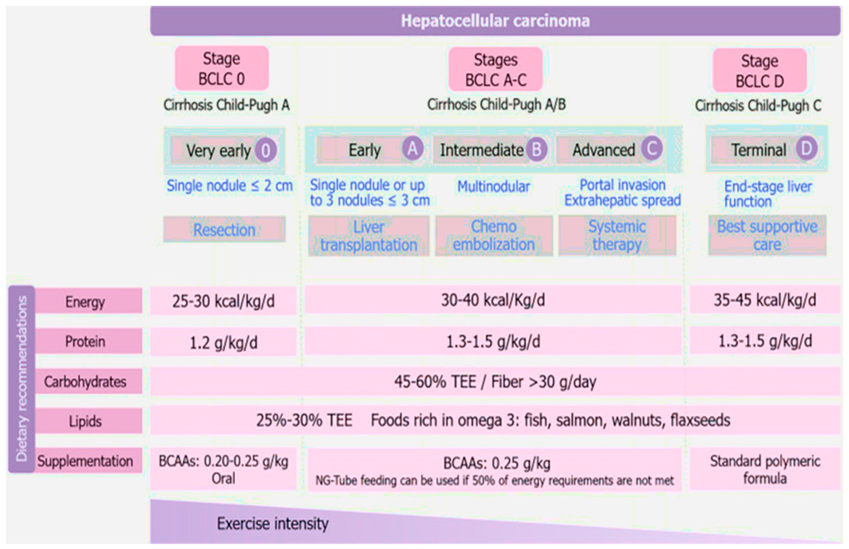
3.13. Nutritional Therapy for Hepatocellular Carcinoma

The most prevalent primary liver cancer, hepatocellular carcinoma [HCC], typically manifests as cirrhosis [175]. In addition to the more well-known risk factors for the development of HCC, such as hepatitis B and C virus infection, age, and alcohol and tobacco use, there are nutritional risk factors related to the development of HCC as well. These include a high intake of saturated fats from red meat, the type of cooking (which produces heterocyclic amines), and aflatoxins contaminating food [176,177,178]. Conversely, dietary components that are protective include those high in fibre, fruits, vegetables, n-3 polyunsaturated fatty acids, and coffee [179,180,181]. A special focus should be given to nutritional support, including appropriate nutritional assessment and therapy by a multidisciplinary team while the patient is being examined for staging and treatment of HCC. It must be taken into account that these patients typically acquire HCC on top of chronic cirrhosis, and, as a result, they may exhibit severe malnutrition. Complications from cirrhosis should be adequately treated and taken into account for nutritional care. In addition to customary techniques [182], among the most helpful tools for nutritional assessment are functional testing, phase angle, and the skeletal muscle index-L3 generated from computed tomography scans [183]. The dietary approach for hepatocellular carcinoma, according to Barcelona Clinic Liver Cancer classification is presented in Figure 1 [184].The main goal of nutritional therapy should be to supply enough protein and energy to meet the increased needs of cirrhosis and cancer. Branched-chain amino acid supplementation is also advised since it enhances survival, nutritional status, and response to treatment. Lastly, physical activity should be promoted and tailored to individual needs [184].

3.14. Hepatitis A Virus (HAV)

One of the well-known viruses that causes hepatitis globally is the hepatitis A virus (HAV). Despite the fact that this disease has declined in industrialised nations as a result of widespread vaccination, the virus continues to plague a large number of undeveloped and underdeveloped nations [185]. HAV infection can be disseminated through oral-fecal contact, and foodborne outbreaks are common [186,187]. The prevalence of the disease has changed globally as a result of improvements in socioeconomic and sanitary conditions [188]. Children under the age of five are typically asymptomatic, but as they get older, the infection symptoms start to show. Fortunately, most patients recover within 2 months of infection, while 10–15% of patients recur within the first 6 months and an acute infection may be self-limiting. Symptoms range from mild inflammation and jaundice to abrupt liver failure in older people [189,190,191,192,193]. A virus hardly ever causes persistent infection or liver damage, but undiagnosed chronic infections and the incorrect use of therapeutic methods based on clinical standards are connected to a higher risk of cirrhosis, hepatocellular cancer, and mortality [194]. Supportive care is the cornerstone on which therapy is founded. According to the Centers for Disease Control and Prevention and the American Academy of Pediatrics, all children between the ages of 12 and 23 months should receive routine vaccinations [195,196,197]. Antigen detection, monitoring liver enzyme levels, and antibody screening are the three main components of the HAV laboratory diagnosis [198,199,200]. A further benefit of polymerase chain reaction (PCR) technology is that it has been used to confirm the presence of HAV in potential dietary sources [185].

3.15. Hepatitis B Virus (HBV)

The hepatitis B virus (HBV) has infected 2 billion individuals worldwide, 360 million of them have a chronic infection, and 600,000 of them pass away each year from hepatocellular carcinoma or liver illness associated with HBV [201].
The endemic hepatitis B virus (HBV) is a major cause of hepatocellular carcinoma and chronic liver damage in many parts of the world [202,203]. The treatment for HBV differs depending on the stage of the illness. Liver transplantation is the best treatment choice for patients with end-stage liver disease [204], although interferons or nucleoside analogues are frequently used to treat HBV infection in chronic patients [205]. However, the majority of HBV patients do not have access to treatment because of the barriers to care and the high cost of antivirals [206]. These issues have prompted researchers to reevaluate therapeutic modalities, such as nutritional therapy. According to recent research, hepatitis B patients may benefit from resveratrol, vitamin E, lactoferrin, selenium, curcumin, luteolin-7-O-glucoside, moringa extracts, chlorogenic acid, and epigallocatechin-3-gallate [207]. Most of these nutrients have been examined in vitro and in animal models for their anti-HBV effects. These nutrients have been linked to a variety of antiviral and hepatoprotective mechanisms, including the induction of autophagosomes, regulation of metabolic homeostasis, epigenetic control, activation of the p53 gene [208], inhibition of oncogenes, inhibition of virus entry [209], and activation of antioxidant and anti-inflammatory pathways [210]. In conclusion, scientific data suggests that nutrition can have a direct impact on HBV replication, transcription, and expression of viral antigens. In the future, these nutrients might be taken into account to create a suitable dietary treatment for hepatitis B patients [207].

3.16. Nutritional Considerations to Prevent and Manage Viral Hepatitis A and B

Patients with viral hepatitis, both acute and chronic, frequently have changed metabolisms that cause them to eat less and become malnourished. These patients may have higher morbidity and mortality rates if they are suffering from protein and calorie deficiencies. Malnutrition’s prognosis and overall survival are improved by early detection and timely treatment [211].
Sanitation and hygiene: International travellers are more likely to contract hepatitis A (HAV) through handling or consuming contaminated raw fruits and vegetables. HAV must be rendered inactive by boiling or heating food and water for 1 min at 85 °C [185 °F] [212].
Avoiding game foods and tainted seafood is important, as the majority of acute HAV infections are brought on by eating infected seafood [212].
The microbial pathogens in saltwater can concentrate when shellfish are harvested from waters that have been polluted by sewage. Clams and oysters, for example, that have been harvested from the coastline are especially likely to be pathogenic. Animal products, particularly wild game and contaminated pig, as well as tainted seafood and produce, have all been found to contain hepatitis E (HEV) [213,214].
Avoiding iron supplements and foods strong in iron is also important, as patients’ hepatitis C progresses as a result of faster hepatic iron intake and the oxidative stress brought on by free radical generation stimulated by iron. A low-iron diet and phlebotomy help these individuals’ risk of developing hepatocellular carcinoma (HCC) [215].
Supplemental nutrition might also be necessary. Weight loss has been documented in 11–29% of treated patients who receive interferon treatment. Treatment with interferon can cause digestive symptoms, which can then lead to a decrease in appetite and food consumption [216].
A diet with reduced fat and cholesterol may be beneficial. Hepatic steatosis risk is increased by chronic hepatitis C (CHC) infection [212,216]. This issue is exacerbated by a larger dietary cholesterol intake, which is also linked to the advancement of liver disease brought on by hepatitis C. Patients with CHC who followed a diet low in fat (23 percent of calories) and cholesterol (185 mg/day) saw a decline in liver enzyme elevations as well as an improvement in immunological abnormalities [TH17/Treg balance], which are known to contribute to liver inflammation [217].
Adequate vitamin D status is important as patients with chronic liver illness frequently have vitamin D shortage and may have a harder time converting vitamin D to its active form [218]. In patients with CHC, vitamin D levels and viral load appear to be inversely correlated. Vitamin D supplementation increases the likelihood that a patient will respond to treatment, but deficiency dramatically reduces the likelihood of a sustained virological response to pegylated interferon and ribavirin [218,219].
Avoiding B12 status extremes is useful because patients with hepatitis C are better able to eliminate the virus from their systems when their B12 levels are adequate. However, very high serum B12 levels have been linked to hepatitis C RNA levels and may also promote viral replication [220].
Chronic hepatitis C and coffee intake are associated because drinking coffee may be beneficial as it decreases oxidative DNA damage, increases the death of virus-infected cells, stabilises chromosomes, and decreases fibrosis [221]. For people with nonalcoholic fatty liver disease (NAFLD), a feasible addition to therapy is a moderate daily intake of unsweetened coffee [222].
We can summarize the information about all mentioned diseases in Table 1, which describes the nutrition dysregulated and the nutrition management required for each disease.

4. Conclusions

Patients with chronic liver dysfunction (CLD) should have as their nutritional goals the restoration of liver function, prevention of related comorbidities, and overcoming malnutrition brought on by nutritional inadequacies. Patients with cirrhosis are more likely to experience micronutrient deficiencies, patients with hepatocellular disease are more likely to experience calorie and protein deficiencies, and patients with cholestasis liver disease are more likely to experience calorie deficits and an increased risk of fat-soluble vitamin deficits. Because the administration of dietary supplements has been shown to be associated with a decreased risk of infection, in-hospital mortality, and improved liver function, early detection of micronutrient and macronutrient deficiencies is crucial. All chronic liver disorder (CLD) patients are screened to determine who is at risk for complications that can be avoided.

Author Contributions

Conceptualization, A.N.A.-F. and N.A.B., investigation, K.M.A., A.-H.A.A. and E.M.E.; resources, A.N.A.-F., N.A.B., K.M.A., A.-H.A.A. and E.M.E.; writing—original draft preparation, A.N.A.-F. and E.M.E.; writing—review and editing, A.N.A.-F. and E.M.E.; visualization, A.N.A.-F., N.A.B., K.M.A. and A.-H.A.A.; supervision, E.M.E. All authors have read and agreed to the published version of the manuscript.

Funding

This research received no external funding.

Institutional Review Board Statement

Not applicable.

Informed Consent Statement

Not applicable.

Data Availability Statement

Not applicable.

Conflicts of Interest

The authors declare no conflict of interest.

References

  1. Mandato, C.; Di Nuzzi, A.; Vajro, P. Nutrition and liver disease. Nutrients 2017, 10, 9. [Google Scholar] [CrossRef] [PubMed]
  2. Silva, M.; Gomes, S.; Peixoto, A.; Torres-Ramalho, P.; Cardoso, H.; Azevedo, R.; Cunha, C.; Macedo, G. Nutrition in chronic liver disease. GE Port. J. Gastroenterol. 2015, 22, 268–276. [Google Scholar] [CrossRef] [PubMed]
  3. Smart, K.M.; Alex, G.; Hardikar, W. Feeding the child with liver disease: A review and practical clinical guide. J. Gastroenterol. Hepatol. 2011, 26, 810–815. [Google Scholar] [CrossRef] [PubMed]
  4. Purnak, T.; Yilmaz, Y. Liver disease and malnutrition. Best Pract. Res. Clin. Gastroenterol. 2013, 27, 619–629. [Google Scholar] [CrossRef]
  5. Shergill, R.; Syed, W.; Rizvi, S.A.; Singh, I. Nutritional support in chronic liver disease and cirrhotics. World J. Hepatol. 2018, 10, 685. [Google Scholar] [CrossRef]
  6. Johnson, T.M.; Overgard, E.B.; Cohen, A.E.; DiBaise, J.K. Nutrition Assessment and Management in Advanced Liver Disease. Nutr. Clin. Pr. 2013, 28, 15–29. [Google Scholar] [CrossRef]
  7. Popovici, P.; Moraru, E. Nutrition in cholestasis and long-term impact on trasnplant. Rev. Med.-Chir. A Soc. Med. Si Nat. Din Iasi 2010, 114, 726–730. [Google Scholar]
  8. Periyalwar, P.; Dasarathy, S. Malnutrition in Cirrhosis: Contribution and Consequences of Sarcopenia on Metabolic and Clinical Responses. Clin. Liver Dis. 2012, 16, 95–131. [Google Scholar] [CrossRef]
  9. Griffith, C.M.; Schenker, S. The Role of Nutritional Therapy in Alcoholic Liver Disease. Alcohol Res. Health 2006, 29, 296–306. [Google Scholar]
  10. Mendenhall, C.L.; Anderson, S.; Weesner, R.E.; Goldberg, S.J.; Crolic, K.A. Protein-calorie malnutrition associated with alcoholic hepatitis: Veterans administration cooperative study group on alcoholic hepatitis. Am. J. Med. 1984, 76, 211–222. [Google Scholar] [CrossRef]
  11. Mezey, E. Interaction Between Alcohol and Nutrition in the Pathogenesis of Alcoholic Liver Disease. Semin. Liver Dis. 1991, 11, 340–348. [Google Scholar] [CrossRef] [PubMed]
  12. Albrecht, J.; Norenberg, M.D. Glutamine: A Trojan horse in ammonia neurotoxicity. Hepatology 2006, 44, 788–794. [Google Scholar] [CrossRef] [PubMed]
  13. Mitchell, M.C.; Aday, A.W. Food for Thought: Importance of Nutrition in Alcoholic Hepatitis. Pract. Gastroenterol. 2018, 31, 30–36. [Google Scholar]
  14. Barnes, P.J.; Adcock, I.M. Glucocorticoid resistance in inflammatory diseases. Lancet 2009, 373, 1905–1917. [Google Scholar] [CrossRef] [PubMed]
  15. Thursz, M.R.; Richardson, P.; Allison, M.; Austin, A.; Bowers, M.; Day, C.P.; Downs, N.; Gleeson, D.; Macgilchrist, A.; Grant, A.; et al. Prednisolone or Pentoxifylline for Alcoholic Hepatitis. N. Engl. J. Med. 2015, 372, 1619–1628. [Google Scholar] [CrossRef]
  16. Singal, A.K.; Walia, I.; Singal, A.; Soloway, R.D. Corticosteroids and pentoxifylline for the treatment of alcoholic hepatitis: Current status. World J. Hepatol. 2011, 3, 205–210. [Google Scholar] [CrossRef]
  17. Mendenhall, C.L.; Moritz, T.E.; Roselle, G.A.; Morgan, T.R.; Nemchausky, B.A.; Tamburro, C.H.; Schiff, E.R.; McClain, C.J.; Marsano, L.S.; Allen, J.I.; et al. Protein Energy Malnutrition in Severe Alcoholic Hepatitis: Diagnosis and Response to Treatment. J. Parenter. Enter. Nutr. 1995, 19, 258–265. [Google Scholar] [CrossRef]
  18. Mendenhall, C.; Bongiovanni, G.; Goldberg, S.; Miller, B.; Moore, J.; Rouster, S.; Schneider, D.; Tamburro, C.; Tosch, T.; Weesner, R.; et al. VA Cooperative Study on Alcoholic Hepatitis III: Changes in Protein-Calorie Malnutrition Associated with 30 Days of Hospitalization with and without Enteral Nutritional Therapy. J. Parenter. Enter. Nutr. 1985, 9, 590–596. [Google Scholar] [CrossRef]
  19. Lucey, M.R.; Mathurin, P.; Morgan, T.R. Alcoholic hepatitis. N. Engl. J. Med. 2009, 360, 2758–2769. [Google Scholar] [CrossRef]
  20. Mendenhall, C.; Roselle, G.A.; Gartside, P.; Moritz, T.; The Veterans Administration Cooperative Study Groups 119 and 275. Relationship of protein calorie malnutrition to alcoholic liver disease: A reexamination of data from two Veterans Administration Cooperative Studies. Alcohol. Clin. Exp. Res. 1995, 19, 635–641. [Google Scholar] [CrossRef]
  21. Lai, J.C.; Tandon, P. Improving nutritional status in patients with cirrhosis. Off. J. Am. Coll. Gastroenterol. ACG 2018, 113, 1574–1576. [Google Scholar] [CrossRef]
  22. Matos, C.; Porayko, M.K.; Francisco-Ziller, N.; DiCecco, S. Nutrition and Chronic Liver Disease. J. Clin. Gastroenterol. 2002, 35, 391–397. [Google Scholar] [CrossRef]
  23. Galati, J.S.; Holdeman, K.P.; Dalrymple, G.V.; Harrison, K.A.; Quigley, E.M. Delayed gastric emptying of both the liquid and solid components of a meal in chronic liver disease. Am. J. Gastroenterol. 1994, 89, 708–711. [Google Scholar] [CrossRef]
  24. Kalaitzakis, E. Gastrointestinal dysfunction in liver cirrhosis. World J. Gastroenterol. 2014, 20, 14686–14695. [Google Scholar] [CrossRef]
  25. Kalaitzakis, E.; Simren, M.; Olsson, R.; Henfridsson, P.; Hugosson, I.; Bengtsson, M.; Björnsson, E. Gastrointestinal symp- toms in patients with liver cirrhosis: Associations with nutritional status and health-related quality of life. Scand. J. Gastroenterol. 2006, 41, 1464–1472. [Google Scholar] [CrossRef]
  26. Kearns, P.J.; Young, H.; Garcia, G.; Blaschke, T.; O’Hanlon, G.; Rinki, M.; Sucher, K.; Gregory, P. Accelerated improvement of alcoholic liver disease with enteral nutrition. Gastroenterology 1992, 102, 200–205. [Google Scholar] [CrossRef]
  27. Smith, J.; Horowitz, J.; Henderson, J.M.; Heymsfield, S. Enteral hyperalimen- tation in undernourished patients with cirrhosis and ascites. Am. J. Clin. Nutr. 1982, 35, 56–72. [Google Scholar] [CrossRef]
  28. Mitchell, M.C.; Friedman, L.S.; McClain, C.J. Medical Management of Severe Alcoholic Hepatitis: Expert Review from the Clinical Practice Updates Committee of the AGA Institute. Clin. Gastroenterol. Hepatol. 2017, 15, 5–12. [Google Scholar] [CrossRef]
  29. McClave, S.A.; Martindale, R.G.; Vanek, V.W.; McCarthy, M.; Roberts, P.; Taylor, B.; American College of Critical Care Medicine. Guidelines for the provision and assessment of nutrition support therapy in the adult critically ill patient: Society of Critical Care Medicine (SCCM) and American Society for Parenteral and Enteral Nutrition (ASPEN). J. Parenter. Enter. Nutri. 2009, 33, 277–316. [Google Scholar] [CrossRef]
  30. Puri, P.; Thursz, M. Intensive Enteral Nutrition in Alcoholic Hepatitis: More Food for Thought. Gastroenterology 2016, 150, 803–805. [Google Scholar] [CrossRef]
  31. Sanchez-Delgado, J.; Miquel, M. Role of rifaximin in the treatment of hepatic encephalopathy. Gastroenterol. Hepatol. 2016, 39, 282–292. [Google Scholar] [CrossRef] [PubMed]
  32. Venu, M.; Martin, E.; Saeian, K.; Gawrieh, S. High prevalence of vitamin A deficiency and vitamin D deficiency in patients evaluated for liver transplantation. Liver Transplant. 2013, 19, 627–633. [Google Scholar] [CrossRef] [PubMed]
  33. European Association for The Study of The Liver. EASL clinical practical guidelines: Management of alcoholic liver disease. J. Hepatol. 2012, 57, 399–420. [Google Scholar] [CrossRef] [PubMed]
  34. Manne, V.; Saab, S. Impact of Nutrition and Obesity on Chronic Liver Disease. Clin. Liver Dis. 2014, 18, 205–218. [Google Scholar] [CrossRef]
  35. Mohammad, M.K.; Zhou, Z.; Cave, M.; Barve, A.; McClain, C.J. Zinc and liver disease. Nutr. Clin. Pract. 2012, 27, 8–20. [Google Scholar] [CrossRef]
  36. Zhou, Z.; Wang, L.; Song, Z.; Saari, J.T.; McClain, C.J.; Kang, Y.J. Zinc Supplementation Prevents Alcoholic Liver Injury in Mice through Attenuation of Oxidative Stress. Am. J. Pathol. 2005, 166, 1681–1690. [Google Scholar] [CrossRef]
  37. Merli, M.; Berzigotti, A.; Zelber-Sagi, S.; Dasarathy, S.; Montagnese, S.; Genton, L.; Parés, A. EASL Clinical Practice Guidelines on nutrition in chronic liver disease. J. Hepatol. 2019, 70, 172–193. [Google Scholar] [CrossRef]
  38. Glass, C.; Hipskind, P.; Tsien, C.; Malin, S.; Kasumov, T.; Shah, S.N.; Kirwan, J.P.; Dasarathy, S. Sarcopenia and a physiologically low respiratory quotient in patients with cirrhosis: A prospective controlled study. J. Appl. Physiol. 2013, 114, 559–565. [Google Scholar] [CrossRef]
  39. Glass, C.; Hipskind, P.; Cole, D.; Lopez, R.; Dasarathy, S. Handheld calorimeter is a valid instrument to quantify resting energy expenditure in hospitalized cirrhotic patients: A prospective study. Nutr. Clin. Pract. 2012, 27, 677–688. [Google Scholar] [CrossRef]
  40. Dasarathy, S.; Merli, M. Sarcopenia from mechanism to diagnosis and treatment in liver disease. J. Hepatol. 2016, 65, 1232–1244. [Google Scholar] [CrossRef]
  41. Dasarathy, J.; McCullough, A.J.; Dasarathy, S. Sarcopenia in Alcoholic Liver Disease: Clinical and Molecular Advances. Alcohol. Clin. Exp. Res. 2017, 41, 1419–1431. [Google Scholar] [CrossRef]
  42. Greco, A.V.; Mingrone, G.; Benedetti, G.; Capristo, E.; Tataranni, P.A.; Gasbarrini, G. Daily energy and substrate metabolism in patients with cirrhosis. Hepatology 1998, 27, 346–350. [Google Scholar] [CrossRef]
  43. Guglielmi, F.W.; Panella, C.; Buda, A.; Budillon, G.; Caregaro, L.; Clerici, C.; Federico, A.; Gasbarrini, G.; Guglielmi, A.; Loguercio, C.; et al. Nutritional state and energy balance in cirrhotic patients with or without hypermetabolism. Multicentre prospective study by the ‘Nutritional Problems in Gastroenterology’ Section of the Italian Society of Gastroenterology (SIGE). Dig. Liver Dis. 2005, 37, 681–688. [Google Scholar] [CrossRef]
  44. Riggio, O.; Angeloni, S.; Ciuffa, L.; Nicolini, G.; Attili, A.; Albanese, C.; Merli, M. Malnutrition is not related to alterations in energy balance in patients with stable liver cirrhosis. Clin. Nutr. 2003, 22, 553–559. [Google Scholar] [CrossRef]
  45. Kondrup, J.; Nielsen, K.; Juul, A. Effect of long-term refeeding on protein metabolism in patients with cirrhosis of the liver. Br. J. Nutr. 1997, 77, 197–212. [Google Scholar] [CrossRef]
  46. Dolz, C.; Raurich, J.M.; Ibanez, J.; Obrador, A.; Marse, P.; Gaya, J. Ascites increases the resting energy expenditure in liver cirrhosis. Gastroenterology 1991, 100, 738–744. [Google Scholar] [CrossRef]
  47. Madden, A.M.; Morgan, M.Y. Resting energy expenditure should be measured in patients with cirrhosis, not predicted. Hepatology 1999, 30, 655–664. [Google Scholar] [CrossRef]
  48. Knudsen, A.W.; Krag, A.; Nordgaard-Lassen, I.; Frandsen, E.; Tofteng, F.; Mortensen, C.; Becker, U. Effect of paracentesis on metabolic activity in patients with advanced cirrhosis and ascites. Scand. J. Gastroenterol. 2015, 51, 601–609. [Google Scholar] [CrossRef]
  49. Müller, M.J.; Böker, K.H.W.; Selberg, O. Are patients with liver cirrhosis hypermetabolic? Clin. Nutr. 1994, 13, 131–144. [Google Scholar] [CrossRef]
  50. Peng, S.; Plank, L.D.; McCall, J.L.; Gillanders, L.K.; McIlroy, K.; Gane, E.J. Body composition, muscle function, and energy expenditure in patients with liver cirrhosis: A comprehensive study. Am. J. Clin. Nutr. 2007, 85, 1257–1266. [Google Scholar] [CrossRef]
  51. Iwasa, M.; Iwata, K.; Hara, N.; Hattori, A.; Ishidome, M.; Sekoguchi-Fujikawa, N.; Mifuji-Moroka, R.; Sugimoto, R.; Fujita, N.; Kobayashi, Y.; et al. Nutrition therapy using a multidisciplinary team improves survival rates in patients with liver cirrhosis. Nutrition 2013, 29, 1418–1421. [Google Scholar] [CrossRef] [PubMed]
  52. Tsien, C.D.; McCullough, A.J.; Dasarathy, S. Late evening snack: Exploiting a period of anabolic opportunity in cirrhosis. J. Gastroenterol. Hepatol. 2012, 27, 430–441. [Google Scholar] [CrossRef] [PubMed]
  53. Vaisman, N.; Katzman, H.; Carmiel-Haggai, M.; Lusthaus, M.; Niv, E. Breakfast improves cognitive function in cirrhotic patients with cognitive impairment. Am. J. Clin. Nutr. 2010, 92, 137–140. [Google Scholar] [CrossRef] [PubMed]
  54. Nielsen, K.; Kondrup, J.; Martinsen, L.; Stilling, B.; Wikman, B. Nutritional assessment and adequacy of dietary intake in hospitalized patients with alcoholic liver cirrhosis. Br. J. Nutr. 1993, 69, 665–679. [Google Scholar] [CrossRef] [PubMed]
  55. Swart, G.R.; van den Berg, J.W.; van Vuure, J.K.; Rietveld, T.; Wattimena, D.L.; Frenkel, M. Minimum protein requirements in liver cirrhosis deter- mined by nitrogen balance measurements at three levels of protein intake. Clin. Nutr. 1989, 8, 329–336. [Google Scholar] [CrossRef]
  56. Fenton, J.; Knight, E.; Humpherson, P. Milk-and-cheese diet in portal-systemic encephalopathy. Lancet 1966, 287, 164–166. [Google Scholar] [CrossRef]
  57. Greenberger, N.J.; Carley, J.; Schenker, S.; Bettinger, I.; Stamnes, C.; Beyer, P. Effect of vegetable and animal protein diets in chronic hepatic encephalopathy. Am. J. Dig. Dis. 1977, 22, 845–855. [Google Scholar] [CrossRef]
  58. Gheorghe, L.; Iacob, R.; Vădan, R.; Iacob, S.; Gheorghe, C. Improvement of hepatic encephalopathy using a modified high-calorie high-protein diet. Rom. J. Gastroenterol. 2005, 14, 231–238. [Google Scholar]
  59. Córdoba, J.; López-Hellín, J.; Planas, M.; Sabín, P.; Sanpedro, F.; Castro, F.; Esteban, R.; Guardia, J. Normal protein diet for episodic hepatic encephalopathy: Results of a randomized study. J. Hepatol. 2004, 41, 38–43. [Google Scholar] [CrossRef]
  60. Nakaya, Y.; Harada, N.; Kakui, S.; Okada, K.; Takahashi, A.; Inoi, J.; Ito, S. Severe catabolic state after prolonged fasting in cirrhotic patients: Effect of oral branched-chain amino-acid-enriched nutrient mixture. J. Gastroenterol. 2002, 37, 531–536. [Google Scholar] [CrossRef]
  61. Yoshida, T.; Muto, Y.; Moriwaki, H.; Yamato, M. Effect of long-term oral supplementation with branched-chain amino acid granules on the prognosis of liver cirrhosis. Gastroenterol. JPN 1989, 24, 692–698. [Google Scholar] [CrossRef]
  62. Fialla, A.D.; Israelsen, M.; Hamberg, O.; Krag, A.; Gluud, L.L. Nutritional therapy in cirrhosis or alcoholic hepatitis: A systematic review and meta-analysis. Liver Int. 2015, 35, 2072–2078. [Google Scholar] [CrossRef]
  63. Koretz, R.L.; Avenell, A.; Lipman, T.O. Nutritional support for liver disease. Cochrane Database Syst. Rev. 2012, 23, CD008344. [Google Scholar] [CrossRef]
  64. Singal, A.K.; Charlton, M.R. Nutrition in alcoholic liver disease. Clin. Liver Dis. 2012, 16, 805–826. [Google Scholar] [CrossRef]
  65. Ayers, P.; Adams, S.; Boullata, J.; Gervasio, J.; Holcombe, B.; Kraft, M.D.; American Society for Parenteral and Enteral Nutrition. ASPEN parenteral nutrition safety consensus recommendations. J. Parenteral Enteral Nutr. 2014, 38, 296–333. [Google Scholar] [CrossRef]
  66. Maharshi, S.; Sharma, B.C.; Sachdeva, S.; Srivastava, S.; Sharma, P. Efficacy of Nutritional Therapy for Patients With Cirrhosis and Minimal Hepatic Encephalopathy in a Randomized Trial. Clin. Gastroenterol. Hepatol. 2015, 14, 454–460.e3. [Google Scholar] [CrossRef]
  67. Calmet, F.; Martin, P.; Pearlman, M. Nutrition in Patients With Cirrhosis. Gastroenterol. Hepatol. 2019, 15, 248–254. [Google Scholar]
  68. Bunchorntavakul, C.; Reddy, K.R. malnutrition/sarcopenia and frailty in patients with cirrhosis. Aliment. Pharmacol. Ther. 2020, 51, 64–77. [Google Scholar] [CrossRef]
  69. Roongpisuthipong, C.; Sobhonslidsuk, A.; Nantiruj, K.; Songchitsomboon, S. Nutritional assessment in various stages of liver cirrhosis. Nutrition 2001, 17, 761–765. [Google Scholar] [CrossRef]
  70. Fried, L.P.; Tangen, C.M.; Walston, J.; Newman, A.B.; Hirsch, C.; Gottdiener, J.; Seeman, T. Cardiovascular Health Study Collaborative Research Group. Frailty in older adults: Evidence for a phenotype. J. Gerontol. A Biol. Sci. Med. Sci. 2001, 56, M146–M156. [Google Scholar] [CrossRef]
  71. Cheung, K.; Lee, S.S.; Raman, M. Prevalence and Mechanisms of Malnutrition in Patients With Advanced Liver Disease, and Nutrition Management Strategies. Clin. Gastroenterol. Hepatol. 2012, 10, 117–125. [Google Scholar] [CrossRef] [PubMed]
  72. Puneeta, T.; Maitreyi, R.; Marina, M.; Manuela, M. A practical approach to nutritional screening and assessment in cirrhosis. Hepatology 2017, 65, 1044–1057. [Google Scholar]
  73. European Association for the Study of the Liver. EASL Clinical Practice Guidelines on nutrition in chronic liver disease. J. Hepatol. 2019, 70, 172–193. [Google Scholar] [CrossRef] [PubMed]
  74. Hsu, H.J.; Yen, C.H.; Wu, I.W.; Liu, M.H.; Cheng, H.Y.; Lin, Y.T.; Wang, C.H. The association between low protein diet and body composition, muscle function, inflammation, and amino acid-based metabolic profile in chronic kidney disease stage 3–5 patients. Clin. Nutr. ESPEN 2021, 46, 405–415. [Google Scholar] [CrossRef]
  75. Müller, M.J.; Böttcher, J.; Selberg, O.; Weselmann, S.; Böker, K.H.; Schwarze, M.; Mühlen, A.V.Z.; Manns, M.P. Hypermetabolism in clinically stable patients with liver cirrhosis. Am. J. Clin. Nutr. 1999, 69, 1194–1201. [Google Scholar] [CrossRef]
  76. Young, S.; Kwarta, E.; Azzam, R.; Sentongo, T. Nutrition assessment and support in children with end-stage liver disease. Nutr. Clin. Prac. 2013, 28, 317–329. [Google Scholar] [CrossRef]
  77. O’Brien, A.; Williams, R. Nutrition in End-Stage Liver Disease: Principles and Practice. Gastroenterology 2008, 134, 1729–1740. [Google Scholar] [CrossRef]
  78. Newsome, P.N.; Beldon, I.; Moussa, Y.; Delahooke, T.E.; Poulopoulos, G.; Hayes, P.C.; Plevris, J.N. Low serum retinol levels are associated with hepatocellular carcinoma in patients with chronic liver disease. Aliment. Pharmacol. Ther. 2000, 14, 1295–1301. [Google Scholar] [CrossRef]
  79. Rahelić, D.; Kujundzić, M.; Romić, Z.; Brkić, K.; Petrovecki, M. Serum concentration of zinc, copper, manganese and magnesium in patients with liver cirrhosis. Coll. Antropol. 2006, 30, 523–528. [Google Scholar]
  80. Vidot, H.; Carey, S.; Allman-Farinelli, M.; Shackel, N. Systematic review: The treatment of muscle cramps in patients with cirrhosis. Aliment. Pharmacol. Ther. 2014, 40, 221–232. [Google Scholar] [CrossRef]
  81. Baskol, M.; Ozbakir, O.; Coşkun, R.; Baskol, G.; Saraymen, R.; Yucesoy, M. The Role of Serum Zinc And Other Factors on the Prevalence of Muscle Cramps in Non-alcoholic Cirrhotic Patients. J. Clin. Gastroenterol. 2004, 38, 524–529. [Google Scholar] [CrossRef]
  82. Plauth, M.; Cabre, E.; Campillo, B.; Kondrup, J.; Marchesini, G.; Schütz, T.; Wendon, J. ESPEN Guidelines on Parenteral Nutrition. Hepatol. Clin. Nutr. 2009, 28, 436–444. [Google Scholar] [CrossRef]
  83. Owen, O.E.; Trapp, V.E.; Reichard, G.A.; Mozzoli, M.A.; Moctezuma, J.; Paul, P.; Boden, G. Nature and quantity of fuels consumed in patients with alcoholic cirrhosis. J. Clin. Investig. 1983, 72, 1821–1832. [Google Scholar] [CrossRef]
  84. Amodio, P.; Bemeur, C.; Butterworth, R.; Cordoba, J.; Kato, A.; Montagnese, S.; Uribe, M.; Vilstrup, H.; Morgan, M.Y. The nutritional management of hepatic encephalopathy in patients with cirrhosis: International society for hepatic encephalopathy and nitrogen metabolism consensus. Hepatology 2013, 58, 325–336. [Google Scholar] [CrossRef]
  85. Plank, L.D.; Gane, E.J.; Peng, S.; Muthu, C.; Mathur, S.; Gillanders, L.; McIlroy, K.; Donaghy, A.J.; McCall, J.L. Nocturnal nutritional supplementation improves total body protein status of patients with liver cirrhosis: A randomized 12-month trial. Hepatology 2008, 48, 557–566. [Google Scholar] [CrossRef]
  86. Merli, M. Nutrition in cirrhosis: Dos and Don’ts. J. Hepatol. 2020, 73, 1563–1565. [Google Scholar] [CrossRef]
  87. Nakaya, Y.; Okita, K.; Suzuki, K.; Moriwaki, H.; Kato, A.; Miwa, Y.; Shiraishi, K.; Okuda, H.; Onji, M.; Kanazawa, H.; et al. BCAA-enriched snack improves nutritional state of cirrhosis. Nutrition 2007, 23, 113–120. [Google Scholar] [CrossRef]
  88. Muto, Y.; Sato, S.; Watanabe, A.; Moriwaki, H.; Suzuki, K.; Kato, A.; Kato, M.; Nakamura, T.; Higuchi, K.; Nishiguchi, S.; et al. Effects of Oral Branched-Chain Amino Acid Granules on Event-Free Survival in Patients with Liver Cirrhosis. Clin. Gastroenterol. Hepatol. 2005, 3, 705–713. [Google Scholar] [CrossRef]
  89. Toshikuni, N.; Arisawa, T.; Tsutsumi, M. Nutrition and exercise in the manage- ment of liver cirrhosis. World J. Gastroenterol. 2014, 20, 7286–7297. [Google Scholar] [CrossRef]
  90. Liberati, A.; Altman, D.G.; Tetzlaff, J.; Mulrow, C.; Gøtzsche, P.C.; Ioannidis, J.P.; Moher, D. The PRISMA statement for reporting systematic reviews and meta-analyses of studies that evaluate health care interventions: Explanation and elaboration. PLoS Med. 2009, 6, e1000100. [Google Scholar] [CrossRef]
  91. Hayashi, F.; Momoki, C.; Yuikawa, M.; Simotani, Y.; Kawamura, E.; Hagihara, A.; Fujii, H.; Kobayashi, S.; Iwai, S.; Morikawa, H.; et al. Nutritional status in relation to lifestyle in patients with compensated viral cirrhosis. World J. Gastroenterol. 2012, 18, 5759–5770. [Google Scholar] [CrossRef] [PubMed]
  92. Campillo, B.; Fouet, P.; Bonnet, J.C.; Atlan, G. Submaximal oxygen consumption in liver cirrhosis. J. Hepatol. 1990, 10, 163–167. [Google Scholar] [CrossRef] [PubMed]
  93. Berzigotti, A.; Garcia-Tsao, G.; Bosch, J.; Grace, N.D.; Burroughs, A.K.; Morillas, R.; Escorsell, A.; Garcia-Pagan, J.C.; Patch, D.; Matloff, D.S.; et al. Obesity is an independent risk factor for clinical decompensation in patients with cirrhosis. Hepatology 2011, 54, 555–561. [Google Scholar] [CrossRef] [PubMed]
  94. Everhart, J.E.; Lok, A.S.; Kim, H.; Morgan, T.R.; Lindsay, K.L.; Chung, R.T.; Bonkovsky, H.L.; Ghany, M.G. Weight-Related Effects on Disease Progression in the Hepatitis C Antiviral Long-Term Treatment Against Cirrhosis Trial. Gastroenterology 2009, 137, 549–557. [Google Scholar] [CrossRef]
  95. Macías-Rodríguez, R.U.; Ilarraza-Lomelí, H.; Ruiz-Margáin, A.; Ponce-De-León-Rosales, S.; Vargas-Vorácková, F.; García-Flores, O.; Torre, A.; Duarte-Rojo, A. Changes in Hepatic Venous Pressure Gradient Induced by Physical Exercise in Cirrhosis: Results of a Pilot Randomized Open Clinical Trial. Clin. Transl. Gastroenterol. 2016, 7, e180. [Google Scholar] [CrossRef]
  96. Zenith, L.; Meena, N.; Ramadi, A.; Yavari, M.; Harvey, A.; Carbonneau, M.; Ma, M.; Gonzalez-Abraldes, J.; Paterson, D.I.; Haykowsky, M.J.; et al. Eight Weeks of Exercise Training Increases Aerobic Capacity and Muscle Mass and Reduces Fatigue in Patients With Cirrhosis. Clin. Gastroenterol. Hepatol. 2014, 12, 1920–1926.e2. [Google Scholar] [CrossRef]
  97. Vidot, H.; Bowen, D.G.; Carey, S.; McCaughan, G.W.; Allman-Farinelli, M.; Shackel, N.A. Aggressive nutrition intervention reduces ascites and frequency of paracentesis in malnourished patients with cirrhosis and ascites. JGH Open 2017, 1, 92–97. [Google Scholar] [CrossRef]
  98. Campillo, B. Nutritional aspects of liver and biliary disease. In Textbook of Hepatology: From Basic Science to Clinical Practice, 3rd ed.; Rodes, J., Benhamou, J.P., Blei, A.T., Reichen, J., Rezzetto, M., Eds.; Blackwell Publishing Ltd.: Oxford, UK, 2007; pp. 1922–1930. [Google Scholar]
  99. Møller, S.; Bendtsen, F.; Christensen, E.; Henriksen, J.H. Prognostic variables in patients with cirrhosis and oesophageal varices without prior bleeding. J. Hepatol. 1994, 21, 940–946. [Google Scholar] [CrossRef]
  100. Lautz, H.U.; Selberg, O.; Körber, J.; Burger, M.; Muller, M.J. Proteincalorie malnutrition in liver cirrhosis. Clin. Investig. 1992, 70, 478–486. [Google Scholar] [CrossRef]
  101. Sinclair, M.; Gow, P.J.; Grossmann, M.; Angus, P.W. Review article: Sarcopenia in cirrhosis—Aetiology, implications and potential therapeutic interventions. Aliment. Pharmacol. Ther. 2016, 43, 765–777. [Google Scholar] [CrossRef]
  102. Montano-Loza, A.J. Clinical relevance of sarcopenia in patients with cirrhosis. World J. Gastroenterol. 2014, 20, 8061–8071. [Google Scholar] [CrossRef]
  103. Jones, J.M. The methodology of nutritional screening and assessment tools. J. Hum. Nutr. Diet. 2002, 15, 59–71. [Google Scholar] [CrossRef]
  104. Bémeur, C.; Butterworth, R.F. Reprint of: Nutrition in the Management of Cirrhosis and its Neurological Complications. J. Clin. Exp. Hepatol. 2015, 5, S131–S140. [Google Scholar] [CrossRef]
  105. Hasse, J.; Strong, S.; Gorman, M.A.; Liepa, G. Subjective global assessment: Alternative nutrition-assessment technique for liver-transplant candidates. Nutrition 1993, 9, 339–343. [Google Scholar]
  106. Bin, C.M.; Flores, C.; Alvares-da-Silva, M.R.; Francesconi, C.F.M. Comparison between handgrip strength, subjective global assessment, anthropometry, and biochemical markers in assessing nutritional status of patients with Crohn’s disease in clinical remission. Dig. Dis. Sci. 2010, 55, 137–144. [Google Scholar] [CrossRef]
  107. Charlton, C.P.; Buchanan, E.; Holden, C.E.; Preece, M.A.; Green, A.; Booth, I.W.; Tarlow, M.J. Intensive enteral feeding in advanced cirrhosis: Reversal of malnutrition without precipitation of hepatic encephalopathy. Arch. Dis. Child. 1992, 67, 603–608. [Google Scholar] [CrossRef]
  108. Tai, M.L.S.; Razlan, H.; Goh, K.L.; Taib, S.H.M.; Huzaini, A.H.M.; Rampal, S.; Mahadeva, S. Short-term nasogastric versus oral feeding in hospitalised patients with advanced cirrhosis. e-SPEN Eur. e-J. Clin. Nutr. Metab. 2011, 6, e242–e247. [Google Scholar] [CrossRef]
  109. Mahadeva, S.; Goh, K.-L. Letter: Enteral nutrition in advanced cirrhosis—A case of missing the boat? Aliment. Pharmacol. Ther. 2012, 36, 205–206. [Google Scholar] [CrossRef]
  110. Arroyo, V.; Gines, P.; Gerbes, A.L.; Dudley, F.J.; Gentilini, P.; Laffi, G.; Schölmerich, J. Definition and diagnostic criteria of refractory ascites and hepatorenal syndrome in cirrhosis. Hepatology 1996, 23, 164–276. [Google Scholar] [CrossRef]
  111. Teiusanu, A.; Andrei, M.; Arbanas, T.; Nicolaie, T.; Diculescu, M. Nutritional status in cirrhotic patients. Maedica 2012, 7, 284. [Google Scholar]
  112. Bémeur, C.; Desjardins, P.; Butterworth, R.F. Role of Nutrition in the Management of Hepatic Encephalopathy in End-Stage Liver Failure. J. Nutr. Metab. 2010, 2010, 1–12. [Google Scholar] [CrossRef] [PubMed]
  113. Campillo, B.; Richardet, J.-P.; Scherman, E.; Bories, P.N. Evaluation of nutritional practice in hospitalized cirrhotic patients: Results of a prospective study. Nutrition 2003, 19, 515–521. [Google Scholar] [CrossRef] [PubMed]
  114. Changani, K.K.; Jalan, R.; Cox, I.J.; Ala-Korpela, M.; Bhakoo, K.; Taylor-Robinson, S.D.; Bell, J.D. Evidence for altered hepatic gluconeogenesis in patients with cirrhosis using in vivo 31-phosphorus magnetic resonance spectroscopy. Gut 2001, 49, 557–564. [Google Scholar] [CrossRef] [PubMed]
  115. Plauth, M.; Cabré, E.; Riggio, O.; Assis-Camilo, M.; Pirlich, M.; Kondrup, J.; Ferenci, P.; Holm, E.; Dahl, S.V.; Müller, M.; et al. ESPEN Guidelines on Enteral Nutrition: Liver disease. Clin. Nutr. 2006, 25, 285–294. [Google Scholar] [CrossRef] [PubMed]
  116. Sherlock, S. Hepatic encephalopathy. In Diseases of the Liver and Biliary System, 8th ed.; Blackwell Scientific: Oxford, UK, 1989; pp. 109–110. [Google Scholar]
  117. Donaghy, A. Issues of malnutrition and bone disease in patients with cirrhosis. J. Gastroenterol. Hepatol. 2002, 17, 462–466. [Google Scholar] [CrossRef]
  118. Heyman, J.K.; Rn, C.J.W.; Brock, K.E.; Mccaughan, G.; Donaghy, A.J. Dietary protein intakes in patients with hepatic encephalopathy and cirrhosis: Current practice in NSW and ACT. Med. J. Aust. 2006, 185, 542–543. [Google Scholar] [CrossRef]
  119. Mullen, K.D.; Dasarathy, S. Protein restriction in hepatic encephalopathy: Necessary evil or illogical dogma? J. Hepatol. 2004, 41, 147–148. [Google Scholar] [CrossRef]
  120. Plauth, M.; Merli, M.; Kondrup, J.; Weimann, A.; Ferenci, P.; Müller, M. ESPEN guidelines for nutrition in liver disease and transplantation. Clin. Nutr. 1997, 16, 43–55. [Google Scholar] [CrossRef]
  121. Plauth, M.; Merli, M.; Kondrup, J. Management of hepatic encephalopathy. N. Engl. J. Med. 1997, 337, 1921–1922. [Google Scholar]
  122. Merli, M.; Riggio, O. Dietary and nutritional indications in hepatic encephalopathy. Metab. Brain Dis. 2008, 24, 211–221. [Google Scholar] [CrossRef]
  123. Keshavarzian, A.; Meek, J.; Sutton, C.; Emery, V.M.; Hughes, E.A.; Hodgson, H.J. Dietary protein supplementation from vegetable sources in the management of chronic portal systemic encephalopathy. Am. J. Gastroenterol. 1984, 79, 945–949. [Google Scholar]
  124. Bianchi, G.P.; Marchesini, G.; Fabbri, A.; Rondelli, A.; Bugianesi, E.; Zoli, M.; Pisi, E. Vegetable versus animal protein diet in cirrhotic patients with chronic encephalopathy. A randomized cross-over comparison. J. Intern. Med. 1993, 233, 385–392. [Google Scholar] [CrossRef]
  125. Louvet, A.; Artru, F.; Canva-Delcambre, V.; Dharancy, S.; Mathurin, P. Maladie alcoolique du foie. La Lett. De L’hépato-Gastroentérologue 2013, 16, 118–122. [Google Scholar]
  126. Sherlock, S. Diseases of the Liver and Biliary System, 9th ed.; Blackwell Scientific: Oxford, UK, 1993. [Google Scholar]
  127. Henkel, A.S.; Buchman, A.L. Nutritional support in patients with chronic liver disease. Nat. Clin. Pr. Gastroenterol. Hepatol. 2006, 3, 202–209. [Google Scholar] [CrossRef]
  128. McCluskey, S.A.; Karkouti, K.; Wijeysundera, D.N.; Kakizawa, K.; Ghannam, M.; Hamdy, A.; Grant, D.; Levy, G. Derivation of a risk index for the prediction of massive blood transfusion in liver transplantation. Liver Transplant. 2006, 12, 1584–1593. [Google Scholar] [CrossRef]
  129. Fusai, G.; Dhaliwal, P.; Rolando, N.; Sabin, C.A.; Patch, D.; Davidson, B.R.; Burroughs, A.K.; Rolles, K. Incidence and risk factors for the development of prolonged and severe intrahepatic cholestasis after liver transplantation. Liver Transplant. 2006, 12, 1626–1633. [Google Scholar] [CrossRef]
  130. Teperman, L.W.; Peyregne, V.P. Considerations on the Impact of Hepatic Encephalopathy Treatments in the Pretransplant Setting. Transplantation 2010, 89, 771–778. [Google Scholar] [CrossRef]
  131. Shaw, B.; Wood, R.; Gordon, R.; Iwatsuki, S.; Gillquist, W.; Starzl, T. Influence of Selected Patient Variables and Operative Blood Loss on Six-Month Survival Following Liver Transplantation. Semin. Liver Dis. 1985, 5, 385–393. [Google Scholar] [CrossRef]
  132. Selberg, O.; Bottcher, J.; Tusch, G.; Pichlmayr, R.; Henkel, E.; Muller, M.J. Identification of high- and low-risk patients before liver transplantation: A prospective cohort study of nutritional and metabolic parameters in 150 patients. Hepatology 1997, 25, 652–657. [Google Scholar] [CrossRef]
  133. Miki, C.; Iriyama, K.; Mayer, A.; Buckels, J.A.; Harrison, J.D.; Suzuki, H.; McMaster, P. Energy storage and cytokine response in patients undergoing liver transplantation. Cytokine 1999, 11, 244–248. [Google Scholar] [CrossRef]
  134. Hasse, J.M.; Blue, L.S.; Liepa, G.U.; Goldstein, R.M.; Jennings, L.W.; Mor, E.; Husberg, B.S.; Levy, M.F.; Gonwa, T.A.; Klintmalm, G.B. Early Enteral Nutrition Support in Patients Undergoing Liver Transplantation. J. Parenter. Enter. Nutr. 1995, 19, 437–443. [Google Scholar] [CrossRef] [PubMed]
  135. Reilly, J.; Mehta, R.; Teperman, L.; Cemaj, S.; Tzakis, A.; Yanaga, K.; Ritter, P.; Rezak, A.; Makowka, L. Nutritional Support after Liver Transplantation: A Randomized Prospective Study. J. Parenter. Enter. Nutr. 1990, 14, 386–391. [Google Scholar] [CrossRef] [PubMed]
  136. Mustafa, K.N. The Role of Some Foods and Diet Habits in Cholecystitis and Gallstones in Mosul in Iraq. J. Cardiovasc Disease Res. 2020, 11, 189–193. [Google Scholar]
  137. Stinton, L.M.; Shaffer, E.A. Epidemiology of Gallbladder Disease: Cholelithiasis and Cancer. Gut Liver 2012, 6, 172–187. [Google Scholar] [CrossRef] [PubMed]
  138. Ahmed, A.; Cheung, R.C.; Keeffe, E.B. Management of gallstones and their complications. Am. Fam. Physician 2000, 61, 1673–1680. [Google Scholar]
  139. Pixley, F.; Wilson, D.; McPherson, K.; Mann, J. Effect of vegetarianism on development of gall stones in women. BMJ 1985, 291, 11–12. [Google Scholar] [CrossRef]
  140. Tsai, C.-J.; Leitzmann, M.F.; Willett, W.C.; Giovannucci, E.L. Fruit and Vegetable Consumption and Risk of Cholecystectomy in Women. Am. J. Med. 2006, 119, 760–767. [Google Scholar] [CrossRef]
  141. Simon, J.A.; Hudes, E.S. Serum ascorbic acid and gallbladder disease prevalence among US adults: The Third National Health and Nutrition Examination Survey (NHANES III). Arch. Intern. Med. 2000, 160, 931–936. [Google Scholar] [CrossRef]
  142. Tsai, C.-J.; Leitzmann, M.F.; Willett, W.C.; Giovannucci, E.L. Dietary Protein and the Risk of Cholecystectomy in a Cohort of US Women: The Nurses’ Health Study. Am. J. Epidemiol. 2004, 160, 11–18. [Google Scholar] [CrossRef]
  143. Maclure, K.M.; Hayes, K.C.; Colditz, G.A.; Stampfer, M.J.; Willett, W.C. Dietary predictors of symptom-associated gallstones in middle-aged women. Am. J. Clin. Nutr. 1990, 52, 916–922. [Google Scholar] [CrossRef]
  144. Tsai, C.J.; Leitzmann, M.F.; Willett, W.C.; Giovannucci, E.L. The effect of long-term intake of cis unsaturated fats on the risk for gallstone disease in men: A prospective cohort study. Ann. Intern. Med. 2004, 141, 514–522. [Google Scholar] [CrossRef]
  145. Tsai, C.-J.; Leitzmann, M.F.; Willett, W.C.; Giovannucci, E.L. Long-term Intake of trans-Fatty Acids and Risk of Gallstone Disease in Men. Arch. Intern. Med. 2005, 165, 1011–1015. [Google Scholar] [CrossRef]
  146. Halldestam, I.; Kullman, E.; Borch, K. Incidence of and potential risk factors for gallstone disease in a general population sample. Br. J. Surg. 2009, 96, 1315–1322. [Google Scholar] [CrossRef]
  147. Erlinger, S. Gallstones in obesity and weight loss. Eur. J. Gastroenterol. Hepatol. 2000, 12, 1347–1352. [Google Scholar] [CrossRef]
  148. Thornton, J.R.; Emmett, P.M.; Heaton, K.W. Diet and gall stones: Effects of refined and unrefined carbohydrate diets on bile cholesterol saturation and bile acid metabolism. Gut 1983, 24, 2–6. [Google Scholar] [CrossRef]
  149. Tsai, C.-J.; Leitzmann, M.F.; Willett, W.C.; Giovannucci, E.L. Dietary carbohydrates and glycaemic load and the incidence of symptomatic gall stone disease in men. Gut 2005, 54, 823–828. [Google Scholar] [CrossRef]
  150. Tsai, C.-J.; Leitzmann, M.F.; Willett, W.C.; Giovannucci, E.L. Long-Term Intake of Dietary Fiber and Decreased Risk of Cholecystectomy in Women. Am. J. Gastroenterol. 2004, 99, 1364–1370. [Google Scholar] [CrossRef]
  151. Attili, A.F.; Scafato, E.; Marchioli, R.; Marfisi, R.M.; Festi, D.; The MICOL Group. Diet and gallstones in italy: The cross-sectional MICOL results. Hepatology 1998, 27, 1492–1498. [Google Scholar] [CrossRef]
  152. Everhart, J.E. Contributions of Obesity and Weight Loss to Gallstone Disease. Ann. Intern. Med. 1993, 119, 1029–1035. [Google Scholar] [CrossRef]
  153. Syngal, S.; Coakley, E.H.; Willett, W.C.; Byers, T.; Williamson, D.F.; Colditz, G. Long-Term Weight Patterns and Risk for Cholecystectomy in Women. Ann. Intern. Med. 1999, 130, 471–477. [Google Scholar] [CrossRef] [PubMed]
  154. Tsai, C.-J.; Leitzmann, M.F.; Willett, W.C.; Giovannucci, E.L. Weight Cycling and Risk of Gallstone Disease in Men. Arch. Intern. Med. 2006, 166, 2369–2374. [Google Scholar] [CrossRef] [PubMed]
  155. Gebhard, R.L.; Prigge, W.F.; Ansel, H.J.; Schlasner, L.; Ketover, S.R.; Sande, D.; Peterson, F.J. The role of gallbladder emptying in gallstone formation during diet-induced rapid weight loss. Hepatology 1996, 24, 544–548. [Google Scholar] [CrossRef] [PubMed]
  156. Lee, R.G. Nonalcoholic steatohepatitis: A study of 49 patients. Hum. Pathol. 1989, 20, 594–598. [Google Scholar] [CrossRef] [PubMed]
  157. Ludwig, J.; Viggiano, T.R.; McGill, D.B.; Oh, B.J. Nonalcoholic steatohepatitis: Mayo Clinic experiences with a hitherto unnamed disease. Mayo Clin. Proc. 1980, 55, 434–438. [Google Scholar]
  158. Bacon, B.R.; Farahvash, M.J.; Janney, C.G.; Neuschwander-Tetri, B.A. Nonalcoholic steatohepatitis: An expanded clinical entity. Gastroenterology 1994, 107, 1103–1109. [Google Scholar] [CrossRef]
  159. Sathya, P.; Martin, S.; Alvarez, F. Nonalcoholic fatty liver disease (NAFLD) in children. Curr. Opin. Pediatr. 2002, 14, 593–600. [Google Scholar] [CrossRef]
  160. Grant, L.M.; Lisker-Melman, M. Nonalcoholic fatty liver disease. Ann. Hepatol. 2004, 3, 93–99. [Google Scholar] [CrossRef]
  161. Gashgari, A.M.N.; Alhusainy, K.M.; Alharbi, F.O.E.; Hani, M.A.Q.F.; Omer, A.M.; Alali, A.H.; Alsharif, M. Nonalcoholic Fatty Liver Disease (NAFLD). Egypt. J. Hosp. Med. 2018, 70, 181–186. [Google Scholar] [CrossRef]
  162. Mirmiran, P.; Amirhamidi, Z.; Ejtahed, H.-S.; Bahadoran, Z.; Azizi, F. Relationship between Diet and Non-alcoholic Fatty Liver Disease: A Review Article. Iran. J. Public Health 2017, 46, 1007–1017. [Google Scholar]
  163. Kalafati, I.P.; Borsa, D.; Dimitriou, M.; Revenas, K.; Kokkinos, A.; Dedoussis, G.V. Dietary patterns and non-alcoholic fatty liver disease in a Greek case–control study. Nutrition 2019, 61, 105–110. [Google Scholar] [CrossRef]
  164. Zelber-Sagi, S.; Ivancovsky-Wajcman, D.; Isakov, N.F.; Webb, M.; Orenstein, D.; Shibolet, O.; Kariv, R. High red and processed meat consumption is associated with non-alcoholic fatty liver disease and insulin resistance. J. Hepatol. 2018, 68, 1239–1246. [Google Scholar] [CrossRef]
  165. He, K.; Li, Y.; Guo, X.; Zhong, L.; Tang, S. Food groups and the likelihood of non-alcoholic fatty liver disease: A systematic review and meta-analysis. Br. J. Nutr. 2020, 124, 1–13. [Google Scholar] [CrossRef]
  166. Luukkonen, P.K.; Sädevirta, S.; Zhou, Y.; Kayser, B.; Ali, A.; Ahonen, L.; Lallukka, S.; Pelloux, V.; Gaggini, M.; Jian, C.; et al. Saturated Fat Is More Metabolically Harmful for the Human Liver Than Unsaturated Fat or Simple Sugars. Diabetes Care 2018, 41, 1732–1739. [Google Scholar] [CrossRef]
  167. Astrup, A.; Magkos, F.; Bier, D.M.; Brenna, J.T.; De Oliveira Otto, M.C.; Hill, J.O.; King, J.C.; Mente, A.; Ordovas, J.M.; Volek, J.S.; et al. Saturated Fats and Health: A Reassessment and Proposal for Food-Based Recommendations: JACC State-of-the-Art Review. J. Am. Coll. Cardiol. 2020, 76, 844–857. [Google Scholar] [CrossRef]
  168. Zou, T.-T.; Zhang, C.; Zhou, Y.-F.; Han, Y.-J.; Xiong, J.-J.; Wu, X.-X.; Chen, Y.-P.; Zheng, M.-H. Lifestyle interventions for patients with nonalcoholic fatty liver disease: A network meta-analysis. Eur. J. Gastroenterol. Hepatol. 2018, 30, 747–755. [Google Scholar] [CrossRef]
  169. Wong, V.W.-S.; Wong, G.L.-H.; Chan, R.S.-M.; Shu, S.S.-T.; Cheung, B.H.-K.; Li, L.S.; Chim, A.M.-L.; Chan, C.K.-M.; Leung, J.K.-Y.; Chu, W.C.-W.; et al. Beneficial effects of lifestyle intervention in non-obese patients with non-alcoholic fatty liver disease. J. Hepatol. 2018, 69, 1349–1356. [Google Scholar] [CrossRef]
  170. Oshakbayev, K.; Bimbetov, B.; Manekenova, K.; Bedelbayeva, G.; Mustafin, K.; Dukenbayeva, B. Severe nonalcoholic steatohepatitis and type 2 diabetes: Liver histology after weight loss therapy in a randomized clinical trial. Curr. Med Res. Opin. 2018, 35, 157–165. [Google Scholar] [CrossRef]
  171. Promrat, K.; Kleiner, D.E.; Niemeier, H.M.; Jackvony, E.; Kearns, M.; Wands, J.R.; Fava, J.L.; Wing, R.R. Randomized controlled trial testing the effects of weight loss on nonalcoholic steatohepatitis. Hepatology 2010, 51, 121–129. [Google Scholar] [CrossRef]
  172. Glass, L.M.; Dickson, R.C.; Anderson, J.C.; Suriawinata, A.A.; Putra, J.; Berk, B.S.; Toor, A. Total Body Weight Loss of ≥10 % Is Associated with Improved Hepatic Fibrosis in Patients with Nonalcoholic Steatohepatitis. Dig. Dis. Sci. 2014, 60, 1024–1030. [Google Scholar] [CrossRef]
  173. Ebbeling, C.B.; Feldman, H.A.; Klein, G.L.; Wong, J.M.W.; Bielak, L.; Steltz, S.K.; Luoto, P.K.; Wolfe, R.R.; Wong, W.W.; Ludwig, D.S. Effects of a low carbohydrate diet on energy expenditure during weight loss maintenance: Randomized trial. BMJ 2018, 363, k4583. [Google Scholar] [CrossRef]
  174. Parra-Vargas, M.; Rodriguez-Echevarria, R.; Jimenez-Chillaron, J.C. Nutritional Approaches for the Management of Nonalcoholic Fatty Liver Disease: An Evidence-Based Review. Nutrients 2020, 12, 3860. [Google Scholar] [CrossRef] [PubMed]
  175. Yang, J.D.; Hainaut, P.; Gores, G.J.; Amadou, A.; Plymoth, A.; Roberts, L.R. A global view of hepatocellular carcinoma: Trends, risk, prevention and management. Nat. Rev. Gastroenterol. Hepatol. 2019, 16, 589–604. [Google Scholar] [CrossRef] [PubMed]
  176. Ziouziou, I.; Shariat, S.F.; Ajdi, F.; Khabbal, Y. Association of Processed Meats and Alcohol Consumption with Renal Cell Carcinoma: A Worldwide Population-Based Study. Nutr. Cancer 2020, 73, 2665–2670. [Google Scholar] [CrossRef] [PubMed]
  177. Golabi, P.; Rhea, L.; Henry, L.; Younossi, Z.M. Hepatocellular carcinoma and non-alcoholic fatty liver disease. Hepatol. Int. 2019, 13, 688–694. [Google Scholar] [CrossRef] [PubMed]
  178. Kulik, L.; El-Serag, H.B. Epidemiology and Management of Hepatocellular Carcinoma. Gastroenterology 2019, 156, 477–491.e1. [Google Scholar] [CrossRef]
  179. Sawada, N.; Inoue, M.; Iwasaki, M.; Sasazuki, S.; Shimazu, T.; Yamaji, T.; Takachi, R.; Tanaka, Y.; Mizokami, M.; Tsugane, S. Consumption of n-3 Fatty Acids and Fish Reduces Risk of Hepatocellular Carcinoma. Gastroenterology 2012, 142, 1468–1475. [Google Scholar] [CrossRef]
  180. Wanders, A.J.; van den Borne, J.J.G.C.; De Graaf, C.; Hulshof, T.; Jonathan, M.C.; Kristensen, M.; Mars, M.; Schols, H.A.; Feskens, E.J.M. Effects of dietary fibre on subjective appetite, energy intake and body weight: A systematic review of randomized controlled trials. Obes. Rev. 2011, 12, 724–739. [Google Scholar] [CrossRef]
  181. Babio, N.; Balanza, R.; Basulto, J.; Bulló, M.; Salas-Salvadó, J. Dietary fibre: Influence on body weight, glycemic control and plasma cholesterol profile. Nutr. Hosp. 2010, 25, 327–340. [Google Scholar]
  182. Duarte-Rojo, A.; Ruiz-Margáin, A.; Montaño-Loza, A.J.; Macías-Rodríguez, R.U.; Ferrando, A.; Kim, W.R. Exercise and physical activity for patients with end-stage liver disease: Improving functional status and sarcopenia while on the transplant waiting list. Liver Transplant. 2017, 24, 122–139. [Google Scholar] [CrossRef]
  183. Montano-Loza, A.J.; Meza-Junco, J.; Prado, C.M.; Lieffers, J.R.; Baracos, V.E.; Bain, V.G.; Sawyer, M.B. Muscle wasting is associated with mortality in patients with cirrhosis. Clin. Gastroenterol. Hepatol. 2012, 10, 166–173. [Google Scholar] [CrossRef]
  184. Ruiz-Margáin, A.; Román-Calleja, B.M.; Moreno-Guillén, P.; González-Regueiro, J.A.; Kúsulas-Delint, D.; Campos-Murguía, A.; Flores-García, N.C.; Macías-Rodríguez, R.U. Nutritional therapy for hepatocellular carcinoma. World J. Gastrointest. Oncol. 2021, 13, 1440–1452. [Google Scholar] [CrossRef]
  185. Gholizadeh, O.; Akbarzadeh, S.; Ghazanfari Hashemi, M.; Gholami, M.; Amini, P.; Yekanipour, Z.; Poortahmasebi, V. Hepatitis A: Viral Structure, Classification, Life Cycle, Clinical Symptoms, Diagnosis Error, and Vaccination. Can. J. Infect. Dis. Med. Microbiol. 2023, 2023, 4263309. [Google Scholar] [CrossRef]
  186. Nelson, N.P.; Weng, M.K.; Hofmeister, M.G.; Moore, K.L.; Doshani, M.; Kamili, S.; Harris, A.M. Prevention of hepatitis A virus infection in the United States: Recommendations of the Advisory Committee on Immunization Practices. MMWR Recomm. Rep. 2020, 69, 1. [Google Scholar] [CrossRef]
  187. Linder, K.A.; Malani, P.N. Hepatitis A. JAMA 2017, 318, 2393. [Google Scholar] [CrossRef]
  188. Noël, L.; Tubiana, R.; Simon, A.; Valantin, M.-A.; Palich, R.; Blanc, C.; Katlama, C.; Marcelin, A.-G.; Calvez, V.; Todesco, E. Low immune response rate of HIV-infected patients to a single injection of hepatitis A vaccine. Infect. Dis. Now 2021, 51, 94–96. [Google Scholar] [CrossRef]
  189. Shinde, R.V.; Shinde, A.R.; Patil, A.D.; Pawar, S.; Mohite, S.; Patil, S. Co-infection of Hepatitis A and Hepatitis E Viruses among the Acute Viral Hepatitis Cases in Tertiary Care Hospital—A Four Years Retrospective Study. J. Pure Appl. Microbiol. 2020, 14, 2047–2051. [Google Scholar] [CrossRef]
  190. Ndumbi, P.; Freidl, G.S.; Williams, C.J.; Mårdh, O.; Varela, C.; Avellón, A.; Severi, E. Members of the european hepatitis a outbreak investigation teamhepatitis a outbreak disproportionately affecting men who have sex with men (MSM) in the European Union and European Economic Area. Euro Surveill. 2018, 23, 1700641. [Google Scholar]
  191. Van Damme, P. Hepatitis a vaccines. In Pediatric Vaccines and Vaccinations; Springer: New York, NY, USA, 2017; pp. 99–108. [Google Scholar]
  192. Sung, P.S.; Hong, S.-H.; Lee, J.; Park, S.-H.; Yoon, S.K.; Chung, W.J.; Shin, E.-C. CXCL10 is produced in hepatitis A virus-infected cells in an IRF3-dependent but IFN-independent manner. Sci. Rep. 2017, 7, 6387. [Google Scholar] [CrossRef]
  193. Arum, C.; Fraser, H.; Artenie, A.A.; Bivegete, S.; Trickey, A.; Alary, M.; Strathdee, S.A. Homelessness, unstable housing, and risk of HIV and hepatitis C virus acquisition among people who inject drugs: A systematic review and meta-analysis. Lancet Public Health 2021, 6, e309–e323. [Google Scholar] [CrossRef]
  194. Win, N.N.; Kanda, T.; Ogawa, M.; Nakamoto, S.; Haga, Y.; Sasaki, R.; Nakamura, M.; Wu, S.; Matsumoto, N.; Matsuoka, S.; et al. Superinfection of hepatitis A virus in hepatocytes infected with hepatitis B virus. Int. J. Med Sci. 2019, 16, 1366–1370. [Google Scholar] [CrossRef]
  195. Abe, H.; Ushijima, Y.; Bikangui, R.; Ondo, G.N.; Zadeh, V.R.; Pemba, C.M.; Mpingabo, P.I.; Igasaki, Y.; De Vries, S.G.; Grobusch, M.P.; et al. First evidence for continuous circulation of hepatitis A virus subgenotype IIA in Central Africa. J. Viral Hepat. 2020, 27, 1234–1242. [Google Scholar] [CrossRef] [PubMed]
  196. Peak, C.M.; Stous, S.S.; Healy, J.M.; Hofmeister, M.G.; Lin, Y.; Ramachandran, S.; McDonald, E.C. Homelessness and hepatitis A—San diego county, 2016–2018. Clin. Infect. Dis. 2019, 71, 14–21. [Google Scholar] [CrossRef] [PubMed]
  197. Ramaswamy, M.; Bruden, D.; Nolen, L.D.; Mosites, E.; Snowball, M.; Nelson, N.P.; Bruce, M.; McMahon, B.J. Hepatitis A vaccine immunogenicity 25 years after vaccination in Alaska. J. Med. Virol. 2020, 93, 3991–3994. [Google Scholar] [CrossRef] [PubMed]
  198. Gedam, S.R.; Dhabarde, A.; Patil, P.S.; Sharma, A.; Kumar, K.; Babar, V. Psychiatric Comorbidity, Severity of Dependence and Liver Enzymes Dysfunction among Alcohol Dependent Individuals: A Cross-sectional Study from Central Rural India. J. Clin. Diagn. Res. 2019, 13, 1–5. [Google Scholar] [CrossRef]
  199. Ribeiro, C.R.D.A.; Amado, L.A.; Tourinho, R.S.; Lima, L.R.P.; Melgaço, J.G.; De Almeida, A.J.; Bastos, L.S.; Lewis-Ximenez, L.L.; de Paula, V. Accuracy of rapid test for diagnosis of hepatitis A with different infection rate settings and with predictive modeling. Futur. Microbiol. 2019, 14, 247–258. [Google Scholar] [CrossRef]
  200. Wahyuddin, D.; Amin, M.; Yamani, L.N.; Utsumi, T.; Sustini, F.; Lusida, M.I. Analysis of genetic and serology of hepatitis A virus infection during and after outbreak in two junior high schools in Surabaya, Indonesia. J. Med Virol. 2019, 91, 1048–1055. [Google Scholar] [CrossRef]
  201. Shepard, C.W.; Simard, E.P.; Finelli, L.; Fiore, A.E.; Bell, B.P. Hepatitis B virus infection: Epidemiology and vaccination. Epidemiol. Rev. 2006, 28, 112–125. [Google Scholar] [CrossRef]
  202. World Health Organization WHO. Hepatitis B. 2020. Available online: https://www.who.int/news-room/fact-sheets/detail/hepatitis-b (accessed on 8 July 2021).
  203. Allain, J.P.; Candotti, D.; Soldan, K.; Sarkodie, F.; Phelps, B.; Giachetti, C.; Opare-Sem, O. The risk of hepatitis B virus infection by transfusion in Kumasi, Ghana. Blood J. Am. Soc. Hematol. 2003, 101, 2419–2425. [Google Scholar] [CrossRef]
  204. Seal, K.H.; Edlin, B.R.; Ochoa, K.C.; Tulsky, J.P.; Moss, A.R.; Hahn, J.A. Risk of hepatitis B infection among young injection drug users in San Francisco: Opportunities for intervention. West. J. Med. 2000, 172, 16. [Google Scholar] [CrossRef]
  205. Nguyen, M.H.; Wong, G.; Gane, E.; Kao, J.-H.; Dusheiko, G. Hepatitis B Virus: Advances in Prevention, Diagnosis, and Therapy. Clin. Microbiol. Rev. 2020, 33, e00046-19. [Google Scholar] [CrossRef]
  206. Koithan, M.; Devika, J. New Approaches to Nutritional Therapy. J. Nurse Pr. 2010, 6, 805–806. [Google Scholar] [CrossRef]
  207. Jose-Abrego, A.; Rivera-Iñiguez, I.; Torres-Reyes, L.A.; Roman, S. Anti-hepatitis B virus activity of food nutrients and potential mechanisms of action. Ann. Hepatol. 2022, 100766. [Google Scholar] [CrossRef]
  208. Finck, B.N.; Kelly, D.P. PGC-1 coactivators: Inducible regulators of energy metabolism in health and disease. J. Clin. Investig. 2006, 116, 615–622. [Google Scholar] [CrossRef]
  209. Teng, C.-F.; Yu, C.-H.; Chang, H.-Y.; Hsieh, W.-C.; Wu, T.-H.; Lin, J.-H.; Wu, H.-C.; Jeng, L.-B.; Su, I.-J. Chemopreventive Effect of Phytosomal Curcumin on Hepatitis B Virus-Related Hepatocellular Carcinoma in A Transgenic Mouse Model. Sci. Rep. 2019, 9, 1–13. [Google Scholar] [CrossRef]
  210. Park, S.; Lim, J.; Kim, J.R.; Cho, S. Inhibitory effects of resveratrol on hepatitis B virus X protein-induced hepatocellular carcinoma. J. Veter-Sci. 2017, 18, 419–429. [Google Scholar] [CrossRef]
  211. Praharaj, D.L.; Anand, A.C. Nutrition in Viral Hepatitis. Curr. Hepatol. Rep. 2023, 22, 9–23. [Google Scholar] [CrossRef]
  212. Wu, D.; Guo, C. Epidemiology and Prevention of Hepatitis A in Travelers. J. Travel Med. 2013, 20, 394–399. [Google Scholar] [CrossRef]
  213. Leblanc, D.; Poitras, E.; Gagné, M.-J.; Ward, P.; Houde, A. Hepatitis E virus load in swine organs and tissues at slaughterhouse determined by real-time RT-PCR. Int. J. Food Microbiol. 2010, 139, 206–209. [Google Scholar] [CrossRef]
  214. Yugo, D.M.; Meng, X.-J. Hepatitis E Virus: Foodborne, Waterborne and Zoonotic Transmission. Int. J. Environ. Res. Public Health 2013, 10, 4507–4533. [Google Scholar] [CrossRef]
  215. Yasutake, K.; Kohjima, M.; Nakashima, M.; Kotoh, K.; Nakamuta, M.; Enjoji, M. Nutrition Therapy for Liver Diseases Based on the Status of Nutritional Intake. Gastroenterol. Res. Pr. 2012, 2012, 1–8. [Google Scholar] [CrossRef]
  216. Oliveira, L.P.; De Jesus, R.P.; Boulhosa, R.S.; Mendes, C.M.C.; Gnoatto, M.C.; Lemaire, D.C.; Toralles, M.B.P.; Cavalcante, L.N.; Lyra, A.C.; Lyra, L.G. Effect of soy protein supplementation in patients with chronic hepatitis C: A randomized clinical trial. World J. Gastroenterol. 2012, 18, 2203–2211. [Google Scholar] [CrossRef]
  217. Maggio, R.; Viscomi, C.; Andreozzi, P.; D’Ettorre, G.; Viscogliosi, G.; Barbaro, B.; Balsano, C. Normocaloric low cholesterol diet modulates Th17/Treg balance in patients with chronic hepatitis C virus infection. PLoS ONE 2014, 9, e112346. [Google Scholar] [CrossRef] [PubMed]
  218. Villar, L.M.; Del Campo, J.A.; Ranchal, I.; Lampe, E.; Romero-Gomez, M. Association between vitamin D and hepatitis C virus infection: A meta-analysis. World J. Gastroenterol. 2013, 19, 5917–5924. [Google Scholar] [CrossRef] [PubMed]
  219. Refaat, B.; El-Shemi, A.G.; Ashshi, A.; Azhar, E. Vitamin D and chronic hepatitis C: Effects on success rate and prevention of side effects associated with pegylated interferon-α and ribavirin. Int. J. Clin. Exp. Med. 2015, 8, 10284–10303. [Google Scholar] [PubMed]
  220. Mechie, N.-C.; Goralzcyk, A.D.; Reinhardt, L.; Mihm, S.; Amanzada, A. Association of serum vitamin B12 levels with stage of liver fibrosis and treatment outcome in patients with chronic hepatitis C virus genotype 1 infection: A retrospective study. BMC Res. Notes 2015, 8, 260. [Google Scholar] [CrossRef]
  221. Cardin, R.; Piciocchi, M.; Martines, D.; Scribano, L.; Petracco, M.; Farinati, F. Effects of coffee consumption in chronic hepatitis C: A randomized controlled trial. Dig. Liver Dis. 2013, 45, 499–504. [Google Scholar] [CrossRef]
  222. Torres, D.M.; Harrison, S.A. Is It Time to Write a Prescription for Coffee? Coffee and Liver Disease. Gastroenterology 2013, 144, 670–672. [Google Scholar] [CrossRef]
  223. Bischoff, S.C.; Singer, P.; Koller, M.; Barazzoni, R.; Cederholm, T.; van Gossum, A. Standard operating procedures for ESPEN guidelines and consensus papers. Clin. Nutr. 2015, 34, 1043–1051. [Google Scholar] [CrossRef]
  224. Schütte, K.; Tippelt, B.; Schulz, C.; Röhl, F.-W.; Feneberg, A.; Seidensticker, R.; Arend, J.; Malfertheiner, P. Malnutrition is a prognostic factor in patients with hepatocellular carcinoma (HCC). Clin. Nutr. 2014, 34, 1122–1127. [Google Scholar] [CrossRef]
  225. Alvares-da-Silva, M.R.; da Silveira, R.T. Comparison between handgrip strength, subjective global assessment, and prognostic nutritional index in assessing malnutrition and predicting clinical outcome in cirrhotic outpatients. Nutrition 2005, 21, 113–117. [Google Scholar] [CrossRef]
  226. Belarmino, G.; Gonzalez, M.C.; Torrinhas, R.S.; Sala, P.; Andraus, W.; D’Albuquerque, L.A.C.; Pereira, R.M.R.; Caparbo, V.F.; Ravacci, G.R.; Damiani, L.; et al. Phase angle obtained by bioelectrical impedance analysis independently predicts mortality in patients with cirrhosis. World J. Hepatol. 2017, 9, 401–408. [Google Scholar] [CrossRef]
  227. Wang, C.W.; Feng, S.; Covinsky, K.E.; Hayssen, H.; Zhou, L.Q.; Yeh, B.M.; Lai, J.C. A comparison of muscle function, mass, and quality in liver transplant can- didates: Results from the functional assessment in liver transplantation study. Transplantation 2016, 100, 1692. [Google Scholar] [CrossRef]
  228. Lai, J.C. Defining the threshold for too sick for transplant. Curr. Opin. Organ Transplant. 2016, 21, 127–132. [Google Scholar] [CrossRef]
  229. Sinclair, M.; Poltavskiy, E.; Dodge, J.L.; Lai, J.C. Frailty is independently associated with increased hospitalisation days in patients on the liver transplant waitlist. World J. Gastroenterol. 2017, 23, 899–905. [Google Scholar] [CrossRef]
  230. Tsien, C.; Shah, S.N.; McCullough, A.J.; Dasarathy, S. Reversal of sarcopenia predicts survival after a transjugular intrahepatic portosystemic stent. Eur. J. Gastroenterol. Hepatol. 2013, 25, 85–93. [Google Scholar] [CrossRef]
  231. Hanai, T.; Shiraki, M.; Nishimura, K.; Imai, K.; Suetsugu, A.; Takai, K.; Shimizu, M.; Naiki, T.; Moriwaki, H. Free fatty acid as a marker of energy malnutrition in liver cirrhosis. Hepatol. Res. 2013, 44, 218–228. [Google Scholar] [CrossRef]
  232. Dasarathy, S. Posttransplant sarcopenia: An underrecognized early conse- quence of liver transplantation. Dig. Dis. Sci. 2013, 58, 3103–3111. [Google Scholar] [CrossRef]
  233. DiMartini, A.; Cruz, R.J., Jr.; Dew, M.A.; Myaskovsky, L.; Goodpaster, B.; Fox, K. Muscle mass predicts outcomes following liver transplantation. Liver Transplant. 2013, 19, 1172–1180. [Google Scholar] [CrossRef]
  234. Englesbe, M.J.; Patel, S.P.; He, K.; Lynch, R.J.; Schaubel, D.E.; Harbaugh, C. Sarcopenia and mortality after liver transplantation. J. Am. Coll. Surg. 2010, 211, 271–278. [Google Scholar] [CrossRef]
  235. Montano-Loza, A.J.; Angulo, P.; Meza-Junco, J.; Prado, C.M.M.; Sawyer, M.B.; Beaumont, C.; Esfandiari, N.; Ma, M.; Baracos, V.E. Sarcopenic obesity and myosteatosis are associated with higher mortality in patients with cirrhosis. J. Cachex-Sarcopenia Muscle 2015, 7, 126–135. [Google Scholar] [CrossRef]
  236. Walsh, T.S.; Wigmore, S.J.; Hopton, P.; Richardson, R.; Lee, A. Energy expenditure in acetaminophen-induced fulminant hepatic failure. Crit. Care Med. 2000, 28, 649–654. [Google Scholar] [CrossRef] [PubMed]
  237. Mathur, S.; Peng, S.; Gane, E.J.; McCall, J.L.; Plank, L.D. Hypermetabolism predicts reduced transplant-free survival independent of MELD and Child-Pugh scores in liver cirrhosis. Nutrition 2007, 23, 398–403. [Google Scholar] [CrossRef] [PubMed]
  238. Hipskind, P.; Glass, C.; Charlton, D.; Nowak, D.; Dasarathy, S. Do handheld calorimeters have a role in assessment of nutrition needs in hospitalized patients? A systematic review of literature. Nutr. Clin. Pract. 2011, 26, 426–433. [Google Scholar] [CrossRef] [PubMed]
  239. Nielsen, K.; Kondrup, J.; Martinsen, L.; Døssing, H.; Larsson, B.; Stilling, B.; Jensen, M.G. Long-term oral refeeding of patients with cirrhosis of the liver. Br. J. Nutr. 1995, 74, 557–567. [Google Scholar] [CrossRef] [PubMed]
  240. Diamond, I.R.; de Silva, N.T.; Tomlinson, G.A.; Pencharz, P.B.; Feldman, B.M.; Moore, A.M. The role of parenteral lipids in the development of advanced intestinal failure-associated liver disease in infants: A multiple- variable analysis. J. Parenter. Enter. Nutr. 2011, 35, 596–602. [Google Scholar] [CrossRef]
  241. Stanko, R.T.; Nathan, G.; Mendelow, H.; Adibi, S.A. Development of hepatic cholestasis and fibrosis in patients with massive loss of intestine supported by prolonged parenteral nutrition. Gastroenterology 1987, 92, 197–202. [Google Scholar] [CrossRef]
  242. Kelly, D.A. Intestinal failure-associated liver disease: What do we know today? Gastroenterology 2006, 130, S70–S77. [Google Scholar] [CrossRef]
  243. Pironi, L.; Arends, J.; Bozzetti, F.; Cuerda, C.; Gillanders, L.; Jeppesen, P.B.; Joly, F.; Kelly, D.; Lal, S.; Staun, M.; et al. ESPEN guidelines on chronic intestinal failure in adults. Clin. Nutr. 2016, 35, 247–307. [Google Scholar] [CrossRef]
  244. Calkins, K.L.; Dunn, J.C.; Shew, S.B.; Reyen, L.; Farmer, D.G.; Devaskar, S.U. Pediatric intestinal failure-associated liver disease is reversed with 6 months of intravenous fish oil. JPEN J. Parenter. Enter. Nutr. 2014, 38, 682–692. [Google Scholar] [CrossRef]
  245. Le, H.D.; de Meijer, V.E.; Zurakowski, D.; Meisel, J.A.; Gura, K.M.; Puder, M. Parenteral Fish Oil as Monotherapy Improves Lipid Profiles in Children With Parenteral Nutrition–Associated Liver Disease. J. Parenter. Enter. Nutr. 2010, 34, 477–484. [Google Scholar] [CrossRef]
  246. Nandivada, P.; Chang, M.I.M.; Potemkin, A.K.B.; Carlson, S.J.M.; Cowan, E.; O’Loughlin, A.A.M.; Mitchell, P.D.M.; Gura, K.M.P.D.; Puder, M. The Natural History of Cirrhosis from Parenteral Nutrition-Associated Liver Disease after Resolution of Cholestasis with Parenteral Fish Oil Therapy. Ann. Surg. 2015, 261, 172–179. [Google Scholar] [CrossRef]
  247. Angsten, G.; Finkel, Y.; Lucas, S.; Kassa, A.M.; Paulsson, M.; Lilja, H.E. Improved outcome in neonatal short bowel syndrome using parenteral fish oil in combination with omega-6/9 lipid emulsions. J. Parenter. Enter. Nutr. 2012, 36, 587–595. [Google Scholar] [CrossRef]
  248. Tomsits, E.; Pataki, M.; Tolgyesi, A.; Fekete, G.; Rischak, K.; Szollar, L. Safety and efficacy of a lipid emulsion containing a mixture of soybean oil, medium- chain triglycerides, olive oil, and fish oil: A randomised, double-blind clin- ical trial in premature infants requiring parenteral nutrition. J. Pediatr. Gastroenterol. Nutr. 2010, 51, 514–521. [Google Scholar] [CrossRef]
  249. Bowyer, B.A.; Fleming, C.R.; Ludwig, J.; Petz, J.; McGill, D.B. Does long-term home parenteral nutrition in adult patients cause chronic liver disease? J. Parenter. Enter. Nutr. 1985, 9, 11–17. [Google Scholar] [CrossRef]
  250. Cavicchi, M.; Beau, P.; Crenn, P.; Degott, C.; Messing, B. Prevalence of Liver Disease and Contributing Factors in Patients Receiving Home Parenteral Nutrition for Permanent Intestinal Failure. Ann. Intern. Med. 2000, 132, 525–532. [Google Scholar] [CrossRef]
  251. Burns, D.L.; Gill, B.M. Reversal of Parenteral Nutrition–Associated Liver Disease With a Fish Oil–Based Lipid Emulsion (Omegaven) in an Adult Dependent on Home Parenteral Nutrition. J. Parenter. Enter. Nutr. 2012, 37, 274–280. [Google Scholar] [CrossRef]
  252. Venecourt-Jackson, E.; Hill, S.J.; Walmsley, R.S. Successful treatment of parenteral nutrition–associated liver disease in an adult by use of a fish oil–based lipid source. Nutrition 2013, 29, 356–358. [Google Scholar] [CrossRef]
  253. Xu, Z.; Li, Y.; Wang, J.; Wu, B.; Li, J. Effect of omega-3 polyunsaturated fatty acids to reverse biopsy-proven parenteral nutrition-associated liver disease in adults. Clin. Nutr. 2011, 31, 217–223. [Google Scholar] [CrossRef]
  254. Pironi, L.; Colecchia, A.; Guidetti, M.; Belluzzi, A.; D’Errico, A. Fish oil-based emulsion for the treatment of parenteral nutrition associated liver disease in an adult patient. e-SPEN Eur. e-J. Clin. Nutr. Metab. 2010, 5, e243–e246. [Google Scholar] [CrossRef]
  255. Clemmesen, J.O.; Kondrup, J.; Ott, P. Splanchnic and leg exchange of amino acids and ammonia in acute liver failure. Gastroenterology 2000, 118, 1131–1139. [Google Scholar] [CrossRef]
  256. Vilstrup, H.; Iversen, J.; Tygstrup, N. Glucoregulation in acute liver failure. Eur. J. Clin. Investig. 1986, 16, 193–197. [Google Scholar] [CrossRef] [PubMed]
  257. Clemmesen, J.O.; Høy, C.-E.; Kondrup, J.; Ott, P. Splanchnic metabolism of fuel substrates in acute liver failure. J. Hepatol. 2000, 33, 941–948. [Google Scholar] [CrossRef] [PubMed]
  258. McClave, S.A.; Taylor, B.E.; Martindale, R.G.; Warren, M.M.; Johnson, D.R.; Braunschweig, C.; McCarthy, M.S.; Davanos, E.; Rice, T.W.; Cresci, G.A.; et al. Guidelines for the provision and assessment of nutrition support therapy in the adult critically ill patient: Society of critical care medicine (SCCM) and American society for parenteral and enteral nutrition (A.S.P.E.N.). J. Parenter. Enter. Nutr. 2016, 40, 159–211. [Google Scholar] [CrossRef] [PubMed]
  259. Rabinowich, L.; Wendon, J.; Bernal, W.; Shibolet, O. Clinical management of acute liver failure: Results of an international multi-center survey. World J. Gastroenterol. 2016, 22, 7595–7603. [Google Scholar] [CrossRef]
  260. Mendenhall, C.L.; Tosch, T.; Weesner, R.E.; Garcia-Pont, P.; Goldberg, S.J.; Kiernan, T.; Seeff, L.B.; Sorell, M.; Tamburro, C.; Zetterman, R.; et al. VA cooperative study on alcoholic hepatitis II: Prognostic significance of protein-calorie malnutrition. Am. J. Clin. Nutr. 1986, 43, 213–218. [Google Scholar] [CrossRef]
  261. Moreno, C.; Deltenre, P.; Senterre, C.; Louvet, A.; Gustot, T.; Bastens, B.; Hittelet, A.; Piquet, M.-A.; Laleman, W.; Orlent, H.; et al. Intensive Enteral Nutrition Is Ineffective for Patients With Severe Alcoholic Hepatitis Treated With Corticosteroids. Gastroenterology 2016, 150, 903–910.e8. [Google Scholar] [CrossRef]
  262. Koretz, R.L. The evidence for the use of nutritional support in liver disease. Curr. Opin. Gastroenterol. 2014, 30, 208–214. [Google Scholar] [CrossRef]
  263. Liu, M.; Shah, V.H. New prospects for medical management of acute alcoholic hepatitis. Clin. Liver Dis. 2019, 13, 131. [Google Scholar] [CrossRef]
  264. Bonkovsky, H.L.; Singh, R.H.; Jafri, I.H.; Fiellin, D.A.; Smith, G.S.; Simon, D. A randomized, controlled trial of treatment of alcoholic hepatitis with parenteral nutrition and oxandrolone. II. Short-term effects on nitrogen metabolism, metabolic balance, and nutrition. Am. J. Gastroenterol. 1991, 86, 1209–1218. [Google Scholar]
  265. Cabre, E.; Rodriguez-Iglesias, P.; Caballeria, J.; Quer, J.C.; Sanchez-Lombrana, J.L.; Pares, A. Short- and long-term outcome of severe alcohol-induced hepatitis treated with steroids or enteral nutrition: A multicenter randomized trial. Hepatology 2000, 32, 36–42. [Google Scholar] [CrossRef]
  266. Antar, R.; Wong, P.; Ghali, P. A Meta-Analysis of Nutritional Supplementation for Management of Hospitalized Alcoholic Hepatitis. Can. J. Gastroenterol. 2012, 26, 463–467. [Google Scholar] [CrossRef]
  267. Foody, W.; Heuman, D.D.; Mihas, A.A.; Schubert, M.L. Nutritional therapy for alcoholic hepatitis: New life for an old idea. Gastroenterology 2001, 120, 1053–1054. [Google Scholar] [CrossRef]
Figure 1. Dietary approach for hepatocellular carcinoma, according to Barcelona Clinic Liver Cancer classification. BCAAs: Branched-chain amino acids; BCLC: Barcelona Clinic Liver Cancer; TEE: Total energy expenditure. Ref. [184].
Livers 03 00013 g001
Table 1. Describing the nutrition dysregulated and the nutrition management required.
Hepatic DysfunctionRequired Dietary ModificationsN. Reference
Patients with (ALF: acute liver failure, ASH: alcoholic steatohepatitis) and cirrhosisREE is usually increased (REE, resting energy expenditure)[222]
patients with NAFLD (non-alcoholic fatty liver disease)Normal REE[222]
Malnutrition and sarcopeniaMalnutrition can impair the whole spectrum of hepatic metabolic-functions, and malnutrition alone can cause severe fatty liver but is not known to cause chronic liver disease[223]
Patients with chronic liver diseases and have a sedentary life styleShould receive a total energy supply of 1.3*REE[224]
Acute Liver
Failure
(ALF)
A severe disorder of carbohydrate, protein, and lipid metabolism should be expected in ALF due to subtotal loss of hepatocellular function and ensuing multi-organ failure. This disorder is characterised by impaired hepatic glucose production and lactate clearance as well as protein catabolism linked to hyper-aminoacidemia and hyper-ammonemia[72]
Acute Liver
Failure
(ALF)
In ALF patients, obesity is associated with an increased risk of death or need for transplantation and increased mortality ofter transplantation[225]
In patients with mild hepatic encephalopathyONS “oral nutritional supplements” should be used when feeding goals cannot be attained by oral nutrition alone[226]
In patients with severe hyper-acute disease with hepatic
encephalopathy and highly elevated arterial ammonia who are at risk of cerebral edema
Nutritional protein support can be deferred for 24–48 h until hyper-ammonemia is controlled. When protein administration is commenced, arterial ammonia should be monitored to ensure no pathological elevation occurs.[227]
Acute Liver
Failure
(ALF) who cannot be fed orally
Should receive EN via nasogastric
Inaso-jejunal tube
[228]
EN: enteral nutrition in Acute Liver Failure (ALF)ALF patients without malnutrition should be provided with nutritional support (preferentially EN) when they are considered unlikely to resume normal oral nutrition within the next 5 to 7 days[72]
EN: enteral nutrition in Acute Liver Failure (ALF)Standard enteral formulas can be given, as there are no data regarding the value of a disease specific composition[229]
PN: parenteral nutrition in Acute Liver Failure (ALF)In malnourished ALF patients, EN and/or PN should be initiated promptly, PN should be used as second-line treatment in patients who cannot be fed adequately by oral and/or EN[228]
Alcoholic Steatohepatitis (ASH)Individualized nutrition counselling should be used in order to improve food intake[230]
Alcoholic Steatohepatitis (ASH)ONS should be used when patients with severe ASH cannot meet their caloric requirements through normal food in order to improve survival:[231]
EN: enteral nutrition in Alcoholic Steatohepatitis (ASH)
  • In order to increase survival and reduce morbidity in patients with severe ASH who are unable to meet their caloric needs through conventional diet and/or ONS, EN should be utilised
  • EN can be used in people with severe ASH to make sure they get enough protein and energy without raising their risk of developing hepatic encephalopathy
  • Standard formulae, preferably those with high energy density (1.5 kcal/mL), should be utilised for ONS or EN in patients with severe ASH
PN: parenteral nutrition in Alcoholic Steatohepatitis (ASH)
  • Patients with severe ASH who are moderately or severely malnourished and who are unable to receive enough nutrition through oral and/or enteral routes must start receiving PN right away
  • Daily administration of electrolytes, trace minerals, and water- and fat-soluble vitamins, starting at the start of PN, is required to meet needs
Non-alcoholic Steatohepatitis (NASH)
  • Patients with NAFL/NASH who are overweight or obese must adhere to a weight-loss plan to lower their risk of comorbidity and to improve their liver enzyme levels and histology (necroinflammation)
  • To improve steatosis and liver biochemistry in overweight or obese NAFL INASH patients, a weight loss of 7–10% should be the goal; a loss of more than 10% should be the goal to improve fibrosis
  • Regardless of the macronutrient composition, a hypocoloric diet must be followed in order to lose weight, according to current obesity guidelines
  • After weight loss diets and extensive lifestyle therapies have failed in otherwise eligible obese NAFL INASH patients without cirrhosis, bariatric surgery should be suggested
Non-alcoholic Steatohepatitis (NASH)
  • To improve liver enzymes and histology in nondiabetic persons with histologically proven NASH, vitamin E (800 /U a-tocopherol daily) should be administered
  • Antioxidants, such as those found in vitamins (resveratrol, anthocyanin, and bayberries), cannot be advised for the treatment of NAFL or NASH until more information on their efficacy is known.
  • Until more information on their effectiveness is available, omega3 fatty acids cannot be advised for the treatment of NAFL or NASH
  • Patients with NAFL or NASH may benefit from nutritional supplements, including specific probiotics or synbiotics to boost their liver enzymes
  • Patients with intercurrent illnesses who are obese and receiving EN or PN should have a target energy intake of 25 kcal/kg IBW/d and a higher target protein intake of 2.0–2.5 g/kg IBW
Liver Cirrhosis (LC)
  • It should be expected that individuals with cirrhosis will have a high prevalence of malnutrition, protein depletion, and trace element deficit
  • To improve patients’ long-term outcome/survival, specific dietary counselling should be applied to cirrhotic patients employing a multidisciplinary team
  • To improve the status of total body protein, a late-night snack should be encouraged. This will help to shorten periods of famine
  • Patients with cirrhosis who are malnourished or experience higher energy consumption (such as acute complications or refractory ascites) should consume more energy
  • Increased energy intake is not advised for individuals who are overweight or obese who have cirrhosis
  • In obese patients with cirrhosis, lifestyle interventions aimed at weight loss’s positive effects, which include reduced portal hypertension
  • Patients with compensated cirrhosis who are not malnourished need to consume 1.2 g/kg/d of protein
  • Patients with cirrhotic malnutrition and/or sarcopenia should consume 1.5 g·kg−1•ct1 of protein to replenish their bodies
  • Patients with cirrhosis who are malnourished and have reduced muscle mass should consume 30–35 kcal per kg and 1.5 g of protein per kg daily
  • Protein catabolism is increased in cirrhotic patients with HE, hence protein intake shouldn’t be restricted
  • Vegetable proteins or BCAAs (0.25 g·kg−1•ctJ) should be given orally in cirrhotic patients who are protein “intolerant” to help them consume enough protein
  • To increase event-free survival or quality of life in patients with advanced cirrhosis, long-term oral BCAA supplements 0.25 g•kg-Lcf l) should be administered
  • When prescribing a low-sodium diet, the moderate benefit in the treatment of ascites should be weighed against the increased risk of even less food consumption. After reducing sodium intake, caution should be exercised to prevent impairing the diet’s palatability
  • EN should be carried out on cirrhotic patients who cannot be fed orally or who do not meet the nutritional aim by oral diet
  • In cirrhotic patients, PN should be utilised if oral and/or EN are ineffective or impractical
Liver Transplantation (LTx) and Surgery
  • Patients with liver cirrhosis who are scheduled for elective surgery or who are on the transplant list should have their nutritional condition promptly examined and assessed in order to address malnutrition prior to surgery and consequently enhance body protein status
  • Aim for a total daily protein intake of 1.2–1.5 g·kg−1·d−1 and a total daily energy intake of 30–35 kcal•Jrg-l.J−1 (126–147 kcal•Jrg-l.J−1). These ranges cover suggested intakes based on therapy objectives, such as nutritional status maintenance or improvement
  • Patients who are obese can get EN and/or PN with an enhanced goal protein intake of 2.0–2.5 g·kg−1 IBW and a target energy intake of 25 kcal·kg−1IBWJ-1
  • The dietary management plan for NASH patients who are overweight or obese and planned for elective surgery should be followed
  • No suggestions can be offered for the conditioning of donors or organs using certain feeding regimens, such as intravenous glutamine or arginine, with the goal of reducing ischemia/reperfusion damage
Liver Transplantation (LTx): Preoperative phase
  • To lower the risk of infection after LTx, normal food and/or EN should be introduced within the first 12 to 24 h following surgery.
  • Aim for an energy intake of 30–35 kcal•krr1•ct1 (126–141 kJ •krr1 •ct) and a protein intake of 1.2–1.5 g kg”1 •ct1 beyond the immediate postoperative phase
Liver Transplantation (LTx): postoperative phase
  • To lower the rate of infection after transplantation, enteral formula should be given in conjunction with specific probiotics
  • Patients with HE who require EN can take formulations that are BCAA-enriched
  • When oral nutrition or EN is neither possible nor practical, PN should be preferred to no feeding in order to lower complication rates, the duration of mechanical ventilation, and the length of hospital stay
  • When the cough and swallow reflexes are impaired, EN is contraindicated, or it is not practical to employ EN, PN should be administered in patients with unprotected airways and HE
Disclaimer/Publisher’s Note: The statements, opinions and data contained in all publications are solely those of the individual author(s) and contributor(s) and not of MDPI and/or the editor(s). MDPI and/or the editor(s) disclaim responsibility for any injury to people or property resulting from any ideas, methods, instructions or products referred to in the content.

