Article

Labeling of Polysaccharides with Biotin and Fluorescent Dyes

1
M. M. Shemyakin–Yu. A. Ovchinnikov Institute of Bioorganic Chemistry, Russian Academy of Sciences, 16/10 Miklukho-Maklaya, 117997 Moscow, Russia
2
Institute of Physiology of Federal Research Centre “Komi Science Centre of the Urals Branch of the Russian Academy of Sciences”, 167982 Syktyvkar, Russia
3
Zelinsky Institute of Organic Chemistry RAS, 47 Leninsky Prosp, 119991 Moscow, Russia
4
Kazan Institute of Biochemistry and Biophysics, Kazan Scientific Center of the Russian Academy of Sciences, Lobachevsky str., 2/31, 420111 Kazan, Russia
*
Author to whom correspondence should be addressed.
Polysaccharides 2024, 5(1), 1-15; https://doi.org/10.3390/polysaccharides5010001
Submission received: 9 October 2023 / Revised: 13 December 2023 / Accepted: 19 December 2023 / Published: 27 December 2023

Abstract

:
Examples of labeling polysaccharides at hydroxyl groups are described in this paper, which are especially in demand for molecules with a blocked reducing end. The protocols presented are suitable for the microscale synthesis of labeled polysaccharides that do not require a chromatography step for isolation. Examples of hydroxyl labeling include (1) direct modification with fluorescein isothiocyanate; (2) reaction with a fluorescein-dichlorotriazine derivative; (3) reaction with biotin-dichlorotriazine; (4) indirect two-step modification (given for glycosphingolipid) with glutaric anhydride followed by amidation with aminospacered BODIPY or SuCy5. The labeling of carboxyl groups of hyaluronic acid with BODIPY is also described. The staining of plant tissue sections with biotinylated polysaccharide versus being fluorescein labeled is compared.

Graphical Abstract

1. Introduction

The labeling of a polysaccharide (PS) with a fluorescent label opens up numerous possibilities for studying bacterial biofilms [1], PS-binding proteins [2], and cellular [3] and other processes. The literature on PS labeling is extensive and comprehensive. The most commonly used method is reductive amination or other reactions of the reducing (aldehyde) group [4,5,6,7,8]. For all its attractiveness, a number of limitations should be noted, in particular that PSs do not always have a free reducing end (or they are supposed to be modified by another tag, or they are intended for immobilization), one mole of label per mole of PS (especially a high MW molecule) is not always sufficient for reliable detection, and reductive amination requires rather harsh conditions and gives far from quantitative yields. Other functional groups in PSs are also subjected to modification. The most accessible for modification are the pre-existing amino groups of a PS, which are present in bacterial PSs obtained by removing lipid A from whole lipopolysaccharide by mild acid hydrolysis, and also chitosans. The amino groups react with N-oxysuccinimide esters, isothiocyanates, and other amino-reactive groups [3,9,10]. The modification of PSs by carboxyl groups is applied to plant PSs containing uronic acids (for example, pectins), hyaluronic acids, and other glycosaminoglycans. To activate carboxyl groups for their subsequent reactions with amines, carbodiimides and 4-(4,6-dimethoxy-1,3,5-triazin-2-yl)-4-methylmorpholinium chloride (DMTMM) were used, and diazomethane derivatives were also applied for modification [11,12,13,14,15,16]. Hydroxyl groups are especially attractive for modification since they are present in any polysaccharide and because there are many of them, including the most reactive primary ones. For the modification of PSs at hydroxyl groups, reactions with isocyanates, isothiocyanates, N,N′-disuccinimidyl carbonate (DSC), dichlorotriazines, and vinylsulfones have been applied [17,18,19,20,21,22,23].
In recent years, in our group, there has been a need to attach a fluorescent label (as well as a biotin label, which makes it possible to indirectly solve the same problem, through labeled streptavidin) to polysaccharides, namely by the hydroxyl group, as well as by the amino group of phosphoethanolamine in the composition of an inner core fragment of bacterial polysaccharides [3]. In this study, we describe practical protocols for the dosed labeling of polysaccharides for hydroxyl groups that have been repeatedly verified.

2. Materials and Methods

2.1. Materials

Sodium hyaluronate (HA Na-salt) 8–15 and 15–30 kDa were purchased from Contipro Biotech (Dolní Dobrouč, Czech Republic). Fluorescent dyes BODIPY and sulfocyanine 5 (herein referred to as SuCy5) in an amino-spacered form (for their chemical structure, see chapter 3.5) were purchased from Lumiprobe https://ru.lumiprobe.com (accessed on 20 December 2023). DTAF (5-(4,6-dichlorotriazinyl)aminofluorescein) was purchased from Molecular Probes (Eugene, OR, USA). Sephadex G-15 was purchased from Amersham Biosciences (Buckinghamshire, United Kingdom). Dowex 50X4-400 was purchased from Acros Organics (Geel, Belgium). Reversed-phase C18 LiChroprep RP-18 (40–63 µm), paraformaldehyde, N-(3-dimethylaminopropyl)-N′-ethylcarbodiimide hydrochloride, N-hydroxysuccinimide, and N,N-diisopropylethylamine were purchased from Sigma-Aldrich (Saint Louis, MO, USA). Tris, DMSO, DMF, Et3N, and Triton-X100 N,N′-dicyclohexylcarbodiimide were purchased from Merck (Darmstadt, Germany). Glycolipid blood group A-Ceramide is a product, CBGSL00012, of Chembind LLC (USA). Thin-layer chromatography was performed using aluminum sheets, Kieselgel 60 F254 (1.05554.0001, Merck, Darmstadt, Germany), with detection made by charring after 7% H3PO4 soaking or with phosphomolybdic acid.

2.2. Methods

2.2.1. Na+ to i-Pr2EtNH+ Ion Replacement in Acidic Polysaccharides with the Example of Sodium Hyaluronate 15–30 kDa

A solution of 15–30 kDa HA Na-salt (5 mg) in 100 μL of water was slowly passed through a Dowex 50W_X2_200-400_mesh column (0.2 mL, i-Pr2EtNH+ form) and then eluted with 20% ethanol (0.8 mL). The eluate was evaporated and the residue was freeze-dried from water. As a result, 6.24 mg (99%) 15–30 kDa HA i-Pr2EtNH+ salt was obtained, the solubility of which in DMSO was >100 mg/mL. Similarly, the exchange of cations was carried out for hyaluronic acid 8–15 kDa and plant polysaccharide from the pulp of baobab fruit Adansonia digitata L. (isolated as previously described [2]).

2.2.2. Direct Labeling of Acidic Polysaccharides with Fluorescein Isothiocyanate (FITC) with the Example of HA15–30kDa i-Pr2EtNH+ Salt

HA15–30kDa i-Pr2EtNH+ salt (2.5 mg) was dissolved in 75 µL of dry DMSO; FITC (0.25 mg in 25 μL DMSO) and i-Pr2EtN (3.1 μL) was added. The solution was kept for 40 h at 50 °C, 2.5 M NaOAc (2 µL) in water/methanol 2:1 was added (this was approximately equivalent to the content of carboxyl groups of hyaluronic acid), and labeled HA was precipitated with 1300 µL of ethyl acetate; after 15 min, the precipitate was separated by centrifugation (14,000× g/7 min). The supernatant was removed by decantation, the precipitate was dissolved in 15 µL of water, 15 µL of methanol was added, and the labeled HA was precipitated by the addition of 650 µL of acetonitrile followed by 650 µL of ethyl acetate. After 15 min, the precipitate was separated by centrifugation (14,000× g/7 min), the supernatant was removed by decantation, the residue was dried on with air and freeze-dried from water. According to TLC, supernatants do not contain polysaccharide material, so the yield of labeled HA can be considered as near to quantitative. The content of the label was spectrophotometrically determined at pH 7.4 by absorption at 492 nm based on the molar extinction for fluorescein ε = 74,000. In the described above case, the label content was found to be 1 eq. per ~30 kDa of HA.
Similarly, labeling was carried out for hyaluronic acid 8–15 kDa and plant polysaccharide from the pulp of baobab fruit Adansonia digitata L.(Table 1).

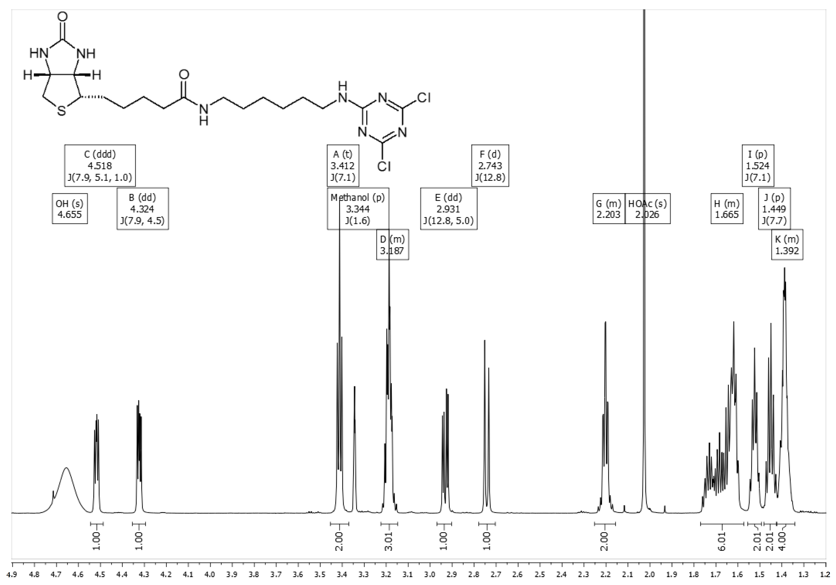
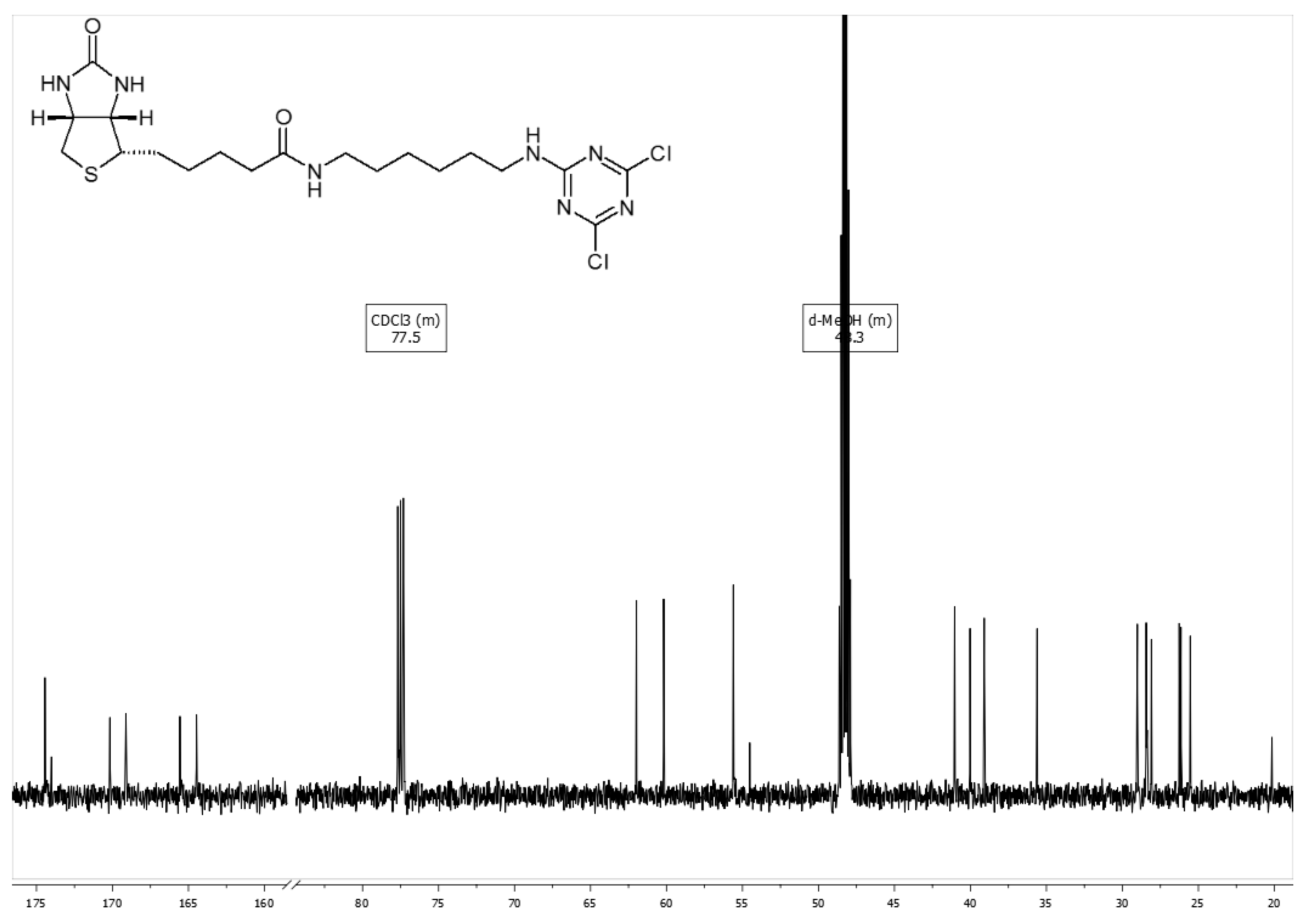
2.2.3. Synthesis of Dichlorotriazine (DCTA) Reagent, biot-DCTA

To a solution of amino-spacered biotin biot-NH-(CH2)6-NH2 × CF3COOH (148 mg, 0.324 mmol) in a mixture of water (2.5 mL) and MeCN (5 mL) i-Pr2EtN (113 μL, 0.648 mmol) was added. The solution was cooled to 0 °C, and trichlorotriazine (59.8 mg, 0.324 mmol) in dichloroethane (1.5 mL) was added with intensive stirring. Reaction mixture was stirred for 10 min at 0 °C, then for 10 min at room temperature. AcOH (217 μL) was added and the mixture was evaporated at 30 °C. The residue was extracted with a mixture of CHCl3 (20 mL), water (20 mL) and AcOH (0.4 mL). Aqueous layer was extracted additionally with CHCl3 (15 mL). Chloroform extracts were filtered (cotton wool) and evaporated to dryness at 30 °C. The yield of vacuum-dried biot-DCTA was 146 mg (92%), white solid powder. TLC (silica gel 60 TLC aluminum sheets): Rf = 0.59 (CHCl3/MeOH 4:1). Stored dry at −18 °C.
1H NMR (700 MHz, CD3OD/CDCl3 1:1 + 0.5% CD3COOD, 30 °C): δ 4.518 (ddd, 1H, J = 7.9, 5.1, 1.0 Hz; N-CH of biot), 4.324 (dd, 1H, J = 7.9, 4.5 Hz; N-CH` of biot), 3.412 (t, 2H, J = 7.1 Hz; CH2N), 3.344 (p; d-MeOH), 3.187 (m, 3H; CH2N and SCH of biot), 2.931 (dd, 1, J = 12.8, 5.0 Hz; SCH` of biot), 2.743 (d, 1H, J = 12.8 Hz; SCH`` of biot), 2.203 (m, 2H; CH2CO of biot), 1.665 (m, 6H; 3 CH2), 1.524 (p, 2H, J = 7.1 Hz; CH2), 1.449 (p, 2H, J = 7.7 Hz; CH2), 1.392 (m, 4H; 2 CH2) ppm (Figure A1).
13C NMR (176 MHz, CD3OD/CDCl3 1:1 + 0.5% CD3COOD, 30 °C) δ 174.4 and 170.2 and 169.1 and 165.6 and 164.5 (2 C=O and 3 C of dichlorotriazine), 77.5 (CDCl3), 61.98 and 60.2 (2 NC of biot), 55.6 (SC of biot), 48.3 (d-MeOH), 41.0, 40.0, 39.1, 35.6, 29.0, 28.4, 28.38, 28.1, 26.3, 26.1, 25.5, 20.2 (Figure A2).
NMR spectra of biot-DCTA and data of its stability are given in Appendix A.

2.2.4. Labeling of Polysaccharides with biot-DCTA on the Example of HA15–30kDa Na Salt

To a solution of sodium hyaluronate 15–30 kDa (4 mg) in a mixture of water and DMSO (120 + 80 μL), a freshly prepared solution of biot-DCTA (0.4 mg) in DMSO (40 μL) and i-Pr2EtN (4.8 μL) was added. The solution was kept for 4 h at room temperature, then a labeled polysaccharide was precipitated with 700 µL of acetonitrile and 700 µL of ethyl acetate. After 15 min, the precipitate was separated by centrifugation (4000× g/8 min), dissolved in 50 μL of water and the procedure of precipitation/centrifugation was repeated. The residue was dried on air and freeze-dried from water. According to NMR data, the content of the biotin label was one biotin residue per 16–17 kDa of PS. Similarly, the labeling was carried out for four plant polysaccharides (Table 2).

2.2.5. Labeling of Bacterial Polysaccharides with DTAF

PS (0.2 mg) was dissolved in a mixture of water (24 μL) and DMSO (16 μL), and a freshly prepared solution of DTAF (0.1 mg) in DMSO (10 μL) and i-Pr2EtN (1 μL) were added. The solution was kept for 4 h at room temperature, then labeled PS was precipitated with 200 µL of acetonitrile and 800 µL of ethyl acetate. After centrifugation, the precipitate was dissolved in 40 μL of water and freeze-dried.
Some of capsular PS in a mixture of water and DMSO formed a gel; however, after adding i-Pr2EtN, a homogeneous solution was obtained.
To reliably determine the content of the label in polysaccharides for three of them, modification was carried out under the same conditions, but in amounts of 1 mg.
A list of bacterial polysaccharides labeled with DTAF is given in Table 3.

2.2.6. Fluorescent Labeling of PSs at Carboxyl Groups

Hyaluronic Acids

To the stirred solution of sodium hyaluronate HA15–30kDa (10 mg, ~26.4 μmol of disacch–ride -[GlcAβ1-3GlcNAcβ]- units) in 0.9 mL of water, a solution of N-(3-dimethylaminopropyl)-N′-ethylcarbodiimide hydrochloride (30 mg/mL in DMF, 147 μL, 23 μmol) and a solution of N-hydroxysuccinimide (40 mg/mL in water, 134 μL, 46 μmol) were added, and pH was adjusted to 5.4. A solution of BODIPY amine (10 mg/mL in DMF, 98 μL, 2.3 μmol) was added, and the mixture was stirred for 7 h at room temperature. The reaction mixture was dialyzed in Spectra/Por® 6 dialysis tubing (MWCO 1000) against 1× PBS (pH 7.4), then 0.15 M NaCl, and finally against distilled water. Freeze-drying gave 9.2 mg of BODIPY-labeled HA15–30kDa sodium salt.
The content of BODIPY in the conjugate was determined by measurement of absorption (ε, l·mol−1·cm−1 = 82,000) at 503 nm in H2O. The resulting value ~0.12 µM/mg corresponds to one BODIPY residue per 20 disacch–ride -[GlcAβ1-3GlcNAcβ]- units, in average three BODIPY residues per one HA15–30kDa molecule.

Xylogalacturonan

Sodium salt of pectin from baobab fruit Adansonia digitata L. (2 mg) was transformed into a i-Pr2EtNH+ salt (as described in Section 2.2.1), yield 2.8 mg. To the stirred solution (slightly opalescent) of the salt (2.8 mg) in dry DMF (1.8 mL) a solution of hydroxybenzotriazole monohydrate (0.56 μmol, 17.2 μL of 5 mg/mL in DMF), a solution of SuCy5 amine (0.14 μmol, 20.7 μL of 5 mg/mL in DMF), a solution of N,N′-dicyclohexylcarbodiimide (0.42 μmol, 17.3 μL of 5 mg/mL in DMF) and a solution of i-Pr2EtNH (0.14 μmol, 2.5 μL of 1% v/v in DMF) were added sequentially (the pH of 2 μL of reaction mixture diluted with 2 μL of water was 6.2–6.4). The reaction mixture was stirred at ~25 °C for 24 h, the reaction completeness was controlled by TLC in CHCl3/MeOH/water 28:16:3, for SuCy5 amine Rf = 0.21, for PS Rf = 0). A solution of 2.5 M NaOAc (3.2 μL, an equivalent per i-Pr2EtNH in 2.8 mg of the pectin salt) was added, the labeled PS was precipitated by addition of 7.4 mL of ethyl acetate/heptane 1:1 v/v followed by centrifugation. The precipitate was dissolved in 0.25 mL of water, 0.75 mL of MeOH and 2 mL of MeCN were added. The precipitated material was isolated by centrifugation, dried in vacuum and freeze-dried from water. The yield of the labeled PS sodium salt was 1.8 mg (contains only traces of free dye). Based on the amounts of the starting PS and the conjugated dye, the content of the dye determined by measurement of absorption (ε, l·mol−1·cm−1 = 271,000) at 646 nm in H2O is estimated as 1 eq. per 15–16 kDa of PS.

2.2.7. Indirect Fluorescent Labeling at Hydroxyl Groups (through the Introduction of the Carboxyl Function)

Glutarate of A-Ceramide
To a solution of blood group A ceramide (herein ref. as A-Ceramide, 0.7 mg, 0.518 μmol) in dry DMF (0.2 mL) glutaric anhydride (4.14 micromole, 0.47 mg in 47 μL of DMF) and i-Pr2EtN (4 μL) were added, and the solution was kept for 1 h at 70 °C. The solution was acidified with AcOH (30 μL) and A-Ceramide glutarate was isolated on Sephadex G-15 column (50 mL) in CHCl3/MeOH/water 1:3:1. The yield of isomeric glutarates containing ~30% of unreacted A-Ceramide was 0.7 mg. TLC in CHCl3/MeOH/water 28:16:3: A-Ceramide Rf = 0.65, its glutarates Rf = 0.57, 0.51 and 0.43.
BODIPY-A-Ceramide
To a solution of A-Ceramide glutarate (0.7 mg, ~0.48 μmol) in dry DMF (0.2 mL) hydroxybenzotriazole monohydrate (3.84 μmol, 0.6 mg in 60 μL of DMF), N,N′-dicyclohexylcarbodiimide (3.84 μmol, 0.8 mg in 80 μL of DMF), BODIPY amine hydrochloride (1.92 μmol, 0.82 mg in 82 μL of DMF) and i-Pr2EtN (1.92 μmol, 6.8 μL of 5% solution in DMF) were added and the solution was kept for 24 h at 37 °C. BODIPY-A-Ceramide was isolated on Sephadex G-15 column (50 mL) in CHCl3/MeOH/water 1:3:1, yield 0.76 mg.
Removal of the label
BODIPY-A-Ceramide (0.58 mg) was dissolved in a mixture of 2-propanol (200 μL), MeOH (400 μL) and water (400 μL). Et3N (50 μL) was added and the solution was kept for 2 h at 50 °C. A-Ceramide was separated from the dye on the reverse phase C18 LiChroprep (1 mL) by sequential elution with water/MeOH 1:1, 1:2, 1:3 and water/MeOH 1:3 + 12% CHCl3, yield 0.45 mg.
SuCy5-A-Ceramide
To a solution of A-Ceramide glutarate obtained from the recovered A-Ceramide as described above (0.35 mg, 0.24 μmol) in dry DMF (0.1 mL) hydroxybenzotriazole monohydrate (1.92 μmol, 0.3 mg in 30 μL of DMF), N,N′-dicyclohexylcarbodiimide (1.92 μmol, 0.4 mg in 40 μL of DMF), SuCy5 amine (0.96 μmol, 0.71 mg in 71 μL of DMF) and i-Pr2EtN (0.96 μmol, 3.4 μL of 5% solution in DMF) were added and the solution was kept for 24 h at 37 °C. SuCy5-A-Ceramide was isolated on Sephadex G-15 (50 mL) in CHCl3/MeOH/water 1:3:1, yield 0.44 mg.

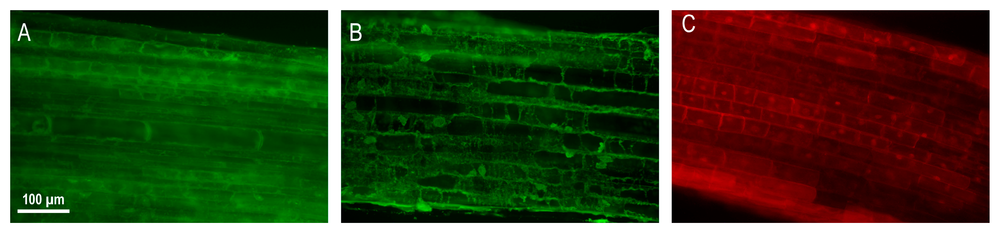
2.2.8. Probing of Cell Sections with Labeled Polysaccharides

Longitudinal sections of the roots of 3–4 day old seedlings of rye Secale cereale L. 50 µm thick, were obtained using a VT1000S vibratome (Leica Biosystems, Wetzlar, Germany). After cutting, the sections were incubated for 15–20 min in paraformaldehyde (PFA) 6%, followed by 3–4 fold washing in TBS buffer (0.02 M Tris, 0.15 M NaCl, pH 7.6 with the addition of 1:1 0.25% Triton-X100). Then, PS labeled with fluorescein, SuCy5, or biotin, diluted with TBS buffer at a ratio of 100 μg/100 μL, was applied to the section. Sections were incubated with the appropriate conjugate for 1 h at room temperature in the dark, after which they were washed 3–4 times with TBS buffer. Sections coated with biotinylated PS were incubated with streptavidin AlexaFluor488 (Invitrogen, Waltham, MA, USA) for 1 h at room temperature in the dark followed by washing 3–4 times with TBS buffer. Sections were observed under a Leica DM1000 fluorescence microscope (Leica Biosystems) at exposures of 5000 ms; for biotinylated and fluorescein PS, an excitation filter 460–500 nm and a barrier filter 512-542 were used; for SuCy5-labeled PS, an excitation filter 540–580 nm and a barrier filter 608-683 were used. The photographs were taken using an AxioCam HRs camera (Zeiss, Oberkochen, Germany).

3. Results and Discussion

For our studies of polysaccharide-binding proteins in mammals and plants, it was necessary to introduce labels into the composition of PSs of various structures. As evidenced by the literature data, the method of reductive amination at the reducing end of PS is best suited for this. Therefore, this way of introducing a fluorescent or biotin label was initially tested on the example of hyaluronic acid 15–30 kDa. However, (data not shown) in the presence of 2-picoline-borane as a reducing agent, the amino- or hydrazide derivatives (in the spacer) of fluorescein and BODIPY degrade, while the non-degrading amino derivative of biotin, even when in several equivalents of excess, inadequately modifies PS. Therefore, further efforts were directed towards labeling at hydroxyl groups, especially since this route enables one to introduce a label in the required amount, regardless of the presence of a reducing end in PS.

3.1. FITC Labeling of PSs at Hydroxyl Groups

Fluorescein derivatives appear to be the most commonly used fluorescent labeling reagent in biology, including PS modification [10,18,19,24,25,26,27]. Methods of labeling polysaccharides at hydroxyl groups with fluorescein isothiocyanate (FITC) are also described in the literature. The reaction is carried out in DMF/DMSO in the presence of NaHCO3 and dibutyltin dilaurate [18,19]. In our case, the last reagent was not applicable, so we modified the technique. The main problem was that acidic polysaccharides in the form of sodium salt are extremely poorly soluble in DMSO. The problem was solved by converting the sodium salt of PS into i-Pr2EtN salt, which was easily and quantitatively performed by passing the PS solution through a Dowex (i-Pr2EtN+ form) microcolumn with subsequent evaporation and lyophilization. The obtained polysaccharide i-Pr2EtN salt in DMSO reacted with FITC in the presence of i-Pr2EtN at 50 °C. To isolate the labeled polysaccharide in a form of sodium salt, NaOAc solution was added and the product was precipitated with an organic solvent. The precipitate contained almost all labeled PS without admixture of free FITC, but it may contain a small amount of NaOAc, (which does not interfere with the use of labeled PS as a probe). The technique was tested on the examples of hyaluronic acid 8–15 and 15–30 kDa, and was further used for labeling plant polysaccharides (Table 1, Scheme 1A).
Spectrophotometric evaluation showed the content of one fluorescein residue per 20–40 kDa PS region. Changing the concentration of FITC in the reaction mixture enables proportionally shifting this range. The degree of substitution was reproduced when the reaction was carried out under identical conditions. The typical absorption and fluorescence spectra for the polysaccharides labeled with FITC are shown in Figure A3, Appendix A.

3.2. Biotinylation of PSs at Hydroxyl Groups

To introduce the biotin tag (Scheme 1B) at hydroxyls, the chemistry described for modification by dichlorotriazinyl aminofluorescein (DTAF) was used [18,26,27]. We modified the described method and applied it to the new reagent biot-DCTA. The reaction is carried out in a water/DMSO mixture (as biot-DCTA does not dissolve without DMSO); instead of sodium sulfate and alkali, i-Pr2EtN was used as a base (pH 9), which made it possible at the isolation step to extract labeled PS by precipitation, namely by adding acetonitrile and ethyl acetate 1:1 to the solution; the final purification was carried out by a similar precipitation from water. The technique was optimized on the example of 15–30 kDa hyaluronic acid and was further used to label plant polysaccharides (Table 2). According to NMR data, in the case of sodium hyaluronate 15–30 kDa labeling, the content of the biotin residue at the selected ratio of reagents (see Experimental) is one biotin residue per 16–17 kDa polysaccharide. Synthesis of the starting reagent biot-DCTA is described in detail in the Experimental.

3.3. Fluorescein Labeling of Bacterial PSs Using DTAF Reagent

Bacterial PSs obtained by gentle hydrolysis of lipopolysaccharides contain an amino group (in the form of an ethanolamine substituent) in their core, which previously enabled us to introduce a fluorescent label selectively for the amino group using BODIPY NHS ester [3]. However, this route is not suitable for labeling PSs that lack an amino group (like capsular PSs, for instance). As a universal method of introducing the fluorescent label, we tested the reaction with dichlorotriazinyl aminofluorescein (DTAF), for which the procedure described in the literature [27] was modified similarly to that described above for labeling with biot-DCTA. The reaction was carried out in aqueous DMSO, with a freshly prepared solution of DTAF in DMSO in the presence of i-Pr2EtN at room temperature; labeled PS was precipitated by adding an organic solvent.
Conclusively, in the case of the PS containing an amino group (see above), both amino and hydroxyl groups react with DTAF (Scheme 2). Spectrophotometric evaluation of the amount of the label (determined by measurement of absorption (ε, l·mol−1·cm−1 = 80,000 at 493 nm in H2O) in the DTAF-labeled PS from E. coli O19ab, #16 in Table 3 (one label per 13 kDa), and comparison of it with the amount of the label in the case of labeling by BODIPY NHS ester by core amino groups only (one label per 32 kDa), showed that the total amount of the label is 2.5 fold greater, that is, one label residue per 32 kDa fragment of PS. This enables estimating the incorporation of the DTAF label by hydroxyl groups under these conditions as approximately one label residue per 20 kDa fragment of PS. Indeed, the determination of the label content in capsule polysaccharides (which do not have a core fragment and an amino group) gave one label per 23.5 kDa for Acinetobacter baumannii LUH5535 K35 (#7 in Table 3) and one label per 21 kDa for Acinetobacter baumannii LUH5550 K42 (#8 in Table 3).
The typical absorption and fluorescence spectra for the polysaccharides labeled with DTAF are shown in Figure A3, Appendix A. The list of PSs labeled with DTAF is shown in Table 3.
Note that the reactions were carried out on a microscale (0.2 mg of PS) and that simple precipitation with acetonitrile/ethyl acetate was used to separate polysaccharide material from the reagent and solvent.

3.4. Fluorescent Labeling of Acidic Polysaccharide by Carboxyl Groups

Labeling of acidic polysaccharides in the form of Na-salt was carried out in an aqueous-organic solution by conjugation of an amino derivative of the dye by carboxylic groups under the action of a water-soluble carbodiimide in the presence of N-hydroxysuccinimide at pH of about 5.4. An acidic polysaccharide can also be converted to i-Pr2EtNH+ salt, which enables the reaction to be carried out in dry DMF using hydroxybenzotriazole and N,N′-dicyclohexylcarbodiimide. In the first case, to isolate the reaction product, it is necessary to carry out gel chromatography or dialysis, while in the second case the isolation is a simple precipitation procedure.
Here, we present as an example BODIPY labeling of hyaluronic acid (earlier, we similarly synthesized lipid derivatives of hyaluronic acid [24]) and SuCy5-labeled acidic pectin xylogalacturonan (Scheme 3). The absorption and fluorescence spectra for the polysaccharides labeled with BODIPY and SuCy5 are shown in Figure A3 in Appendix A.
This method is simple, and enables one to dose the degree of substitution of the polysaccharide. Therefore, we consider it as the simplest labeling route for acidic PSs.

3.5. Indirect Fluorescent Labeling at Hydroxyl Groups (through the Introduction of the Caboxyl Function)

The task of synthesizing a glyco-probe with the optimal content of a fluorescent label, noted above, becomes much more complicated if the amount of the initial glyco-molecule is very small, or the researcher, for other reasons, cannot afford to expend some of it on optimizing the labeling reaction in order to confidently introduce exactly the amount of label that is needed. Without optimizing the ratio of reagents during synthesis, there is a risk of ruining the entire substance, and with optimization, a significant part of it. The solution here is a two-stage modification (Scheme 4), where in the first stage the polyol (PS or glycolipid) is modified with glutaric anhydride, and in the second stage, an aminospacer-dye is quantitatively attached to the introduced carboxyl group. Previously (unpublished data), we found that glutaric anhydride used in reasonable excess and under relatively mild conditions acylates natural ceramide almost exclusively via the primary hydroxyl, without affecting the secondary hydroxyl of sphingosine. The advantage of the glutaric bridge is that the regeneration of the original polyol takes place under relatively mild basic conditions and, therefore, the remains of unsuccessful modification can be quantitatively recovered. We used this approach to modify the glycosphingolipid A-Ceramide (Scheme 4) and GalNAcα1-3(Fucα1-2)Galβ1-4Glcβ-Ceramide, a representative polysaccharide.
Our specific approach developed as follows. An attempt to label this glycosylceramide with FITC under the conditions described in the Section 3.1 showed that the amount of the label introduced per whole tetrasaccharide was extremely small. We then carried out the modification with glutaric anhydride, followed by conjugation with BODIPY amine, where the yield was high; however, the individual properties of BODIPY dye did not suit experiments with cells (very rapid internalization of labeled ceramide was observed). This approach was abandoned and SuCy5 was introduced instead. These syntheses (see Experimental) gave BODIPY and SuCy5 derivatives containing approx. one residue of the label; note that this entire chain of events was undertaken with approximately 1 mg of the initial A-Ceramide.

3.6. Evaluation of the Label Content

The amount of the label in the composition of the obtained glycoconjugates was determined spectrophotometrically by the absorption of the dye or by 1H NMR spectra (for biotin-labeled hyaluronic acid). Labeling with FITC, under the conditions found it possible to introduce one eq. label calculated per region of the polysaccharide ~20–40 kDa, when labeled with the biot-DCTA reagent—per region ~20 kDa. When labeling bacterial PS with DTAF, there were up to two fluorescein moieties per region ~20 kDa due to additional labeling of core amino groups. Indirect fluorescent labeling after the introduction of carboxyl groups makes it possible to introduce the required amount of the label, the maximum of which is determined by the content of the primary hydroxyl groups of the polyol.

4. Application of the Synthesized Probes, and General Conclusions

Polysaccharides of infectious bacteria are a key target for the human immune system—not only for the antibody response, but also for the cellular response. Previously [3], we have used fluorescently labeled PS to study the interaction of human dendritic cells with bacterial PS and found that not all PS are recognized by these cells; now, using the synthetic work described above, obtaining these kinds of molecular probes has become routine, which opens up the possibility of continuing and expanding the study of the specificity of dendritic cells.
The diversity of plant cell polysaccharides suggests the presence of many PS-recognizing proteins, such as lectins, glycosidases, and glycosyl transferases. However, such well-characterized proteins are still surprisingly few. At the very beginning of this study, we faced the problem of non-specific binding of the fluorescein label to preparations of plant tissue sections. In order to advance in this work, it was necessary to (1) find another (or several) appropriate tag, (2) learn how to optimize the amount of the tag introduced into the PS molecule, and (3) have several labeling methods, since the structure of plant PS is very diverse, which suggests the possibility of using several alternative chemistries. Problems 2 and 3 have been solved, as described above. The solution to the first problem is illustrated in Figure 1, where three versions of a polysaccharide probe, each with a different tag, were compared for specific staining of rye root cells.
The FITC-labeled probe gives a less defined picture and more pronounced background staining. When using the biotin probe, background staining is lower, but flocculent structures are observed. The sulfocyanine probe gives a clearer and more specific pattern of fluorescence distribution. According to the literature data, xylogalacturonan binding sites (PS used as a probe is xylogalacturonan) are present in rye roots in the cell walls of vessels and cells of the inner cortex in the zone of root hairs [28]; such sites were not found in other root zones. In our experiment, staining was observed in the nuclei and cell walls of all cells in the meristem (data not shown) and cell elongation zones (Figure 1B,C).

Author Contributions

Conceptualization, N.B. and T.G.; methodology, A.T.; investigation, A.T., O.P., T.C., Y.K., N.S., T.O. and A.N.; data curation, N.S., T.O., O.P. and T.C.; writing—original draft preparation, A.T.; writing—review and editing, A.T. and N.B.; supervision, N.B.; project administration, N.B.; funding acquisition, T.G., O.P. and N.B. All authors have read and agreed to the published version of the manuscript.

Funding

This work was financially supported by the Russian Science Foundation projects #20-63-47110 (printing and working with glycan arrays) and #20-64-47036 (purification, separation and characterization of polysaccharides), as well as by the State Assignment #AAAA-A18-118022790083-9 (characterization of commercial polysaccharides).

Institutional Review Board Statement

Not applicable.

Data Availability Statement

The data presented in this study are available on request from the corresponding author.

Conflicts of Interest

The authors declare no conflict of interest.

Appendix A

  • NMR spectra of biot-DCTA
Figure A1. 1H NMR spectrum of biot-DCTA (CD3OD/CDCl3 1:1 + 0.5% d-AcOH, 700 MHz, 303 K).
Polysaccharides 05 00001 g0a1
Figure A2. 13C NMR spectrum of biot-DCTA (CD3OD/CDCl3 1:1 + 0.5% d-AcOH, 176 MHz, 303 K).
Polysaccharides 05 00001 g0a2
  • Stability of Biot-DCTA
According to TLC and 1H NMR data, biot-DCTA dissolved in dry DMSO is stable at room temperature for at least 20 h. However, in a sample that was stored at room temperature for 5 days, some changes are observed in the 1H NMR spectrum, and so it is recommended that fresh reagents are used.
It was shown that the one chlorine atom remaining after the reaction of the dichlorotriazine fragment of biot-DCTA with a hydroxyl group of alcohol is stable and does not react with other hydroxyls under the described conditions of the labeling procedure. To achieve this, the reaction mixture was kept for 4 or 24 h, evaporated, dried and analyzed by 1H NMR. In the sample prepared according to the described procedure with the replacement of the water/DMSO mixture with methanol (without adding polysaccharide), after 4 h at room temperature, the binding of ~ 0.83 equivalent of MeOH (2.5 H of methyl ether) was observed in the NMR spectrum. In the reaction mixture kept for 24 h at room temperature, a complete binding of one equivalent of MeOH (3 H of methyl ether) was observed in the NMR spectrum.
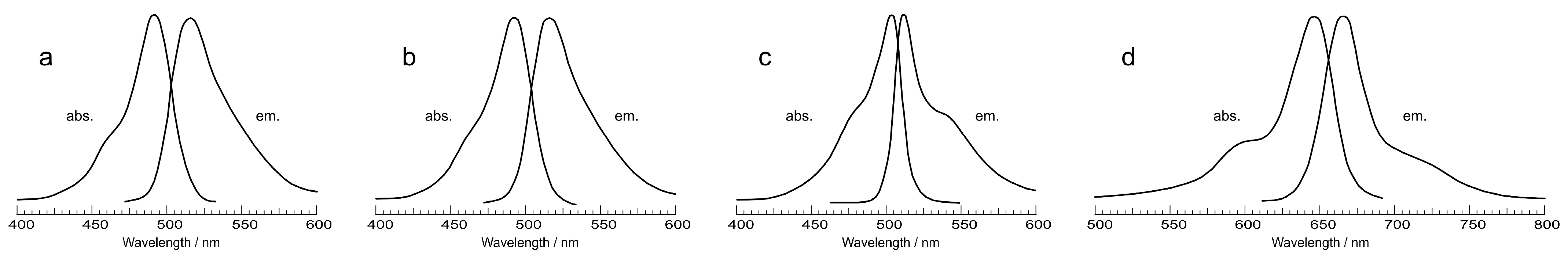
  • Absorption and fluorescence spectra for the labeled polysaccharides
Figure A3. The absorption and emission spectra for the polysaccharides labeled with. (a) FITC (hyaluronic acid 8–15 kDa, #1 in Table 1), (b) DTAF (Escherichia coli O19ab, #16 in Table 3), (c) BODIPY (hyaluronic acid 15–30 kDa), and (d) SuCy5 (xylogalacturonan, #9 in Table 2).
Polysaccharides 05 00001 g0a3
There is no absorption or emission in these areas for the initial polysaccharides.

References

  1. Balducci, E.; Papi, F.; Capialbi, D.E.; Del Bino, L. Polysaccharides’ Structures and Functions in Biofilm Architecture of Antimicrobial-Resistant (AMR) Pathogens. Int. J. Mol. Sci. 2023, 24, 4030. [Google Scholar] [CrossRef] [PubMed]
  2. Nikiforova, A.V.; Golovchenko, V.V.; Mikshina, P.V.; Patova, O.A.; Gorshkova, T.A.; Bovin, N.V.; Shilova, N.V. Plant Polysaccharide Array for Studying Carbohydrate-Binding Proteins. Biochemistry 2022, 87, 890–902. [Google Scholar] [CrossRef] [PubMed]
  3. Tuzikov, A.B.; Rapoport, E.M.; Khaidukov, S.V.; Nokel, E.A.; Knirel, Y.A.; Bovin, N.V. Synthesis of Bodipy-Labeled Bacterial Polysaccharides and Their Interaction with Human Dendritic Cells. Glycoconj. J. 2021, 38, 369–374. [Google Scholar] [CrossRef] [PubMed]
  4. Meunier, F.; Wilkinson, K.J. Nonperturbing Fluorescent Labeling of Polysaccharides. Biomacromolecules 2002, 3, 857–864. [Google Scholar] [CrossRef] [PubMed]
  5. Nakajima, K.; Oda, Y.; Kinoshita, M.; Masuko, T.; Kakehi, K. Time-Resolved Fluorometric Analysis of Carbohydrates Labeled with Amino-Aromatic Compounds by Reductive Amination. Analyst 2002, 127, 972–976. [Google Scholar] [CrossRef] [PubMed]
  6. Zhang, Y.; Wang, Z.; Zhang, X.; Zhou, W.; Huang, L. One-Pot Fluorescent Labeling of Saccharides with Fluorescein-5-Thiosemicarbazide for Imaging Polysaccharides Transported in Living Cells. Carbohydr. Res. 2011, 346, 2156–2164. [Google Scholar] [CrossRef]
  7. Ruhaak, L.R.; Steenvoorden, E.; Koeleman, C.A.M.; Deelder, A.M.; Wuhrer, M. 2-Picoline-Borane: A Non-Toxic Reducing Agent for Oligosaccharide Labeling by Reductive Amination. Proteomics 2010, 10, 2330–2336. [Google Scholar] [CrossRef]
  8. Cosenza, V.A.; Navarro, D.A.; Stortz, C.A. Usage of α-Picoline Borane for the Reductive Amination of Carbohydrates. Arkivoc 2011, 2011, 182–194. [Google Scholar] [CrossRef]
  9. Zhao, J.; Wu, J. Preparation and Characterization of the Fluorescent Chitosan Nanoparticle Probe. Chin. J. Anal. Chem. 2006, 34, 1555–1559. [Google Scholar] [CrossRef]
  10. Moussa, S.H.; Tayel, A.A.; Al-Turki, A.I. Evaluation of Fungal Chitosan as a Biocontrol and Antibacterial Agent Using Fluorescence-Labeling. Int. J. Biol. Macromol. 2013, 54, 204–208. [Google Scholar] [CrossRef]
  11. Kobayashi, M.; Ichishima, E. Fluorescent Labeling of Sugar-Carboxylate with Water-Soluble Carbodiimide. Biosci. Biotechnol. Biochem. 1992, 56, 186–189. [Google Scholar] [CrossRef]
  12. Han, Z.R.; Wang, Y.F.; Liu, X.; Wu, J.D.; Cao, H.; Zhao, X.; Chai, W.G.; Yu, G.L. Fluorescent Labeling of Several Glycosaminoglycans and Their Interaction with Anti-Chondroitin Sulfate Antibody. Chin. J. Anal. Chem. 2011, 39, 1352–1357. [Google Scholar] [CrossRef]
  13. Kunishima, M.; Kawachi, C.; Monta, J.; Terao, K.; Iwasaki, F.; Tani, S. 4-(4,6-Dimethoxy-1,3,5-Triazin-2-Yl)-4-Methyl-Morpholinium Chloride: An Efficient Condensing Agent Leading to the Formation of Amides and Esters. Tetrahedron 1999, 55, 13159–13170. [Google Scholar] [CrossRef]
  14. D’Este, M.; Eglin, D.; Alini, M. A Systematic Analysis of DMTMM vs EDC/NHS for Ligation of Amines to Hyaluronan in Water. Carbohydr. Polym. 2014, 108, 239–246. [Google Scholar] [CrossRef] [PubMed]
  15. Bohrn, R.; Potthast, A.; Schiehser, S.; Rosenau, T.; Sixta, H.; Kosma, P. The FDAM Method: Determination of Carboxyl Profiles in Cellulosic Materials by Combining Group-Selective Fluorescence Labelling with GPC. Biomacromolecules 2006, 7, 1743–1750. [Google Scholar] [CrossRef] [PubMed]
  16. Hutterer, C.; Fackler, K.; Potthast, A. The Fate of 4-O-Methyl Glucuronic Acid in Hardwood Xylan during Alkaline Extraction. ACS Sustain. Chem. Eng. 2017, 5, 1818–1823. [Google Scholar] [CrossRef]
  17. Abushammala, H.; Mao, J. A Review of the Surface Modification of Cellulose and Nanocellulose Using Aliphatic and Aromatic Mono- and Di-Isocyanates. Molecules 2019, 24, 2782. [Google Scholar] [CrossRef]
  18. de Belder, A.N.; Granath, K. Preparation and Properties of Fluorescein-Labelled Dextrans. Carbohydr. Res. 1973, 30, 375–378. [Google Scholar] [CrossRef]
  19. Unnikrishnan, B.S.; Preethi, G.U.; Sreeranganathan, M.; Syama, H.P.; Archana, M.G.; Sreelekha, T.T. Fabrication of Fluorescein Labeled Galactoxyloglucan Polysaccharide for Tumor and Macrophage Tagging. J. Drug Deliv. Sci. Technol. 2019, 52, 863–869. [Google Scholar] [CrossRef]
  20. Sun, M.; Su, F.; Yang, J.; Gao, Z.; Geng, Y. Fluorescent Labeling of Polysaccharides from Masson Pine Pollen and Its Effect on RAW264.7 Macrophages. Polymers 2018, 10, 372. [Google Scholar] [CrossRef]
  21. Alasel, M.; Dassinger, N.; Vornicescu, D.; Keusgen, M. Development of a Borreliosis Assay: Mannan Coated Polyethylene Sinter Bodies as a New Platform Technology. Anal. Biochem. 2018, 543, 55–61. [Google Scholar] [CrossRef] [PubMed]
  22. Li, T.; Vanderah, D. Solubility of N, N′-Disuccinimidyl Carbonate and Its Relevance to Polysaccharide Functionalization. Anal. Biochem. 2021, 626, 114250. [Google Scholar] [CrossRef] [PubMed]
  23. Heyna, J. Reactive Dyes Containing Vinylsulfonyl Groups. Angew. Chem. Int. Ed. Engl. 1963, 2, 20–23. [Google Scholar] [CrossRef]
  24. Rapoport, E.M.; Khasbiullina, N.R.; Komarova, V.A.; Ryzhov, I.M.; Gorbatch, M.M.; Tuzikov, A.B.; Khaidukov, S.V.; Popova, I.S.; Korchagina, E.Y.; Henry, S.M.; et al. Localization of Synthetic Glycolipids in the Cell and the Dynamics of Their Insertion/Loss. Biochim. Biophys. Acta Biomembr. 2021, 1863, 183645. [Google Scholar] [CrossRef] [PubMed]
  25. Mahmoud, K.A.; Mena, J.A.; Male, K.B.; Hrapovic, S.; Kamen, A.; Luong, J.H.T. Effect of Surface Charge on the Cellular Uptake and Cytotoxicity of Fluorescent Labeled Cellulose Nanocrystals. ACS Appl. Mater. Interfaces 2010, 2, 2924–2932. [Google Scholar] [CrossRef]
  26. Hess, R.; Pearse, A.G.E. Labelling of Proteins with Cellulose-Reactive Dyes. Nature 1959, 183, 260–261. [Google Scholar] [CrossRef]
  27. Abitbol, T.; Palermo, A.; Moran-Mirabal, J.M.; Cranston, E.D. Fluorescent Labeling and Characterization of Cellulose Nanocrystals with Varying Charge Contents. Biomacromolecules 2013, 14, 3278–3284. [Google Scholar] [CrossRef]
  28. Petrova, A.; Sibgatullina, G.; Gorshkova, T.; Kozlova, L. Dynamics of Cell Wall Polysaccharides during the Elongation Growth of Rye Primary Roots. Planta 2022, 255, 108. [Google Scholar] [CrossRef]
Scheme 1. Polysaccharides (A), xylogalacturonan from baobab fruit Adansonia digitata L.; (B) pectin from sugar beet Beta vulgaris L. labeled with fluorescein (Polysaccharides 05 00001 i001
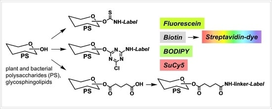
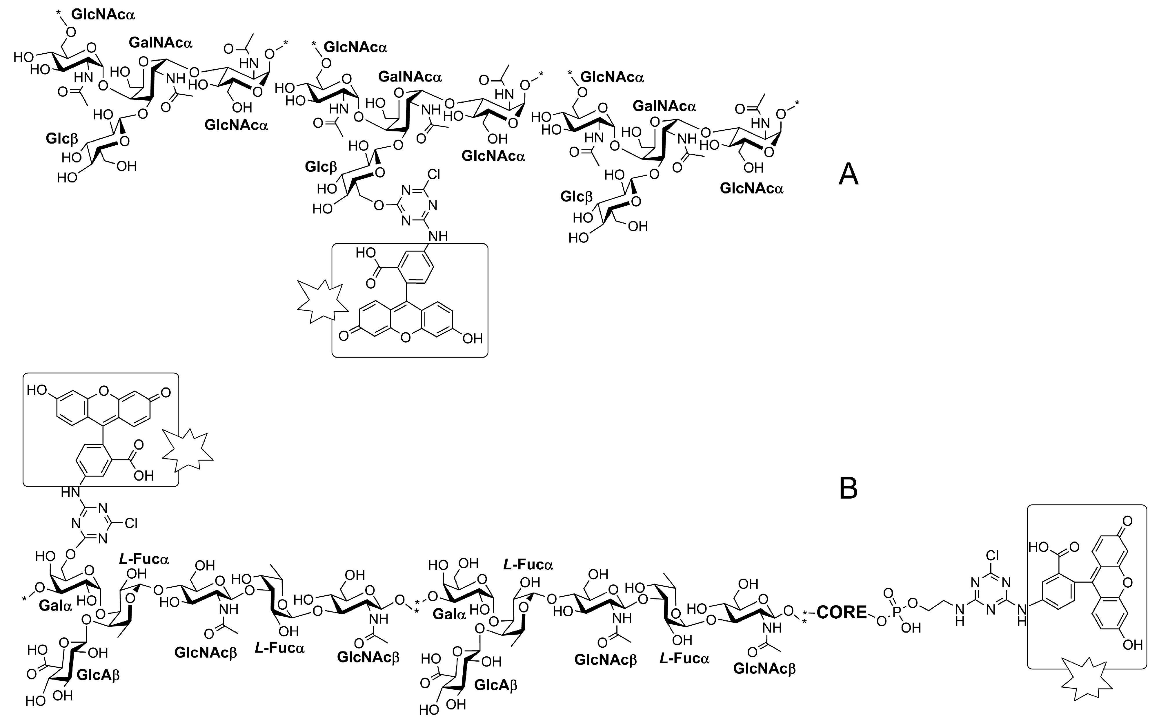
) and biotin tags. Reagents and conditions: (A) PS in the form of i-Pr2EtN salt in DMSO, FITC, i-Pr2EtN, 40 h/50 °C; (B) native PS, biot-DCTA, water/DMSO 1:1, i-Pr2EtN (pH~9), 4 h/20 °C.
Polysaccharides 05 00001 sch001
Scheme 2. Bacterial polysaccharides labeled with DTAF (Polysaccharides 05 00001 i001
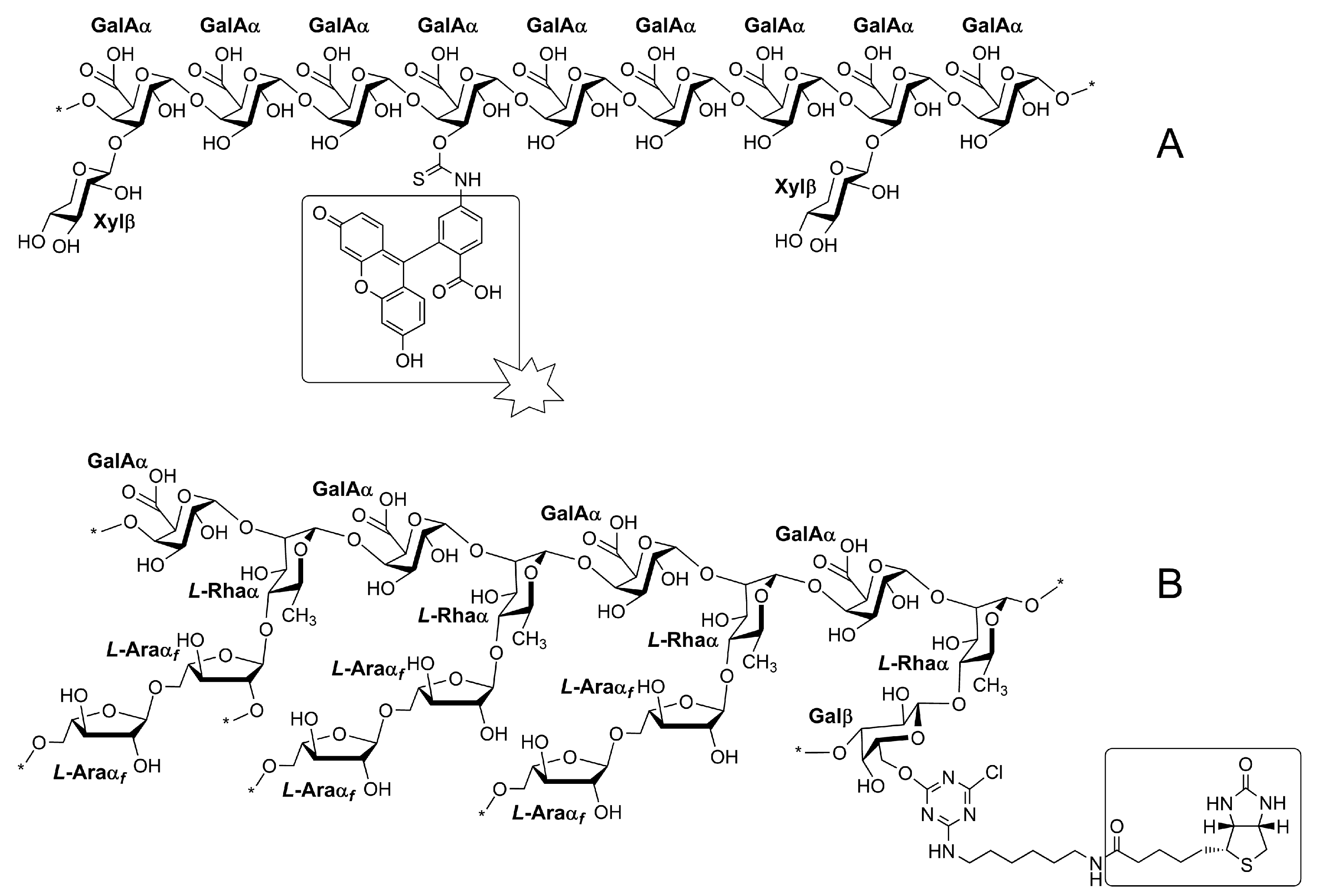
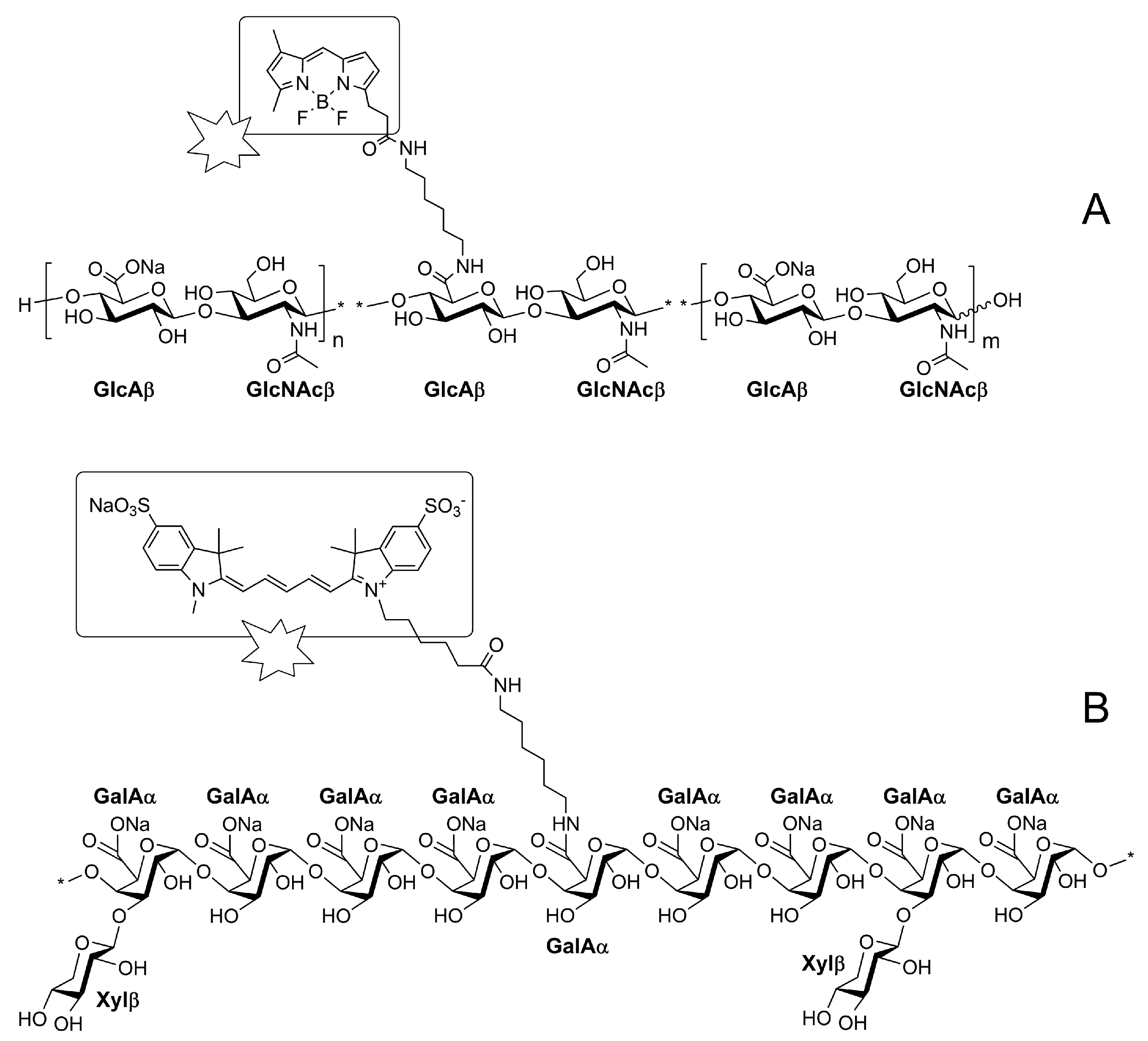
). (A) Capsular polysaccharide (CPS Acinetobacter baumannii NIPH60 K43). (B) Polysaccharide with O-antigen (O-PS Escherichia coli O41), containing an amino group in the CORE (inner core part of the source LPS from which the PS was obtained). Reagents and conditions for (A,B): DTAF, water/DMSO 1:1, i-Pr2EtN (pH~9), 4 h/20 °C.
Polysaccharides 05 00001 sch002
Scheme 3. (A) BODIPY (Polysaccharides 05 00001 i001
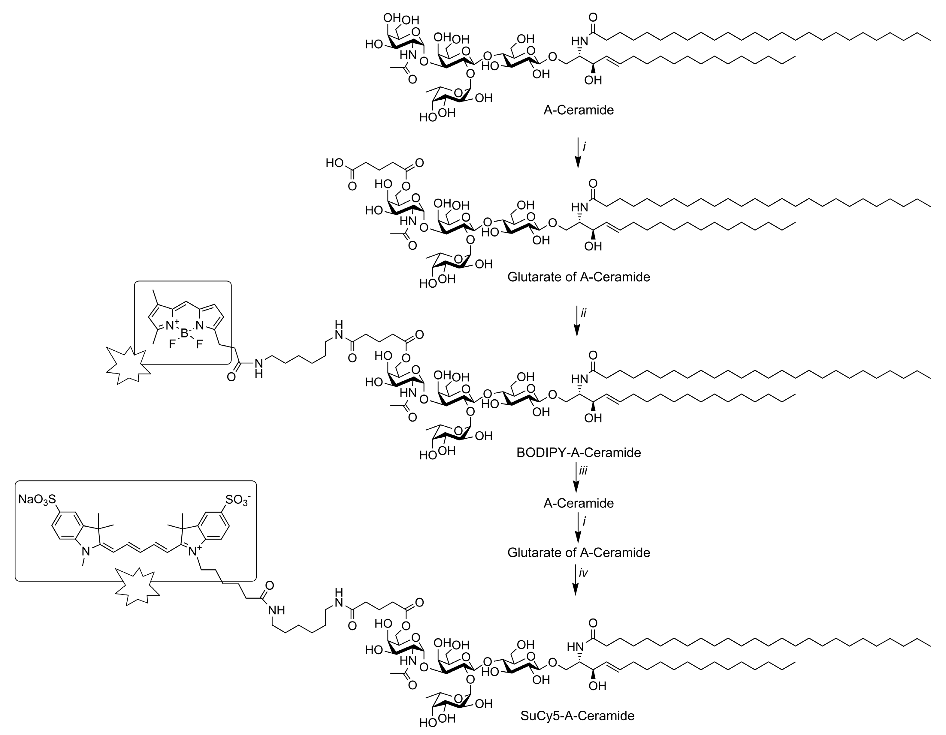
)-labeled hyaluronic acid (15–30 kDa), reagents and conditions: sodium hyaluronate, BODIPY amine, HONSu, EDAC, water/DMF, 7 h/20° C. (B) SuCy5-labeled (Polysaccharides 05 00001 i001) xylogalacturonan from baobab fruit Adansonia digitata L., reagents and conditions: i-Pr2EtNH+ salt of xylogalacturonan, SuCy5 amine, HOBT, DCC, DMF, 24 h/25 °C.
Polysaccharides 05 00001 sch003
Scheme 4. Two-stage labeling of A-Ceramide (shown only one of the isomers of the glutarate location). Reagents and conditions: i, glutaric anhydride, i-Pr2EtN, DMF, 1 h/70 °C; ii, BODIPY (Polysaccharides 05 00001 i001) amine, DCC, HOBt, i-Pr2EtN, DMF, 24 h/37 °C; iii, 2-propanol/MeOH/water, Et3N, 2 h/50 °C; iv, SuCy5 (Polysaccharides 05 00001 i001) amine, DCC, HOBt, i-Pr2EtN, DMF, 24 h/37 °C. For details see Section 2.2.7.
Polysaccharides 05 00001 sch004
Figure 1. Comparison of the binding of polysaccharide probes, each with a different tag, to a plant section (cell elongation zone of rye root), using confocal fluorescence microscopy. Probing with: (A) FITC-labeled polysaccharide; (B) biotin labeled polysaccharide; (C) SuCy5 labeled polysaccharide. Xylogalacturonan from baobab (Adansonia digitate) was the labeled polysaccharide in all three cases.
Polysaccharides 05 00001 g001
Table 1. Polysaccharides labeled using conjugation with FITC.
#StructureSourceType
1Hyaluronic acid 8–15 kDa
-4GlcAβ1-3GlcNAcβ1-
Streptococcus equi, zooepidemicus
Contipro Biotech (Czech Republic)
Glycosaminoglycan
(GAG)
2Hyaluronic acid 15–30 kDa
-4GlcAβ1-3GlcNAcβ1-
Streptococcus equi, zooepidemicus
Contipro Biotech (Czech Republic)
GAG
3XylogalacturonanAdansonia digitata L., baobab fruitpectin
Table 2. Polysaccharides labeled with biot-DCTA.
#StructureSourceType
4Hyaluronic acid 15–30 kDa
-4GlcAβ1-3GlcNAcβ1-
Streptococcus equi, zooepidemicus
Contipro Biotech (Czech Republic)
GAG
5Homogalacturonan predominanceMalus sp., apple, Fluka76282pectin
6Ara predominanceBeta vulgaris L., sugar beet, Megazyme 11078-27-6pectin
7Ara and Gal predominanceLarix sp. larch, Megazyme 9036-66-2pectin
8Ara and Gal predominanceAcacia sp. acacia, Sigma G9752pectin
9Xylogalacturonan *Adansonia digitata L., baobab fruitpectin
* Xylogalacturonan was also labeled with SuCy5; see Xylogalacturonan.
Table 3. Bacterial polysaccharides labeled using DTAF reagent.
#Structure *SourceType **
1-2Glcβ1-6GlcNAcα1-3FucNAcα1-GlcNAcβ1-Escherichia coli O12O-PS
2Galα1-2Galα1-2(Galβ1-4)Glcα1-3Glcα1-/inner core-lipid A/Escherichia coli O14LPS(R)
3-3Galα1-3(GlcAβ1-4)Fucα1-4GlcNAcβ1-3Fucα1-3GlcNAcβ1-Escherichia coli O41O-PS
4-4GalAα1-2Rhaα1-2Ribfβ1-4Galβ1-3GlcNAcβ1-Escherichia coli O54O-PS
5-5(Glcα1-2)Galfβ1-5(Glcα1-2)Galfβ1-3Galfβ1-Escherichia coli O62O-PS
6-4(Aci5Ac7Acα)Galα1-3FucNAcα1-3FucNAcα1-Acinetobacter baumannii UMB001 K13CPS
7-3GalNAcA4Acα1-4GalNAcA3(%)Acα1-4GalNAcA3(%)Acα1-3QuiNAc4NAcβ1-Acinetobacter baumannii LUH5535 K35CPS
8-3(Pse5Ac7RHbα2-4)Ribβ1-3GalNAcβ1-Acinetobacter baumannii LUH5550 K42CPS
9-6GlcNAcα1-4(Glcβ1-3)GalNAcα1-3GlcNAcα1-Acinetobacter baumannii NIPH60 K43CPS
10-4ManA2Acβ1-4(GalNAcα1-3)FucNAcα1-3DFucNAcα1-Acinetobacter baumannii MAR13-1452 K125CPS
11-2Rha3Acα1-2Rhaα1-4GalAβ1-3GalNAcβ1-Shigella flexneri type 6 (5-F6 51579)O-PS
12-2Rhaα1-2Rhaα1-3Rhaα1-3GlcNAcβ1-Shigella flexneri type Y (51581 Y)O-PS
13-2(EtN1-P-3)Rhaα1-2(EtN1-P-3)Rhaα1-3Rhaα1-3GlcNAcβ1- Shigella flexneri Yv (Yv)O-PS
14-2Rha3Acα1-2Rhaα1-3(Glcα1-4)Rhaα1-3GlcNAc6Acβ1-Shigella flexneri 2a2-2 (2a)O-PS
15-3GlcNAcβ1-2Rha3Acα1-2(Glcα1-3)Rhaα1-3Rhaα1-Shigella flexneri type 5a1 (F-5a)O-PS
16-2Rhaα1-2Rhaα1-2Rhaα1-2Glcα1-3GlcNAc6Acα1-Escherichia coli O19abO-PS
* Monosaccharides are pyranoses except as specified; fucose and rhamnose are L-sugars (except as specified), and all others are D-sugars. Structures of polysaccharides are shown in “glycan form” for convenience of comparison with oligosaccharides; it means that we omitted designations of ring size (pyranose/furanose) and in most cases configuration (D/L). Other abbreviations were used according to the database of polysaccharides http://csdb.glycoscience.ru/ (accessed on 20 December 2023). ** The designation O-PS means polysaccharide with O-antigen, containing an amino group in the inner core part; CPS means capsular polysaccharide. LPS(R) means truncated form of lipopolysaccharide.
Disclaimer/Publisher’s Note: The statements, opinions and data contained in all publications are solely those of the individual author(s) and contributor(s) and not of MDPI and/or the editor(s). MDPI and/or the editor(s) disclaim responsibility for any injury to people or property resulting from any ideas, methods, instructions or products referred to in the content.

