Article

The Use of Indocyanine Green to Visualize the Thoracic Duct and Evaluate Gastric Conduit Perfusion in Esophagectomy

by
Katherine Aw
1,2,3,4,†,
Aziza Al Rawahi
2,3,4,†,
Rebecca Lau
1,2,3,4,†,
Sami Aftab Abdul
1,2,3,4,†,
Caitlin Anstee
2,4,
Sebastien Gilbert
1,2,3,4,
Daniel Jones
1,2,3,4,
Andrew J. E. Seely
1,2,3,4,
Ramanadhan Sudhir Sundaresan
1,2,3,4,
Patrick James Villeneuve
1,2,3,4 and
Donna Elizabeth Maziak
1,2,3,4,5,6,*
1
Faculty of Medicine, University of Ottawa, 451 Smyth Road, Ottawa, ON K1H 8L1, Canada
2
Division of Thoracic Surgery, The Ottawa Hospital, General Campus, 501 Smyth Road, Ottawa, ON K1H 8L6, Canada
3
Division of Thoracic Surgery, Department of Surgery, Faculty of Medicine, University of Ottawa, 501 Smyth Road, Ottawa, ON K1H 8L6, Canada
4
Ottawa Hospital Research Institute, 501 Smyth Road, Ottawa, ON K1H 8L6, Canada
5
Ontario Health (Cancer Care Ontario), Toronto, ON M5G 2L3, Canada
6
Telfer School of Management, University of Ottawa, 55 Laurier Ave E, Ottawa, ON K1N 6N5, Canada
*
Author to whom correspondence should be addressed.
These authors contributed equally to this work.
Surgeries 2023, 4(4), 579-589; https://doi.org/10.3390/surgeries4040056
Submission received: 14 September 2023 / Revised: 27 October 2023 / Accepted: 2 November 2023 / Published: 6 November 2023

Abstract

:
Background: In this study, we investigate indocyanine green (ICG) dye visualization of the thoracic duct (TD) and conduit perfusion during esophagectomy to reduce anastomotic leak (AL) and chylothorax adverse events (AEs). Methods: Retrospective data of adult patients who underwent esophagectomy for esophageal carcinoma between July 2019 and 2022 were included (n = 105). ICG was delivered intravenously (2 mL, 2.5 mg/mL) to assess conduit perfusion into the small bowel mesentery, inguinal lymph nodes, or foot web spaces for TD visualization using fluorescence imaging. Incidence of TD injury, chylothorax, AL, and AEs were collected. Results: A total of 23 patients received ICG (ICG for TD and perfusion (n = 12) and perfusion only (n = 11)), while 82 patients were controls. TD was visualized in 6 of 12 patients who received ICG for TD. No intraoperative TD injuries or postoperative chylothoraces occurred in these patients. Non-ICG patients had 1 (1.22%) intraoperative TD injury and 10 (12.2%) postoperative chylothoraces (grade I–IIIb). While 10 non-ICG patients (12.2%) developed AL (grade I–IVb), only 2 (8.7%) ICG patients developed AL (grade IIIa). Conclusions: This study demonstrates the utility of ICG fluorescence in intraoperative TD and conduit perfusion assessment for limiting AEs. Standard incorporation of ICG in esophagectomy may help surgeons improve the quality of care in this patient population.

1. Introduction

An esophagectomy is the mainstay surgical procedure used to treat local–regional esophageal cancer [1]. However, this operative undertaking is associated with significant morbidity as well as a potentially high mortality rate [1,2]. Two major complications that have profound impacts on patient quality of life (QOL) include anastomotic leak (AL) and chylothorax.
An anastomotic leak occurs in 3–25% of esophagectomy patients [3] and is associated with a high mortality rate, prolonged length of stay (LOS), decreased QOL, and increased hospital costs [4,5,6,7]. Ischemia has been identified as a major contributor to AL risk [8,9]. To assess vascularity, surgeons traditionally rely on color, pulsation, temperature, or Doppler probes. These techniques, however, are limited by surgeon subjectivity, lack of reproducibility, and weak correlation with AL incidence [10,11,12]. Assessing gastric conduit perfusion (GCP) with a novel intraoperative visualization tool before anastomosis may therefore help prevent AL events.
Iatrogenic trauma to the thoracic duct (TD) and subsequent chylothorax—chyle leak into the pleural space—is another concerning complication post-esophagectomy, with rates upwards of 2–9% [13,14,15]. Chylothorax can precipitate other life-threatening postoperative complications, including respiratory failure, hypovolemia, malnutrition, coagulopathy, and sepsis [13,14]. Traditional techniques to visualize the TD, such as preoperative lymphoscintigraphy or oral administration of lipid-rich creams and oils, are limited by their ability to be transferred to the intraoperative setting and their need for preoperative preparation, respectively [16,17]. As such, there is a need for a reliable intraoperative technique to prophylactically visualize the TD to prevent TD damage and subsequent chylothorax.
Fluorescent imaging using indocyanine green (ICG) dye and near-infrared spectroscopy has emerged as a real-time visualization technique to assist surgeons in identifying anatomical structures intraoperatively. ICG, a cyanine dye with a well-documented safety profile, can be injected to assess vascularity or the TD and its tributaries. The dye fluoresces under near-infrared stimulation and is detected by specific cameras. Current applications of ICG include sentinel lymph node mapping [18], liver perfusion assessment [19], and visualization of vascular pathologies in neurosurgery [20]. A systematic review and meta-analysis demonstrated that ICG for assessment of GCP before anastomosis is safe and reduces AL secondary to esophagectomy [21]. With regard to TD visualization, existing studies have shown the utility of ICG in animal models [22] and post-esophagectomy during chylothorax re-operation [23,24,25]. Although limited, existing literature on the prophylactic use of ICG for TD visualization in esophagectomy patients suggests the utility and effectiveness of ICG in preventing or managing TD injury [16,26,27,28,29]. To our knowledge, however, there are no studies that examine ICG use for both TD and GCP during esophagectomy. It should be noted that while prospective studies exist in the literature to define the scope of ICG use, there are limited clinical investigations offered from the Canadian patient population context. While a retrospective series is limited by methodological rigor due to selection bias and inference, often such a series is needed to showcase local value to pursue prospective investigations. As such, our study provides a window into the Canadian landscape.
The purpose of this study is to assess the clinical utility and feasibility of implementing indocyanine green dye as an intra-operative tool in the visualization of the thoracic duct and gastric conduit perfusion. Indocyanine green dye use may reduce the risk of intra-operative and post-operative adverse events (AEs), specifically chylothorax and anastomotic leak, in patients undergoing esophagectomy. Our study is conducted in the Canadian healthcare context and offers a retrospective series to draw on for a future clinical investigation. We present the following article in accordance with the STROBE reporting checklist.

2. Materials and Methods

2.1. Study Design

We performed a single-center retrospective cohort study. All patients who underwent elective esophagectomy at the Ottawa Hospital between 1 July 2019 and 1 July 2022 were identified retrospectively through the “Thoracic Surgery Quality, monitoring, Information management, and Clinical” software system [30]. Patients with biopsy-proven esophageal carcinoma who underwent esophagectomy with gastric conduit anastomosis were included. Patients who were less than 18 years old, did not receive any form of neoadjuvant chemoradiotherapy, and had an iodine allergy were excluded from the study.

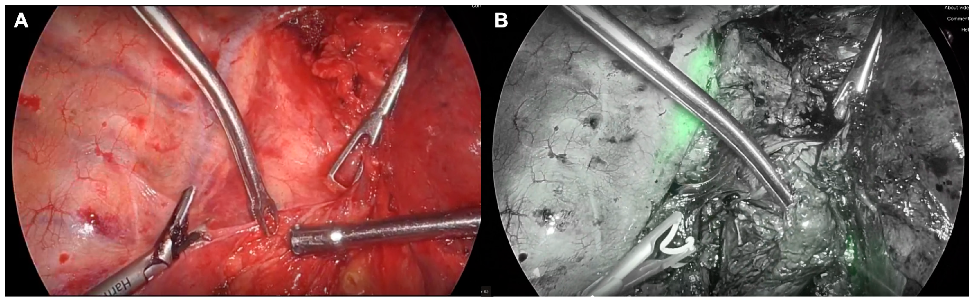
2.2. Surgical Technique and Indocyanine Green

Esophagectomy was performed with an open approach, minimally invasive esophagectomy (MIE), or hybrid minimally invasive approach. Open esophagectomy involved a right thoracotomy and laparotomy, and MIE involved a right video-assisted thoracoscopy and laparoscopy. Hybrid MIE involved either a uni- or bilateral neck dissection, right thoracoscopy, and laparotomy (three-field) approach or a right video-assisted thoracoscopy and open laparotomy (hybrid). All surgical techniques involved cervical or thoracic anastomosis.
A total of 2 mL of 2.5 mg/mL of SPY AGENTTM GREEN ICG (Stryker ©, Kalamazoo, MI, USA) was injected intravenously, followed by a 10 cc normal saline flush to visualize the GCP. ICG fluorescence was detected using the Stryker SPY-PHI™ system (Kalamazoo, MI, USA). Endoscopic examination and insufflation with the anastomosis submerged in saline were used to evaluate the integrity of the seal. An additional 2 mL of ICG (2.5 mg/mL) was injected into the small bowel mesentery, the inguinal region, or web spaces of the foot to visualize the TD. Any ICG leakage into the thoracic space was noted to assess TD injury 1–2 h post-injection. Surgical cases were distributed across five surgeons. The decision to use ICG and surgical treatment plan were determined at the surgeon’s discretion.

2.3. Data Collection

Primary outcomes included frequencies of intraoperative TD injury, postoperative chylothorax, and AL. Secondary outcomes included estimated blood loss, operating time, conversion to open, intraoperative AEs, LOS, 30- and 90-day mortality, and postoperative AEs. Postoperative AEs, including chylothorax and AL, are classified according to the Thoracic Morbidity and Mortality (TM&M) classification schema as minor (grades I–II) and major (grades III–V) [30]. Grade I complications do not and Grade II do require pharmacological treatment or intervention. Grade III complications require surgical, radiological, endoscopic intervention, or multiple therapies with (IIIb) or without (IIIa) general anesthesia. Grade IV complications lead to single-organ (IVa) or multi-organ (IVb) failure and require ICU care and life support. Grade V complications lead to death. All data were collected through retrospective patient chart review with review by the clinical team. All data were stored in a database on a secure server in the Division of Thoracic Surgery in accordance with local patient data regulations.

2.4. Statistical Analysis

Given the limited sample size between groups, univariate descriptive statistics (frequencies, proportions, medians, and interquartile ranges) were computed and tabulated without 95% confidence intervals. Sample size determination and power to evaluate statistical significance is limited due to recent adoption of ICG for TD and GCP at our center; as such, all patients meeting the inclusion criteria were included in the final analysis. Continuous variables were analyzed using a one-way analysis of variance (ANOVA) test across the three cohorts (ICG for TD and GCP, ICG for GCP only, and no ICG). Nominal categorical variables were analyzed using a Pearson Chi-Square test of independence and ordinal categorical data using an independent-samples Kruskal–Wallis test according to the presence or absence of ICG for gastric conduit perfusion and thoracic duct visualization. Statistical significance was determined at a p value less than 0.05 for all analyses without correction for multiple comparisons using SPSS (Version 28, IBM, Armonk, NY, USA) or RStudio (Version 1.3.1091, 2020 PBC, Boston, MA, USA) statistical software.

3. Results

3.1. Cohort Characteristics

Of 175 patients identified, a total of 105 patients with a mean age of 67.8 ± 9.8 years and male to female ratio of 2.89:1 were included. A total of 82 patients did not receive ICG (78.0%), and 23 patients received ICG. In the ICG group, 12 patients received ICG for both the visualization of the TD and assessment of GCP (11.4), and 11(10.5) patients received ICG for the assessment of GCP only (Table 1). The decision to use ICG was made at the discretion of the operating surgeon. Most patients had adenocarcinoma (80.0%) and Siewert I or II esophageal tumors (69.5%) (Table 2). The predominant surgical approach was MIE (42.8%), with 11 conversions from the originally planned MIE approach.

3.2. Thoracic Duct Injury and Chylothorax

ICG was delivered via the small bowel mesentery (n = 9), the inguinal lymph nodes (n = 2), or feet web spaces (n = 1) with a 50% TD visualization rate (Table 3). While there is a statistically significant difference between ICG injection sites for small bowel mesentery and inguinal lymph nodes, this is primarily due to the reduced sample size in the ICG cohort. The TD was visualized after approximately 1 h for the small bowel mesentery and 2 h for inguinal nodes (Figure 1). Of the six ICG patients where the TD was successfully visualized, there were no intraoperative TD injuries or postoperative chylothoraces (Table 3). In the other six patients, one grade II AE chylothorax was reported. The TD was injured and repaired via ligation in one non-ICG patient. Ten chylothoraces (grade I–IIIb) were reported in non-ICG patients. Grade I and II chylothoraces were treated with dietary modification. Of the four non-ICG patients with grade IIIa chylothorax, three required total parenteral nutrition and one required lymphangiogram with TD embolization. One patient receiving ICG for GCP assessment and four non-ICG patients with grade IIIb chylothoraces required surgical intervention to repair the TD. No patients had a grade IV (organ failure) or grade V (death) chylothorax.

3.3. Anastomotic Leak

The intraoperative AL test was positive in one ICG (4.3%) and two non-ICG patients (2.4%) (Table 4). The anastomosis was repaired by placing interrupted sutures at the area of the leak, and none of these patients developed AL during their postoperative course. While 10 patients (12.2%) of the non-ICG group developed AL (grade I–IVb), only 2 (8.7%) of all patients who received ICG for perfusion developed AL (grade IIIa). The non-ICG patients with grade I AL required no clinical or surgical intervention. The seven non-ICG (8.5%) and two ICG (8.7%)) patients with grade IIIa AL were managed using routine endoscopic assessment. The one non-ICG patient with grade IIIb AL required stent placement under general anesthesia. One non-ICG patient developed grade IVb AL with multi-organ failure. No grade V (death) complication for AL occurred in any group. The two ICG (8.7%) patients with AL received a thoracic anastomosis. Nine non-ICG (10.9%) patients with AL received a thoracic anastomosis, and one patient received a cervical anastomosis.

3.4. Adverse Events

The median estimated blood loss and operative time for the ICG and non-ICG groups were comparable (p = 0.112). Six ICG (50.0%) patients and fourteen non-ICG (17.1%) patients had intraoperative complications. Most intraoperative complications were iatrogenic injuries to the spleen, lung, colon, or vasculature. Fifteen ICG (65.2%) and fifty-four non-ICG (65.8%) patients had at least one postoperative AE. The median LOS (days) (IQR) was 10 [8,9,10,11,12,13] for ICG and 11 [8,9,10,11,12,13,14,15,16,17,18] for non-ICG (p = 0.551). While no 30- or 90-day mortalities were observed in the ICG group, two non-ICG patients were deceased by the 30-day follow-up period due to tracheoesophageal fistula and pneumonia.

4. Discussion

4.1. Key Findings

Our initial results demonstrate the clinical utility of ICG in showing that ICG can be a safe, simple way to visualize the gastric conduit before anastomosis and to identify the TD to both prevent and identify iatrogenic TD injury. Our results illustrate the value of implementing ICG prophylactically in the TD. In the ICG patients where the TD was successfully visualized, no postoperative chylothoraces were reported. Anastomotic leak occurred in 2 ICG (8.7%) and 10 non-ICG (12.2%) patients, with non-ICG patients having higher-grade adverse-event-based ALs.

4.2. Study Limitations

This is the first study exploring ICG use for both TD visualization and assessment of GCP during esophagectomy. However, our findings are limited by the retrospective design of this study. Due to this retrospective design, we were unable to randomize ICG use and to collect quantitative outcomes of interest, including ICG fluorescence intensity, flow speed, and total volume of ICG used. These metrics would enable better methodological design of prospective studies and remain an area of the literature where surgeon preference and discretion are the practice in the intraoperative setting rather than an evidence-based decision. Inter-surgeon variability in implementation, variability of resection techniques, and inconsistent ICG injection sites may have introduced bias. We were unable to optimize ICG administration as the TD was visualized in only 50% of patients. Furthermore, the generalizability of our results is limited by the small sample size and single-institution design. Given the relative novelty of prophylactic ICG use during esophagectomy, however, we believe that our study supplements the limited Canadian literature on this topic and will help guide future prospective feasibility studies on ICG use and development of local protocols prior to future clinical investigations to reduce variability in adoption and implementation.

4.3. Comparison to Existing Literature

The current literature supports the utility of ICG in identifying and repairing active chyle leaks when they occur during esophagectomy [26,27,28,29]. As such, the utility of ICG remains as an active tool to correct or repair surgical intervention, expending resources, workforce, and time to recovery. Previous studies report no postoperative chylothoraces in ICG-receiving esophagectomy patients [16,27,28]. Barnes et al. report 1 postoperative chylothorax in their sample of 17 ICG patients [26]. These studies showcase the clinical benefit and utility of ICG in reducing and preventing chylothoraces. In the present study, no postoperative chylothoraces occurred in the ICG patients where the TD was visualized. However, one chylothorax occurred in a patient receiving ICG for the TD where the TD was not visualized. Thus, the TD needs to be successfully visualized by the operating team for ICG to exert its benefits. While time to visualization was not recorded, such an outcome should be incorporated in future prospective series to quantify the duration to visualization in various injection sites to inform future clinical practice guidelines and to allow anticipation of operating times to encourage the uptake of ICG in the surgical setting.
Unlike the TD, ICG is well established as a tool for reducing AL. Systematic reviews have shown that intraoperative intervention using ICG to assess GCP reduces AL rates post-esophagectomy [21,31,32,33]. For example, Ladak et al. demonstrated an absolute risk reduction of 69% for AL in patients using ICG, with an AL rate of 5.7% and 22.9% in patients with and without ICG use, respectively [32]. Casas et al., however, reported no reduction in AL risk in ICG patients undergoing MIE with thoracic anastomosis [34]. One key difference between these two studies is the anastomotic technique used—cervical anastomosis (Ladak et al.) versus thoracic anastomosis (Casas et al.). Of note, intrathoracic anastomosis has a lower incidence of AL when compared with cervical anastomosis [35], and we included patients receiving both techniques. Only 1 of the 12 patients with AL in our study received cervical anastomosis; however, only 14.3% of all patients had cervical anastomosis (three-field). As such, the utility of ICG may depend on the anastomosis location, surgical approach, and other perioperative factors. Controversy remains regarding the optimal site for esophagogastric anastomosis, and many trials have compared cervical to thoracic regions, concluding a similar safety profile across morbidity and mortality. That being said, there may be room to adopt ICG use in those patients where AL may pose a greater risk, such as those undergoing cervical anastomosis in a prophylactic sense. In our retrospective series, we are limited by the quality of data surrounding ICG use from chart review; however, future series should examine duration and time to visualization by injection site. In this study, we injected ICG in the small bowel mesentery, inguinal lymph nodes, and web spaces of the feet. However, there is no objective metric to indicate or recommend an optimal site. This should be of consideration for future prospective studies to inform clinical practice guidelines and may be an area of research given the gap in the literature.

4.4. Explanation of Findings

In this study, we had a varied method of delivery of ICG: via the small bowel mesentery, the inguinal lymph nodes, or the web space of feet. Only 50% of the ICG patients had TD visualization: five with small bowel mesentery and one with inguinal lymph node delivery. There is no established literature or consensus on the optimal site, duration, or volume for ICG administration. Barnes et al. reported 80% TD visualization when injecting 1.5–2.0 mL of ICG (2.5 mg/mL) into the small bowel mesentery [26], while Varshney et al. reported 100% TD visualization when injecting a 2 mL (1 mg/mL) solution into inguinal lymph nodes [28]. Optimization of ICG administration technique, injection site, and dosage may help ensure adequate TD visualization and reduce inter-surgeon variability in the delivery of care. Blind dissection may also be required for TD identification prior to injection if it is embedded in thickened mediastinal fat or fascia.
Patients with chylothorax will often require intervention and a prolonged LOS [14]. In the present study, one ICG patient and four non-ICG patients developed major grade IIIb chylothoraces, which required patients to return to the operating room for thoracotomy and TD ligation. The remaining patients with chylothoraces (grades I–IIIa) were treated with diet modification, and one non-ICG patient (grade IIIa chylothorax) was treated with a lymphangiogram with TD embolization. Prevention of chylothorax via ICG may therefore reduce re-operative interventions, LOS, and hospital resource and financial expenditures.
In our study, two ICG and seven non-ICG patients developed grade IIIa ALs, which were all treated by routine endoscopic assessment. Higher-grade ALs were reported in non-ICG patients. One non-ICG patient developed grade IIIb AL, which required stent placement and a prolonged LOS (65 days). Another non-ICG patient developed grade IVb AL causing septic shock, abdominal compartment syndrome, acute kidney injury, and respiratory failure. This patient was discharged 161 days post-esophagectomy. Reducing the risk of high-grade ALs with routine ICG use may simplify and expedite patients’ postoperative recovery as well as reduce operational costs associated with AE management.
The patients studied in this retrospective series were comparable in age and Charlson Comorbidity Index. A total of 69 patients (65.7%) experienced postoperative AEs, compared to the 33–63% post-esophagectomy morbidity reported in the literature [2,36,37]. The median number (IQR) of AEs was 1 [0–2], with the predominant AEs being pleural effusion, atrial arrhythmia, pneumonia, chylothorax, and AL. Interestingly, four patients (one ICG and three non-ICG) developed both chylothorax and AL and experienced a prolonged LOS.

4.5. Study Implications and Next Steps

Patients undergoing an esophagectomy for esophageal cancer are at high risk for postoperative AEs, morbidity, and mortality. Specifically, chylothorax and anastomotic leak represent two concerning complications that can significantly worsen patient QOL by increasing their LOS and the potential need for reoperation, as well as increasing healthcare costs and resource expenditures [14,34]. Standard incorporation of ICG in esophagectomy for TD visualization and assessment of GCP before anastomosis may help reduce these complications and simplify patients’ postoperative recovery. However, additional studies involving larger patient populations are needed to strengthen the external validity of the results and further support prophylactic ICG use in esophagectomy. Future studies need to address the standardization of an ICG protocol by transparently reporting and collecting the dose, duration to visualization, flow, intensity, method of injection, and site of injection when implementing prophylactic ICG use. Addressing such factors enables objective reporting of optimal characteristics and clinical outcomes attributable to ICG use, reducing the need for inter-surgeon and inter-hospital variability in the adoption and implementation of IGC. Additionally, conducting future prospective studies will allow for the investigation of additional outcomes such as ICG fluorescence intensity, flow speed, total ICG used, and optimal ICG administration.

5. Conclusions

In summary, our study demonstrates the clinical utility of ICG fluorescence in visualizing the TD and GCP, with the potential for limiting chyle leak and AL. Given the limited literature on prophylactic ICG use during esophagectomy, we hope our findings add insight into the utility of ICG in preventing these AEs and guide future prospective feasibility studies. The incorporation of ICG in esophagectomy has the potential to improve the quality of care in this patient population.

Author Contributions

Conceptualization, K.A., A.A.R., R.L., S.A.A. and D.E.M.; Data curation, K.A., A.A.R., R.L., S.A.A., C.A. and D.E.M.; Formal analysis, K.A., A.A.R., R.L., S.A.A., C.A. and D.E.M.; Investigation, K.A., A.A.R., R.L., S.A.A. and D.E.M.; Methodology, K.A., A.A.R., R.L., S.A.A. and D.E.M.; Project administration, K.A., A.A.R., R.L., S.A.A., C.A., S.G., D.J., A.J.E.S., R.S.S., P.J.V. and D.E.M.; Resources; Software, K.A., A.A.R., R.L., S.A.A. and D.E.M.; Supervision, D.E.M.; Validation, K.A., A.A.R., R.L., S.A.A., C.A. and D.E.M.; Visualization, K.A., A.A.R., R.L., S.A.A. and D.E.M.; Writing—original draft, K.A., A.A.R., R.L., S.A.A. and D.E.M.; Writing—review and editing, K.A., A.A.R., R.L., S.A.A., C.A., S.G., D.J., A.J.E.S., R.S.S., P.J.V. and D.E.M.; Principal investigator, D.E.M. All authors have read and agreed to the published version of the manuscript.

Funding

This research received no external funding.

Institutional Review Board Statement

This study was conducted in accordance with the Declaration of Helsinki and approved by the Ottawa Health Science Network Research Ethics Board and the Ottawa Hospital Research Institute (protocol code 20220427-01H and date of approval 4 July 2022).

Informed Consent Statement

The Ottawa Health Science Network Research Ethics Board and the Ottawa Hospital Research Institute approved the collection of thoracic patient chart data through waived consent.

Data Availability Statement

The data presented in this study are available upon request from the corresponding author. The data are not publicly available due to privacy and ethical constraints.

Acknowledgments

The authors would like to thank Anna Fazekas for her administrative support.

Conflicts of Interest

The authors declare no conflict of interest.

References

  1. Manghelli, J.L.; Ceppa, D.P.; Greenberg, J.W.; Blitzer, D.; Hicks, A.; Rieger, K.M.; Birdas, T.J. Management of anastomotic leaks following esophagectomy: When to intervene? J. Thorac. Dis. 2019, 11, 131–137. [Google Scholar] [CrossRef] [PubMed]
  2. Raymond, D.P.; Seder, C.W.; Wright, C.D.; Magee, M.J.; Kosinski, A.S.; Cassivi, S.D.; Grogan, E.L.; Blackmon, S.H.; Allen, M.S.; Park, B.J.; et al. Predictors of Major Morbidity or Mortality after Resection for Esophageal Cancer: A Society of Thoracic Surgeons General Thoracic Surgery Database Risk Adjustment Model. Ann. Thorac. Surg. 2016, 102, 207–214. [Google Scholar] [CrossRef] [PubMed]
  3. Turkyilmaz, A.; Eroglu, A.; Aydin, Y.; Tekinbas, C.; Erol, M.M.; Karaoglanoglu, N. The management of esophagogastric anastomotic leak after esophagectomy for esophageal carcinoma. Dis. Esophagus 2009, 22, 119–126. [Google Scholar] [CrossRef] [PubMed]
  4. Biere, S.; Maas, K.; Cuesta, M.; van der Peet, D. Cervical or Thoracic Anastomosis after Esophagectomy for Cancer: A Systematic Review and Meta-Analysis. Dig. Surg. 2011, 28, 29–35. [Google Scholar] [CrossRef] [PubMed]
  5. AlAnezi, K.; Urschel, J.D. Mortality secondary to esophageal anastomotic leak. Ann. Thorac. Cardiovasc. Surg. 2004, 10, 71–75. [Google Scholar]
  6. Williams, R.N.; Hall, A.W.; Sutton, C.D.; Ubhi, S.S.; Bowrey, D.J. Management of Esophageal Perforation and Anastomotic Leak by Transluminal Drainage. J. Gastrointest. Surg. 2011, 15, 777–781. [Google Scholar] [CrossRef]
  7. Weidenhagen, R.; Hartl, W.H.; Gruetzner, K.U.; Eichhorn, M.E.; Spelsberg, F.; Jauch, K.W. Anastomotic Leakage after Esophageal Resection: New Treatment Options by Endoluminal Vacuum Therapy. Ann. Thorac. Surg. 2010, 90, 1674–1681. [Google Scholar] [CrossRef]
  8. Briel, J.W.; Tamhankar, A.P.; Hagen, J.A.; DeMeester, S.R.; Johansson, J.; Choustoulakis, E.; Prters, J.H.; Bremner, C.G.; DeMeester, T.M. Prevalence and risk factors for ischemia, leak, and stricture of esophageal anastomosis: Gastric pull-up versus colon interposition. J. Am. Coll. Surg. 2004, 198, 536–542. [Google Scholar] [CrossRef]
  9. Zehetner, J.; DeMeester, S.R.; Alicuben, E.T.; Oh, D.S.; Lipham, J.C.; Hagen, J.A.; DeMeester, T.R. Intraoperative Assessment of Perfusion of the Gastric Graft and Correlation with Anastomotic Leaks after Esophagectomy. Ann. Surg. 2015, 262, 74–78. [Google Scholar] [CrossRef]
  10. Karliczek, A.; Harlaar, N.J.; Zeebregts, C.J.; Wiggers, T.; Baas, P.C.; Van Dam, G.M. Surgeons lack predictive accuracy for anastomotic leakage in gastrointestinal surgery. Int. J. Color. Dis. 2009, 24, 569–576. [Google Scholar] [CrossRef]
  11. Ikeda, Y.; Niimi, M.; Kan, S.; Shatari, T.; Takami, H.; Kodaira, S. Clinical significance of tissue blood flow during esophagectomy by laser Doppler flowmetry. J. Thorac. Cardiovasc. Surg. 2001, 122, 1101–1106. [Google Scholar] [CrossRef] [PubMed]
  12. Miyazaki, T.; Kuwano, H.; Kato, H.; Yoshikawa, M.; Ojima, H.; Tsukada, K. Predictive value of blood flow in the gastric tube in anastomotic insufficiency after thoracic esophagectomy. World J. Surg. 2002, 26, 1319–1323. [Google Scholar] [CrossRef]
  13. Miao, L.; Zhang, Y.; Hu, H.; Ma, L.; Shun, Y.; Xiang, J.; Chen, H. Incidence and management of chylothorax after esophagectomy. Thorac. Cancer 2015, 6, 354–358. [Google Scholar] [CrossRef] [PubMed]
  14. Shah, R.D.; Luketich, J.D.; Schuchert, M.J.; Christie, N.A.; Pennathur, A.; Landreneau, R.J.; Nason, K.S. Postesophagectomy Chylothorax: Incidence, Risk Factors, and Outcomes. Ann. Thorac. Surg. 2012, 93, 897–904. [Google Scholar] [CrossRef] [PubMed]
  15. Bender, B.; Murthy, V.; Chamberlain, R.S. The changing management of chylothorax in the modern era. Eur. J. Cardio-Thorac. Surg. 2015, 49, 18–24. [Google Scholar] [CrossRef] [PubMed]
  16. Vecchiato, M.; Martino, A.; Sponza, M.; Uzzau, A.; Ziccarelli, A.; Marchesi, F.; Petri, R. Thoracic duct identification with indocyanine green fluorescence during minimally invasive esophagectomy with patient in prone position. Dis. Esophagus 2020, 33, doaa030. [Google Scholar] [CrossRef]
  17. Du, Z.-S.; Li, X.-Y.; Luo, H.-S.; Wu, S.-X.; Zheng, C.-P.; Li, Z.-Y.; Fu, J.-H. Preoperative Administration of Olive Oil Reduces Chylothorax after Minimally Invasive Esophagectomy. Ann. Thorac. Surg. 2019, 107, 1540–1543. [Google Scholar] [CrossRef]
  18. Grischke, E.-M.; Röhm, C.; Hahn, M.; Helms, G.; Brucker, S.; Wallwiener, D. ICG Fluorescence Technique for the Detection of Sentinel Lymph Nodes in Breast Cancer: Results of a Prospective Open-label Clinical Trial. Geburtshilfe Frauenheilkd. 2015, 75, 935–940. [Google Scholar] [CrossRef]
  19. Zhang, Y.M.; Shi, R.; Hou, J.C.; Liu, Z.R.; Cui, Z.L.; Li, Y.; Wu, D.; Shi, Y.; Shen, Z.Y. Liver tumor boundaries identified intraoperatively using real-time indocyanine green fluorescence imaging. J. Cancer Res. Clin. Oncol. 2017, 143, 51–58. [Google Scholar] [CrossRef]
  20. Raabe, A.; Nakaji, P.; Beck, J.; Kim, L.J.; Hsu, F.P.K.; Kamerman, J.D.; Seifert, V.; Spetzler, R.F. Prospective evaluation of surgical microscope-integrated intraoperative near-infrared indocyanine green videoangiography during aneurysm surgery. J. Neurosurg. 2005, 103, 982–989. [Google Scholar] [CrossRef]
  21. Slooter, M.D.; Eshuis, W.J.; Cuesta, M.A.; Gisbertz, S.S.; Henegouwen, M.I.V.B. Fluorescent imaging using indocyanine green during esophagectomy to prevent surgical morbidity: A systematic review and meta-analysis. J. Thorac. Dis. 2019, 11, S755–S765. [Google Scholar] [CrossRef] [PubMed]
  22. Ashitate, Y.; Tanaka, E.; Stockdale, A.; Choi, H.S.; Frangioni, J.V. Near-infrared fluorescence imaging of thoracic duct anatomy and function in open surgery and video-assisted thoracic surgery. J. Thorac. Cardiovasc. Surg. 2011, 142, 31–38.e2. [Google Scholar] [CrossRef] [PubMed]
  23. Yang, F.; Zhou, J.; Li, H.; Yang, F.; Xiao, R.; Chi, C.; Tian, J.; Wang, J. Near-infrared fluorescence-guided thoracoscopic surgical intervention for postoperative chylothorax. Interact. Cardiovasc. Thorac. Surg. 2018, 26, 171–175. [Google Scholar] [CrossRef] [PubMed]
  24. Londero, F.; Grossi, W.; Vecchiato, M.; Martino, A.; Ziccarelli, A.; Petri, R.; Morelli, A. Fluorescence-Guided Identification of the Thoracic Duct by VATS for Treatment of Postoperative Chylothorax: A Short Case Series. Front. Surg. 2022, 9, 912351. [Google Scholar] [CrossRef] [PubMed]
  25. Kato, M.; Nomura, K.; Ko, Y.; Kinami, H.; Tanami, Y.; Watanabe, S.; Watanabe, A.; Utsunomiya, H.; Fujisawa, K. The use of indocyanine green lymphography for the treatment of postoperative chylothorax with lipiodol lymphangiography in a 2-year-old child. J. Pediatr. Surg. Case Rep. 2017, 23, 46–49. [Google Scholar] [CrossRef]
  26. Barnes, T.G.; MacGregor, T.; Sgromo, B.; Maynard, N.D.; Gillies, R.S. Near infra-red fluorescence identification of the thoracic duct to prevent chyle leaks during oesophagectomy. Surg. Endosc. 2022, 36, 5319–5325. [Google Scholar] [CrossRef]
  27. Barbato, G.; Cammelli, F.; Braccini, G.; Staderini, F.; Cianchi, F.; Coratti, F. Fluorescent lymphography for thoracic duct identification: Initial experience of a simplified and feasible ICG administration. Int. J. Med. Robot. 2022, 18, e2380. [Google Scholar] [CrossRef]
  28. Varshney, V.K.; Nayar, R.; Soni, S.C.; Selvakumar, B.; Garg, P.K.; Varshney, P.; Khera, P.S. Intra-Nodal Indocyanine Green Injection to Delineate Thoracic Duct During Minimally Invasive Esophagectomy. J. Gastrointest. Surg. 2022, 26, 1559–1565. [Google Scholar] [CrossRef]
  29. Tokumaru, S.; Kitazawa, M.; Nakamura, S.; Koyama, M.; Soejima, Y. Intraoperative visualization of morphological patterns of the thoracic duct by subcutaneous inguinal injection of indocyanine green in esophagectomy for esophageal cancer. Ann. Gastroenterol. Surg. 2022, 6, 873–879. [Google Scholar] [CrossRef]
  30. Seely, A.J.; Ivanovic, J.; Threader, J.; Al-Hussaini, A.; Al-Shehab, D.; Ramsay, T.; Gilbert, S.; Maziak, D.E.; Shamji, F.M.; Sundaresan, R.S. Systematic Classification of Morbidity and Mortality after Thoracic Surgery. Ann. Thorac. Surg. 2010, 90, 936–942. [Google Scholar] [CrossRef]
  31. Koyanagi, K.; Ozawa, S.; Ninomiya, Y.; Yarabe, K.; Higuchi, T.; Yamamoto, M.; Kanamori, K.; Tajima, K. Indocyanine green fluorescence imaging for evaluating blood flow in the reconstructed conduit after esophageal cancer surgery. Surg. Today 2022, 52, 369–376. [Google Scholar] [CrossRef] [PubMed]
  32. Ladak, F.; Dang, J.T.; Switzer, N.; Mocanu, V.; Tian, C.; Birch, D.; Turner, S.R.; Karmali, S. Indocyanine green for the prevention of anastomotic leaks following esophagectomy: A meta-analysis. Surg. Endosc. 2018, 33, 384–394. [Google Scholar] [CrossRef]
  33. Van Daele, E.; Van Nieuwenhove, Y.; Ceelen, W.; Vanhove, C.; Braeckman, B.P.; Hoorens, A.; Van Limmen, J.; Varin, O.; Van de Putte, D.; Willaert, W.; et al. Near-infrared fluorescence guided esophageal reconstructive surgery: A systematic review. World J. Gastrointest. Oncol. 2019, 11, 250–263. [Google Scholar] [CrossRef] [PubMed]
  34. Casas, M.A.; Angeramo, C.A.; Harriott, C.B.; Dreifuss, N.H.; Schlottmann, F. Indocyanine green (ICG) fluorescence imaging for prevention of anastomotic leak in totally minimally invasive Ivor Lewis esophagectomy: A systematic review and meta-analysis. Dis. Esophagus 2021, 35, doab056. [Google Scholar] [CrossRef] [PubMed]
  35. Gooszen, J.A.H.; Goense, L.; Gisbertz, S.S.; Ruurda, J.P.; van Hillegersberg, R.; Henegouwen, M.I.V.B. Intrathoracic versus cervical anastomosis and predictors of anastomotic leakage after oesophagectomy for cancer. Br. J. Surg. 2018, 105, 552–560. [Google Scholar] [CrossRef]
  36. Bailey, S.H.; Bull, D.A.; Harpole, D.H.; Rentz, J.J.; Neumayer, L.A.; Pappas, T.N.; Daley, J.; Henderson, W.G.; Krasnicka, B.; Khuri, S.F. Outcomes after esophagectomy: A ten-year prospective cohort. Ann. Thorac. Surg. 2003, 75, 217–222. [Google Scholar] [CrossRef]
  37. Shen, K.R.; Harrison-Phipps, K.M.; Cassivi, S.D.; Wigle, D.; Nichols, F.C.; Allen, M.S.; Wood, C.M.; Deschamps, C. Esophagectomy after anti-reflux surgery. J. Thorac. Cardiovasc. Surg. 2010, 139, 969–975. [Google Scholar] [CrossRef]
Figure 1. (A) Thoracoscopic image of the thoracic duct before ICG injection. (B) Thoracic duct visualized by ICG with near-infrared fluorescent imaging.
Surgeries 04 00056 g001
Table 1. Demographic characteristics.
CharacteristicICG for TD and GCP
(n = 12)
ICG for GCP Only
(n = 11)
No ICG
(n = 82)
p Value
Age, y, median (IQR)74 (66–76)66 (59–72)67 (63–74)0.219
Sex, M:F2:12.7:13.1:10.797
BMI, kg/m2, median (IQR)26.7 (25.2–28.4)26.2 (24.2–28.8)24.8 (22.1–28.0)0.645
CCI, median (IQR)5 (5–6)4 (3–5)5 (4–6)0.150
Previous surgery, n (%)
  Thoracic1 (8.3)--3 (3.7)0.574
  Abdominal4 (33.3)4 (36.4)22 (26.8)0.747
Smoking history, n (%)
  Current 2 (16.7)--15 (18.3)0.302
  Former8 (66.7)6 (54.4)53 (64.6)0.788
Never smoked2 (16.7)5 (45.5)14 (17.1)0.083
BMI, body mass index; CCI, Charlson Comorbidity Index; GCP, gastric conduit perfusion; ICG, indocyanine green; IQR, interquartile range; M:F, male to female ratio.
Table 2. Esophageal tumor characteristics and clinical data.
CharacteristicICG for TD and GCP
(n = 12)
ICG for GCP Only
(n = 11)
No ICG
(n = 82)
p Value
Tumor type †, n (%)
   Adenocarcinoma10 (83.3)9 (81.8)65 (79.3)0.935
   SCC2 (16.7)2 (18.2)17 (20.7)0.935
Clinical T stage ‡, n (%)
   T1----2 (2.4)1.000
   T21 (8.3)--10 (12.2)0.502
   T310 (83.3)9 (81.8)58 (70.1)0.521
   T4----2 (2.4)0.751
   Unknown1 (8.3)2 (18.2)10 (12.2)0.251
Clinical N stage ‡, n (%)
   N06 (50.0)3 (27.3)32 (39.0)0.802
   N+6 (50.0)6 (54.5)46 (56.1)0.841
   Unknown--2 (18.2)4 (4.9)0.135
Location, n (%)
   Upper----1 (1.2)0.868
   Middle4 (33.3)2 (18.2)10 (12.2)0.157
   Lower2 (16.7)013 (15.9)0.358
   GEJ (Siewert 1 and 2)6 (50.0)9 (81.8)58 (70.7)0.223
Neoadjuvant therapy, n (%)
   CROSS11 (91.7)8 (72.7)75 (91.5)0.157
   FLOT chemotherapy1 (8.3)2 (18.2)7 (8.5)0.585
   Radiation01 (9.1)00.013
Surgical approach, n (%)
   MIE7 (58.3)5 (45.5)33 (40.2)0.489
   Three-field2 (16.7)5 (45.5)8 (9.8)0.006
   Open2 (16.7)1 (9.1)32 (39.0)0.061
   Hybrid1 (8.3)--9 (11.0)0.502
Conversion §, n (%)1 (8.3)1 (9.1)9 (11.0)0.779
   Open1 (8.3)--6 (7.3)0.378
   Three-field--1 (9.1)2 (2.4)0.639
   Hybrid----1 (1.2)0.868
† Tumor type was determined using final surgical pathology reports. ‡ Cancer staging in accordance with the 8th edition of the American Joint Committee on Cancer staging system. § Conversion presented as the surgical approach after converting from minimally invasive esophagectomy. CROSS, chemoradiotherapy for oesophageal cancer followed by surgery study; FLOT, fluorouracil, leucovorin, oxaliplatin, docetaxel; GEJ, gastroesophageal junction; GCP, gastric conduit perfusion; ICG, indocyanine green; MIE, minimally invasive esophagectomy; N stage, nodal stage; SCC, squamous cell carcinoma; T stage, tumor stage.
Table 3. Thoracic duct injury and chylothorax outcomes.
VariableICG for TD and GCP
(n = 12)
ICG for GCP Only
(n = 11)
No ICG
(n = 82)
p Value
ICG injection site, n (%)
  Small bowel mesentery9 (75.0)----<0.001
  Inguinal lymph nodes2 (16.7)----0.020
  Web space of feet1 (8.3)----1.000
TD visualized via ICG, n (%)6 (50.0)----1.000
Intraoperative TD injury, n (%)---- 1 (1.2)0.867
Chylothorax †, n (%)1 (8.3) 1 (9.1)10 (12.2)0.873
Minor Grades 0.437
   I----1 (1.2)
   II1 (8.3)--1 (1.2)
Major Grades 0.532
   IIIa---- 4 (4.9)
   IIIb--1 (9.1)4 (4.9)
   IVa-- ----
   IVb-- ----
   V-- ----
† Chylothorax severity is classified by the Thoracic Morbidity and Mortality schema. GCP, gastric conduit perfusion; ICG, indocyanine green; TD, thoracic duct.
Table 4. Anastomotic leak outcomes.
OutcomeICG for GCP
(n = 23)
No ICG
(n = 82)
p Value
Positive leak test, n (%)1 (4.3)2 (2.4)
Anastomotic leak †, n (%)2 (8.7)10 (12.2)0.409
Minor Grades 0.869
   I--1 (1.2)
   II-- --
Major Grades 0.410
   IIIa2 (8.7)7 (8.5)
   IIIb--1 (1.2)
   IVa----
   IVb--1 (1.2)
   V----
† Anastomotic leak severity is classified by the Thoracic Morbidity and Mortality schema ICG, indocyanine green; GCP, gastric conduit perfusion.
Disclaimer/Publisher’s Note: The statements, opinions and data contained in all publications are solely those of the individual author(s) and contributor(s) and not of MDPI and/or the editor(s). MDPI and/or the editor(s) disclaim responsibility for any injury to people or property resulting from any ideas, methods, instructions or products referred to in the content.

