Article

Short-Term Postoperative Outcome of Baerveldt Glaucoma Implant with Two Tubes Inserted into the Vitreous Cavity

Osaka Red Cross Hospital, 5-30 Fudegasakicho, Tennojuku, Osaka 543-8555, Japan
*
Author to whom correspondence should be addressed.
Surgeries 2022, 3(4), 323-333; https://doi.org/10.3390/surgeries3040035
Submission received: 9 September 2022 / Revised: 4 November 2022 / Accepted: 16 November 2022 / Published: 19 November 2022

Abstract

:
Here, we report a new surgical technique designed to increase filtration volume and reduce intraocular pressure (IOP) in glaucoma and its one-year outcome. Two tubes were created from a single Baerveldt glaucoma implant (BI) by folding the tube in a U-shape and incising only the outer edge of the stretched loop tip. The tubes were placed into the vitreous cavity via the pars plana through a long scleral tunnel, without a scleral valve or graft patch. Twenty eyes of 18 patients with neovascular glaucoma were included. This technique was performed in 10 eyes of 10 patients (double group), and outcomes were compared to 10 eyes of eight patients in which a single tube BI was inserted (single group). The primary outcome measures included IOP, supplemental medical therapy score (SMTS), and intraoperative and postoperative complications before and after surgery at 12 months. The mean IOP (SMTS) were 32.0 ± 11.33 mmHg (4.1) in the double group and 29.7 ± 6.31 mmHg (5.7) in the single group, preoperatively reduced to 11.8 ± 2.70 mmHg (0.2) (63% reduction, p < 0.004) and 14.2 ± 4.05 mmHg (1.1) (52% reduction, p < 0.002) after 12 months, respectively. SMTS showed 95% (p = 0.005) and 89% (p = 0.005) reductions, respectively. Although there was no significant difference in IOP between the two groups at 12 months (p = 0.16), there were significant differences in the SMTS between the two groups before, and 6 and 12 months after, surgery (p = 0.01, 0.04 and 0.04, respectively). A reduction in the SMTS suggests that increasing filtration volume by placing two tubes has the potential to further reduce IOP as compared with a single tube.

1. Introduction

Tube-shunt surgery has been considered the optimal option for eyes in which trabeculectomy has failed, as well as for refractory glaucoma such as neovascular glaucoma (NVG) and secondary glaucoma [1]. Landmark trials Tube Versus Trabeculectomy (TVT) and Primary Tube Versus Trabeculectomy (PTVT) reported that tube-shunt surgery yields a superior revision outcome compared to trabeculectomy when the outcome of the initial surgery is equivalent [2,3]. Based on these results, even though trabeculectomy has been the gold standard in glaucoma surgery, tube shunt surgery is becoming increasingly popular. Recent statistics reveal that trabeculectomy and tube-shunt surgeries are performed in equal numbers in United States and Australia [4,5].
Regarding intraocular pressure (IOP) reduction, both the TVT and PTVT studies reported that the mean postoperative IOP tends to be lower after trabeculectomy than after tube-shunt surgery [2,3], although the outcome is superior following the latter. Since greater IOP reduction may slow down the progression of glaucoma [6], the discrepancy between postoperative outcome and IOP reduction prompted us to develop a surgical technique. In trabeculectomy, the filtration volume can be increased depending on the extent or amount of trabecular meshwork resection [7]. In contrast, tube-shunt surgery cannot increase the filtration volume because the inner diameter is fixed. Here, we attempt to achieve greater IOP reduction with tube-shunt surgery, which often necessitates a smaller resection compared to trabeculectomy, in which a large resection helps attain a lower IOP.
We explored how to increase the filtration volume in tube-shunt surgery and invented a novel technique involving the obtention of two tubes from a Baerveldt glaucoma implant (BI) and their simultaneous placement into the eye. In this study, we compared the outcomes of the double-tube technique with BI for NVG cases with the outcomes of the normal single-tube technique.

2. Materials and Methods

This study was a hospital-based, single-center case series. Written informed consent was obtained from each patient prior to the original surgery. The procedures used were approved by the Ethics Committee of Osaka Red Cross Hospital (protocol code 951). This study adhered to the tenets of the Declaration of Helsinki and was retrospectively registered in UMIN Clinical Trials Registry (UMIN000047298). The study sample comprised 20 eyes of 18 patients with NVG from 2017 to 2021.
We performed simultaneous insertion of two tubes into the vitreous cavity on ten eyes of ten patients. These cases were referred to as the double group. We retrospectively compared the new technique with the classic single tube procedure by reviewing the medical records of eight patients (total ten eyes) who underwent insertion of a single tube into the vitreous cavity through a 4 mm-long scleral tunnel, created with a 24 G catheter needle without using a Hoffman elbow [8,9]. These patients were referred to as the single group. Pars plana vitrectomy was performed simultaneously with tube-shunt surgery in eyes that had not previously undergone vitrectomy.
All patients underwent comprehensive ophthalmologic examinations prior to surgery, and both groups were operated on by the same surgeon (MA). Furthermore, all patients received medical checkups for 12 months following the surgery.

2.1. Primary Outcome Measures

We measured IOP before surgery and at 1, 2, 3, 6, 9 and 12 months after surgery.
The supplemental medical therapy score (SMTS) was calculated as follows: one point for one type of glaucoma eye drop, two points for combination eye drops, and two points for oral acetazolamide. Intraoperative and postoperative complications were noted.

2.2. Statistical Analyses

Based on a previous report [2], we considered a 13.0 mmHg reduction in IOP and a 1.9 reduction in medical therapy score to be desirable outcomes in both groups. Four and six patients each were needed to establish the superiority of the test with 80% power at a 5% two-sided significance level. To verify that the data were normally distributed, the Kolmogorov–Smirnov test was performed. For those with a normal distribution, we conducted a paired t-test for comparison before and after surgery, and an unpaired t-test for comparison between the two groups. For those that did not have a normal distribution, we conducted the Wilcoxon signed-rank test for comparison before and after surgery and the Mann–Whitney U test for comparison between the two groups. EZR (Easy R) was used for statistical analysis [10]. Statistical significance was set at p < 0.05.

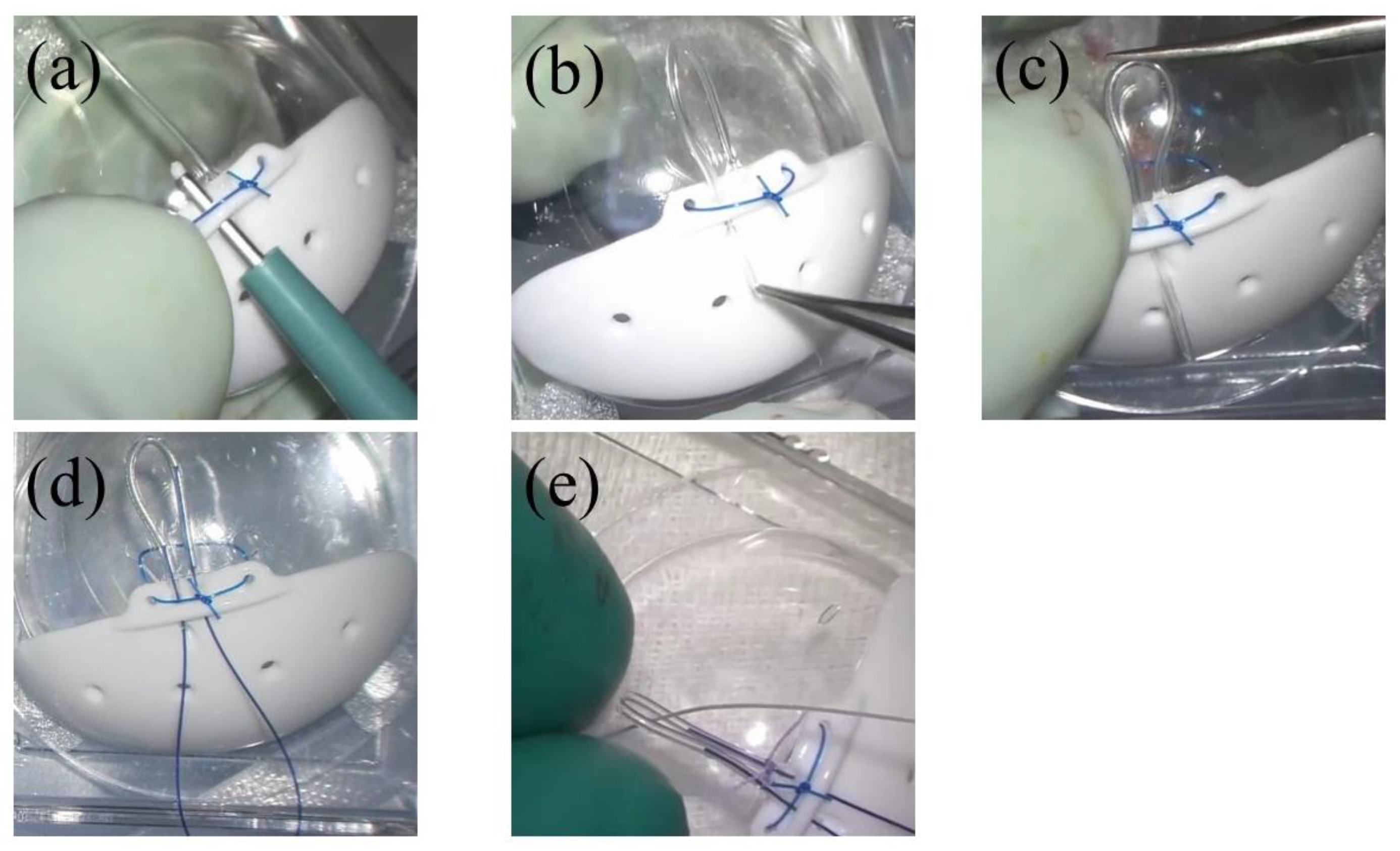
2.3. Surgical Technique
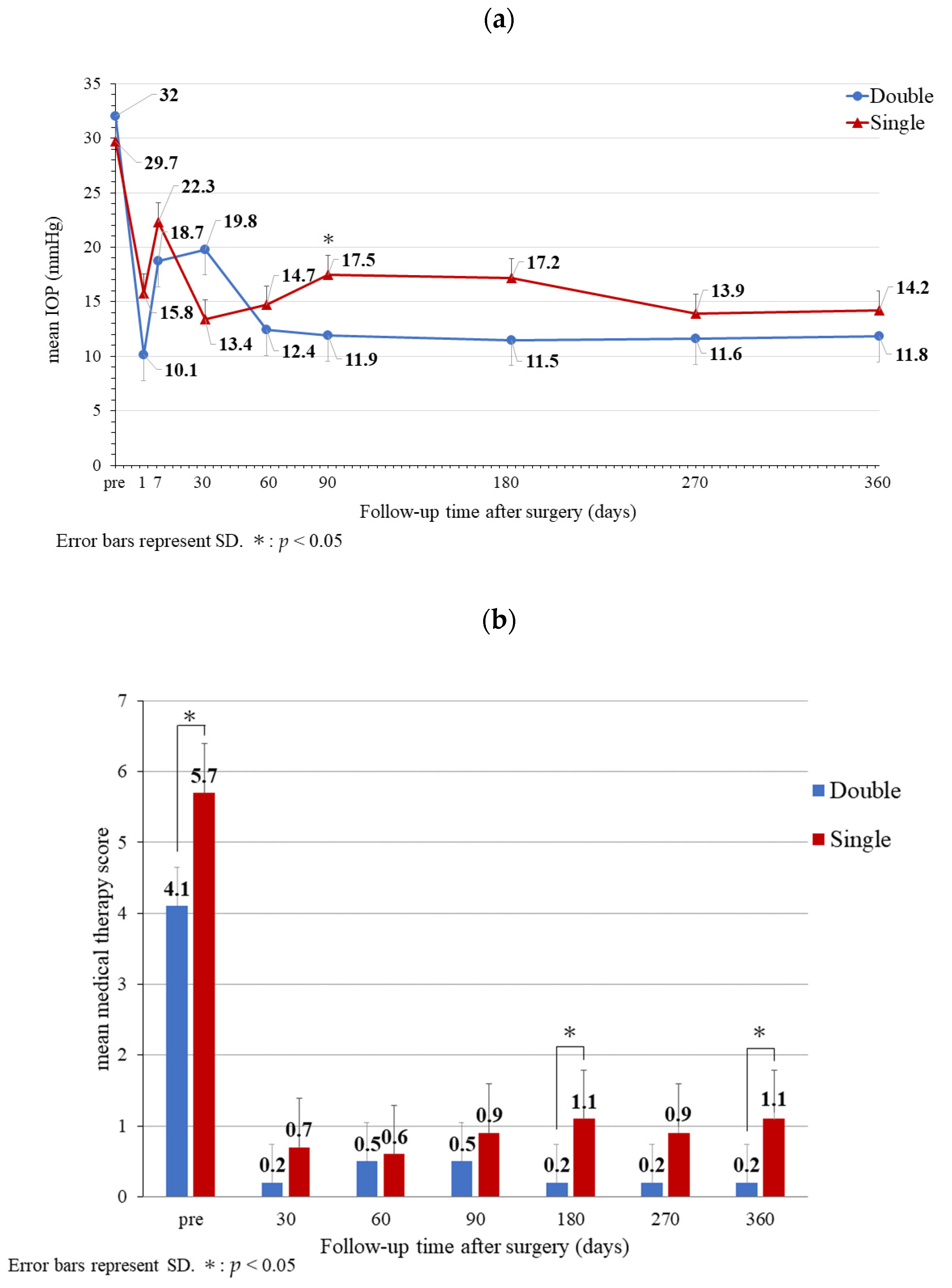
Supplementary Video S1 summarizes the steps of the surgery. Briefly: prior to the surgery, we modified the BI. We created a parallel tunnel in the plate adjacent to the tube using a 1.0 mm biopsy trephine (Figure 1a). The tip of the tube was inserted into the tunnel to create a U-shaped loop (Figure 1b). The length of this loop was set to 7.5 mm from the root. The outer wall of the tip of the loop and the end of the tube were then cut with scissors (Figure 1c). To avoid unwanted hypotony, 3-0 nylon was inserted into both tubes as a stent suture (Figure 1d). Each root of the tube was fastened using a 6-0 absorbable suture. A 4-0 silk thread was then passed through the loop of the tube (Figure 1e).
Because the outer diameter of the tube measures 0.61 mm, when two tubes are placed side-by-side, their total width is calculated to be 1.57 mm (Figure 2). To minimize leakage of intraocular fluid through the gap, a 1.5 mm scleral tunnel was designed. Finally, the placement of this looped tube created two drainage paths.
After modifying the BI, we initiated the surgical procedure. Following a conjunctival incision at the limbus, a wide and bare scleral field was secured. We marked the sclera at 4 mm and 8 mm posterior to the corneal limbus; then, an 8-0 nylon needle pierced the sclera at a point of 4 mm as a marker for penetration (Figure 3a). Next, we created a 4 mm long scleral tunnel with a 1.5 mm slit knife between the marks (Figure 3b). The slit knife was advanced until the marker needle began swaying. Once movement of the marker needle was confirmed, the slit knife was pointed towards the vitreous cavity and penetrated the pars plana. Both tips of the silk thread were grabbed with fine forceps and placed into the scleral tunnel until they were visible through the pupil (Figure 3c). After another sclerotomy was performed with a 20-Gauge MVR, both tips of the silk thread were removed from the sclerotomy with fine forceps. A U-shaped tube was introduced into the scleral tunnel by pulling the guidance silk thread (Figure 3d). Once the tip of the looped tube was confirmed through the pupil, the guidance silk thread was cut and removed after securing the plate (Figure 3e). The implant plate was placed between the external muscles. The stent suture was fixed near the corneal limbus for ease of removal (Figure 3f). The Tenon’s capsule and conjunctival incisions were sutured using absorbable sutures. The stent thread was removed when IOP increased to >21 mmHg or 2–4 weeks after surgery.

3. Results

A summary of both groups’ demographic data is presented in Table 1a,b.
Proliferative diabetic retinopathy was the most common cause of NVG in both groups. Four eyes and three eyes had undergone previous glaucoma surgery in the double and single groups, respectively.
The mean preoperative IOPs (SMTS) were 32.0 ± 11.33 mmHg (4.1) and 29.7 ± 6.31 mmHg (5.7) in the double and single groups, respectively (Figure 4a,b). The mean postoperative IOP were 19.8 ± 14.82 mmHg (0.2) and 13.4 ± 3.89 mmHg (0.7); 12.4 ± 3.95 mmHg (0.5) and 14.7 ± 4.42 (0.6); 11.9 ± 3.38 mmHg (0.5), and 17.5 ± 4.71 mmHg (0.9); 11.5 ± 4.30 mmHg (0.2) and 17.2 ± 5.82 mmHg (1.1); 11.6 ± 4.32 mmHg (0.2) and 13.9 ± 5.30 mmHg (0.9), at 1, 2, 3, 6 and 9 months, respectively. At 12 months, the mean IOP were 11.8 ± 2.70 mmHg (63% reduction, p < 0.004) and 14.2 ± 4.05 mmHg (52% reduction, p < 0.002). The medical therapy scores were 95% (p = 0.005) and 89% (p = 0.005), respectively. There was a significant difference in IOP between the two groups at 3 months (p = 0.01). However, there was no significant difference in IOP between the two groups at 12 months (p = 0.16).
There were significant differences in the SMTS between the two groups before surgery, at 6 and 12 months (p = 0.01, 0.04 and 0.04, respectively).
Intraoperatively, one eye in the double group presented iridodialysis, which was resolved by performing iridoplasty immediately. Postoperative complications in the double group included vitreous hemorrhage in nine eyes, macular edema in two eyes, rhegmatogenous retinal detachment in one eye, epiretinal membrane in one eye, and choroidal detachment in one eye. Postoperative hypotony (<5 mmHg) was observed in three eyes on postoperative day 7. One eye required reoperation because of rhegmatogenous retinal detachment. In addition, reoperation was required in two eyes because of rhegmatogenous retinal detachment and iris bombe in the single group. Vitreous hemorrhage was spontaneously absorbed in all the cases. There were no incidents of postoperative tube exposure, implant exposure, or endophthalmitis in either of the groups.
Two eyes in the double group and one eye in the single group were confirmed to have no light perception at 12 months. The preoperative visual acuity of four eyes in the double group and three eyes in the single group were below counting fingers. Corneal endothelial cell density was not recorded at 12 months in the double group because the eye had gone through phthisis bulbi. Finally, one eye was not measurable before surgery in the single group due to corneal edema.

4. Discussion

In this study, no statistically significant difference between the two groups was identified for IOP reduction, but the SMTS was reduced between the two groups. Therefore, the use of a double tube with modified BI may facilitate reducing IOP in NVG eyes compared to a single tube. The mean postoperative IOP and medical therapy scores at one year following this novel surgical technique were lower than those reported in previous studies [2,3,11].
The mechanism by which the final IOP level is regulated during tube-shunt surgery remains unknown [12]. Controversies exist regarding whether plate size correlates with IOP reduction. Heuer et al. reported that double-plate Molteno implantation (274 mm2) may provide better IOP control than single-plate Molteno implantation (137 mm2) [13]; additionally, Souza et al. reported double-plate Molteno implantation (274 mm2) more frequently affords IOP control than Ahmed valve implant (184 mm2) [14]. Based on these reports, several devices with larger plates have been developed. However, other studies have reported that plate area does not strictly correlate with the degree of IOP reduction with BI [15,16].
According to fluid dynamics, the aqueous flow through the chambers of the human eye ranges from 1.8 to 4.3 μL/min [17]. The diameter of the BI was designed to function effectively under these conditions [18]. According to a former study that employed a numerical model and computational fluid dynamics simulation, the tube diameter is a determinant of hypotensive efficacy [19]. One report indicated that, the larger the inner diameter of the tube, the greater the probability of clinical IOP reduction [20]. In contrast, there is a report that the filtration rate declines when the inner diameter of the tubing is reduced in BI [21]. An in vivo study showed that a large-lumen glaucoma drainage device equipped with a flow regulator can provide both prevention of immediate postoperative hypotony and progressive IOP reduction on demand [22]. A similar technique of inserting a double XEN to improve filtration was reported at the 9th World Glaucoma E-Congress [23]. The authors intended to increase the filtration volume to achieve a greater reduction in IOP. Our novel surgical technique is the only one focused on increasing the filtration volume with BI. Enlarging the outer diameter of the tube may not only increase the filtration volume, but also the risk of tube exposure, which is a serious complication [2,3]. We previously invented a new technique in which a single tube was introduced into the anterior chamber [9], or the vitreous cavity [8], through a long scleral tunnel created by a 24 g catheter needle, without a scleral flap or patch. Because the outer shell of a 24 g catheter needle has approximately the same diameter as the tube of the BI, the catheter could be used as a guide to introduce the tube into a long scleral tunnel, and we did not observe tube exposure with this method.
In light of this experience, we found that the width of two parallel tubes, measuring approximately 1.5 mm, can be introduced into a long scleral tunnel created by a 1.5 mm slit knife (Figure 2); in this process, the height of the tunnel remains the same as during insertion of a single tube, and this would hypothetically double the filtration volume. We also noticed that if we bent the tube in a U-shape and cut only the outer wall of the tube at the bottom of the U-shape while the inner wall remained connected (Figure 1c), the cut exposed the orifices of two parallel connected tubes, which could be easily introduced into the vitreous cavity through a scleral tunnel using a guidance thread pulling on the U-shaped part (Figure 3d). Tunnel incision may also contribute to prevention of plate exposure by fixing the plate away from the limbus.
While our results are encouraging, we could not completely eliminate postoperative adverse effects. We recorded nine eyes with vitreous hemorrhage in the double group. Vitreous hemorrhage appears in approximately 20% of cases of NVG with tube-shunt surgery [24,25]; however, this study found it to be more common than in previous reports.
Some studies have shown that vitreous hemorrhage in tube-shunt surgery arises due to intraoperative blood entering the eye through the sclerotomy and scleral tunnel [9,26]. In our case, this may have occurred partly because we created a 1.5 × 4 mm scleral tunnel in which cauterization was not possible in the presence of damage to the scleral vessels. It is also important to treat retinal ischemia, which induces further neovascularization, using anti-vascular endothelial growth factor therapy and pan-retinal photocoagulation [27]. Reduction in postoperative vitreous hemorrhage has been reported with the use of bevacizumab for NVG, before or after tube-shunt surgery [28,29]. Thus, anti-vascular endothelial growth factor therapy should be performed proactively. Fortunately, vitreous hemorrhage was spontaneously absorbed without reoperation in both groups.
The insertion of the U-shaped tube into the vitreous cavity using silk thread is one of the features of this technique; however, iridodialysis occurred in one case. In this case, the clearance between the silk thread and ciliary tissue was insufficient because the scleral incision for extruding the guidance silk thread was abutting the scleral tunnel to facilitate tube placement in the same quadrant.
Although a 3-0 nylon was inserted within each tube as a stent suture to prevent overfiltration [30], and each root of the tube was fastened using an absorbable suture [31], three eyes exhibited postoperative hypotony (<5 mmHg) in the double group. While our surgical procedure aimed to minimize leakage from each tube, leakage may inevitably occur through the gap between the scleral tunnel and the tubes, as indicated in gray in Figure 2. Although hypotony was observed before stent suture removal, it may be better to set stent sutures separately to each tube and to remove them sequentially to prevent excessive reduction of IOP afterwards.
The present study has some limitations. First, to prove the hypothesis that increasing filtration volume achieves more reduction of IOP, it is necessary to evaluate this surgical technique using bleb fluid images [32,33]. In addition, magnetic resonance imaging can be performed after surgery to check the condition inside the capsule; in our study, imaging studies were not performed due to limited medical resources. Second, our number of patients was relatively small, and this was a single-institution study. Therefore, the number of cases was insufficient to detect statistically significant differences in the double group, which was shown in the present study to exhibit greater reduction in IOP than the single group. Third, because the primary outcome in this study focused on IOP and medical therapy, we did not evaluate the postoperative visual acuity and visual field associated with IOP reduction. Fourth, we were not able to directly compare the outcome of this novel technique with that of trabeculectomy. The double group may have experienced greater reduction in IOP than the single group; however, it has not been proven whether this IOP reduction necessarily leads to better clinical outcomes than the IOP reduction achieved using trabeculectomy. Finally, we used this technique only for patients with NVG. Further studies are required to evaluate the long-term outcomes for these patients and further evaluation of this technique is necessary to establish reproducibility of these results in other types of glaucoma.

5. Conclusions

There are many strategies for tube-shunt surgery, but it remains unclear which surgical approach leads to optimal patient outcomes. We hypothesized that placing two tubes simultaneously may facilitate IOP reduction.

Supplementary Materials

The following supporting information can be downloaded at: https://www.mdpi.com/article/10.3390/surgeries3040035/s1, Video S1: The procedures of the representative cases in this study were summarized.

Author Contributions

K.T. wrote the manuscript; M.A. designed the experiments and performed surgeries; K.T., R.A., K.M., M.K. and M.A. discussed the procedure, assisted with the surgery and examined the patients. All authors have read and agreed to the published version of the manuscript.

Funding

This research received no external funding.

Institutional Review Board Statement

The study was conducted in accordance with the Declaration of Helsinki and approved by the Ethics Committee of Osaka Red Cross Hospital (protocol code 951 and date of approval 26 April 2019).

Informed Consent Statement

Informed consent was obtained from all subjects involved in the study.

Data Availability Statement

The data presented in this study are available on request from the corresponding author. The data are not publicly available due to restrictions privacy.

Acknowledgments

The 75th Annual Congress of Japan Clinical Ophthalmology, Fukuoka, Japan, October 2021; The 9th World Glaucoma E-Congress, Kyoto, Japan, July 2021.

Conflicts of Interest

M.A. is a consultant at Kowa Co., Ltd. No conflicting relationships exist for any other authors. The funders/companies had no role in the design of the study; in the collection, analyses, or interpretation of data; in the writing of the manuscript, or in the decision to publish the results.

References

  1. Sidoti, P.A.; Dunphy, T.R.; Baerveldt, G.; LaBree, L.; Minckler, D.S.; Lee, P.P.; Heuer, D.K. Experience with the Baerveldt Glaucoma Implant in Treating Neovascular Glaucoma. Ophthalmology 1995, 102, 1107–1118. [Google Scholar] [CrossRef]
  2. Gedde, S.J.; Schiffman, J.C.; Feuer, W.J.; Herndon, L.W.; Brandt, J.D.; Budenz, D.L. Tube versus Trabeculectomy Study Group. Treatment Outcomes in the Tube Versus Trabeculectomy (TVT) Study After Five Years of Follow-Up. Am. J. Ophthalmol. 2012, 153, 789–803.e2. [Google Scholar] [CrossRef] [PubMed] [Green Version]
  3. Gedde, S.J.; Feuer, W.J.; Lim, K.S.; Barton, K.; Goyal, S.; Ahmed, I.I.; Brandt, J.D. Treatment Outcomes in the Primary Tube Versus Trabeculectomy Study After 3 Years of Follow-Up. Ophthalmology 2020, 127, 333–345. [Google Scholar] [CrossRef] [PubMed]
  4. Ma, A.K.; Lee, J.H.; Warren, J.L.; Teng, C.C. GlaucoMap—Distribution of Glaucoma Surgical Procedures in the United States. Clin Ophthalmol. 2020, 14, 2551–2560. [Google Scholar] [CrossRef] [PubMed]
  5. Sun, M.T.; Madike, R.; Huang, S.; Cameron, C.; Selva, D.; Casson, R.J.; Wong, C.X. Changing Trends in Glaucoma Surgery within Australia. Br. J. Ophthalmol. 2021, 106, 957–961. [Google Scholar] [CrossRef] [PubMed]
  6. Heijl, A.; Leske, M.C.; Bengtsson, B.; Hyman, L.; Bengtsson, B.; Hussein, M.; Early Manifest Glaucoma Trial Group. Reduction of Intraocular Pressure and Glaucoma Progression: Results from the Early Manifest Glaucoma Trial. Arch. Ophthalmol. 2002, 120, 1268–1279. [Google Scholar] [CrossRef] [PubMed]
  7. Samsudin, A.B. An Assessment of Flow and Pressure Control in Experimental Models of Glaucoma Drainage Surgery. Ph.D. Thesis, UCL (University College London), London, UK, 2014. [Google Scholar]
  8. Kusaka, M.; Kujime, Y.; Yamakawa, M.; Akimoto, M. Baerveldt Tube Shunt Implantation Through a Long Scleral Tunnel. Eur. J. Ophthalmol. 2019, 29, 458–463. [Google Scholar] [CrossRef] [PubMed]
  9. Akamine, R.; Miyamoto, N.; Kusaka, M.; Akimoto, M. Tube Placement into the Long Scleral Tunnel with a Catheter Needle without a Scleral Valve and/or Graft Patch: 1-Year Outcome. EC Ophthalmol. 2022, 13, 1–9. [Google Scholar]
  10. Kanda, Y. Investigation of the Freely Available Easy-to-Use Software ‘EZR’for Medical Statistics. Bone Marrow Transplant. 2013, 48, 452–458. [Google Scholar] [CrossRef] [PubMed] [Green Version]
  11. Christakis, P.G.; Kalenak, J.W.; Tsai, J.C.; Zurakowski, D.; Kammer, J.A.; Harasymowycz, P.J.; Mura, J.J.; Cantor, L.B.; Ahmed, I.I. The Ahmed Versus Baerveldt Study: Five-Year Treatment Outcomes. Ophthalmology 2016, 123, 2093–2102. [Google Scholar] [CrossRef] [PubMed]
  12. Lavin, M.J.; Franks, W.A.; Wormald, R.P.; Hitchings, R.A. Clinical Risk Factors for Failure in Glaucoma Tube Surgery: A Comparison of Three Tube Designs. Arch. Ophthalmol. 1992, 110, 480–485. [Google Scholar] [CrossRef] [PubMed]
  13. Heuer, D.K.; Lloyd, M.A.; Abrams, D.A.; Baerveldt, G.; Minckler, D.S.; Lee, M.B.; Martone, J.F. Which Is Better? One or Two? A Randomized Clinical Trial of Single-Plate Versus Double-Plate Molteno Implantation for Glaucomas in Aphakia and Pseudophakia. Ophthalmology 1992, 99, 1512–1519. [Google Scholar] [CrossRef]
  14. Souza, C.; Tran, D.H.; Loman, J.; Law, S.K.; Coleman, A.L.; Caprioli, J. Long-Term Outcomes of Ahmed Glaucoma Valve Implantation in Refractory Glaucomas. Am. J. Ophthalmol. 2007, 144, 893–900. [Google Scholar] [CrossRef] [PubMed]
  15. Britt, M.T.; LaBree, L.D.; Lloyd, M.A.; Minckler, D.S.; Heuer, D.K.; Baerveldt, G.; Varma, R. Randomized Clinical Trial of the 350-mm2 Versus the 500-mm2 Baerveldt Implant: Longer Term Results: Is Bigger Better? Ophthalmology 1999, 106, 2312–2318. [Google Scholar] [CrossRef]
  16. Lloyd, M.A.; Baerveldt, G.; Fellenbaum, P.S.; Sidoti, P.A.; Minckler, D.S.; Martone, J.F.; LaBree, L.; Heuer, D.K. Intermediate-Term Results of a Randomized Clinical Trial of the 350-Versus-the 500-mm2 Baerveldt Implant. Ophthalmology 1994, 101, 1456–1463; discussion 1463. [Google Scholar] [CrossRef]
  17. Sheybani, A.; Reitsamer, H.; Ahmed, I.I. Fluid Dynamics of a Novel Micro-Fistula Implant for the Surgical Treatment of Glaucoma. Investig. Ophthalmol. Vis. Sci. 2015, 56, 4789–4795. [Google Scholar] [CrossRef] [PubMed]
  18. Brubaker, R.F. Flow of Aqueous Humor in Humans [The Friedenwald Lecture]. Investig. Ophthalmol. Vis. Sci. 1991, 32, 3145–3166. [Google Scholar] [PubMed]
  19. Kudsieh, B.; Fernández-Vigo, J.I.; Agujetas, R.; Montanero, J.M.; Ruiz-Moreno, J.M.; Fernández-Vigo, J.Á.; García-Feijóo, J. Numerical Model to Predict and Compare the Hypotensive Efficacy and Safety of Minimally Invasive Glaucoma Surgery Devices. PLoS ONE 2020, 15, e0239324. [Google Scholar] [CrossRef] [PubMed]
  20. Fea, A.M.; Menchini, M.; Rossi, A.; Posarelli, C.; Malinverni, L.; Figus, M. Early Experience with the New XEN63 Implant in Primary Open-Angle Glaucoma Patients: Clinical Outcomes. J. Clin. Med. 2021, 10, 1628. [Google Scholar] [CrossRef] [PubMed]
  21. Breckenridge, R.R.; Bartholomew, L.R.; Crosson, C.E.; Kent, A.R. Outflow resistance of the Baerveldt glaucoma drainage implant and modifications for early postoperative intraocular pressure control. J. Glaucoma 2004, 13, 396–399. [Google Scholar] [CrossRef] [PubMed]
  22. Olson, J.L.; Groman-Lupa, S. Design and Performance of a Large Lumen Glaucoma Drainage Device. Eye 2017, 31, 152–156. [Google Scholar] [CrossRef]
  23. Salazar, D.; Shah, M. Double XEN implant in Childhood glaucoma. In Proceedings of the 9th World Glaucoma E-Congress, Kyoto, Japan, 30 June–3 July 2021. [Google Scholar]
  24. Kolomeyer, A.M.; Seery, C.W.; Emami-Naeimi, P.; Zarbin, M.A.; Fechtner, R.D.; Bhagat, N. Combined Pars Plana Vitrectomy and Pars Plana Baerveldt Tube Placement in Eyes with Neovascular Glaucoma. Retina 2015, 35, 17–28. [Google Scholar] [CrossRef] [PubMed]
  25. Nishitsuka, K.; Sugano, A.; Matsushita, T.; Nishi, K.; Yamashita, H. Surgical Outcomes After Primary Baerveldt Glaucoma Implant Surgery with Vitrectomy for Neovascular Glaucoma. PLoS ONE 2021, 16, e0249898. [Google Scholar] [CrossRef] [PubMed]
  26. Go, M.; Ulrich, J.N.; Fleischman, D. Intraocular and Extraocular Hemorrhage Associated with Ligature Release of Non-Valved Glaucoma Drainage Implant. Am. J. Ophthalmol. Case Rep. 2017, 5, 114–116. [Google Scholar] [CrossRef]
  27. Takayama, K.; Someya, H.; Yokoyama, H.; Takamura, Y.; Morioka, M.; Sameshima, S.; Ueda, T.; Kitano, S.; Tashiro, M.; Sugimoto, M.; et al. Risk Factors of Neovascular Glaucoma After 25-Gauge Vitrectomy for Proliferative Diabetic Retinopathy with Vitreous Hemorrhage: A Retrospective Multicenter Study. Sci. Rep. 2019, 9, 14858. [Google Scholar] [CrossRef] [PubMed] [Green Version]
  28. Hwang, H.B.; Han, J.W.; Yim, H.B.; Lee, N.Y. Beneficial Effects of Adjuvant Intravitreal Bevacizumab Injection on Outcomes of Ahmed Glaucoma Valve Implantation in Patients with Neovascular Glaucoma: Systematic Literature Review. J. Ocul. Pharmacol. Ther. 2015, 31, 198–203. [Google Scholar] [CrossRef] [PubMed]
  29. Zhou, M.; Xu, X.; Zhang, X.; Sun, X. Clinical Outcomes of Ahmed Glaucoma Valve Implantation with or without Intravitreal Bevacizumab Pretreatment for Neovascular Glaucoma: A Systematic Review and Meta-Analysis. J. Glaucoma 2016, 25, 551–557. [Google Scholar] [CrossRef] [PubMed]
  30. Sherwood, M.B.; Smith, M.F. Prevention of Early Hypotony Associated with Molteno Implants by a New Occluding Stent Technique. Ophthalmology 1993, 100, 85–90. [Google Scholar] [CrossRef]
  31. Trible, J.R.; Brown, D.B. Occlusive Ligature and Standardized Fenestration of a Baerveldt Tube with and without Antimetabolites for Early Postoperative Intraocular Pressure Control. Ophthalmology 1998, 105, 2243–2250. [Google Scholar] [CrossRef]
  32. Ferreira, J.; Fernandes, F.; Patricio, M.; Brás, A.; Rios, C.; Stalmans, I.; Pinto, L.A. Magnetic Resonance Imaging Study on Blebs Morphology of Ahmed Valves. J. Curr. Glaucoma Pract. 2015, 9, 1–5. [Google Scholar] [CrossRef] [PubMed]
  33. Sano, I.; Tanito, M.; Uchida, K.; Katsube, T.; Kitagaki, H.; Ohira, A. Assessment of Filtration Bleb and Endplate Positioning Using Magnetic Resonance Imaging in Eyes Implanted with Long-Tube Glaucoma Drainage Devices. PLoS ONE 2015, 10, e0144595. [Google Scholar] [CrossRef] [PubMed]
Figure 1. Preparation of the Baerveldt glaucoma implant; (a) a parallel tunnel in the plate adjacent to the tube using a 1.0 mm biopsy trephine; (b) the tip of the tube was inserted into the tunnel to create a U-shaped loop; (c) the outer wall of the tip of the loop was cut with scissors; (d) a 3-0 nylon was inserted into both tubes as a stent suture; (e) each root of the tube was fastened using 6-0 absorbable sutures. A 4-0 silk thread was then passed through the tube loop.
Surgeries 03 00035 g001
Figure 2. Calculation of the width of two tubes placed side by side; The outer diameter of the tube is 0.61 mm, and when two tubes are placed side by side, their width is 1.22 mm, and the half of the outer circumference calculated to be 1.57 mm. The hypothetical gap when the tubes are placed in the scleral tunnel is indicated in gray.
Surgeries 03 00035 g002
Figure 3. Surgical Procedure; (a) a needle penetrated the sclera 4 mm posterior to the corneal limbus; (b) a 4 mm long scleral tunnel was created with a 1.5 mm slit knife between the marks; (c) both tips of the silk thread were grabbed with fine forceps and inserted into the scleral tunnel; (d) the U-shaped tube was introduced into the scleral tunnel by pulling the guidance silk thread; (e) the tip of looped tube was confirmed through the pupil (green arrow); (f) the stent suture was fixed near the corneal limbus.
Surgeries 03 00035 g003
Figure 4. Intraocular pressure and medical therapy score; (a) the mean intraocular pressure of the double group (Double) and the single group (Single); (b) the mean medical therapy score of the double group (Double) and the single group (Single).
Surgeries 03 00035 g004
Table 1. (a) Summary of the double group. (b) Summary of the single group.
(a)
Preoperative Postoperative
(12 Months)
Surgical Complications
CaseAge
(Years)
SexEyeTypePrevious SurgeriesAntiplatelet
Drug
SMTSIOP
(mmHg)
VACECD
(Cells/mm2)
SMTSIOP
(mmHg)
VACECD
(Cells/mm2)
Intraoperative ComplicationsPostoperative Complications
149MLOISPEA + IOL, PPV + SFIOL 53110/200229901620/2001252 VH
288MRCRVOPEA + IOL, PPV 232HM1887010NLP1761 VH, ME
360MRPDRPEA + IOL + LET, PPV 62530/200225721330/2002304 VH
484MLPDRPEA + IOL, PPVClopidogrel,
Aspirin
22220/200220801160/2002273IridodialysisVH
557MLPDRPEA + IOL, PPV, LET 435CF24810740/2001497 VH, CD
666MRPDRPEA + IOL, PPV, LET 434CF874011NLPNR VH, RRD, PB
772MLPDRPEA + IOL, PPV, LETAspirin330HM20920148/2002088 VH
865MLPDRPEA + IOL 51310/2002381015160/2002179 VH
966MRCRVOPEA + IOL, PPV 5442/20014140108/2002309 ME, ERM
1041MLPDRPEA + IOL, PPV 554100/200133001110/2001117 VH
(b)
Preoperative Postoperative
(12 Months)
Surgical Complications
CaseAge
(Years)
SexEyeTypePrevious SurgeriesAntiplatelet
Drug
SMTSIOP
(mmHg)
VACECD
(cells/mm2)
SMTSIOP
(mmHg)
VACECD
(cells/mm2)
Intraoperative ComplicationsPostoperative Complications
149WLPDRPEA + IOL + PPV, LET, BN 7292/200238711630/2002028 ME
249WRPDRPEA + IOL + PPV 623120/2001818118200/2002358
346MRPDRPEA + IOL + PPV + LET 4228/20021372730/2002049
467MRPDRPEA + IOL, PPV 63220/200NR111HM2053 RRD
551MLPDRPEA + IOL + PPV, PI 740CF185901160/2002632 IB
675MLOISPEA + SFIOL + PPV 534LPNM218NLP1222 VH
751MRPDRPEA + IOL + PPV 530CF277001860/2002198 VH, ME
854MRPDRPEA + IOL + PPV 63820/200NR41610/2002257
957MLPDRPEA + IOL + PPV, LET 524120/200168401060/2001529 VH
1053MLPDRPEA + IOL + PPV 62560/200263901760/2002653 ME
(a) M = male, F = female, OIS = ocular ischemic syndrome, CRVO = central retinal vein occlusion, PDR = proliferative diabetic retinopathy, PEA + IOL = phacoemulsification/aspiration and intraocular lens implantation, PPV = pars plana vitrectomy, SFIOL = scleral-fixated intraocular lens, LET = trabeculectomy, SMTS = supplemental medical therapy score, IOP = intraocular pressure, VA = visual acuity, HM = hand motion, CF = counting fingers, IV = intravitreal injection with anti-vascular endothelial growth factor therapy, CECD = corneal endothelial cell density, NLP = no light perception, NR = not recorded, VH = vitreous hemorrhage, ME = macular edema, CD = choroidal detachment, RRD = rhegmatogenous retinal detachment, ERM = epiretinal membrane, PB = phthisis bulbi. (b) BN = bleb needling, PI = peripheral iridotomy, LP = light perception, NM = not measurable, IB = iris bombe.
Publisher’s Note: MDPI stays neutral with regard to jurisdictional claims in published maps and institutional affiliations.

