Article

Multi-Level Fibrotomy for Pediatric Patients with Cerebral Palsy: A Cohort Study

1
Department of Orthopedics, Children’s Hospital Bambino Gesù, Palidoro, 00165 Rome, Italy
2
Department of Orthopaedic and Trauma Surgery, Campus Bio-Medico, University of Rome, 00128 Rome, Italy
*
Author to whom correspondence should be addressed.
Osteology 2021, 1(3), 141-148; https://doi.org/10.3390/osteology1030015
Submission received: 6 June 2021 / Revised: 12 July 2021 / Accepted: 14 July 2021 / Published: 16 August 2021
(This article belongs to the Special Issue Feature Papers in Osteology)

Abstract

:
Muscle retraction in Cerebral Palsy (CP) often requires surgical treatment. Multilevel procedures (using open or percutaneous techniques) are commonly performed in the ambulant patient with CP. The necessity to find new surgical techniques, reduce postoperative discomfort, and accelerate the healing process and rehabilitation is mandatory for these patients. A retrospective cohort study with 189 pediatric patients with CP was performed. The multilevel gradual fibrotomy of Ulzibat was modified using an ophthalmic knife. No significant complications were reported using our technique. Opioid drugs were not necessary, and casting time was reduced at the first 24 h. A significant Range of Motion recovery was assessed post-operatory and maintained at the last follow-up. Mean days of hospitalization were 2.2. The mean follow-up was 39 months (6–64 months). The modified multilevel fibrotomy reduces postoperative pain with easier patient management, resulting in a faster discharge from the hospital. However, the retrospective nature and the lack of a control group of the present study did not allow the authors to report significant results. Further studies with longer follow-up are in progress to obtain more certain data that confirm our preliminary results.

1. Introduction

Muscle retraction in Cerebral Palsy (CP) often requires surgical treatment [1]. Single-event multi-level surgery (SEMS) is commonly performed in the ambulant patient with CP [2], involving hips adductors, ilio-psoas tendon, hamstrings, and Achilleus tendons [3,4]. The surgical procedures usually include tenotomies and lengthening of the retracted and spastic muscles. These procedures can be performed with open or percutaneous techniques [5,6,7,8,9,10,11]. The surgical incision, the type of procedure, and the post-surgical rehabilitation and casting time could be variable [12], depending on the surgical technique adopted. In ambulant children with CP, SEMS is one of the most common treatments performed [2]. To correct severe lower-limb deformity, improve gait, and limit the children’s exposure to recurrent surgeries, general anaesthesia, and rehabilitation, SEMS could be used in bone and soft-tissue treatments [13]. Although this procedure is beneficial in treating children with CP, recovery of function after surgery is a long and arduous process. Derotation osteotomies of the femur and tibia are also commonly performed in CP patients [14].
Nevertheless, osteotomies generally require from 4.5 to 7.6 weeks of post-operative immobilization [14]. Furthermore, the failure of fixation after early weight-bearing and nonunion, infection, and inadequate correction are additional problems. Furthermore, postoperative discomfort and immobility caused by significant muscle dissection could exacerbate pre-existing weakness [14]. Muscle–tendon recession or partial lengthening are commonly used to correct lower-limb contractures in CP [15]. Muscle–tendon lengthening techniques are known to decrease muscular strength; therefore, lengthening at the musculotendinous junction has progressively been adopted to solve this problem [16]. Lengthening the musculotendinous junction is a popular approach for minimizing strength loss and accelerating hospital discharge.
Combined bone and soft-tissue surgery usually require more days of hospitalizations compared to soft tissue SEMS. Moreover, the rehabilitation time is also higher.
CP patients usually underwent numerous surgical procedures during their lives [17]; therefore, surgical techniques and post-operative rehabilitation must be updated to improve CP patients’ outcomes. Therefore, it could be helpful to find a new minimally invasive surgical technique reducing the postoperative discomfort, and accelerating the healing process and the rehabilitation processes [18]. The use of minimally invasive SEMS could solve this problem.
This retrospective cohort study aimed to provide the efficacy of our modified multilevel fibrotomy technique to treat muscle retraction in pediatric patients with CP.

2. Materials and Methods

A novel technique for treating muscle retraction in CP patients was developed, modifying the original multilevel percutaneous procedure [19,20,21]. A mini-invasive ophthalmic knife was used to perform a multilevel approach. The study was conducted according to The Strengthening the Reporting of Observational Studies in Epidemiology (STROBE) statement guidelines [22].

2.1. Study Design

A retrospective cohort study was performed. All the patients were treated in our pediatric hospital of Rome between March 2017 and November 2019. Only patients with a minimum follow-up of 6 months were included.

2.2. Participants

Inclusion criteria were as follows: patients ranged from level II to III of the Gross Motor Functional Classification System (GMFCS) [23], affected by diparetic and quadriparetic cerebral palsy. All the patients were clinically observed once per month after the hospital discharge for at least six months. Patients older than 18 years old or with a history of previous muscle/tendon surgery were excluded. The study was performed following the ethical standards as laid down in the 1964 Declaration of Helsinki and its later amendments or comparable ethical standards.

2.3. Variables and Measurement

Outcome measures evaluated were: drug consumption after surgery, days of hospitalization; casting time; complications (bleeding, infections, vascular and neurological injuries, relapse, others); operation time, blood loss, open surgery conversion, local hematomas and Range of Motion (ROM) of hip, knee, and ankle. The postoperative pain was measured by the Visual Analog Scale (VAS) [24] at different follow-up periods only in patients older than 7 years, as the reliability of this score in younger patients has not been validated [25]. All the parameters were evaluated preoperatory, after surgery, and at last follow-up.

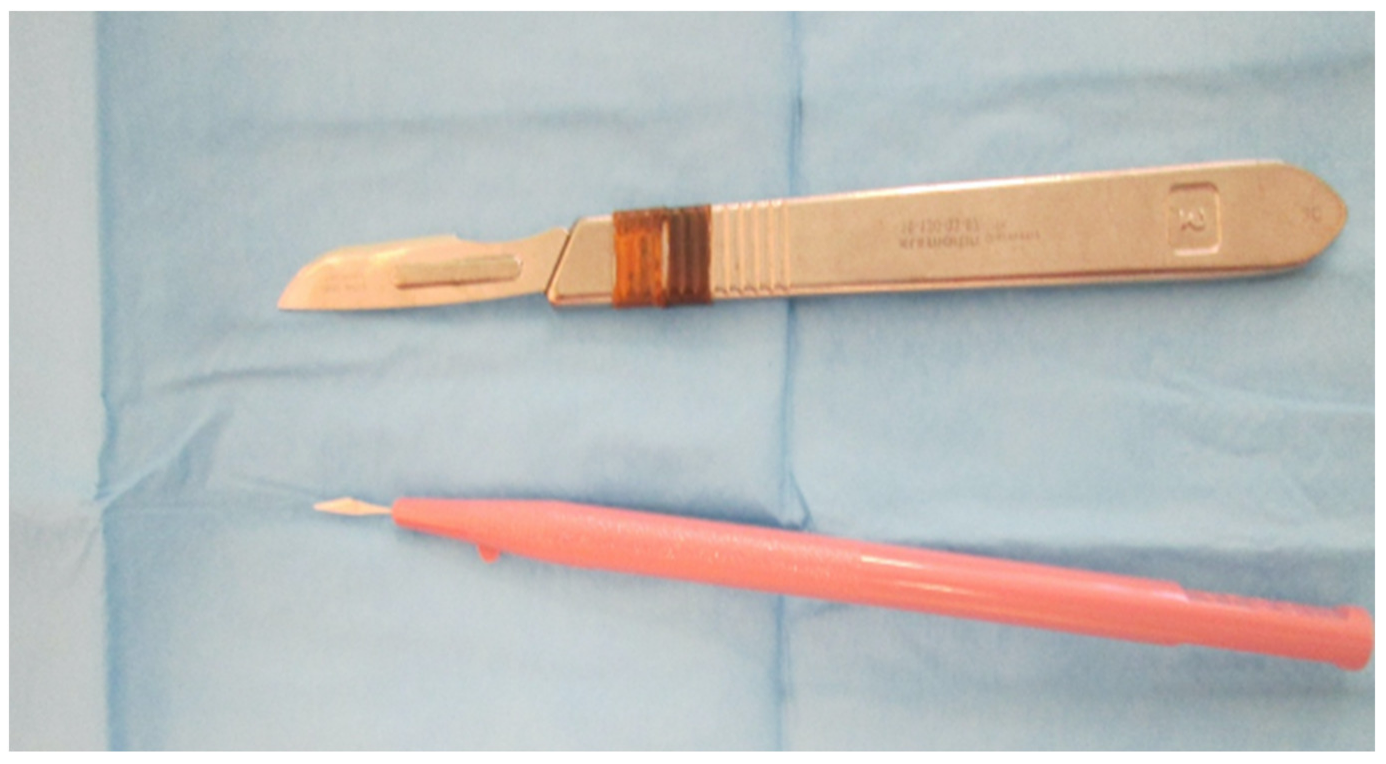
2.4. Surgical Devices

The procedure was performed using an ophthalmic Knife A-OK® 1.52 mm Stainless Steel, with a straight sharp pointed tip angled 15° (Alcon©, United States of America). This knife is approved for medical uses and widely adopted in ophthalmic surgery (Figure 1).

2.5. Surgical Technique

The surgical technique adopted was inspired by the gradual fibrotomy of Ulzibat, described by Gomez-Andres et al. [26]. All patients underwent multi-level fibrotomy of the lower limbs including adductors of the hip, hamstring of the knee, tibialis posterior, and Achilles tendon [27]. The same surgeon performed all the procedures. The patients underwent only percutaneous soft-tissue procedures. The number of levels for each child was decided on pre-operative clinical examination.
For better comprehension, we here firstly describe our adductor fibrotomy technique. The patient was lying supine in general anaesthesia. The right-handed surgeon stood to the patient’s left side, delimiting the adductor longus origin between his fingers. The surgeon introduced an ophthalmic knife A-OK® 1.52 mm Stainless Steel, with a straight sharp pointed tip angled 15° underneath the tendon fascia from lateral to medial. The blade was turned 90° upward cutting the adductor longus fascia and muscular junction from deep to superficial. No more than three cuts were performed for a single level. The second surgeon then divided in a closed manner the remaining fibers by back and forth movements while applying pressure with gauze.
The technique by Gordon et al. [28] was used for the hamstrings fibrotomy. One operator flexed the hip to 90° with the patient lying supine and extended the knee under tension with the contralateral limb in extension. The first operator felt the semitendinosus and gracilis tendon by palpation. The ophthalmic blade was turned 90° and inserted at the junction between the muscle and the tendon. The operator started the release of the structures with no more than three cuts. When the second operator felt the release, the blade was turned back 90° and removed from the incision. A similar approach was performed for the tibialis posterior tendon and Achilles tendon.
The traditional minimally invasive approach and the modified Ulzibat technique reported some fundamental differences. With the Ulzibat technique, the cutting scars are within the muscle and were provided by a specific fibrotomic blade [29]. In the minimally invasive technique, the tendon was cut; instead, in the Ulzibat technique, the muscle fibers and the fascia were cut at the level of the muscle–tendon junction. Lastly, the Ulzibat technique could be less invasive than the traditional approaches.
A plaster cast was applied for the first 24 h and used intermittently after the first night to allow a gradual mobilization for the physiotherapy. No opioid drugs were used during or after the procedure. Paracetamol and local anaesthetic infiltration after surgery were used for pain management.

3. Results

3.1. Participants

A total of 212 patients were enrolled in the study, but only 189 completed the six months follow-up. Among these, 110 were male, and 73 were female. The mean age of the patients was 10.9 ± 4.35 (ranges from 2 to 18). The mean follow-up was 39 months (6–64 months). A total of 485 levels were treated: 149 adductors of the hip, 158 hamstrings, 14 posterior tibialis, and 164 Achilleus tendons. Among these, 207 levels were bilateral. The mean levels treated for a patient was 2.5 ± 1.4. Results are reported in Table 1.

3.2. Main Results

Postoperative pain could be evaluated only in responding patients aged more than 7 years (84 patients). All the other outcomes were assessed in the whole cohort. Postoperative pain, evaluated by VAS scale, was 3.8 ± 1.2 points. Drug consumption after surgery was low: 2.5 mg/kg of Naropine 7.5% (maximum 150 mg) was used for intraoperative injection in all sites treated. Paracetamol (15 mg/kg for a maximum of four daily doses) was used in a standard manner to control the pain; in 7 cases, Ketorolac (0.5 mg/kg for a maximum of three daily doses) was necessary for the first postoperative days. No opioid drugs were used during the hospitalization. Days of hospitalization were on average 2.2 (ranges from 1 to 4). Casting time was reduced at the first 24 h, and it was adopted intermittently after the first night, allowing an early mobilization and physiotherapy treatment. No significant vascular or neurological lesions occurred. Operation time was an average of 15.6 ± 12.3 min; blood loss was not evaluable (always less than 20 cc), and there was no case of bleeding that required open surgery. There was not a necessity to convert in open procedure in any circumstances. The surgical ophthalmic knife had no breakage, and it was never necessary to use different surgical devices. Local hematomas occurred only in 15 patients, treated successfully by ice, rest, and anti-oedema ointment applications (Glicosamminoglicanpolisulphate 25.000 UI).
A significant and stable improvement (p = 0.0028) of the passive and active articular ROM of the levels treated at all considered follow-up periods was obtained. The ROM of hip, knee, and ankle was also maintained at the last follow-up (detailed information is reported in Table 2).

4. Discussion

Alteration of walking in patients with CP results from contractures across joints, muscle spasticity [30], and physically inappropriate muscle action. All the joints could be involved, but usually, lower extremities are mostly affected. The advantages of minimally invasive percutaneous multilevel surgery for the treatment of CP are widely treated in the literature [19,20,28,31,32,33,34,35,36,37].
Single event multilevel surgery (SEMS) for CP patients was first described in 1985 by Norlin and Tkaczuk [38]. McGinley and colleagues performed a systematic review on the use of SEMS in CP patients, reporting a lack of high-quality clinical study and high heterogeneity in terms of surgical techniques and levels treated [39]. Thomason and colleagues performed a randomized controlled trial on the use of SEMS in 19 CP patients [40]. They performed muscle–tendon lengthening, rotational osteotomy, and tendon transfers. The levels treated were decided preoperatively, after a clinical examination by an experienced surgeon. Schranz investigated the long-term outcomes of SEMS surgery in patients with spastic hemiplegia, reporting good results at last follow-up [41].
The first who introduced the concept of “gradual fibrotomy” was Ulzibat, and his technique is reported in the study by Gomez-Andres et al. [26]. Thompson et al. [2], instead, described a multilevel approach for the treatment of CP. The surgical technique described in the present study reported some advantages compared to other techniques.
The advantages of our modified technique are the low rate of complications, the possibility to perform SEMS, and the reduced casting time and drugs consumption.
Edwards and colleagues performed a comparative study between conventional and standard minimally invasive surgery for CP patients [18]. They reported comparable results in gait kinematics and walking speeds, with a reduced complication rate in the minimally invasive group. Edwards reported 37% and 36% of complication rates in the convectional and minimally invasive groups, respectively [18]. Despite the fact that the authors reported similar results in the overall rate of complications, they also found that the severity of adverse events in the minimally invasive group was lower than in the conventional group [18]. The rate of recurrence was 21% in conventional and 36% in the traditional group. Thus, the results described in the literature were higher compared to the present study. However, a direct statistical comparison was not possible due to the lack of Edward’s database.
There was also a reduction in economic costs as the procedure needs a few operatory devices and the hospital stay was about 2.2 days. In the literature, the risk of compartmental syndrome after percutaneous tendon lengthening was fully described [42]; however, this complication was not reported in our patients. The ROM recovery after fibrotomy was significant, and no loss of ROM was noticed during the follow-up (Figure 2).
In case of recurrence, the efficacy and the reduced rate of complications of this procedure allow the surgeon to perform multilevel fibrotomy in complicated patients.
This study has some limitations. Firstly, this is a retrospective study without a control group; therefore, no comparative data with a similar sample size has been obtained. Moreover, this technique was applied to all the patients included (GMFC from II to III). Further studies with a more detailed layering of the patients need to be performed. Moreover, the lack of a long follow-up (maximum 64 months) could underestimate the number of recurrences. Lastly, pain assessment was performed only in children without cognitive problems.

5. Conclusions

The modified minimally invasive multilevel fibrotomy seems to be a valuable and safe technique for treating muscle retraction in CP, with some advantages compared to the standard procedures. This technique could reduce postoperative drug consumption, resulting in a faster discharge from the hospital. Moreover, with a low rate of surgical complications, it could be possible to perform a multilevel approach, reducing hospitalizations. However, the retrospective nature and the lack of a control group of the present study did not allow the authors to report significant results. Further studies with longer follow-up periods are in progress to obtain more certain data that confirm our preliminary results.

Author Contributions

Conceptualization, P.F.C. and S.D.S.; Data curation, R.R. and F.D.; Investigation, F.D.; Methodology, S.D.S.; Supervision, L.R.; Visualization, P.F.C. and L.O.; Writing—original draft, S.D.S. and A.B.; Writing—review & editing, S.D.S. and L.O. All authors have read and agreed to the published version of the manuscript.

Funding

No funds were received in support of this study.

Institutional Review Board Statement

The study was conducted according to the guidelines of the Declaration of Helsinki.

Informed Consent Statement

Informed consent was obtained from all subjects involved in the study.

Data Availability Statement

The datasets used and/or analyzed during the current study are available from the corresponding author on reasonable request.

Conflicts of Interest

The authors declare that they have no conflict of interest.

References

  1. Vlachou, M.; Pierce, R.; Davis, R.M.; Sussman, M. Does Tendon Lengthening Surgery Affect Muscle Tone in Children with Cerebral Palsy? Acta Orthop. Belg. 2009, 75, 808–814. [Google Scholar]
  2. Thompson, N.; Stebbins, J.; Seniorou, M.; Wainwright, A.M.; Newham, D.J.; Theologis, T.N. The Use of Minimally Invasive Techniques in Multi-Level Surgery for Children with Cerebral Palsy: PRELIMINARY RESULTS. J. Bone Joint Surg. Br. 2010, 92-B, 1442–1448. [Google Scholar] [CrossRef]
  3. Genêt, F.; Denormandie, P.; Keenan, M.A. Orthopaedic Surgery for Patients with Central Nervous System Lesions: Concepts and Techniques. Ann. Phys. Rehabil. Med. 2019, 62, 225–233. [Google Scholar] [CrossRef]
  4. Longo, U.G.; Salvatore, G.; Risi Ambrogioni, L.; Cella, E.; Candela, V.; Carnevale, A.; Schena, E.; Ciccozzi, M.; Maffulli, N.; Denaro, V. Epidemiology of Achilles Tendon Surgery in Italy: A Nationwide Registry Study, from 2001 through 2015. BMC Musculoskelet. Disord. 2020, 21, 687. [Google Scholar] [CrossRef]
  5. Haberfehlner, H.; Jaspers, R.T.; Rutz, E.; Harlaar, J.; van der Sluijs, J.A.; Witbreuk, M.M.; van Hutten, K.; Romkes, J.; Freslier, M.; Brunner, R.; et al. Outcome of Medial Hamstring Lengthening in Children with Spastic Paresis: A Biomechanical and Morphological Observational Study. PLoS ONE 2018, 13, e0192573. [Google Scholar] [CrossRef]
  6. Jaddue, D.; Abbas, M.; Sayed-Noor, A. Open versus Percutaneous Tendo-Achilles Lengthening in Spastic Cerebral Palsy with Equines Deformity of the Foot in Children. J. Surg. Orthop. Adv. 2010, 19, 196–199. [Google Scholar]
  7. Khaje Mozafari, J.; Pisoudeh, K.; Gharanizade, K.; Abolghasemian, M. Percutaneous Versus Open Hamstring Lengthening in Spastic Diplegic Cerebral Palsy. Arch. Bone Jt. Surg. 2019, 7, 373–378. [Google Scholar]
  8. Kläusler, M.; Speth, B.M.; Brunner, R.; Tirosh, O.; Camathias, C.; Rutz, E. Long-Term Follow-up after Tibialis Anterior Tendon Shortening in Combination with Achilles Tendon Lengthening in Spastic Equinus in Cerebral Palsy. Gait Posture 2017, 58, 457–462. [Google Scholar] [CrossRef]
  9. Lin, C.-L.; Lin, C.-J.; Huang, M.-T.; Su, W.-R.; Wu, T.-T. Mesh Achilles Tendon Lengthening--a New Method to Treat Equinus Deformity in Patients with Spastic Cerebral Palsy: Surgical Technique and Early Results. J. Pediatr. Orthop. Part B 2013, 22, 14–19. [Google Scholar] [CrossRef]
  10. Nazareth, A.; Rethlefsen, S.; Sousa, T.C.; Mueske, N.M.; Wren, T.A.L.; Kay, R.M. Percutaneous Hamstring Lengthening Surgery Is as Effective as Open Lengthening in Children With Cerebral Palsy. J. Pediatr. Orthop. 2019, 39, 366–371. [Google Scholar] [CrossRef]
  11. Turhan, Y.; Arıcan, M. Clinical Comparison of Percutaneous and Open Hamstring Lengthening in Children with Spastic Cerebral Palsy. Medeni. Med. J. 2019, 34, 182–187. [Google Scholar] [CrossRef]
  12. Katz, K.; Arbel, N.; Apter, N.; Soudry, M. Early Mobilization after Sliding Achilles Tendon Lengthening in Children with Spastic Cerebral Palsy. Foot Ankle Int. 2000, 21, 1011–1014. [Google Scholar] [CrossRef]
  13. Comparison of Proximal and Distal Rotational Femoral Osteotomy in Children with Cerebral Palsy—PubMed. Available online: https://pubmed.ncbi.nlm.nih.gov/12604941/ (accessed on 1 July 2021).
  14. Pirpiris, M.; Trivett, A.; Baker, R.; Rodda, J.; Nattrass, G.R.; Graham, H.K. Femoral Derotation Osteotomy in Spastic Diplegia. Proximal or Distal? J. Bone Joint Surg. Br. 2003, 85, 265–272. [Google Scholar] [CrossRef] [Green Version]
  15. Damiano, D.L.; Abel, M.F.; Pannunzio, M.; Romano, J.P. Interrelationships of Strength and Gait before and after Hamstrings Lengthening. J. Pediatr. Orthop. 1999, 19, 352–358. [Google Scholar] [CrossRef] [PubMed]
  16. Brunner, R. [Muscle physiology and the effect of muscle-tendon surgery in cerebral palsy]. Orthopade 2004, 33, 1096–1102. [Google Scholar] [CrossRef]
  17. Gillani, S.F.U.H.S.; Rafique, A.; Taqi, M.; Chatta, M.A.U.H.; Masood, F.; Ahmad Blouch, T.; Awais, S.M. Effectiveness of Treatment in Children With Cerebral Palsy. Cureus 2021, 13, e13754. [Google Scholar] [CrossRef]
  18. Edwards, T.A.; Thompson, N.; Prescott, R.J.; Stebbins, J.; Wright, J.G.; Theologis, T. A Comparison of Conventional and Minimally Invasive Multilevel Surgery for Children with Diplegic Cerebral Palsy. Bone Jt. J. 2021, 103-B, 192–197. [Google Scholar] [CrossRef]
  19. El Hage, S.; Rachkidi, R.; Noun, Z.; Haidar, R.; Dagher, F.; Kharrat, K.; Ghanem, I. Is Percutaneous Adductor Tenotomy as Effective and Safe as the Open Procedure? J. Pediatr. Orthop. 2010, 30, 485–488. [Google Scholar] [CrossRef] [Green Version]
  20. De Pavía-Mota, E.; Neri-Gámez, S.; Reyes-Contreras, G.; Valencia-Posadas, M. Percutaneous tenotomy and aponeurotomy (PTA) for knee flexor contracture in children with spastic cerebral palsy. Acta Ortop. Mex. 2013, 27, 109–113. [Google Scholar]
  21. Bozinovski, Z.; Popovski, N. Operative Treatment of the Knee Contractures in Cerebral Palsy Patients. Med. Arch. Sarajevo Bosnia Herzeg. 2014, 68, 182–183. [Google Scholar] [CrossRef] [Green Version]
  22. Von Elm, E.; Altman, D.G.; Egger, M.; Pocock, S.J.; Gøtzsche, P.C.; Vandenbroucke, J.P. STROBE Initiative The Strengthening the Reporting of Observational Studies in Epidemiology (STROBE) Statement: Guidelines for Reporting Observational Studies. Ann. Intern. Med. 2007, 147, 573–577. [Google Scholar] [CrossRef] [PubMed] [Green Version]
  23. Wood, E.; Rosenbaum, P. The Gross Motor Function Classification System for Cerebral Palsy: A Study of Reliability and Stability over Time. Dev. Med. Child Neurol. 2007, 42, 292–296. [Google Scholar] [CrossRef]
  24. Faiz, K.W. [VAS--visual analog scale]. Tidsskr. Den Nor. Laegeforening Tidsskr. Prakt. Med. Ny Raekke 2014, 134, 323. [Google Scholar] [CrossRef]
  25. Shields, B.J.; Palermo, T.M.; Powers, J.D.; Grewe, S.D.; Smith, G.A. Predictors of a Child’s Ability to Use a Visual Analogue Scale. Child Care Health Dev. 2003, 29, 281–290. [Google Scholar] [CrossRef]
  26. Gómez-Andrés, D.; Pulido-Valdeolivas, I.; Martín-Gonzalo, J.A.; López-López, J.; Martínez-Caballero, I.; Gómez-Barrena, E.; Rausell, E. [External evaluation of gait and functional changes after a single-session multiple myofibrotenotomy in school-aged children with spastic diplegia]. Rev. Neurol. 2014, 58, 247–254. [Google Scholar]
  27. Dietz, F.R.; Albright, J.C.; Dolan, L. Medium Term Follow-up of Achilles Tendon Lengthening in the Treatment of Ankle Equinus in Cerebral Palsy. Iowa Orthop. J. 2006, 26, 27–32. [Google Scholar] [PubMed]
  28. Gordon, A.B.; Baird, G.O.; McMulkin, M.L.; Caskey, P.M.; Ferguson, R.L. Gait Analysis Outcomes of Percutaneous Medial Hamstring Tenotomies in Children with Cerebral Palsy. J. Pediatr. Orthop. 2008, 28, 324–329. [Google Scholar] [CrossRef] [PubMed]
  29. Bernius, P. The Ulzibat Method – a New Surgical Technique. Neuropediatrics 2013, 44, PS11_1127. [Google Scholar] [CrossRef]
  30. Bar-On, L.; Molenaers, G.; Aertbeliën, E.; Van Campenhout, A.; Feys, H.; Nuttin, B.; Desloovere, K. Spasticity and Its Contribution to Hypertonia in Cerebral Palsy. BioMed Res. Int. 2015, 2015. [Google Scholar] [CrossRef]
  31. Anand, N.; Baron, E.M.; Thaiyananthan, G.; Khalsa, K.; Goldstein, T.B. Minimally Invasive Multilevel Percutaneous Correction and Fusion for Adult Lumbar Degenerative Scoliosis: A Technique and Feasibility Study. J. Spinal Disord. Tech. 2008, 21, 459–467. [Google Scholar] [CrossRef]
  32. Kalkman, B.M.; Bar-On, L.; Cenni, F.; Maganaris, C.N.; Bass, A.; Holmes, G.; Desloovere, K.; Barton, G.J.; O’Brien, T.D. Muscle and Tendon Lengthening Behaviour of the Medial Gastrocnemius during Ankle Joint Rotation in Children with Cerebral Palsy. Exp. Physiol. 2018, 103, 1367–1376. [Google Scholar] [CrossRef] [Green Version]
  33. Krupiński, M.; Borowski, A.; Synder, M. Long Term Follow-up of Subcutaneous Achilles Tendon Lengthening in the Treatment of Spastic Equinus Foot in Patients with Cerebral Palsy. Ortop. Traumatol. Rehabil. 2015, 17, 155–161. [Google Scholar] [CrossRef] [PubMed] [Green Version]
  34. Mansour, T.; Derienne, J.; Daher, M.; Sarraf, D.; Zoghbi, Y.; Ghanem, I. Is Percutaneous Medial Hamstring Myofascial Lengthening as Anatomically Effective and Safe as the Open Procedure? J. Child. Orthop. 2017, 11, 15–19. [Google Scholar] [CrossRef] [PubMed]
  35. Nakagawa, S.; Mutsuzaki, H.; Mataki, Y.; Takeuchi, R.; Kamada, H. A New Tendon-Lengthening Technique Using a Tendon Stripper for Knee Flexion Contracture in a Cerebral Palsy Patient. J. Orthop. 2020, 18, 110–112. [Google Scholar] [CrossRef]
  36. Sahu, R.L. Percutaneous Adductor Release in Nonambulant Children with Cerebral Palsy. JNMA J. Nepal Med. Assoc. 2014, 52, 702–706. [Google Scholar] [CrossRef]
  37. Uyttendaele, D.; Burssens, P.; Pollefliet, A.; Claessens, H. Simultaneous Achilles and Tibialis Posterior Tendon Lengthening in Cerebral Palsy. Acta Orthop. Belg. 1989, 55, 62–66. [Google Scholar] [PubMed]
  38. Norlin, R.; Tkaczuk, H. One-Session Surgery for Correction of Lower Extremity Deformities in Children with Cerebral Palsy. J. Pediatr. Orthop. 1985, 5, 208–211. [Google Scholar] [CrossRef] [PubMed]
  39. McGinley, J.L.; Dobson, F.; Ganeshalingam, R.; Shore, B.J.; Rutz, E.; Graham, H.K. Single-Event Multilevel Surgery for Children with Cerebral Palsy: A Systematic Review. Dev. Med. Child Neurol. 2012, 54, 117–128. [Google Scholar] [CrossRef] [PubMed] [Green Version]
  40. Thomason, P.; Selber, P.; Graham, H.K. Single Event Multilevel Surgery in Children with Bilateral Spastic Cerebral Palsy: A 5 Year Prospective Cohort Study. Gait Posture 2013, 37, 23–28. [Google Scholar] [CrossRef] [PubMed]
  41. Schranz, C.; Kruse, A.; Kraus, T.; Steinwender, G.; Svehlik, M. Does Unilateral Single-Event Multilevel Surgery Improve Gait in Children with Spastic Hemiplegia? A Retrospective Analysis of a Long-Term Follow-Up. Gait Posture 2017, 52, 135–139. [Google Scholar] [CrossRef]
  42. Carbonell, P.G. Compartmental Pressure after Percutaneous Tenotomy of the Achilles Tendon in Children with Infantile Cerebral Palsy. Childs Nerv. Syst. ChNS Off. J. Int. Soc. Pediatr. Neurosurg. 2015, 31, 297–300. [Google Scholar] [CrossRef] [PubMed]
Figure 1. Ophthalmic knife compared to the classical knife.
Osteology 01 00015 g001
Figure 2. A case example of a patient treated by multilevel fibrotomy.
Osteology 01 00015 g002
Table 1. Patients Demographics and Surgical Procedures.
Patients Demographics
Sex (M/F) 110/79
GMFCS level (II/III) 92/97
Age at operation (y) 12.81 ± 6.47 (2–18)
Mean Follow-up duration (months) 20.6 (6–64)
Surgical procedures
Adductors of the hip 149 (R: 2; L: 3; B: 72)
Hamstrings 158 (R: 8; L: 4; B: 73)
Posterior Tibialis 14 (R: 3; L: 3; B: 4)
Achilleus tendons 164 (R: 29; L: 21; B: 57)
Mean preoperative ROM
Hip abduction 11.3 ± 6.4°
Knee extension −81 ± 14.6°
Ankle dorsiflexion 26 ± 4.9°
Total 485 (2.5 ± 1.4 per patient)
Data are presented as mean SD.
GMFCS indicates Gross Motor Function Classification System.
R: right; L: left; B: bilateral
Table 2. Results of outcomes after surgery.
Results of Outcomes after Surgery
Operation time: 15.6 ± 12,3 min per site
Blood loss: not evaluable (less than 20 cc)
Postoperative pain (VAS assessed in 84 eligible patients): 3.8 ± 1.2
Drug consumption after surgery: paracetamol and 7 ketorolac (no opioids)
Casting time: 24 h
Days of hospitalization: 2.2 (range: 1–4)
ROM mean change
Hip abduction: + 27.9 ± 1.0°; p < 0.05
Knee extension: + 21.5 ± 0.5°; p < 0.05
Ankle extension: + 17.0 ± 1.7°; p < 0.05
Data presented as mean and SD.
Publisher’s Note: MDPI stays neutral with regard to jurisdictional claims in published maps and institutional affiliations.

