Article

The IGF1/FSH Ratio Correlates with Sperm Count and Testicular Volume

by
Rossella Cannarella
*,
Sandro La Vignera
*,
Rosita A. Condorelli
and
Aldo E. Calogero
Department of Clinical and Experimental Medicine, University of Catania, 95123 Catania, Italy
*
Authors to whom correspondence should be addressed.
Endocrines 2022, 3(4), 624-632; https://doi.org/10.3390/endocrines3040053
Submission received: 10 August 2022 / Revised: 15 September 2022 / Accepted: 8 October 2022 / Published: 11 October 2022
(This article belongs to the Special Issue Male Infertility and Endocrine Metabolism)

Abstract

:
BACKGROUND. Several studies have already investigated the relationship between IGF1 and semen parameters. However, clinical studies rarely concluded on the existence of a relationship between IGF1 and the sperm number, and whether the IGF1 serum levels have a practical value in the diagnostic work-up of patients with oligozoospermia is still unclear. OBJECTIVE. Molecular evidence reported that IGF1 and FSH belongs to the same molecular pathway. The aim of this study is to assess whether insulin-like growth factor-1 (IGF1)/follicle-stimulating hormone (FSH) ratio has an impact on testicular function and, specifically, on sperm number and testicular volume in a cohort of unselected men. METHODS. This is a cross-sectional study on 59 patients who attended the Seminology laboratory of the Division of Endocrinology of the University of Catania (Catania, Italy) for semen analysis. Data were analyzed to evaluate the relationships between IGF1 or IGF1/FSH ratio and sperm concentration, total sperm count (TSC), and testicular volume (TV). We also evaluated the occurrence of any difference in IGF1 and FSH serum levels and the IGF1/FSH ratio in patients with oligozoospermia and those with a TSC > 39 million/ejaculate. MAIN RESULTS AND ROLE OF CHANGE. Patients had a mean age of 31.0 ± 8.5 years. The mean FSH and IGF1 levels were 3.95 ± 2.55 mIU/mL and 232.59 ± 65.13 ng/mL, respectively. IGF1 serum levels did not correlate with sperm concentration, TSC, and TV. The IGF1/FSH ratio showed a positive correlation with sperm concentration (r = 0.408; p = 0.004), TSC (r = 0.468; p = 0.001), and TV (0.463; p = 0.002). Patients with oligozoospermia (Group 1, 23.7%, n = 14) had a significant lower IGF1/FSH ratio (57.9 ± 9.5 vs. 94.1 ± 8.7; p = 0.03) compared to those with TSC > 39 million/ejaculate (Group 2, 76.3%, n = 45). They did not differ significantly for neither IGF1 nor FSH serum levels. CONCLUSION. We found a positive correlation between the IGF1/FSH ratio and sperm concentration, TSC and TV. Furthermore, patients with oligozoospermia showed a significantly lower ratio compared to those with a normal TSC, while neither IGF1 nor FSH differed significantly in the two groups. Our results may reflect the existence of a molecular pathway to which IGF1 and FSH belongs. However, further studies are needed.

1. Introduction

Spermatogenesis is a complex mechanism that involves several steps and molecular factors [1], and that is affected by the driving of various hormones. For about 30 years, the role of the insulin-like growth factor 1 (IGF1) in the sperm physiology and function has been investigated. A pioneer study published in 1994 on hypophysectomized rats suggested an effect of this hormone on the last step of spermatogenesis [2]. Accordingly, the authors reported that both spermatogenesis and the levels of SC-released IGF1 were restored following the combined administration of both follicle-stimulating hormone (FSH) and testosterone [2]. At that time, it was unknown that spermatozoa expressed the IGF1 receptor (IGF1R). The latter was identified for the first time only in 1999, and, since IGF1 is present in seminal plasma and the IGF1R has a tyrosine kinase activity, the authors speculated its role in sperm capacitation [3]. Later on, the role of the IGF1 in seminal plasma on sperm motility and viability was confirmed [4].
Over the years, several lines of evidence have accumulated on the influence of IGF1 in testicular development and function in humans (for review: [5]. Data collected from a cohort of 1030 healthy children, adolescents, and adults have shown the presence of a positive correlation between IGF1 serum levels and testicular volume (TV) [6]. Accordingly, IGF1 administration in male patients with primary GH resistance (Laron syndrome) increases TV and penis size [7]. Furthermore, low TVs, cryptorchidism, and genital abnormalities have often been described in patients with 15q chromosome structural abnormalities involving the IGF1 receptor (IGF1R) locus (15q26.3), suggesting a possible role for the IGF1 signaling pathway in testicular development, descent, and growth, and in genital differentiation [8]. Supporting these findings, several in vitro studies on both non-mammalian and mammalian species demonstrated that IGF1 can stimulate Sertoli cell (SC) proliferation and differentiation [5].
Lately, molecular studies have interestingly reported the existence of a common molecular pathway involving both FSH and IGF1. Indeed, IGF1 has been shown to be required for FSH-mediated SC or spermatogonia proliferation in male newts, zebrafish, and mice [9,10,11]. Specifically, IRS1 has been proposed as the hub linking between the FSH and the IGF1 molecular pathways in granulosa cells [12] (Law and Hunzicker-Dunn, 2016). Accordingly, the inibition of the phosphoprotein phosphatase 1 β (PP1 β), which is activated by the IGF1R-IRS, blocks the ability of FSH to trigger the downstream signaling pathway [12].
Despite this knowledge, the repercussions of basic information in the clinical practice are unknown. IGF1 may influence FSH signaling in human SCs, as already suggested [13] and, therefore, also human sperm output. However, no study so far has attempted to understand the influence, if any, of the IGF1/FSH ratio (which may be used as a marker to concomitantly evaluate both IGF1 and FSH levels) on human sperm count. The objective of the present study was to assess whether IGF1/FSH serum levels may have an impact on testicular function and, specifically, sperm number and TV in a cohort of unselected men. To accomplish this, we evaluated whether IGF1 serum levels were related to those of FSH, sperm concentration, total sperm count (TSC), and TV in the whole cohort. Due to the evidence pointing to the presence of a common molecular pathway between IGF1 and FSH, we then analyzed if a correlation between the IGF1/FSH ratio and sperm concentration, TSC, and TV does exist. Finally, we also evaluated whether any difference in IGF1, FSH, and IGF1/FSH ratio occurs between men with a total sperm count < 39 million/ejaculate (Group 1) and men with a total sperm count ≥ 39 million/ejaculate (Group 2).

2. Subjects and Methods

2.1. Patient Selection

This is a cross-sectional study performed on men who were referred to the Division of Endocrinology, Metabolic Diseases and Nutrition of the University-Teaching Hospital Policlinico “G. Rodolico—San Marco”, University of Catania, for semen analysis. Specifically, the Center is devoted to the care of patients with infertility, andrology care, and well-being (including prevention and follow-up of healthy men), and patients with endocrinology diseases (e.g., patient with thyroid nodules, obesity, etc, which can be fertile). We consecutively recruited 59 Caucasian men older than 18 years (mean ± SD age 30.95 ± 8.4 years) of which information on semen analysis was available. Participants were divided into two groups based on the TSC: patients with a TSC < 39 million/ejaculate (Group 1) and controls with a TSC ≥ 39 million/ejaculate (Group 2).
Selected subjects had no azoospermia, head injury, endocrine disorders (hypogonadism, hyperprolactinemia, Cushing syndrome, acromegaly, hypopituitarism), abnormal FSH levels, systemic diseases (kidney and/or liver diseases), and genetic disorders.

2.2. Hormonal Measurements

Each man underwent blood testing for the measurement of LH, FSH, T, GH, and IGF1 serum levels. The hormone evaluation was performed by electrochemiluminescence (Hitachi-Roche equipment, Cobas 6000, Roche Diagnostics, Indianapolis, IN, USA). The reference values were the following: LH 1.14–8.75 mIU/mL, FSH 0.95–11.95 mIU/mL, total testosterone (TT) 0.478–9.8 ng/mL, and growth hormone (GH) 0.02–1.23 ng/mL. The reference values of IGF1 are shown in Table 1.

2.3. Semen Analysis

Semen parameters were analyzed in the Seminology Unit of the Division of Endocrinology, Metabolic Diseases, and Nutrition, University of Catania. For each patient, semen samples were collected by masturbation into a sterile container after 2–7 days of sexual abstinence and were analyzed immediately after liquefaction. According to the 2010 WHO guidelines, each sample was evaluated for conventional parameters. More in detail, the outcomes of the study were sperm concentration, total sperm count, sperm progressive motility, sperm total motility, sperm morphology, and semen leukocyte concentration. For the assessment of sperm concentration and sperm motility, 10 µL of well-mixed semen sample was loaded in a clean Makler chamber, maintained at the temperature of 37 °C, gently covered with a glass cover, and examined using an ×200 magnification. Ten of the 100 squares in the microscope field were randomly skimmed. Sperm morphology was evaluated using the Papanicolaou staining procedure, while the vitality was assessed using eosin staining. Finally, leukocyte concentration was assessed using the cellular peroxidase staining by ortho-toluidine, according to the WHO 2010 manual recommendations [14].

2.4. Scrotal Ultrasound Evaluation

The ultrasound examination was performed with a GX Megas Esaote (Esaote SpA, Genoa, Italy) device, equipped with linear, high-resolution, and high-frequency (7.5 to 14 MHz) probes dedicated to the study of soft body areas, with color Doppler for detecting slow flow and a scanning surface of at least 5 cm. The TV was calculated using the ellipsoid formula (length × width × thickness × 0.52). The testis was considered normal in size when it had a volume between 15 and 25 cm3, low normal when it had a volume between 10 and 12 cm3, and hypotrophic when it had a volume lower than 10 cm3 [15,16]. TV was evaluated by adding the volumes of the right and left testes.

2.5. Statistical Analysis

Results are reported as mean ± SD throughout the study.
TSC was chosen for power calculation. The study required a total of 28 participants (14 per group) to preserve a 90% power to detect a 5% absolute difference for a 5% two-sided test while accounting for a 5% dropout rate.
The normality of the variables was evaluated with the Shapiro–Wilks test. The association of IGF1 and IGF/FSH ratio with sperm concentration, TSC, and TV was analyzed with Pearson’s correlation or Spearman rank correlation according to the normal distribution of the variables, and the results are reported as correlation coefficient (r) and p-value. Data collected from Groups 1 and 2 were analyzed by Student t-test. Statistical analysis was performed using SPSS 22.0 for Windows (SPSS Inc., Chicago, IL, USA). A p-value lower than 0.05 was accepted as statistically significant. A trend was assumed for p values ranging from 0.05 to 0.099.

2.6. Ethical Approval
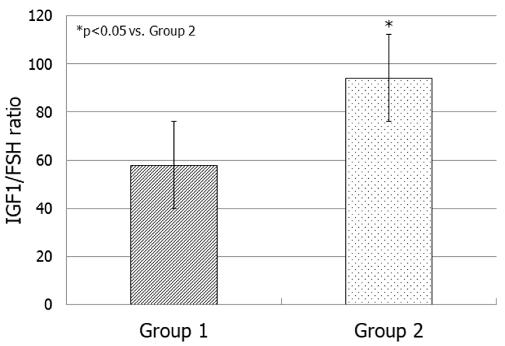
This study was conducted at the Division of Endocrinology, Metabolic Diseases and Nutrition of the University-Teaching Hospital Policlinico “G. Rodolico—San Marco”, University of Catania (Catania, Italy). The protocol was approved by the internal Institutional Review Board. Informed written consent was obtained from each participant after a full explanation of the purpose and nature of all procedures used. The study has been conducted following the principles expressed in the Declaration of Helsinki.

3. Results

Table 2 shows age, hormone values, TV, and conventional sperm parameters of the entire cohort.
We did not find any correlation between IGF1 and FSH serum values, nor between IGF1 and sperm concentration, TSC, or TV. Interestingly, the IGF1/FSH ratio showed a positive correlation with sperm concentration (r = 0.408; p = 0.004) (Figure 1, panel A), TSC (r = 0.468; p = 0.001) (Figure 1, panel B), and TV (0.463; p = 0.002) (Figure 1, panel C).
We then analyzed data from Group 1 (n = 14) and Group 2 (n = 45). Their baseline values are reported in Table 3. Group 1 showed a mean IGF1 value of 217.48 ± 11.01 ng/mL, while Group 2 presented a mean IGF1 value of 237.72 ± 11.13 ng/mL. These values did not significantly differ. These groups did not differ either for FSH levels (Table 3).
The IGF1/FSH ratio was significantly lower in Group 1 compared to Group 2 (57.9 ± 9.5 vs. 94.1 ± 8.7; p = 0.03) (Figure 2).

4. Discussion

The present study aimed at assessing the influence of IGF1 and of the IGF1/FSH ratio on sperm count and TV. The rational for the study lies in the emerging evidence coming from both granulosa and SCs and linking the IGF1 and FSH pathways together [12]. In greater detail, IGF1 seems to be required for FSH-mediated SC or spermatogonia proliferation in male newts, zebrafish, and mice [9,10,11]. Additionally, inhibition of IGF1R or of the INS1,which is linked to the IGF1R, has been found to reduce the FSH-signaling cascade [12,13]. In addition, while incubation of SCs with FSH only did not increase cell proliferation, incubation with IGF1 showed an increase in it. Interestingly, the extent of proliferation was even greater when both FSH and IGF1 were added to the culture, thus suggesting a synergic effect resulting from co-incubation with both hormones [17]. So far, no direct repercussion of these basic findings was reported in the clinical practice. For the first time in the literature, our study uses the IGF1/FSH ratio as an indicator of the level of both hormones. We found a positive correlation between the IGF1/FSH ratio and sperm concentration, TSC, and TV. Furthermore, patients with oligozoospermia showed a significantly lower ratio compared to those with a normal TSC, while neither IGF1 nor FSH differed significantly in the two groups. This may indicate that, in adulthood, IGF1 may influence spermatogenesis through a modulation of FSH signaling and effect in SCs.
The role of IGF1 in testicular function has been deeply investigated over the years, as supported by the huge amount of in vitro data showing the involvement of IGF1 in testicular development and function [5]. However, whether the IGF1 serum levels have a practical value in the diagnostic work-up of patients with oligozoospermia is still unclear. Clinical studies rarely concluded on the existence of a relationship between IGF1 and the sperm number. In a cohort of 202 Polish men, IGF1 has been reported to positively correlate with the sperm morphology, but, in agreement with our findings, no relationship with the sperm number was found [18]. In addition, a study carried out in 15 oligozoospermic patients and 15 normozoospermic controls found the presence of an altered responsiveness of the pituitary gland to the administration of the growth hormone-releasing hormone in patients than controls [19], suggesting the presence of a dysfunction of the GH-IGF1 axis in the oligozoospermic group. Futhermore, a study on 28 infertile patients and 20 fertile controls found an association between decreased sperm motility (but not sperm count) in asthenozoospermic group and levels of IGF1 in the seminal fluid [20].
Evidence for the involvement of IGF1 in the determination of the final TV is greater. Indeed, IGF1 serum levels positively correlate with TV in healthy men [6]. Furthermore, IGF1 administration in patients with Laron syndrome increases TV, penile size, gonadotropin, and testosterone serum levels [7]. IGF1 has also been related with testicular distance to pubic bone, which has been proposed as a marker of postnatal testicular function reflecting SC and Leydig cell health [21]. Moreover, an observational study found statistically different levels in IGF1/body mass index ratio among patients with congenital hypogonadotropic hypogonadism and constitutional delay of puberty [22], suggesting a role for IGF1 in human testicular function.
IGF1 seems to facilitate the onset of puberty, thus leading to an increase in TV. This is supported by the observation that normal children show physiologically high levels of IGF1—defined by the authors as “acromegalic” [23]. A retrospective analysis carried out in 93 patients with GH deficiency was performed to analyze whether treatment with GH could influence puberty onset and testicular growth in these patients. The authors found a significant TV growth over time, though no growth was observed in the age-matched and not yet treated controls, even when the age was compatible with a spontaneous start of puberty. Additionally, the duration of treatment and the mean dose of GH predicted the percentage of TV increase over time [24]. Likewise, patients with Laron syndrome (which are resistant to IGF1) show puberty delay and lower TV [25]. In vitro data on gonadotropin hormone-releasing hormone (GnRH) neurons found that incubation with IGF1 is capable of inducing both migration and GnRH secretion [26]. Therefore, the mechanisms by which IGF1 can induce TV growth during adolescence could be the following: (1) the induction of SC proliferation by a direct interaction with the FSH signaling [13,17]; the induction of GnRH neuron firing, which leads to the release of GnRH and gonadotropins [25,26].
The relevance of IGF1 in human fertility has been investigated also in women. IGF1, which is present in the follicular fluid, acts in combination with FSH [27], enhancing granulosa cell proliferation and aromatase activity and inhibiting follicle apoptosis [28]. Follicular development seems to be, at least partially, regulated by the IGF system. Indeed, IGF1 acts as a local amplifier of FSH action in the follicular fluid. Additionally, poor FSH responders have lower intra-follicular IGF1 concentrations [29], which might influence the ability of the follicles to respond to FSH. Treatment with IGF1 is costly. Therefore, GH administration has been alternately used to induce an increase in IGF1 levels in patients with poor ovarian response. Summarizing the evidence coming from meta-analysis of randomized controlled trials (RCTs), the probability of clinical pregnancy is increased with the addition of GH to FSH in poor ovarian responders [30,31]. Furthermore, the beneficial effects of GH on oocyte competence and embryo quality may be applied also to normal ovarian responder women [32].
In men, the effectiveness of GH administration in sperm output has been investigated, but its inefficacy has been documented most times. Only one study reported a normalization of conventional sperm parameters after GH therapy in boys with non-GH deficient short stature and constitutional delay of puberty [33]. Furthermore, combined treatment with gonadotropins and GH increased TV in four azoospermic patients with hypogonadotropic hypogonadism, but no result on azoospermia was achieved [34].
Study limitations include the relatively small cohort, although power analysis supports that the sample size is large enough to allow statistically significant conclusions. In addition, IGF1 isoforms could not be measured, thus adding another limitation to the interpretation of our data. Lastly, we did not assess IGF1 levels in the seminal fluid. Although this may be considered a limitation, IGF1 may influence the sperm counts likely acting on SCs. The latter are not exposed to the IGF1 levels in the seminal fluid due to the testis–blood barrier, but to the levels in the bloodstream. Further studies on larger cohorts and intervention studies should be encouraged to confirm our findings. In addition, our results may not be generalizable to men from the general population. Cross-sectional study design limits causal inference.

5. Conclusions

In conclusion, several studies have already investigated the relationship between IGF1 and semen parameters. However, clinical studies rarely concluded on the existence of a relationship between IGF1 and the sperm number, and whether the IGF1 serum levels have a practical value in the diagnostic work-up of patients with oligozoospermia is still unclear. For the first time in the literature, our study uses the IGF1/FSH ratio as an indicator of the level of both hormones. We found a positive correlation between the IGF1/FSH ratio and sperm concentration, TSC, and TV. Furthermore, patients with oligozoospermia showed a significantly lower ratio compared to those with a normal TSC, while neither IGF1 nor FSH differed significantly in the two groups. Our results may reflect the existence of a molecular pathway to which IGF1 and FSH belong and through which IGF1 might exert its influence on spermatogenesis. However, further studies are needed.

Author Contributions

R.C. selected the subjects, critically analyzed the data, drafted and wrote the manuscript. S.L.V. and R.A.C. performed the clinical analysis, contributed to interpretation of data and to the writing of manuscript. A.E.C. drafted and supervised the project and revised the manuscript critically. All authors have read and agreed to the published version of the manuscript.

Funding

This research did not receive any specific grant from funding agencies in the public, commercial, or not-for-profit sectors.

Institutional Review Board Statement

The study was conducted in accordance with the Declaration of Helsinki, and approved by the internal Institutional Review Board of the Division of Endocrinology, Metabolic Diseases and Nutrition of the University-Teaching Hospital Policlinico “G. Rodolico—San Marco”, University of Catania (protocol code 2021/03).

Informed Consent Statement

Informed consent was obtained from all subjects involved in the study.

Data Availability Statement

Data are available upon request to the corresponding author.

Conflicts of Interest

The authors declare that there is no conflict of interest that could be perceived as prejudicing the impartiality of the research reported.

References

  1. Cannarella, R.; Condorelli, R.A.; Duca, Y.; La Vignera, S.; Calogero, A.E. New insights into the genetics of spermatogenic failure: A review of the literature. Hum. Genet. 2019, 138, 125–140. [Google Scholar] [CrossRef] [PubMed]
  2. Itoh, N.; Nanbu, A.; Tachiki, H.; Akagashi, K.; Nitta, T.; Mikuma, N.; Tsukamoto, T.; Kumamoto, Y. Restoration of testicular transferrin, insulin-like growth factor-1 (IGF-1), and spermatogenesis by exogenously administered purified FSH and testosterone in medically hypophysectomized rats. Arch. Androl. 1994, 33, 169–177. [Google Scholar] [CrossRef] [PubMed] [Green Version]
  3. Naz, R.K.; Padman, P. Identification of insulin-like growth factor (IGF)-1 receptor in human sperm cell. Arch. Androl. 1999, 43, 153–159. [Google Scholar] [CrossRef] [PubMed]
  4. Minelli, A.; Moroni, M.; Castellini, C. Isolation and purification of the IGF-I protein complex from rabbit seminal plasma: Effects on sperm motility and viability. J. Exp. Zool. 2001, 290, 279–290. [Google Scholar] [CrossRef]
  5. Cannarella, R.; Condorelli, R.A.; La Vignera, S.; Calogero, A.E. Effects of the insulin-like growth factor system on testicular differentiation and function: A review of the literature. Andrology 2018, 6, 3–9. [Google Scholar] [CrossRef] [Green Version]
  6. Juul, A.; Bang, P.; Hertel, N.T.; Main, K.; Dalgaard, P.; Jørgensen, K.; Müller, J.; Hall, K.; Skakkebaek, N.E. Serum insulin-like growth factor-I in 1030 healthy children, adolescents, and adults: Relation to age, sex, stage of puberty, testicular size, and body mass index. J. Clin. Endocrinol. Metab. 1994, 78, 744–752. [Google Scholar]
  7. Laron, Z.; Klinger, B. Effect of insulin-like growth factor-I treatment on serum androgens and testicular and penile size in males with Laron syndrome (primary growth hormone resistance). Eur. J. Endocrinol. 1998, 138, 176–180. [Google Scholar] [CrossRef] [Green Version]
  8. Cannarella, R.; Mattina, T.; Condorelli, R.A.; Mongioì, L.M.; Pandini, G.; La Vignera, S.; Calogero, A.E. Chromosome 15 structural abnormalities: Effect on the IGF1R gene expression and function. Endocr. Connect. 2017, 6, 528–539. [Google Scholar] [CrossRef]
  9. Yamamoto, T.; Nakayama, Y.; Abé, S.I. Mammalian follicle-stimulating hormone and insulin-like growth factor I (IGF-I) up-regulate IGF-I gene expression in organ culture of newt testis. Mol. Reprod. Dev. 2001, 60, 56–64. [Google Scholar] [CrossRef]
  10. Pitetti, J.L.; Calvel, P.; Romero, Y.; Conne, B.; Truong, V.; Papaioannou, M.D.; Schaad, O.; Docquier, M.; Herrera, P.L.; Wilhelm, D.; et al. Insulin and IGF1 receptors are essential for XX and XY gonadal differentiation and adrenal development in mice. PLoS Genet. 2013, 9, e1003160. [Google Scholar] [CrossRef] [Green Version]
  11. Nóbrega, R.H.; Morais, R.D.; Crespo, D.; de Waal, P.P.; de França, L.R.; Schulz, R.W.; Bogerd, J. Fsh Stimulates Spermatogonial Proliferation and Differentiation in Zebrafish via Igf3. Endocrinology 2015, 156, 3804–3817. [Google Scholar] [CrossRef] [PubMed]
  12. Law, N.C.; Hunzicker-Dunn, M.E. Insulin Receptor Substrate 1, the Hub Linking Follicle-stimulating Hormone to Phosphatidylinositol 3-Kinase Activation. J. Biol. Chem. 2016, 29, 4547–4560. [Google Scholar] [CrossRef] [PubMed] [Green Version]
  13. Cannarella, R.; Arato, I.; Condorelli, R.A.; Luca, G.; Barbagallo, F.; Alamo, A.; Bellucci, C.; Lilli, C.; La Vignera, S.; Calafiore, R.; et al. The IGF1 Receptor Is Involved in Follicle-Stimulating Hormone Signaling in Porcine Neonatal Sertoli Cells. J. Clin. Med. 2019, 27, 577. [Google Scholar] [CrossRef] [Green Version]
  14. World Health Organization. WHO Laboratory Manual for the Examination and Processing of Human Semen, 5th ed.; Cambridge University Press: Cambridge, UK, 2010. [Google Scholar]
  15. Sakamoto, H.; Yajima, T.; Nagata, M.; Okumura, T.; Suzuki, K.; Ogawa, Y. Relationship between testicular size by ultrasonography and testicular function: Measurement of testicular length, width, and depth in patients with infertility. Int. J. Urol. 2008, 15, 529–533. [Google Scholar] [CrossRef] [PubMed]
  16. Pilatz, A.; Rusz, A.; Wagenlehner, F.; Weidner, W.; Altinkilic, B. Reference values for testicular volume, epididymal head size and peak systolic velocity of the testicular artery in adult males measured by ultrasonography. Ultraschall Med. 2013, 34, 349–354. [Google Scholar] [CrossRef] [PubMed]
  17. Cannarella, R.; Mancuso, F.; Condorelli, R.A.; Arato, I.; Mongioì, L.M.; Giacone, F.; Lilli, C.; Bellucci, C.; La Vignera, S.; Calafiore, R.; et al. Effects of GH and IGF1 on Basal and FSH-Modulated Porcine Sertoli Cells In-Vitro. J. Clin. Med. 2019, 8, 811. [Google Scholar] [CrossRef] [Green Version]
  18. Kamieniczna, M.; Fraczek, M.; Malcher, A.; Rozwadowska, N.; Czernikiewicz, A.; Jedrzejczak, P.; Semczuk, M.; Kurpisz, M. Semen Quality, Hormonal Levels, and Androgen Receptor Gene Polymorphisms in a Population of Young Male Volunteers from Two Different Regions of Poland. Med. Sci. Monit. 2015, 21, 2494–2504. [Google Scholar] [CrossRef] [Green Version]
  19. Rajmil, O.; Rodríguez-Espinosa, J.; Sarquella, J.; Castellet, R.; Oliver, A.; Queraltó, J.M. Growth hormone response to growth hormone-releasing hormone stimulation in oligozoospermic patients. Fertil. Steril. 1994, 62, 1039–1043. [Google Scholar] [CrossRef]
  20. Fu, L.; Yuen, K.C.J.; Tint, A.N.; Hoffman, A.R.; Bongso, A.T.; Lee, K.O. Association of decreased sperm motility and increased seminal plasma IGF-I, IGF-II, IGFBP-2, and PSA levels in infertile men. Endocrine 2021, 74, 698–706. [Google Scholar] [CrossRef]
  21. Koskenniemi, J.J.; Virtanen, H.E.; Wohlfahrt-Veje, C.; Löyttyniemi, E.; Skakkebaek, N.E.; Juul, A.; Andersson, A.M.; Main, K.M.; Toppari, J. Postnatal changes in testicular position are associated with IGF-I and Function of Sertoli and Leydig Cells. J. Clin. Endocrinol. Metab. 2018, 103, 1429–1437. [Google Scholar] [CrossRef] [Green Version]
  22. Rohayem, J.; Nieschlag, E.; Kliesch, S.; Zitzmann, M. Inhibin B, AMH, but not INSL3, IGF1 or DHEAS support differentiation between constitutional delay of growth and puberty and hypogonadotropic hypogonadism. Andrology 2015, 3, 882–887. [Google Scholar] [CrossRef] [PubMed]
  23. Juul, A.; Skakkebæk, N.E. Why Do Normal Children Have Acromegalic Levels of IGF-I During Puberty? J. Clin. Endocrinol. Metab. 2019, 104, 2770–2776. [Google Scholar] [CrossRef]
  24. Cannarella, R.; Caruso, M.; Crafa, A.; Timpanaro, T.A.; Lo Bianco, M.; Presti, S.; Condorelli, R.A.; La Vignera, S.; Calogero, A.E. Testicular Growth and Pubertal Onset in GH-Deficient Children Treated With Growth Hormone: A Retrospective Study. Front. Endocrinol. 2021, 12, 619895. [Google Scholar] [CrossRef] [PubMed]
  25. Cannarella, R.; Crafa, A.; La Vignera, S.; Condorelli, R.A.; Calogero, A.E. Role of the GH-IGF1 axis on the hypothalamus-pituitary-testicular axis function: Lessons from Laron syndrome. Endocr. Connect. 2021, 10, 1006–1017. [Google Scholar] [CrossRef] [PubMed]
  26. Cannarella, R.; Paganoni, A.J.J.; Cicolari, S.; Oleari, R.; Condorelli, R.A.; La Vignera, S.; Cariboni, A.; Calogero, A.E.; Magni, P. Anti-Müllerian Hormone, Growth Hormone, and Insulin-Like Growth Factor 1 Modulate the Migratory and Secretory Patterns of GnRH Neurons. Int. J. Mol. Sci. 2021, 22, 2445. [Google Scholar] [CrossRef] [PubMed]
  27. Adashi, E.Y.; Resnick, C.E.; D’Ercole, A.J.; Svoboda, M.E.; Van Wyk, J.J. Insulin-like growth factors as intraovarian regulators of granulosa cell growth and function. Endocr. Rev. 1985, 6, 400–420. [Google Scholar] [CrossRef] [PubMed]
  28. Hsueh, A.J.; Eisenhauer, K.; Chun, S.Y.; Hsu, S.Y.; Billig, H. Gonadal cell apoptosis. Recent Prog. Horm Res. 1996, 51, 433–455. [Google Scholar]
  29. Oosterhuis, G.J.; Vermes, I.; Lambalk, C.B.; Michgelsen, H.W.; Schoemaker, J. Insulin-like growth factor (IGF)-I and IGF binding protein-3 concentrations in fluid from human stimulated follicles. Hum. Reprod. 1998, 13, 285–289. [Google Scholar] [CrossRef] [Green Version]
  30. Kolibianakis, E.M.; Venetis, C.A.; Diedrich, K.; Tarlatzis, B.C.; Griesinger, G. Addition of growth hormone to gonadotrophins in ovarian stimulation of poor responders treated by in-vitro fertilization: A systematic review and meta-analysis. Hum. Reprod. Update 2009, 15, 613–622. [Google Scholar] [CrossRef] [Green Version]
  31. Li, X.L.; Wang, L.; Lv, F.; Huang, X.M.; Wang, L.P.; Pan, Y.; Zhang, X.M. The influence of different growth hormone addition protocols to poor ovarian responders on clinical outcomes in controlled ovary stimulation cycles: A systematic review and meta-analysis. Medicine 2017, 96, e6443. [Google Scholar] [CrossRef]
  32. Bosch, E.; Labarta, E.; Kolibianakis, E.; Rosen, M.; Meldrum, D. Regimen of ovarian stimulation affects oocyte and therefore embryo quality. Fertil. Steril. 2016, 105, 560–570. [Google Scholar] [CrossRef] [PubMed] [Green Version]
  33. Radicioni, A.F.; Paris, E.; De Marco, E.; Anzuini, A.; Gandini, L.; Lenzi, A. Testicular function in boys previously treated with recombinant-human growth hormone for non-growth hormone-deficient short stature. J. Endocrinol. Investig. 2007, 30, 931–936. [Google Scholar] [CrossRef] [PubMed]
  34. Giagulli, V.A. Absence of effect of recombinant growth hormone to classic gonadotropin treatment on spermatogenesis of patients with severe hypogonadotropic hypogonadism. Arch. Androl. 1999, 43, 47–53. [Google Scholar] [CrossRef] [PubMed]
Figure 1. Correlation of IGF1/FSH ratio and sperm concentration, total sperm count, and testicular volume. IGF1/FSH positively correlate with sperm concentration (panel A), total sperm count (panel B), and testicular volume (panel C) in the entire cohort (n = 59).
Endocrines 03 00053 g001
Figure 2. IGF1/FSH ratio in Group 1 and Group 2. The IGF1/FSH ratio is lower in patients with oligozoospermia (Group 1, n = 14), compared to men with normal total sperm count (Group 2, n = 45).
Endocrines 03 00053 g002
Table 1. Reference values.
IGF1
(Male, Age)
Normal Range
16–20 years119–395 ng/mL
20–25 years127–298 ng/mL
21–40 years99–238 ng/mL
41–55 years82–214 ng/mL
>55 years61–177 ng/mL
Table 2. Range and baseline values of the entire cohort.
ParametersBaseline Value
(Mean ± SD)
Range of Values
(Min-max)
Age (year)30.95 ± 8.417–54
GH (ng/mL)0.32 ± 0.430.05–1.42
IGF1 (ng/mL)232.59 ± 65.13125.6–401
FSH (mIU/mL)3.95 ± 2.550.15–15.34
LH (mIU/mL)3.36 ± 1.820.15–11
TT (ng/mL)5.54 ± 2.150.22–11.81
Total TV (mL)27.72 ± 7.896.6–47.5
Sperm concentration (million/mL)40.94 ± 34.061–150
Total sperm count (million/ejaculate)107.54 ± 84.490.2–380
Sperm progressive motility (%)21.92 ± 8.723–41
Sperm total motility (%)57 ± 10.5210–74
Sperm normal morphology (%)7.34 ± 4.611–21
Abbreviations: FSH = follicle-stimulating hormone; LH = luteinizing hormone; TT = total testosterone; TV = testicular volume.
Table 3. Range and baseline values of Groups 1 and 2.
ParametersGroup 1
(TSC < 39 Million/Ejaculate)
Group 2
(TSC ≥ 39 Million/Ejaculate)
Age (year)29.13 ± 8.4431.43 ± 8.57
FSH (mIU/mL)4.80 ± 23.78 ± 2.61
LH (mIU/mL)3.46 ± 1.833.39 ± 1.77
TT (ng/mL)4.87 ± 2.215.84 ± 1.95
Total TV (mL)24.45 ± 10.0628.84 ± 6.81
Sperm concentration (million/mL)8.59 ± 7.99 *51.01 ± 32.76
Total sperm count (million/ejaculate)17.92 ± 10.55 *135.42 ± 77.64
Sperm progressive motility (%)19.63 ± 12.0822.53 ± 7.74
Sperm total motility (%)50.25 ± 17.82 *58.8 ± 6.98
Sperm morphology (%)6.13 ± 3.987.67 ± 4.77
Abbreviations: FSH = follicle-stimulating hormone; LH = luteinizing hormone; TSC = total sperm count; TT = total testosterone; TV = testicular volume. Results are expressed as mean value ± standard deviation. * p < 0.05.
Publisher’s Note: MDPI stays neutral with regard to jurisdictional claims in published maps and institutional affiliations.

