Review

Different Mechanisms in Doxorubicin-Induced Cardiomyopathy: Impact of BRCA1 and BRCA2 Mutations

by
Hien C. Nguyen
,
Jefferson C. Frisbee
and
Krishna K. Singh
*
Department of Medical Biophysics, Schulich School of Medicine and Dentistry, University of Western Ontario, 1151 Richmond St. N., London, ON N6A 5C1, Canada
*
Author to whom correspondence should be addressed.
Hearts 2024, 5(1), 54-74; https://doi.org/10.3390/hearts5010005
Submission received: 23 December 2023 / Revised: 16 January 2024 / Accepted: 18 January 2024 / Published: 23 January 2024
(This article belongs to the Collection Feature Papers from Hearts Editorial Board Members)

Abstract

:
Germline mutations in Breast cancer susceptibility genes 1 and 2 (BRCA1 and BRCA2) cause breast, ovarian, and other cancers, and the chemotherapeutic drug doxorubicin (Dox) is widely used to treat these cancers. However, Dox use is limited by the latent induction of severe cardiotoxicity known as Dox-induced cardiomyopathy, for which there are no specific treatments currently available. Dox is administered into the systemic circulation, where it readily translocates into sub-cellular compartments and disrupts the integrity of DNA. Accumulating evidence indicates that oxidative stress, DNA damage, inflammation, and apoptosis all play a central role in Dox-induced cardiomyopathy. The BRCA1 and BRCA2 proteins are distinct as they perform crucial yet separate roles in the homologous recombination repair of DNA double-strand breaks, thereby maintaining genomic integrity. Additionally, both BRCA1 and BRCA2 mitigate oxidative stress and apoptosis in both cardiomyocytes and endothelial cells. Accordingly, BRCA1 and BRCA2 are essential regulators of pathways that are central to the development of cardiomyopathy induced by Doxorubicin. Despite extensive investigations, there exists a gap in knowledge about the role of BRCA1 and BRCA2 in Doxorubicin-induced cardiomyopathy. Here, we review the previous findings and associations about the expected role and associated mechanisms of BRCA1 and 2 in Dox-induced cardiomyopathy and future perspectives.

1. Introduction

Doxorubicin (Dox), an anthracycline antibiotic derived from Streptomyces peucetius var. caesius, is a potent geno- and cytotoxic medication used to treat various cancers. Dox is commonly prescribed with a strict regimen for, including but not limited to, breast, lung, and ovarian cancers [1]. Approximately 75% of administered Dox circulates in the blood via binding to plasma proteins [2]. Dox’s average terminal half-life is 20–48 h [3,4]. Upon clearance, ~50% of the administered dose is eliminated from the body unchanged, with 5–12% appearing in urine and 40% in bile within a week. Notably, men have higher Dox clearance compared to women, indicating a higher vulnerability to Dox effects in women vs. men [5]. Dox is designed to target rapidly dividing cells, but the cytotoxicity induced by Dox is wide-scale, devastating virtually all cellular growth, survival, and proliferating processes. The drug’s potent cytotoxic mechanisms are mainly summarized as two tiers: (1) DNA impairment and (2) inducing oxidative stress. Accordingly, multiple novel mechanisms in these categories have recently emerged to substantiate Dox’s potent toxicity in recipients [6]. The genomic impacts of Dox are more specific to the drug’s effects and encompass torsional stress, bombarding DNA strand breaks, and impairing replication, topoisomerase’s activity, DNA repair, and gene expression, ultimately inducing cell cycle arrest and apoptosis.
Although the oxidative impact is less specific to Dox’s pharmacodynamics, Dox-induced oxidative stress is a common feature of the drug’s cytotoxicity, featuring a notable excessive and devastating promotion of reactive oxygen species (ROS) and ROS-associated radicals, such as reactive nitrogen species (RNS), superoxide anions, hydrogen peroxide, etc. For instance, Dox-induced apoptosis results in the assembly of micronuclei, which are clusters of genomic fragments known to activate the cGAS-STING pathway that upregulates the inflammatory machinery and, thereby, ROS production [7]. In other cases, as elaborated later, Dox itself upregulates the bioactivities of radicals. While the cells innately metabolize and neutralize radicals via antioxidant enzymes such as catalase and superoxide dismutase, adverse phenotypes typically indicate that radical accumulation has overwhelmed the antioxidant’s mechanisms. Typical symptoms of Dox-induced oxidative stress include cellular injuries characterized by the destruction of cellular components at all biomolecular levels (e.g., membrane damage, lipid peroxidation, DNA damage, and oxidized protein aggregates) [8].
Notably, the body metabolizes Dox into doxorubicinol, which can undergo spontaneous reduction to form doxorubicin-deoxyaglycone and then quinone-methide, which potently produce radicals from covalently binding to DNA. Moreover, Dox is also metabolized into the doxorubicin-semiquinone radical by enzymes including cytochrome p450, xanthine oxidase, NADPH oxidases (NOX), and NO synthases (NOS) [7,8]. In addition to being a source of ROS, accumulating radicals can re-oxidize the semiquinone radicals back to doxorubicin, amplifying the drug’s cytotoxicity [9].
In this sense, Dox’s cytotoxicity is non-specific, and the adverse clinical implications of Dox’s molecular mechanisms remain a subject of intensive research. Indeed, Dox has been associated with an increased risk of secondary malignancies, reproductive organ toxicity, and infertility. Most notable are complications in women, which include amenorrhea, premature menopause, fetal harm in administered pregnant women, and birth defects such as prepubertal growth failure and gonadal impairment [6,10]. Among a conglomeration of reported adverse events, Dox-induced cardiotoxicity is the most concerning complication, as it is relatively common and leads to cardiovascular dysfunction. For this reason, Dox is excluded for patients with poor heart function, and treatments are discontinued once the maximally tolerated cumulative dose is reached [8].

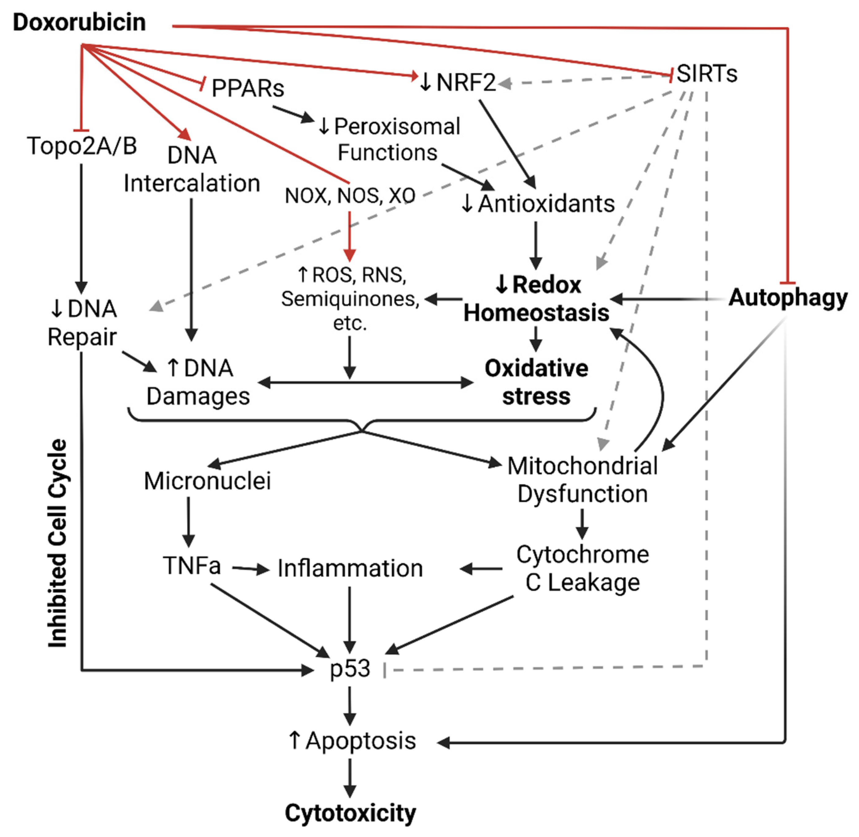
1.1. Dox-Induced Cardiotoxicity (DIC)

Dox-induced cardiotoxicity (DIC) is notably common in patients receiving Dox treatment and typically manifests as cardiomyocyte injuries subjected to the mechanisms of Dox that were meant for neoplastic cells. Clinically, DIC is either acute or chronic, depending on the dosing regimen, age, and cardiovascular health of the patients. The recent incidence rate of acute DIC is ~30% and typically detected 2–3 days after Dox administration, with ~50% mortality after 1-year diagnosis. Acute DIC is reversible and often manifests with tachycardia and ventricular premature beats shortly following administration. Histological features of acute DIC in the myocardium include interruption of myofibrils, cytoplasmic vacuolation, and sparsity in cardiomyocytes [11]. The incidence of chronic DIC is ~2–20% and typically manifests as a complication of breaching Dox dosage tolerance after weeks or months of administration, featuring irreversible cardiomyopathy, prominent left ventricular enlargement, and heart failure [10,12,13]. The mechanisms of DIC are summarized in Figure 1.

1.2. Intercalation of DNA and Topoisomerase II Inhibition

It has been generally established that free radical-associated DNA damage is more likely to occur when Dox concentration breaches the tolerance dosage [14]. Instead, Dox-associated cytotoxic impact on the cell’s genome is often attributed to DNA intercalation and Dox-dependent topoisomerase II inhibition [15,16]. Notably, considering the highly oxidative environment subjected to cardiac tissues by default, both mechanisms likely damage genomic integrity to a similar degree of significance in DIC.
The drug’s potent mechanism is DNA intercalation, achieved via its anthraquinone ring. Dox inserts itself between DNA bases and stabilizes the interaction through hydrogen bonding, facilitated by its hydroxyl and daunosamine sugar’s amino groups. Dox-DNA complexes induce torsional stress in the DNAs superhelical structures, leading to double-strand breaks and apoptosis. In addition, Dox can bind and inhibit DNA and RNA polymerases, disrupting replication, transcription, and DNA repair [16].
An additional Dox-associated cytotoxic impact on the cell’s genome is topoisomerase II inhibition [15]. Topoisomerase II (TopoII) is a well-characterized enzyme that functions in replication (TopoIIa) or DNA repair and gene transcription (TopoIIb). Typically, it is recruited to associate with the DNA strand and induce coordinated DNA double-strand breaks as a response to alleviate torsional stress, relax positive supercoils, and unlink intertwined strands, in turn stabilizing the DNAs superhelical state. TopoII inhibition is also Dox’s mechanism designed for rapidly proliferating cells. Dox binds and inhibits both TopoIIa and IIb during their actions, forming Dox-TopoII-DNA complexes that induce unaccounted-for double-strand breaks, hindering replication and gene expression, and resulting in cell death [15]. Indeed, Dox efficacy is dependent on TopoII concentration in the cells [2]. Dox-TopoII-DNA complexes can be reversed by the dissociation of Dox upon turnover, suggesting that Dox exposure duration is a key factor in Dox cytotoxicity [17].
The genomic impacts of Dox are typically observed at 0.01–5 μM Dox concentrations, followed by p53 upregulation [2]. p53 is constitutively expressed and preferably suppressed in cells. Its activation regulatory competes with various cell growth and DNA repair pathways; hindrance in the cell’s growth/survival (e.g., downregulated E2F, the growth transcription factor) and/or improper DNA repair promote p53 activation [18,19]. Activated p53 facilitates cell cycle arrest and programmed cell death [20]. While Dox is designed to tarnish cellular replication and prompt p53’s pro-apoptotic action in the neoplastic cell population, in DIC, Dox-treated cardiac tissues demonstrate impaired vital gene expression, DNA damage, and dysregulated p53, among other complications [21,22]. In this notion, genetic and pharmacologic inhibition of p53 have been shown to attenuate acute DIC [23,24,25,26,27]; however, disrupted p53 activity has been associated with exacerbated cardiac dysfunction in animal models [22] and is also a feature of Dox-resistant tumor cells [28], suggesting a selectivity challenge for p53 targeting therapeutic strategies.
Moreover, in DIC, while cardiomyocytes are non-dividing cells, they preferentially express TopoIIb over TopoIIa. This expression is adopted by both the cardiomyocytes’ genomes and mitochondria. A single cardiomyocyte can have between 5000 and 8000 mitochondria, and mitochondria are vital for cardiac function, regulating essential lipid oxidation and redox balance [29]. Therefore, Dox’s inhibiting TopoIIb and the formation of Dox-TopoIIb-DNA complexes in cardiomyocytes promote DNA damage and oxidative stress as a feature of DIC.

1.3. NADPH Oxidases, Nitric Oxide Synthases, and Xanthine Oxidase

DICs oxidative stress has been well characterized by three enzymes: NADPH oxidases (NOXs), nitric oxide synthases (NOSs), and xanthine oxidases (XOs), which were previously mentioned as metabolizers of Dox into semiquinone radicals. In addition to metabolizing Dox into radical intermediates, these enzymes pathologically contribute to oxidative stress by facilitating the production of oxidative radicals [30,31,32]. However, targeting these enzymes clinically remains under consideration due to conflicting observations attributed to their physiological oxidative significance. For instance, as XO mediates oxygen radical generation and exacerbates DIC in Dox-treated mice [33], febuxostat, an inhibitor of XO that reduces ROS production in the myocardium, has yet to be used clinically [34,35]. The NOXs enzymes are significant sources of superoxide production, with NOX2 and NOX4 being the primary contributors to Dox-induced oxidative stress [36]. The antioxidant treatments irisin and osteocrin inhibit NOX2 and NOX4, respectively, and result in the attenuation of Dox-induced oxidative stress in the heart [37,38]. However, NOXs inhibitors have yet to be considered clinically as their oxidative roles have physiological significance [39]. NOX2-deficient mice on high-fat diets developed severe glucose metabolism disorders, suggesting that the NOX enzymes may be a sensitive target to modulate Dox cardiotoxicity [40].
Nitric oxide (NO) derived from NOS enzymes can interact with concurrent radicals to produce more radicals, such as the peroxynitrite anion (ONOO-). Indeed, available antioxidant treatments for Dox-induced cardiotoxicity involve altering NOSs expression, such as levosimendan [41] and vitamin C [42]. Dox-treated cardiomyocytes show increased inducible NOS (iNOS) expression, and iNOS-deficient mice show attenuated Dox-induced generation of ONOO [43]. However, studies have also shown that the lack of NO bioavailability exacerbates Dox-induced cardiotoxicity [44]. Notably, endothelial NOS (eNOS) catalyzes the biological synthesis of NO, which is an essential regulator of endothelial function and vasotone [45]. Indeed, eNOS-deficient and overexpressing mice showed reduced and increased susceptibility to Dox-induced oxidative stress, respectively [46]. On the other hand, Dox has been shown to impair eNOS activation while facilitating ROS-mediated oxidative stress [47]. Moreover, eNOS-deficient female mice were shown to have aggravated Dox-induced oxidative stress and cellular damage [48].
Overall, it appears that by engaging in oxidation and Dox metabolism, NOX, NOS, and XO may undesirably exacerbate Dox-induced oxidative stress. However, whether Dox induces the activities of these enzymes requires further investigation, and conflicting results have yet to point out a clear implication for modulating these enzymes as a strategy to protect against DIC.

1.4. Antioxidants in Dox-Induced Cardiotoxicity

Relatively, cardiac tissues engage in an overall highly oxidative metabolic environment to sustain their essential systemic function. Approximately 70% of the energy in the heart is derived from the oxidation of fatty acids within the mitochondria and peroxisomes, which rely heavily on lipid-trafficking mechanisms [49]. Naturally, cardiomyocytes express sensitive levels of endogenous antioxidant enzymes (e.g., superoxide dismutase, glutathione peroxidase, glutathione S-transferase, heme oxygenase-1, catalase, etc.) [50]. Tipping the redox balance, as in antioxidant deficiency, easily renders cardiac tissues vulnerable to oxidative stress. Indeed, Dox impairs the antioxidants’ bioavailability in cardiomyocytes, depending on the duration and dosage of Dox exposure [51]. Conversely, increasing antioxidants’ activity enhances the cells’ redox capacity, obstructs free radical-related injury to DNA, and attenuates DIC [52,53]. However, adjusting antioxidant bioavailability remains a challenge in the clinical setting of DIC. For instance, apigenin, which enhances antioxidants in cardiomyocytes, has yet to be considered for clinical practice [54]. Nrf2, a transcription factor regulating antioxidant enzymes, is downregulated in DIC, which is rescued by the yet-to-be clinically implemented irisin, a benefit additional to the drug’s inhibition of NOX2 that mitigates oxidative DIC [55].

1.5. Peroxisomes in Dox-Induced Cardiotoxicity

Cellular peroxisomes are organelles that maintain redox and lipid homeostasis via fatty acid β-oxidation and detoxification of metabolic byproducts, such as hydrogen peroxide (H2O2), polyamines, and glyoxylate, as well as several xenobiotics [56]. Notable peroxisomal enzymes include Catalase and Peroxidases, such as the Peroxiredoxins, that process various oxidative radicals into water and oxygen, thereby maintaining oxidative balance. Dysfunctional peroxisomes, as in developing Niemann–Pick type C disease [57], lead to radical accumulation, oxidative stress, and the downstream risk of multiple-organ failures. Indeed, aging cells, which become progressively peroxisomal inefficient, feature elevated ROS levels [58]. Conversely, overexpressing peroxiredoxin-1 attenuates oxidative stress and DIC in cardiomyocytes [59,60].
As oxidative stress and inflammation typically occur together, peroxisomal functions are closely linked to the inflammatory response. Likewise, chronic inflammation also features elevated ROS and oxidative cellular injuries. Moreover, peroxisomal enzymes also facilitate the degradation of pro-inflammatory mediators (e.g., prostaglandins, thromboxanes, leukotrienes, and prostacyclins) and the biogenesis of anti-inflammatory metabolites (e.g., omega-3 fatty acids) [61]. Notably, omega-3 fatty acids are precursors of potent anti-inflammatory factors such as resolvins, maresins, and protectins [62]; enhancing the conversion of omega-3 fatty acids to these molecules is a feature of non-steroidal anti-inflammatory drugs (NSAIDs), such as aspirin [63].
Mitigating DIC approaches from peroxisome functions have heavily focused on the peroxisome proliferator-activated receptors (PPARs). The PPARs are a family of nuclear transcription factors that, upon activation, upregulate peroxisome proliferators, which induce an increase in cellular peroxisome number, size, and functions [64]. In humans, three closely related PPAR subtypes have been identified. PPAR-δ is expressed ubiquitously and at higher levels than the other two. PPAR-γ is mainly found in adipose tissues and, to a lesser extent, immune cells (monocytes, macrophages, etc.). PPAR-α is rich in hepatocytes, cardiomyocytes, skeletal muscles, and other peripheral tissues with active lipid oxidation. Notably, of the three, PPAR-α exerts the highest affinity for lipids, regulating the escorts of unsaturated and saturated fatty acids via cytosolic fatty-acid binding proteins to the peroxisomes and mitochondria for lipid β-oxidation [65].
Dox inhibits PPARγ in the adipose tissues of mice, leading to the loss of the storage of blood glucose and lipid, thereby causing hyperglycemia and hyperlipidemia, which are high-risk factors for insulin resistance, atherogenesis, and cardiovascular diseases [66]. PPAR-α-null mice exhibit a loss of fatty acid oxidative capacity, leading to increased lipid accumulation, reduced ketone bodies, a lack of gluconeogenesis, and metabolic switching to fatty acid usage in the heart during starvation. These culminate in cardiac dysfunction, myocardial damage, and fibrosis [67]. The heart of tumor-bearing Dox-treated mice also showed inhibited PPARα, but such is absent in their tumors; conversely, the same paper demonstrates that fenofibrate (FENO) treatment, an agonist of PPARα, and overexpression of PPARα in these mice enhanced cardiac function and salvaged DIC without affecting tumor progression [68]. Notably, FENO was shown to attenuate DIC in mice by improving endothelial function and upregulating eNOS expression and activation via Akt [69]. Outside of modulating the PPARs, there are little to no studies addressing the role of peroxisomes more directly for clinical applications against DIC [60].

1.6. Sirtuins Deacetylate Dox-Induced Cardiotoxicity

The Sir2 and Sir2-like proteins, together referred to as sirtuins (SIRTs), are NAD-dependent deacetylase enzymes that make up the evolutionarily conserved class III histone-deacetylases in humans [70]. A distinct NAD/FAD-binding domain characterizes the class III HDACs. In humans, seven sirtuins have been reported and localized: the nucleus (SIRT1, SIRT2, SIRT3, SIRT6, SIRT7), cytoplasm (SIRT1, SIRT2), and mitochondria (SIRT3, SIRT4, SIRT5) [70].
The SIRTs have been reported to regulate aging, metabolism, inflammation, apoptosis, and maintaining redox balance in cardiac cells [71]. SIRT1 has been linked to cardiovascular diseases [72]. SIRT3, 6, and 7 regulate aging, apoptosis, and oxidative stress in cardiomyocytes and are linked to cardiac hypertrophy [73,74,75]. The roles of SIRT4 and 5 in the heart remain under-investigated.
SIRT3 deacetylase is expressed abundantly in cardiomyocytes and plays a crucial role in regulating mitochondrial function, proliferation, and maintenance of the mitochondrial genome, all of which are essential for cardiac metabolism. The mitochondria also engage in lipid oxidation, similar to peroxisomes, regulating the redox balance. Notably, ~20% of mitochondrial proteins are regulated via reversible lysine acetylation [76], and the mitochondria contain high levels of NAD and NADH [77]. Mitochondrial acyl-CoA dehydrogenase and synthase facilitate long-chain fatty acid oxidation and, thereby, lipid metabolism in cardiomyocytes [78]. SIRT3 regulates these enzymes and also those of the tricarboxylic acid cycle, electron transport chain subunits, and ATP synthase [79]. SIRT3 also regulates mitochondrial ROS formation via the antioxidants SOD2 [80] and Ku70, a factor of the non-homologous end-joining DNA repair pathway [81]. SIRT3 knock-down animals exhibit a high risk for cardiac hypertrophy, oxidative stress, diminished cardiac ATP, and increased mitochondrial fragmentation [82,83,84]. Dox-treated cardiomyocytes exhibit mitochondrial dysfunction that contributes to oxidative stress and impaired lipid metabolism, as featured in DIC [85]. Resveratrol, which activates SIRT3, in co-treatment with Dox, attenuates mitochondrial ROS production [86,87].
Cardiomyocytes adopt protein acetylation/deacetylation in metabolic regulation. Deacetylation of p53 prompts its ubiquitination and subsequent degradation, thereby promoting survival, and class III deacetylases also regulate several antioxidant enzymes. Dox significantly suppressed several SIRT deacetylases in the myocardium, abolishing antioxidants and exacerbating ROS production and apoptosis [88]. Upregulating the SIRT enzymes restores p53 ubiquitination, reduces caspase-3 activation, promotes Nrf2 and antioxidant enzymes, attenuates Dox-induced oxidative stress, and salvages DIC [88,89,90]. Interestingly, endothelial cells also highly express the SIRT enzymes [91]. Overall, the SIRT enzymes regulate cardiomyocytes’ metabolism, redox, and genomic integrity, all of which are aspects impaired in Dox-induced cardiotoxicity. Despite these findings, there remains a lack of SIRT activators developed for clinical therapy.

1.7. Dox Impairs Autophagy

2-Hydroxypropyl-β-cyclodextrin (HPβCD), a proposed treatment for Niemann–Pick type C disease characterized by impaired lipid metabolism, is an activator of the transcription factor TFEB that upregulates autophagy [92]. Co-treatment of HPβCD in Dox-treated neurons, thereby activating autophagy, attenuates peroxisome-associated ROS accumulation, reducing neurotoxicity [93].
All cells continually engage in autophagy (“self-eating”), an essential process that encompasses the encapsulation of the cell’s own macromolecules and organelles in the cytoplasm, followed by the lysosomal digestion of these biowastes into metabolic materials (e.g., amino acids, nucleic acids, phospholipids, etc.) [94,95]. Biowastes of autophagy include mRNAs, turnover proteins, toxic misfolded aggregates, desensitized receptors, and dysfunctional organelles such as abnormally proliferated mitochondria and peroxisomes. Disruption of autophagy leads to accumulating cytotoxicity, resulting in dysfunction and rapidly incapacitating metabolically stressed cells. As apoptosis is an energy-demanding process, this resulting cytotoxicity likely prompts necrosis and tissue inflammation, culminating in organ failures [96,97].
Autophagy is an emerging field in DIC research. Dox impairs autophagic flux at all stages. Dox inhibits various stress response factors that inhibit mTOR [98], thereby inhibiting autophagy. Dox-treated myocardium shows reduced numbers of autophagosomes and autolysosomes. On the other hand, Dox can also inhibit mTOR itself or modulate intermediate mediators, leading to excessive autophagosomes and autolysosomes, thereby dysregulating autophagy [87,99]. Dox-induced ROS accumulation also impairs lysosomal acidification and enzyme activity, resulting in the accumulation of autolysosomes [87]. Dysregulated autophagy, in turn, leads to mitochondrial dysfunction, impaired cellular metabolism, and apoptosis.

1.8. Dox Induces Sarcoplasm Leakage

Under Dox exposure, the myocardium exhibits cellular swelling, cytoplasmic vacuolation, myofibril disruption, and other characteristics of cardiac dysfunction [100]. Most notable is the severe dilation of the sarcoplasm (SR), followed by Ca2+ leakage into the cytoplasm, upsetting the cell ion-tonicity, impairing contractility, and potentiating ROS production [101]. In cardiomyocytes, Dox can increase CaMKII phosphorylation, which promotes the opening of RyR2 clusters on the SR, enabling Ca2+ leakage [102].

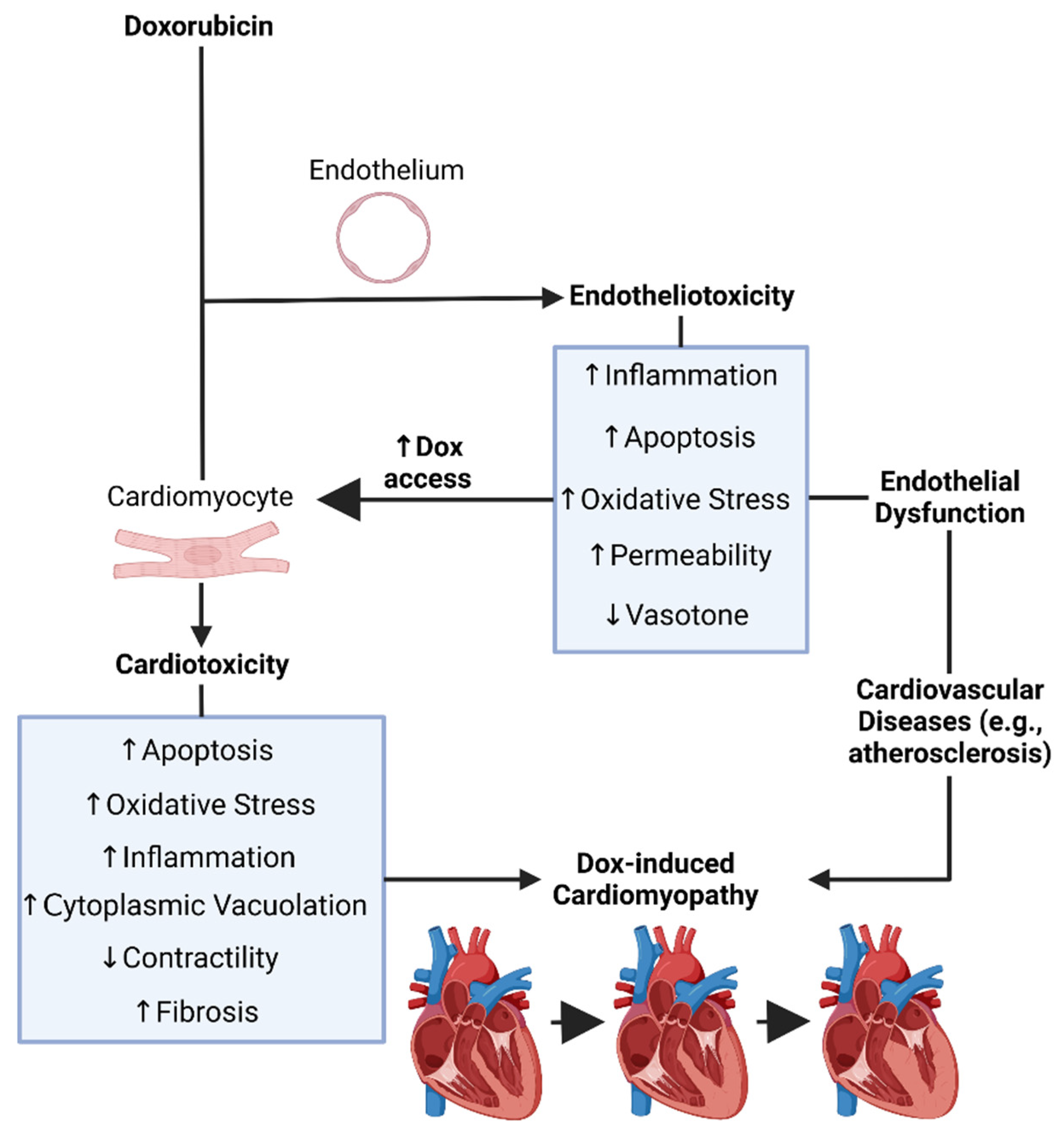
1.9. Dox-Induced Endotheliotoxicity (DIE)

The heart is a complex multicellular organ, comprising of cardiomyocytes—the main parenchymal cells behind cardiac pace-making and atrial/ventricular blood pumping—ECs (5% lymphatic, 95% vascular); vascular smooth muscle cells; fibroblasts; and pericytes. Cardiac ECs lining the endocardium and coronary vessels are known to establish regulatory cross-talk with other cell type populations to regulate vasomotor tone, blood flow, and angiogenesis [103], thereby being subjected to a high-energy oxidative metabolic environment, mitochondrial and peroxisomal redox regulation, and lipid oxidation, which influence cardiac function [104,105].
Virtually all cytotoxic aspects through which cardiomyocytes are subjected to Dox’s cytotoxicity also apply to endothelial cells (ECs). Since the ratio of ECs to cardiomyocytes in cardiac tissues is ~1.5:1, ECs are about 1.5-fold more susceptible to Dox’s genomic and oxidative impacts than cardiomyocytes [106]. Therefore, a comprehensive understanding of the mechanisms that alter EC function, leading to DIC, is warranted.
Recent findings have linked Dox-induced endothelial dysfunction, or Dox-induced endotheliotoxicity (DIE), to the susceptibility and severity of DIC. In this sense, both DIE and DIC may in fact contribute cumulatively to cardiovascular failure (Figure 2). Moreover, ECs are the first surface to interact with all circulatory entities, thereby the first to interact with Dox following systemic administration [107].

1.10. The Endothelium

ECs comprise the endothelium, which is the simple, squamous, specialized epithelia that lines the inner luminal walls of blood vessels, such as those in the endocardium, coronary arteries, and veins. The endothelium adopts systemic structural and functional heterogeneity, reflecting the various functions through which different organ systems interact with the circulatory system. Structurally, ECs can be continuous, fenestrated, or discontinuous, establishing the organ’s unique permeability to blood (e.g., blood-brain barrier vs. spleen ECs) [108]. Other heterogenous structural features include EC endocytic patterns (e.g., clathrin-based or caveolin-based) and junction types (e.g., tight junction, cadherin-based, gap-junction, etc.). Notably, hemodynamic stress and vasoactive agents (e.g., NO, ET-1, histamine, etc.) can modulate endothelial permeability by influencing EC tight junctions. Functionally, ECs adopt extensive regulatory capacities that vary across organ systems and strictly determine vascular homeostasis. Endothelial functions oversee the production of signaling agents that control or maintain vasotone (vasodilation vs. vasoconstriction) and vessel compliance, barrier/exchange permeability, blood fluidity, inflammation, wound healing, angiogenesis, and thrombosis [108].

1.11. Endothelial Dysfunction

Physiologically, ECs maintain a non-thrombogenic and non-inflammatory blood–tissue interface with regulated selective permeability. ECs also maintain a redox balance in their vasotone regulation through nitric oxide production. Upon physical and chemical stresses, such as hemodynamic, metabolic, oxidative, or infectious stresses, the injured ECs undergo reversible endothelial activation, favoring a state of increased permeability, pro-inflammation, thrombosis, and vasoconstriction [109]. Endothelial activation notably involves the secretion of inflammatory cytokines, growth factors, and coagulative factors from ECs that upregulate inflammation, angiogenesis, and thrombosis [110,111]. Prolonged or more severe endothelial activation can introduce irreversible injuries, upon which ECs sustain endothelial dysfunction and malfunction in regulating vascular homeostasis. In addition to hypercholesterolemia, endothelial dysfunction is a key driving mechanism underlying atherosclerosis and cardiovascular diseases [112]. Endothelial dysfunction is characterized by aspects that reflect a deviated endothelial activation: impaired permeability (“leaky endothelium”); pro-inflammatory, proliferative, and hypercoagulative expressions; enhanced apoptosis; free radical production and oxidative stress; dysregulated vasoactive factors (e.g., NO, ET-1); and, thus, impaired vasotone [112].

1.12. Dox-Induced Endothelial Dysfunction, Endotheliotoxicity, and Cardiotoxicity

Recently, endothelial dysfunction has emerged as a novel mechanism contributing to DIC. As mentioned previously, the ECs to cardiomyocyte ratio in the heart is ~1.5:1, suggesting a higher vulnerability of cardiac ECs to Dox in various aspects compared to myocardiocytes. Moreover, Dox likely induces endothelial dysfunction as its cytotoxic mechanisms appear to impair crucial components of endothelial function (Figure 2).
ECs express both TopoIIa and IIb as they are proliferative in comparison to terminally differentiated non-proliferative cardiomyocytes, especially during stress-induced endothelial activation, wound healing, and angiogenesis [113]. Therefore, they are more vulnerable to Dox-inhibition of TopoII in addition to Dox-induced DNA intercalation and micronuclei generation. Cytoplasmic micronuclei signal the cGAS-STING pathway that promotes pro-inflammatory and apoptotic IFN/TNFa signaling [63,114]. In addition, stressed and activated ECs also upregulate inflammation. Therefore, Dox-induced DNA damage in ECs can exacerbate inflammatory cardiac tissue injury. Indeed, the amount of micronuclei in ECs correlates directly with increasing Dox concentration and inversely with EC survival following Dox treatment [115]. Additionally, endothelial tight junction proteins such as zona occludens-1 (ZO-1) make the cardiac endothelium impermeable to prevent exposure of cardiomyocytes to harmful compounds. Dox inhibits ZO-1 expression in coronary ECs, leading to a leaky endothelium and increasing access to Dox in cardiomyocytes [107].
Dox inhibits and dysregulates the bioactivities of eNOS and endothelin-1, disrupting vascular tone and inducing RNS-mediated oxidative stress [116]. The restoration of cardiac NO levels preserves heart function in Dox-treated mice [117]. Dox also hinders the pro-growth factor neuregulin-1, which is expressed in ECs and cardiomyocytes and plays crucial roles in cellular stress responses [118,119]. Likewise, ECs express and regulate NOS, eNOS, and XO, which are the sources of oxidative radical production following Dox-treatment [120,121,122]. Additionally, PPARα, PPARβ/δ, and PPARγ expression have been found in ECs and are crucial in endothelial function, regulating cell proliferation, angiogenesis, inflammation, thrombosis, and coagulation [123]. As Dox is known to inhibit PPARy and PPARα, the drug, therefore, potently induces endothelial dysfunction; this notion, however, remains novel [47].
ECs express multiple SIRT family members that are also involved in endothelial function [124]. For instance, endothelial SIRT1 expression is positively associated with eNOS; overexpressing endothelial SIRT1 enhances vasorelaxation [72,125]. ECs SIRT1 also deacetylates and inhibits p53 [126]. Endothelial SIRT1 and 4 inhibit pro-inflammatory signaling of NF-κB, ICAM-1, and VCAM-1 [127,128]. Endothelial SIRT3-dependent antioxidants preserve mitochondrial function and endothelial redox balance [129]. Notably, downregulated endothelial SIRT1 is associated with oxidative stress, inflammation, and hypertension [130]. Moreover, loss of endothelial SIRT1 leads to MMP-14 downregulation, which normally inhibits the collagen-crosslinking enzyme transglutaminase-2 [131], suggesting that Dox-inhibition of SIRT1 in ECs and cardiomyocytes may give rise to cardiac fibrosis.
Autophagy in ECs strictly determines endothelial function and its metabolic implications. Indeed, loss of autophagy induces endothelial dysfunction; interestingly, aging-associated loss of autophagy closely correlates with loss of eNOS regulation and endothelial function [94]. As Dox impairs autophagy in various stages in cardiomyocytes, ECs likely incur similar damages, suggesting that Dox may also induce endothelial dysfunction through impairing endothelial autophagy. However, this research remains novel. A recent finding by Graziani 2022 demonstrated autophagy upregulation in ECs upon Dox treatment, indicated by an increased LC3II-LC3I ratio even under the autophagy inhibitor chloroquine [132]. While they did not measure Dox’s effects on crucial ATG proteins and p62, which would indicate whether complete and functional autophagy occurred, Dox was shown to induce the aberrant inhibition of mTOR, thereby sustaining autophagy, and the suppression of VEGFR2, which are receptors for VEGFa, an essential endothelial function regulator that also regulates autophagy [133]. Ultimately, Graziani 2022 reinforces that Dox induces endothelial dysfunction and impairs endothelial autophagy. Additionally, another recently published article shows that endothelial cell-specific loss of autophagy exacerbates Dox-induced cardiac dysfunction in mice [134], suggesting an essential role of endothelial autophagy in DIC.
Overall, Dox-induced endothelial dysfunction appears to be a crucial factor in developing DIC, and EC functional alterations, as under Dox treatment, can have significant implications for cardiac health. Therefore, a better understanding of the mechanisms underlying the susceptibility of ECs to Dox-induced endotheliotoxicity is essential for treating DIC in cancer patients undergoing chemotherapy with Dox. The mechanism of Dox-induced endotheliotoxicity is summarized in Figure 2.

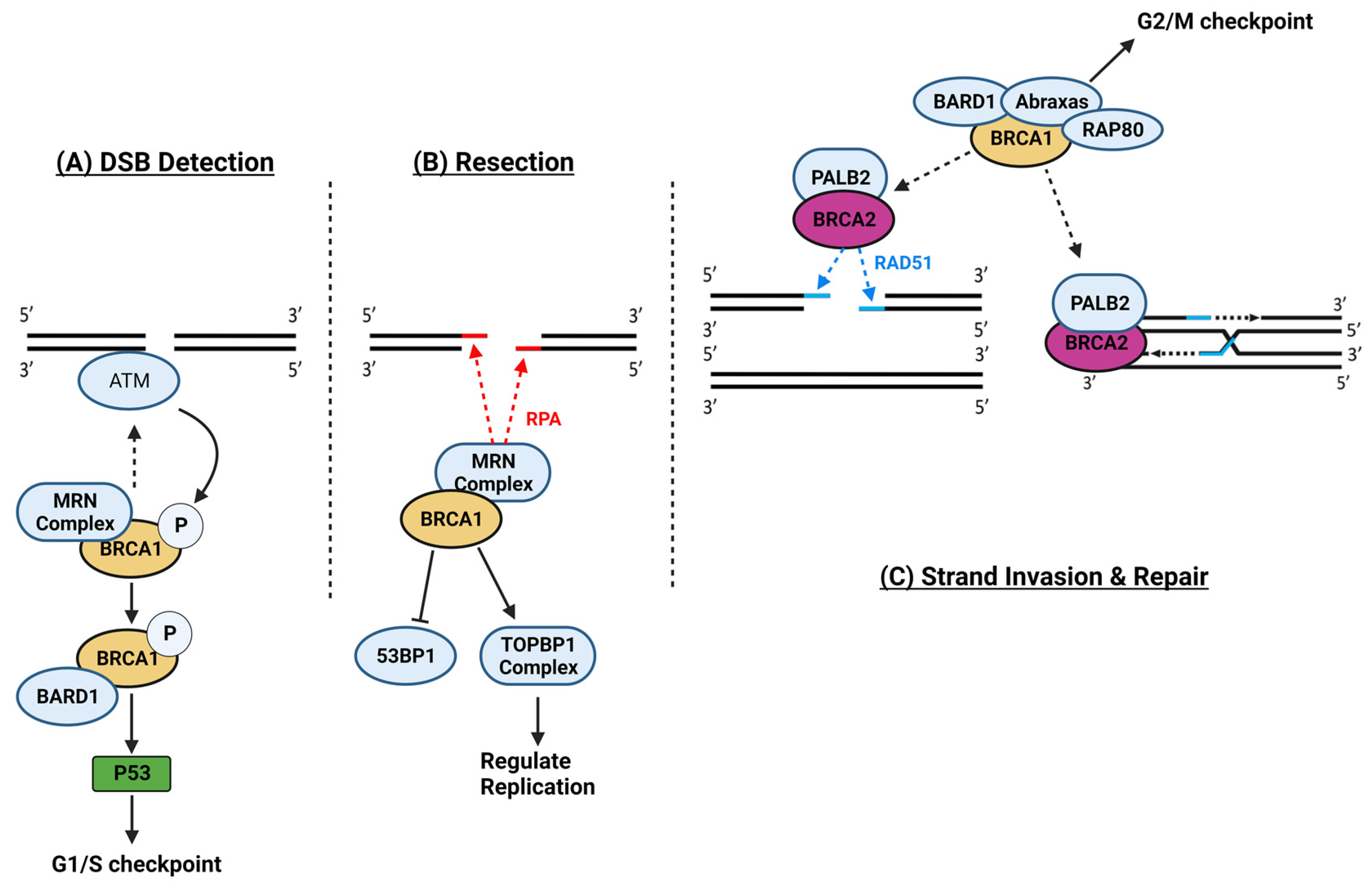
1.13. Breast Cancer Genes 1 and 2

Breast cancer is the most common cancer among Canadian women, and by 2020, it will be the most common cancer globally [135]. Breast cancers are largely sporadic, with tumorigenesis often attributable to the combined effects of genetic and environmental factors. On the other hand, 5–10% of breast cancers are familial and often due to mutations in the BReast-CAncer (BRCA) genes, i.e., BRCA1 and/or BRCA2 genes. The BRCA mutation carriers are predisposed to known increased lifetime risks for mainly breast and ovarian cancer, along with various other cancers such as prostrate or colorectal cancer [136]. Identifying BRCA mutations forms the diagnostic basis of the Hereditary Breast and Ovarian Cancer (HBOC) syndrome [137], and the genes’ association with cancer development risk has prompted intensive investigation into the roles of BRCA1 and 2 in cancer and health.
BRCA1 and BRCA2 are tumor suppressor genes encoding for two critical and non-redundant mediators of the homologous recombination DNA damage repair pathway [138]. BRCA1 and BRCA2 are completely distinct genes, as BRCA1 is located on chromosome 17q21, contains 24 exons, and spans 100 kilobases, while BRCA2 is on chromosome 13q12.3, with 27 exons spanning ~70 kilobases. BRCA1 (220 kDa) and BRCA2 (384 kDa) proteins are also completely distinct in structures and functions; they have different direct and indirect protein partners and, thereby, engage in unique roles in the DNA Damage Response (DDR) pathway [138]. Cells are constantly exposed to DNA damage due to exogenous (e.g., radiation) and endogenous (e.g., metabolite byproducts such as ROS) stressors that, if unaccounted for, would otherwise rapidly compromise their genomic integrity and stability. Genomic impairments are known to underlie cancer, neurodegenerative diseases, premature aging, autoimmune disorders, and cardiovascular diseases [139,140]. In sustaining constant and rapidly arising DNA damage, most cells conduct the DDR pathway, a complex and dynamically fine-tuned error-free signaling package of DNA repair pathways [141,142].
Among the various types of DNA damage, double-strand breaks (DSBs) are the most harmful and require the most energy to repair; several factors regulating DSB repairs also regulate the cell-cycle and apoptosis [143]. In DSB repair, DDRs sensors—ATM and ATR kinases—detect the lesion; promote the phosphorylation of local histone H2AX; and induce a unique chromatin modification that recruits and activates MDC1; which then recruits the initiators of DSB repairs. In addition, activated ATM and ATR interact with the CHK proteins to stabilize p53, which upregulates cell cycle inhibitors and checkpoint activators. If DSBs are not repaired, p53’s continuous activity will eventually upregulate the molecular trigger of apoptosis—BAX; BAM; and PUMA (28). The DDR package devotes two separate repair mechanisms for DSBs. Non-homologous end joining (NHEJ) and Homologous Recombination (HR).
Non-homologous end joining (NHEJ) does not use a homologous chromosomal copy and, therefore, cannot recover the genetic information lost to DSBs; therefore, it is an error-prone method for DNA repair. However, NHEJ is a more immediate response that operates throughout all cell cycle phases, aiming to quickly resolve DSBs and prevent apoptosis or genotypic aberrations due to deletion. The initiator of NHEJ is 53BP1, which mediates the binding of the Ku70-Ku80 heterodimer (Ku) to the DSB lesion, followed by stabilizing the broken ends, alignment by DNA-PK, and ligation by the DNA ligase IV-XRCC4-XLF-PAXX complex. Notably, Ku70 is regulated by SIRT3 deacetylation, which is impaired by Dox treatment [144]. Moreover, DNA-PK facilitates p53’s upregulation should NHEJ fail in its goal [145,146,147,148,149].
Homologous Recombination (HR) is restricted to late S or G2 phases, requiring a completely replicated ‘homologous’ sister chromatid. Compared to NHEJ, it is more sophisticated and time- and energy-consuming. The initiator of HR repair is BRCA1 (Figure 3). Upon activation by the DSB detection complex, BRCA1 mediates the binding of the MRN complex to broken ends. The MRN complex then produces the unique 5′ to 3′ resection and 3′-overhangs; these formations also replace any binding Ku proteins with RPA heterotrimers, inhibiting NHEJ. Indeed, BRCA1 is also known to antagonize 53BP1 [150], and BRCA1’s bioavailability relative to 53BP1 determines whether NHEJ or HR is executed to repair DSBs [151]. Next, PALB2, with the help of BRC1, mediates BRCA2’s disassembling of RAD51 heptamers and loading their monomers onto the 3′-end overhangs and the resected regions, replacing the RPAs. BRCA2 then (1) guides the invasion of the RAD51-single strand formation to the correct copied regions on the homologous sister chromatid and (2) mediates the homologous template-dependent strand extension to re-synthesize the lost base-pairs (Figure 3). Similar to NHEJs DNA-PK, BRCA1 also regulates p53, dictating apoptosis based on the result of repair [152,153,154].
Moreover, BRCA1 also complexes with several factors to regulate the activation of cell cycle checkpoints. In the G1/S-checkpoint, activated ATM phosphorylates BRCA1, which requires complexing with BARD1 to facilitate the stabilization of p53, in turn inducing p21, a cell cycle inhibitor. During S-phase, BRCA1 also interacts with TopoII via the BRCA1–BRIP1–DNA topoisomerase 2-binding protein 1 (TOPBP1) complex to regulate replication. The G2/M checkpoint requires BRCA1 complexing with RAP80, Abraxas, and BARD1 [155].
The broader range of functions of BRCA1 implies that it is the more critical counterpart, although it is an entirely distinct protein relative to BRCA2 [138], and they only overlap in HR repairs. Accordingly, more severe genomic instability is found in tumors associated with defective BRCA1 than in those with faulty BRCA2 [156], which may explain the higher lifetime risks for developing cancers (breast and ovarian cancers) in women carrying BRCA1 mutations. Interestingly, while this is the case in women, recent patient studies suggest that in men, higher cancer risk (breast, pancreas, and prostate cancers) is associated with BRCA2 mutation carriers instead, although the phenotypical connection between BRCA2’s function remains to be investigated [157,158]. Considering the current state of what is known about BRCA2 vs. BRCA1 and that BRCA2 is essential for proper HR repair and, thereby, the cells’ cycle and genomic integrity, this excitingly suggests that there is still a vacuum of BRCA2s’ important mechanisms to be unearthed.

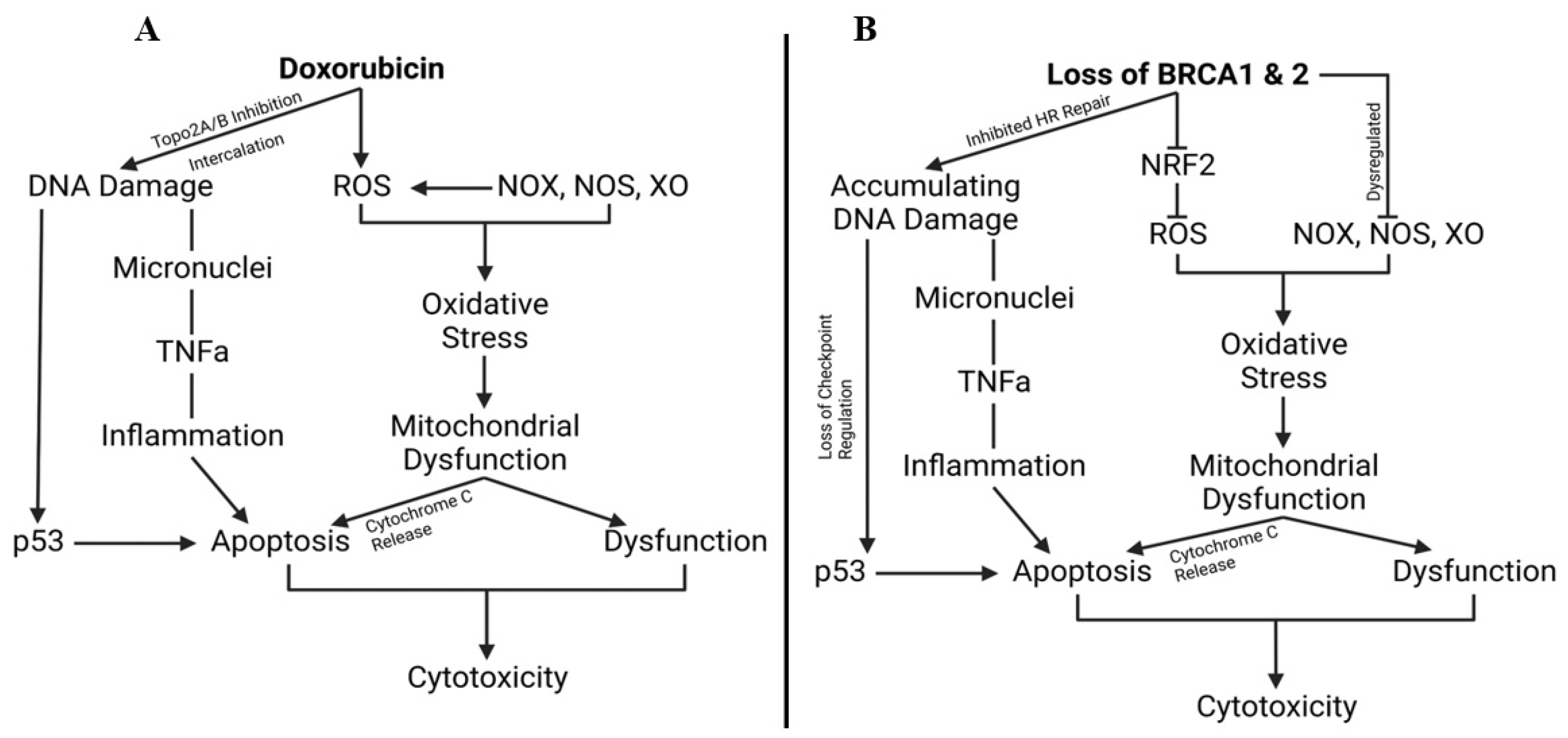
1.14. Breast Cancer Gene, Doxorubicin, and Cardiovascular Limitations, Implications, and Prospects

Considering the essential roles of BRCA1 and BRCA2 in determining cellular survival and genomic integrity and the wide-scale detrimental cytotoxic mechanisms of Dox, there remains a critical lack of investigation into the effects of BRCA1 and BRCA2 functions on Dox-induced cytotoxicity and DIC.
Moreover, the current diagnosing approach for identifying BRCA mutations lacks consideration for haplo-insufficiency in either BRCA genes, whereby the BRCA-haplo-insufficient individuals carry malfunctional BRCA-related phenotypes. This has led to a considerable oversight in clinical examination for the implications related to impaired BRCA functions. For instance, as Dox’s genomic impacts encompass DNA intercalation and TopoII inhibition, thereby impairing replication, gene expression, and DNA repair, it is still currently not known how Dox affects the BRCA’s related pathways. Moreover, the SIRT proteins are Class III deacetylases and, therefore, important regulators of epigenetics and histone modifications. While Dox is known to inhibit SIRT3 and indirectly impair the Ku proteins, thereby inhibiting NHEJ, still little is known about Dox affecting any of the factors related to HR repair other than suppressing DNA synthesis.
In addition, as HBOC diagnosis is directed toward examining the risk of cancer in BRCA mutation carriers, the current clinical approach to BRCA mutations largely overlooks the non-neoplastic implications of haploinsufficiency in the BRCA gene. Notably, BRCA haploinsufficiency can be caused by multiple factors (transcription factors, genetic modifiers, hormonal regulation, SNPs in promoter/5′ UTR) independent of BRCA mutations. This is reinforced by the fact that the estimated general population prevalence of high-risk BRCA1/2 mutations is 1 in 400 [159], and HBOC only accounts for ~6 in 100 cases of breast and ovarian cancers [160]. In other words, not all BRCA1/2 mutation carriers will develop cancer, and most breast and ovarian cancers are not due to HBOC. Therefore, the clinical prospect of compromised BRCA genes in an individual’s health and pathophysiology remains vague outside the focus of cancer, especially in assessing non-specific Dox’s cytotoxicity.
Interestingly, a single clinical retrospective study in 2009 found a noticeably higher risk of mortality associated with carriers of the BRCA mutation in the absence of cancer [161]. This notion is further elaborated by the fact that the roles of BRCA are commonly studied in the context of a general, non-specific eukaryotic cell. The latest update on BRCA expression pattern from The Human Protein Atlas indicates a global expression trend in the human body; a majority of organ systems express BRCA1 and 2 [162]. Likewise, 58% of deaths in breast cancer patients are due to complications related to organ dysfunction; leading causes include pulmonic insufficiency, infection, and hepatic insufficiency [163]. Notably, cancer patients have recently been characterized by higher cardiovascular risk [164].
BRCA1 and 2 maintain genomic integrity and regulate the cell cycle, metabolism, and function, while Dox targets and impairs all the same aspects. This is summarized in Figure 4 and suggests that the mechanisms of Dox may conflict with the essential pathways regulated by BRCA1 and 2. Yet, despite intensive research that has revealed a vast multitude of cellular functions impaired by Dox, particularly in the heart, there remains a critical lack of investigation into Dox’s functional connection to BRCA1 and BRCA2.
Nevertheless, recent research has begun to demonstrate the essential roles of BRCA genes in endothelial and cardiac function. For instance, markedly greater cardiac dysfunction has been observed in the left ventricular sections of Dox-treated cardiomyocyte-specific BRCA2 knock-out mice compared to vehicle-treated controls. Cardiac samples from these Dox-treated mice also exhibit enhanced apoptosis, elevated phosphorylated histone H2Ax (DSB markers), increased expression and activities of p53, PUMA, and Bax, which are pro-apoptotic proteins, and increased cytochrome C release, a marker of mitochondrial dysfunction and apoptosis. Notably, the samples also show reduced RAD51 formation, suggesting that Dox impacts BRCA2’s related DNA repair pathway [165].
In vivo, overexpressing BRCA1 in ECs protects against Dox-induced inflammation in association with reduced ROS levels and upregulated eNOS and VEGFa in vitro [166]. On the other hand, silencing endothelial BRCA1 aggravates inflammation under Dox treatment. In the same study, EC-specific BRCA1-overexpressing ApoE-null mice subjected to hind-limb ischemia, a model of endothelial activation, exhibited improved capillary density and better vascular recovery compared to controls. Under Western diets, modeling atherogenesis, BRCA1-overexpressing mice show a significant reduction in aortic plaque lesions, macrophage infiltration, and oxidative stress vs. controls. In contrast, BRCA1-null mice demonstrated greater inflammation, apoptosis, and impaired endothelial function in lung and aortic sections. Lastly, this study also observed that BRCA1 expression was suppressed in the plaque region of human atherosclerotic carotid artery samples compared to the adjacent plaque-free area [166]. Similarly, EC-specific loss of BRCA2 was found to exacerbate oxidative stress-induced DNA damage, apoptosis, and endothelial dysfunction [109].

2. Conclusions

In conclusion, doxorubicin is a cytotoxic cancer medication with devastating cardiotoxicity side effects that have been well characterized for two modes: genomic impairment and oxidative stress. The molecular mechanisms of Dox remain a subject of intensive research, with novel mechanisms being discovered that fortify the drug’s potency as well as its roles in DIC. In addition to impairing cardiomyocytes’ genomic integrity, redox balance, autophagy, and metabolism, recent research on Dox’s impacts in the endothelia of cardiac tissues has suggested the critical role of endotheliotoxicity in DIC. Moreover, the impact of Dox on the functions of BRCA1 and BRCA2 remains largely unexplored. The current diagnostic approach for identifying BRCA mutations lacks consideration for haploinsufficiency, leading to compulsory oversights on the clinical implications related to impaired BRCA functions. Recent studies have shown the essential roles of BRCA genes in endothelial and cardiac function, and the dysregulation of these genes has cardiovascular and non-neoplastic implications. Accordingly, there are conflicting reports about the role of BRCA in an increased risk for cardiovascular diseases, most importantly regarding DIC [167,168]. Given the noticeable overlap in the mechanisms governing the loss of BRCA-associated phenotype and DIC, further investigation into the functional connections between Dox and BRCA1/2 is warranted to better understand the mechanisms of Dox-induced cardiotoxicity and endotheliotoxicity in order to promote personalized therapy in BRCA-mutant cancer patients.

Author Contributions

Conceptualization, J.C.F. and K.K.S.; resources, H.C.N. and K.K.S.; writing—original draft preparation, H.C.N.; writing—review and editing, H.C.N. and K.K.S.; visualization, H.C.N. and J.C.F.; supervision, K.K.S.; and funding acquisition, K.K.S. All authors have read and agreed to the published version of the manuscript.

Funding

This research was funded by the Heart and Stroke Foundation, grant number G-22-0032104.

Conflicts of Interest

The authors declare no conflicts of interest.

Abbreviation

Dox = Doxorubicin; ROS = Reactive oxygen species; RNS = Reactive nitrogen species; NOX = NAPDH oxidases; NOS = Nitric oxide synthase; DIC = Dox-induced cardiotoxicity; TopoIIa/b = Topoisomerase IIa/b; NO = Nitric oxide; ONOO- = Peroxynitrite anion; iNOS = Inducible NOS; eNOS = Endothelial NOS; NSAIDs = Non-steroidal anti-inflammatory drugs; PPARs = Peroxisome proliferator-activated receptors; FENO = Fenofibrate; SIRTs = Sirtuins; HPβCD = 2-Hydroxypropyl-β-cyclodextrin; SR = Sarcoplasm; DIE = Dox-induced endotheliotoxicity; ECs = Endothelial cells; ZO-1 = Zona occludens-1; BRCA1/2 = BReast-CAncer 1/2 genes; HBOC = Hereditary Breast and Ovarian Cancer Syndrome; DDR = DNA Damage Response; DSBs = Double-strand breaks; NHEJ = Non-homologous end joining; HR = Homologous Recombination; Ku = Ku70-Ku80 heterodimer; TOPBP1 = BRCA1–BRIP1–DNA topoisomerase 2-binding protein 1 complex.

References

  1. Sritharan, S.; Sivalingam, N. A Comprehensive Review on Time-Tested Anticancer Drug Doxorubicin. Life Sci. 2021, 278, 119527. [Google Scholar] [CrossRef] [PubMed]
  2. Nicoletto, R.E.; Ofner, C.M. Cytotoxic Mechanisms of Doxorubicin at Clinically Relevant Concentrations in Breast Cancer Cells. Cancer Chemother. Pharmacol. 2022, 89, 285–311. [Google Scholar] [CrossRef] [PubMed]
  3. Gustafson, D.L.; Rastatter, J.C.; Colombo, T.; Long, M.E. Doxorubicin Pharmacokinetics: Macromolecule Binding, Metabolism, and Excretion in the Context of a Physiologic Model. J. Pharm. Sci. 2002, 91, 1488–1501. [Google Scholar] [CrossRef]
  4. Speth, P.A.; van Hoesel, Q.G.; Haanen, C. Clinical Pharmacokinetics of Doxorubicin. Clin. Pharmacokinet. 1988, 15, 15–31. [Google Scholar] [CrossRef] [PubMed]
  5. Dobbs, N.A.; Twelves, C.J.; Gillies, H.; James, C.A.; Harper, P.G.; Rubens, R.D. Gender Affects Doxorubicin Pharmacokinetics in Patients with Normal Liver Biochemistry. Cancer Chemother. Pharmacol. 1995, 36, 473–476. [Google Scholar] [CrossRef] [PubMed]
  6. Tacar, O.; Sriamornsak, P.; Dass, C.R. Doxorubicin: An Update on Anticancer Molecular Action, Toxicity and Novel Drug Delivery Systems. J. Pharm. Pharmacol. 2013, 65, 157–170. [Google Scholar] [CrossRef] [PubMed]
  7. Thorn, C.F.; Oshiro, C.; Marsh, S.; Hernandez-Boussard, T.; McLeod, H.; Klein, T.E.; Altman, R.B. Doxorubicin Pathways: Pharmacodynamics and Adverse Effects. Pharmacogenet Genom. 2011, 21, 440–446. [Google Scholar] [CrossRef]
  8. Kong, C.-Y.; Guo, Z.; Song, P.; Zhang, X.; Yuan, Y.-P.; Teng, T.; Yan, L.; Tang, Q.-Z. Underlying the Mechanisms of Doxorubicin-Induced Acute Cardiotoxicity: Oxidative Stress and Cell Death. Int. J. Biol. Sci. 2022, 18, 760–770. [Google Scholar] [CrossRef]
  9. Minotti, G.; Recalcati, S.; Mordente, A.; Liberi, G.; Calafiore, A.M.; Mancuso, C.; Preziosi, P.; Cairo, G. The Secondary Alcohol Metabolite of Doxorubicin Irreversibly Inactivates Aconitase/Iron Regulatory Protein-1 in Cytosolic Fractions from Human Myocardium. FASEB J. 1998, 12, 541–552. [Google Scholar] [CrossRef]
  10. Sheibani, M.; Azizi, Y.; Shayan, M.; Nezamoleslami, S.; Eslami, F.; Farjoo, M.H.; Dehpour, A.R. Doxorubicin-Induced Cardiotoxicity: An Overview on Pre-Clinical Therapeutic Approaches. Cardiovasc. Toxicol. 2022, 22, 292–310. [Google Scholar] [CrossRef]
  11. Ibrahim, M.M.; Khedr, M.M.; Morsy, M.H.; Badae, N.M.; Elatrebi, S. A Comparative Study of the Cardioprotective Effect of Metformin, Sitagliptin and Dapagliflozin on Isoprenaline Induced Myocardial Infarction in Non-Diabetic Rats. Bull. Natl. Res. Cent. 2022, 46, 123. [Google Scholar] [CrossRef]
  12. Aloss, K.; Hamar, P. Recent Preclinical and Clinical Progress in Liposomal Doxorubicin. Pharmaceutics 2023, 15, 893. [Google Scholar] [CrossRef] [PubMed]
  13. Hoeger, C.W.; Turissini, C.; Asnani, A. Doxorubicin Cardiotoxicity: Pathophysiology Updates. Curr. Treat. Options Cardio Med. 2020, 22, 52. [Google Scholar] [CrossRef]
  14. Carvalho, C.; Santos, R.X.; Cardoso, S.; Correia, S.; Oliveira, P.J.; Santos, M.S.; Moreira, P.I. Doxorubicin: The Good, the Bad and the Ugly Effect. Curr. Med. Chem. 2009, 16, 3267–3285. [Google Scholar] [CrossRef] [PubMed]
  15. Zhu, H.; Sarkar, S.; Scott, L.; Danelisen, I.; Trush, M.A.; Jia, Z.; Li, Y.R. Doxorubicin Redox Biology: Redox Cycling, Topoisomerase Inhibition, and Oxidative Stress. React. Oxyg. Species 2016, 1, 189–198. [Google Scholar] [CrossRef] [PubMed]
  16. Agudelo, D.; Bourassa, P.; Bérubé, G.; Tajmir-Riahi, H.-A. Intercalation of Antitumor Drug Doxorubicin and Its Analogue by DNA Duplex: Structural Features and Biological Implications. Int. J. Biol. Macromol. 2014, 66, 144–150. [Google Scholar] [CrossRef] [PubMed]
  17. Zhang, S.; Liu, X.; Bawa-Khalfe, T.; Lu, L.-S.; Lyu, Y.L.; Liu, L.F.; Yeh, E.T.H. Identification of the Molecular Basis of Doxorubicin-Induced Cardiotoxicity. Nat. Med. 2012, 18, 1639–1642. [Google Scholar] [CrossRef]
  18. Engeland, K. Cell Cycle Regulation: P53-P21-RB Signaling. Cell Death Differ. 2022, 29, 946–960. [Google Scholar] [CrossRef]
  19. Polager, S.; Ginsberg, D. P53 and E2f: Partners in Life and Death. Nat. Rev. Cancer 2009, 9, 738–748. [Google Scholar] [CrossRef]
  20. Aubrey, B.J.; Kelly, G.L.; Janic, A.; Herold, M.J.; Strasser, A. How Does P53 Induce Apoptosis and How Does This Relate to P53-Mediated Tumour Suppression? Cell Death Differ. 2018, 25, 104–113. [Google Scholar] [CrossRef]
  21. Zhu, W.; Soonpaa, M.H.; Chen, H.; Shen, W.; Payne, R.M.; Liechty, E.A.; Caldwell, R.L.; Shou, W.; Field, L.J. Acute Doxorubicin Cardiotoxicity Is Associated with P53-Induced Inhibition of the Mammalian Target of Rapamycin Pathway. Circulation 2009, 119, 99–106. [Google Scholar] [CrossRef] [PubMed]
  22. Li, J.; Wang, P.-Y.; Long, N.A.; Zhuang, J.; Springer, D.A.; Zou, J.; Lin, Y.; Bleck, C.K.E.; Park, J.-H.; Kang, J.-G.; et al. P53 Prevents Doxorubicin Cardiotoxicity Independently of Its Prototypical Tumor Suppressor Activities. Proc. Natl. Acad. Sci. USA 2019, 116, 19626–19634. [Google Scholar] [CrossRef] [PubMed]
  23. Zhu, W.; Zhang, W.; Shou, W.; Field, L.J. P53 Inhibition Exacerbates Late-Stage Anthracycline Cardiotoxicity. Cardiovasc. Res. 2014, 103, 81–89. [Google Scholar] [CrossRef] [PubMed]
  24. Sin, T.K.; Tam, B.T.; Yung, B.Y.; Yip, S.P.; Chan, L.W.; Wong, C.S.; Ying, M.; Rudd, J.A.; Siu, P.M. Resveratrol Protects against Doxorubicin-Induced Cardiotoxicity in Aged Hearts through the SIRT1-USP7 Axis. J. Physiol. 2015, 593, 1887–1899. [Google Scholar] [CrossRef]
  25. Zheng, W.; Lu, Y.-B.; Liang, S.-T.; Zhang, Q.-J.; Xu, J.; She, Z.-G.; Zhang, Z.-Q.; Yang, R.-F.; Mao, B.-B.; Xu, Z.; et al. SIRT1 Mediates the Protective Function of Nkx2.5 during Stress in Cardiomyocytes. Basic. Res. Cardiol. 2013, 108, 364. [Google Scholar] [CrossRef]
  26. Wu, S.; Lan, J.; Li, L.; Wang, X.; Tong, M.; Fu, L.; Zhang, Y.; Xu, J.; Chen, X.; Chen, H.; et al. Sirt6 Protects Cardiomyocytes against Doxorubicin-Induced Cardiotoxicity by Inhibiting P53/Fas-Dependent Cell Death and Augmenting Endogenous Antioxidant Defense Mechanisms. Cell Biol. Toxicol. 2023, 39, 237–258. [Google Scholar] [CrossRef]
  27. Hori, Y.S.; Kuno, A.; Hosoda, R.; Horio, Y. Regulation of FOXOs and P53 by SIRT1 Modulators under Oxidative Stress. PLoS ONE 2013, 8, e73875. [Google Scholar] [CrossRef]
  28. Guo, Y.; Tang, Y.; Lu, G.; Gu, J. P53 at the Crossroads between Doxorubicin-Induced Cardiotoxicity and Resistance: A Nutritional Balancing Act. Nutrients 2023, 15, 2259. [Google Scholar] [CrossRef]
  29. Li, A.; Gao, M.; Jiang, W.; Qin, Y.; Gong, G. Mitochondrial Dynamics in Adult Cardiomyocytes and Heart Diseases. Front. Cell Dev. Biol. 2020, 8, 584800. [Google Scholar] [CrossRef]
  30. Begum, R.; Thota, S.; Abdulkadir, A.; Kaur, G.; Bagam, P.; Batra, S. NADPH Oxidase Family Proteins: Signaling Dynamics to Disease Management. Cell Mol. Immunol. 2022, 19, 660–686. [Google Scholar] [CrossRef]
  31. Förstermann, U.; Sessa, W.C. Nitric Oxide Synthases: Regulation and Function. Eur. Heart J. 2012, 33, 829–837. [Google Scholar] [CrossRef] [PubMed]
  32. Chung, H.Y.; Baek, B.S.; Song, S.H.; Kim, M.S.; Huh, J.I.; Shim, K.H.; Kim, K.W.; Lee, K.H. Xanthine Dehydrogenase/Xanthine Oxidase and Oxidative Stress. Age 1997, 20, 127–140. [Google Scholar] [CrossRef] [PubMed]
  33. Gustafson, D.L.; Swanson, J.D.; Pritsos, C.A. Role of Xanthine Oxidase in the Potentiation of Doxorubicin-Induced Cardiotoxicity by Mitomycin C. Cancer Commun. 1991, 3, 299–304. [Google Scholar] [CrossRef] [PubMed]
  34. Febuxostat Ameliorates Doxorubicin-Induced Cardiotoxicity in Rats-PubMed. Available online: https://pubmed.ncbi.nlm.nih.gov/26036690/ (accessed on 18 November 2023).
  35. Kondo, M.; Imanishi, M.; Fukushima, K.; Ikuto, R.; Murai, Y.; Horinouchi, Y.; Izawa-Ishizawa, Y.; Goda, M.; Zamami, Y.; Takechi, K.; et al. Xanthine Oxidase Inhibition by Febuxostat in Macrophages Suppresses Angiotensin II-Induced Aortic Fibrosis. Am. J. Hypertens. 2019, 32, 249–256. [Google Scholar] [CrossRef] [PubMed]
  36. Cappetta, D.; De Angelis, A.; Sapio, L.; Prezioso, L.; Illiano, M.; Quaini, F.; Rossi, F.; Berrino, L.; Naviglio, S.; Urbanek, K. Oxidative Stress and Cellular Response to Doxorubicin: A Common Factor in the Complex Milieu of Anthracycline Cardiotoxicity. Oxid. Med. Cell Longev. 2017, 2017, 1521020. [Google Scholar] [CrossRef] [PubMed]
  37. Zhuo, C.; Xin, J.; Huang, W.; Zhang, D.; Yan, X.; Li, R.; Li, H.; Lan, J.; Lin, L.; Li, L.; et al. Irisin Protects against Doxorubicin-Induced Cardiotoxicity by Improving AMPK-Nrf2 Dependent Mitochondrial Fusion and Strengthening Endogenous Anti-Oxidant Defense Mechanisms. Toxicology 2023, 494, 153597. [Google Scholar] [CrossRef] [PubMed]
  38. Hu, C.; Zhang, X.; Zhang, N.; Wei, W.-Y.; Li, L.-L.; Ma, Z.-G.; Tang, Q.-Z. Osteocrin Attenuates Inflammation, Oxidative Stress, Apoptosis, and Cardiac Dysfunction in Doxorubicin-Induced Cardiotoxicity. Clin. Transl. Med. 2020, 10, e124. [Google Scholar] [CrossRef]
  39. Lassègue, B.; San Martín, A.; Griendling, K.K. Biochemistry, Physiology, and Pathophysiology of NADPH Oxidases in the Cardiovascular System. Circ. Res. 2012, 110, 1364–1390. [Google Scholar] [CrossRef]
  40. Henriquez-Olguin, C.; Meneses-Valdes, R.; Raun, S.H.; Gallero, S.; Knudsen, J.R.; Li, Z.; Li, J.; Sylow, L.; Jaimovich, E.; Jensen, T.E. NOX2 Deficiency Exacerbates Diet-Induced Obesity and Impairs Molecular Training Adaptations in Skeletal Muscle. Redox Biol. 2023, 65, 102842. [Google Scholar] [CrossRef]
  41. Efentakis, P.; Varela, A.; Chavdoula, E.; Sigala, F.; Sanoudou, D.; Tenta, R.; Gioti, K.; Kostomitsopoulos, N.; Papapetropoulos, A.; Tasouli, A.; et al. Levosimendan Prevents Doxorubicin-Induced Cardiotoxicity in Time- and Dose-Dependent Manner: Implications for Inotropy. Cardiovasc. Res. 2020, 116, 576–591. [Google Scholar] [CrossRef]
  42. Akolkar, G.; Bagchi, A.K.; Ayyappan, P.; Jassal, D.S.; Singal, P.K. Doxorubicin-Induced Nitrosative Stress Is Mitigated by Vitamin C via the Modulation of Nitric Oxide Synthases. Am. J. Physiol. Cell Physiol. 2017, 312, C418–C427. [Google Scholar] [CrossRef]
  43. Wang, J.; Yao, L.; Wu, X.; Guo, Q.; Sun, S.; Li, J.; Shi, G.; Caldwell, R.B.; Caldwell, R.W.; Chen, Y. Protection against Doxorubicin-Induced Cardiotoxicity through Modulating iNOS/ARG 2 Balance by Electroacupuncture at PC6. Oxidative Med. Cell. Longev. 2021, 2021, e6628957. [Google Scholar] [CrossRef] [PubMed]
  44. Sangweni, N.F.; van Vuuren, D.; Mabasa, L.; Gabuza, K.; Huisamen, B.; Naidoo, S.; Barry, R.; Johnson, R. Prevention of Anthracycline-Induced Cardiotoxicity: The Good and Bad of Current and Alternative Therapies. Front. Cardiovasc. Med. 2022, 9, 907266. [Google Scholar] [CrossRef] [PubMed]
  45. Tran, N.; Garcia, T.; Aniqa, M.; Ali, S.; Ally, A.; Nauli, S. Endothelial Nitric Oxide Synthase (eNOS) and the Cardiovascular System: In Physiology and in Disease States. Am. J. Biomed. Sci. Res. 2022, 15, 153–177. [Google Scholar] [PubMed]
  46. Neilan, T.G.; Blake, S.L.; Ichinose, F.; Raher, M.J.; Buys, E.S.; Jassal, D.S.; Furutani, E.; Perez-Sanz, T.M.; Graveline, A.; Janssens, S.P.; et al. Disruption of Nitric Oxide Synthase 3 Protects against the Cardiac Injury, Dysfunction, and Mortality Induced by Doxorubicin. Circulation 2007, 116, 506–514. [Google Scholar] [CrossRef]
  47. He, H.; Wang, L.; Qiao, Y.; Zhou, Q.; Li, H.; Chen, S.; Yin, D.; Huang, Q.; He, M. Doxorubicin Induces Endotheliotoxicity and Mitochondrial Dysfunction via ROS/eNOS/NO Pathway. Front. Pharmacol. 2020, 10, 1531. [Google Scholar] [CrossRef]
  48. Zeglinski, M.; Premecz, S.; Lerner, J.; Wtorek, P.; Dasilva, M.; Hasanally, D.; Chaudhary, R.; Sharma, A.; Thliveris, J.; Ravandi, A.; et al. Congenital Absence of Nitric Oxide Synthase 3 Potentiates Cardiac Dysfunction and Reduces Survival in Doxorubicin- and Trastuzumab-Mediated Cardiomyopathy. Can. J. Cardiol. 2014, 30, 359–367. [Google Scholar] [CrossRef]
  49. Stanley, W.C.; Recchia, F.A.; Lopaschuk, G.D. Myocardial Substrate Metabolism in the Normal and Failing Heart. Physiol. Rev. 2005, 85, 1093–1129. [Google Scholar] [CrossRef]
  50. Marzoog, B.A.; Vlasova, T.I. Myocardiocyte Autophagy in the Context of Myocardiocytes Regeneration: A Potential Novel Therapeutic Strategy. Egypt. J. Med. Human. Genet. 2022, 23, 41. [Google Scholar] [CrossRef]
  51. Afonso, A.I.; Amaro-Leal, Â.; Machado, F.; Rocha, I.; Geraldes, V. Doxorubicin Dose-Dependent Impact on Physiological Balance—A Holistic Approach in a Rat Model. Biology 2023, 12, 1031. [Google Scholar] [CrossRef]
  52. Dallons, M.; Schepkens, C.; Dupuis, A.; Tagliatti, V.; Colet, J.-M. New Insights About Doxorubicin-Induced Toxicity to Cardiomyoblast-Derived H9C2 Cells and Dexrazoxane Cytoprotective Effect: Contribution of In Vitro 1H-NMR Metabonomics. Front. Pharmacol. 2020, 11, 79. [Google Scholar] [CrossRef] [PubMed]
  53. Qiao, X.; Van Der Zanden, S.Y.; Wander, D.P.A.; Borràs, D.M.; Song, J.-Y.; Li, X.; Van Duikeren, S.; Van Gils, N.; Rutten, A.; Van Herwaarden, T.; et al. Uncoupling DNA Damage from Chromatin Damage to Detoxify Doxorubicin. Proc. Natl. Acad. Sci. USA 2020, 117, 15182–15192. [Google Scholar] [CrossRef] [PubMed]
  54. Kashyap, P.; Shikha, D.; Thakur, M.; Aneja, A. Functionality of Apigenin as a Potent Antioxidant with Emphasis on Bioavailability, Metabolism, Action Mechanism and in Vitro and in Vivo Studies: A Review. J. Food Biochem. 2022, 46, e13950. [Google Scholar] [CrossRef] [PubMed]
  55. Huo, C.-J.; Yu, X.-J.; Sun, Y.-J.; Li, H.-B.; Su, Q.; Bai, J.; Li, Y.; Liu, K.-L.; Qi, J.; Zhou, S.-W.; et al. Irisin Lowers Blood Pressure by Activating the Nrf2 Signaling Pathway in the Hypothalamic Paraventricular Nucleus of Spontaneously Hypertensive Rats. Toxicol. Appl. Pharmacol. 2020, 394, 114953. [Google Scholar] [CrossRef] [PubMed]
  56. He, A.; Dean, J.M.; Lodhi, I.J. Peroxisomes as Cellular Adaptors to Metabolic and Environmental Stress. Trends Cell Biol. 2021, 31, 656–670. [Google Scholar] [CrossRef] [PubMed]
  57. Schedin, S.; Sindelar, P.J.; Pentchev, P.; Brunk, U.; Dallner, G. Peroxisomal Impairment in Niemann-Pick Type C Disease. J. Biol. Chem. 1997, 272, 6245–6251. [Google Scholar] [CrossRef]
  58. Kawałek, A.; Lefevre, S.D.; Veenhuis, M.; Klei, I.J. van der Peroxisomal Catalase Deficiency Modulates Yeast Lifespan Depending on Growth Conditions. Aging 2013, 5, 67–83. [Google Scholar] [CrossRef]
  59. Han, Y.-H.; Lian, X.-D.; Lee, S.-J.; Li, W.-L.; Sun, H.-N.; Jin, M.-H.; Kwon, T. Regulatory Effect of Peroxiredoxin 1 (PRDX1) on Doxorubicin-Induced Apoptosis in Triple Negative Breast Cancer Cells. Appl. Biol. Chem. 2022, 65, 63. [Google Scholar] [CrossRef]
  60. Jiang, L.; Gong, Y.; Hu, Y.; You, Y.; Wang, J.; Zhang, Z.; Wei, Z.; Tang, C. Peroxiredoxin-1 Overexpression Attenuates Doxorubicin-Induced Cardiotoxicity by Inhibiting Oxidative Stress and Cardiomyocyte Apoptosis. Oxid. Med. Cell Longev. 2020, 2020, 2405135. [Google Scholar] [CrossRef]
  61. Wanders, R.J.A.; Waterham, H.R. Peroxisomal Disorders: The Single Peroxisomal Enzyme Deficiencies. Biochim. Biophys. Acta (BBA)-Mol. Cell Res. 2006, 1763, 1707–1720. [Google Scholar] [CrossRef]
  62. Serhan, C.N.; Chiang, N.; Dalli, J.; Levy, B.D. Lipid Mediators in the Resolution of Inflammation. Cold Spring Harb. Perspect. Biol. 2014, 7, a016311. [Google Scholar] [CrossRef]
  63. Endres, S.; Ghorbani, R.; Kelley, V.E.; Georgilis, K.; Lonnemann, G.; van der Meer, J.W.; Cannon, J.G.; Rogers, T.S.; Klempner, M.S.; Weber, P.C. The Effect of Dietary Supplementation with N-3 Polyunsaturated Fatty Acids on the Synthesis of Interleukin-1 and Tumor Necrosis Factor by Mononuclear Cells. N. Engl. J. Med. 1989, 320, 265–271. [Google Scholar] [CrossRef]
  64. Reddy, J.K.; Hashimoto, T. Peroxisomal Beta-Oxidation and Peroxisome Proliferator-Activated Receptor Alpha: An Adaptive Metabolic System. Annu. Rev. Nutr. 2001, 21, 193–230. [Google Scholar] [CrossRef]
  65. Kersten, S. Peroxisome Proliferator Activated Receptors and Lipoprotein Metabolism. PPAR Res. 2008, 2008, 132960. [Google Scholar] [CrossRef]
  66. Arunachalam, S.; Tirupathi Pichiah, P.B.; Achiraman, S. Doxorubicin Treatment Inhibits PPARγ and May Induce Lipotoxicity by Mimicking a Type 2 Diabetes-like Condition in Rodent Models. FEBS Lett. 2013, 587, 105–110. [Google Scholar] [CrossRef]
  67. Lin, Y.; Liu, R.; Huang, Y.; Yang, Z.; Xian, J.; Huang, J.; Qiu, Z.; Lin, X.; Zhang, M.; Chen, H.; et al. Reactivation of PPARα Alleviates Myocardial Lipid Accumulation and Cardiac Dysfunction by Improving Fatty Acid β-Oxidation in Dsg2-Deficient Arrhythmogenic Cardiomyopathy. Acta Pharm. Sin. B 2023, 13, 192–203. [Google Scholar] [CrossRef]
  68. Wang, W.; Fang, Q.; Zhang, Z.; Wang, D.; Wu, L.; Wang, Y. PPARα Ameliorates Doxorubicin-Induced Cardiotoxicity by Reducing Mitochondria-Dependent Apoptosis via Regulating MEOX1. Front. Pharmacol. 2020, 11, 528267. [Google Scholar] [CrossRef]
  69. Huang, W.-P.; Yin, W.-H.; Chen, J.-S.; Huang, P.-H.; Chen, J.-W.; Lin, S.-J. Fenofibrate Attenuates Doxorubicin-Induced Cardiac Dysfunction in Mice via Activating the eNOS/EPC Pathway. Sci. Rep. 2021, 11, 1159. [Google Scholar] [CrossRef]
  70. North, B.J.; Verdin, E. Sirtuins: Sir2-Related NAD-Dependent Protein Deacetylases. Genome Biol. 2004, 5, 224. [Google Scholar] [CrossRef]
  71. Matsushima, S.; Sadoshima, J. The Role of Sirtuins in Cardiac Disease. Am. J. Physiol.-Heart Circ. Physiol. 2015, 309, H1375–H1389. [Google Scholar] [CrossRef]
  72. Ministrini, S.; Puspitasari, Y.M.; Beer, G.; Liberale, L.; Montecucco, F.; Camici, G.G. Sirtuin 1 in Endothelial Dysfunction and Cardiovascular Aging. Front. Physiol. 2021, 12, 733696. [Google Scholar] [CrossRef]
  73. Murugasamy, K.; Munjal, A.; Sundaresan, N.R. Emerging Roles of SIRT3 in Cardiac Metabolism. Front. Cardiovasc. Med. 2022, 9, 850340. [Google Scholar] [CrossRef]
  74. Saiyang, X.; Deng, W.; Qizhu, T. Sirtuin 6: A Potential Therapeutic Target for Cardiovascular Diseases. Pharmacol. Res. 2021, 163, 105214. [Google Scholar] [CrossRef]
  75. Yamamura, S.; Izumiya, Y.; Araki, S.; Nakamura, T.; Kimura, Y.; Hanatani, S.; Yamada, T.; Ishida, T.; Yamamoto, M.; Onoue, Y.; et al. Cardiomyocyte Sirt (Sirtuin) 7 Ameliorates Stress-Induced Cardiac Hypertrophy by Interacting With and Deacetylating GATA4. Hypertension 2020, 75, 98–108. [Google Scholar] [CrossRef]
  76. Anderson, K.A.; Hirschey, M.D. Mitochondrial Protein Acetylation Regulates Metabolism. Essays Biochem. 2012, 52, 23–32. [Google Scholar] [CrossRef]
  77. Stein, L.R.; Imai, S. The Dynamic Regulation of NAD Metabolism in Mitochondria. Trends Endocrinol. Metab. 2012, 23, 420–428. [Google Scholar] [CrossRef]
  78. Zhang, Y.; Bharathi, S.S.; Beck, M.E.; Goetzman, E.S. The Fatty Acid Oxidation Enzyme Long-Chain Acyl-CoA Dehydrogenase Can Be a Source of Mitochondrial Hydrogen Peroxide. Redox Biol. 2019, 26, 101253. [Google Scholar] [CrossRef]
  79. Parodi-Rullán, R.M.; Chapa-Dubocq, X.R.; Javadov, S. Acetylation of Mitochondrial Proteins in the Heart: The Role of SIRT3. Front. Physiol. 2018, 9, 1094. [Google Scholar] [CrossRef]
  80. Dikalova, A.E.; Itani, H.A.; Nazarewicz, R.R.; McMaster, W.G.; Flynn, C.R.; Uzhachenko, R.; Fessel, J.P.; Gamboa, J.L.; Harrison, D.G.; Dikalov, S.I. Sirt3 Impairment and SOD2 Hyperacetylation in Vascular Oxidative Stress and Hypertension. Circ. Res. 2017, 121, 564–574. [Google Scholar] [CrossRef]
  81. Sundaresan, N.R.; Samant, S.A.; Pillai, V.B.; Rajamohan, S.B.; Gupta, M.P. SIRT3 Is a Stress-Responsive Deacetylase in Cardiomyocytes That Protects Cells from Stress-Mediated Cell Death by Deacetylation of Ku70. Mol. Cell Biol. 2008, 28, 6384–6401. [Google Scholar] [CrossRef]
  82. Su, H.; Cantrell, A.C.; Chen, J.-X.; Gu, W.; Zeng, H. SIRT3 Deficiency Enhances Ferroptosis and Promotes Cardiac Fibrosis via P53 Acetylation. Cells 2023, 12, 1428. [Google Scholar] [CrossRef]
  83. Li, Z.; Lu, G.; Lu, J.; Wang, P.; Zhang, X.; Zou, Y.; Liu, P. SZC-6, a Small-Molecule Activator of SIRT3, Attenuates Cardiac Hypertrophy in Mice. Acta Pharmacol. Sin. 2023, 44, 546–560. [Google Scholar] [CrossRef]
  84. Wang, M.; Ding, Y.; Hu, Y.; Li, Z.; Luo, W.; Liu, P.; Li, Z. SIRT3 Improved Peroxisomes-Mitochondria Interplay and Prevented Cardiac Hypertrophy via Preserving PEX5 Expression. Redox Biol. 2023, 62, 102652. [Google Scholar] [CrossRef]
  85. Ling, G.; Wang, X.; Tan, N.; Cao, J.; Li, W.; Zhang, Y.; Jiang, J.; Sun, Q.; Jiang, Y.; Wang, W.; et al. Mechanisms and Drug Intervention for Doxorubicin-Induced Cardiotoxicity Based on Mitochondrial Bioenergetics. Oxidative Med. Cell. Longev. 2022, 2022, e7176282. [Google Scholar] [CrossRef]
  86. Wang, X.; Shen, T.; Lian, J.; Deng, K.; Qu, C.; Li, E.; Li, G.; Ren, Y.; Wang, Z.; Jiang, Z.; et al. Resveratrol Reduces ROS-Induced Ferroptosis by Activating SIRT3 and Compensating the GSH/GPX4 Pathway. Mol. Med. 2023, 29, 137. [Google Scholar] [CrossRef]
  87. Christidi, E.; Brunham, L.R. Regulated Cell Death Pathways in Doxorubicin-Induced Cardiotoxicity. Cell Death Dis. 2021, 12, 1–15. [Google Scholar] [CrossRef]
  88. Li, D.; Yang, Y.; Wang, S.; He, X.; Liu, M.; Bai, B.; Tian, C.; Sun, R.; Yu, T.; Chu, X. Role of Acetylation in Doxorubicin-Induced Cardiotoxicity. Redox Biol. 2021, 46, 102089. [Google Scholar] [CrossRef]
  89. Nagasaka, M.; Miyajima, C.; Aoki, H.; Aoyama, M.; Morishita, D.; Inoue, Y.; Hayashi, H. Insights into Regulators of P53 Acetylation. Cells 2022, 11, 3825. [Google Scholar] [CrossRef]
  90. Chen, J.; Chen, S.; Zhang, B.; Liu, J. SIRT3 as a Potential Therapeutic Target for Heart Failure. Pharmacol. Res. 2021, 165, 105432. [Google Scholar] [CrossRef]
  91. Zhang, H.-N.; Dai, Y.; Zhang, C.-H.; Omondi, A.M.; Ghosh, A.; Khanra, I.; Chakraborty, M.; Yu, X.-B.; Liang, J. Sirtuins Family as a Target in Endothelial Cell Dysfunction: Implications for Vascular Ageing. Biogerontology 2020, 21, 495–516. [Google Scholar] [CrossRef]
  92. Song, W.; Wang, F.; Lotfi, P.; Sardiello, M.; Segatori, L. 2-Hydroxypropyl-β-Cyclodextrin Promotes Transcription Factor EB-Mediated Activation of Autophagy: Implications for Therapy. J. Biol. Chem. 2014, 289, 10211–10222. [Google Scholar] [CrossRef] [PubMed]
  93. Moruno-Manchon, J.F.; Uzor, N.-E.; Kesler, S.R.; Wefel, J.S.; Townley, D.M.; Nagaraja, A.S.; Pradeep, S.; Mangala, L.S.; Sood, A.K.; Tsvetkov, A.S. Peroxisomes Contribute to Oxidative Stress in Neurons during Doxorubicin-Based Chemotherapy. Mol. Cell Neurosci. 2018, 86, 65–71. [Google Scholar] [CrossRef] [PubMed]
  94. Bu, S.; Joseph, J.J.; Nguyen, H.C.; Ehsan, M.; Rasheed, B.; Singh, A.; Qadura, M.; Frisbee, J.C.; Singh, K.K. MicroRNA miR-378-3p Is a Novel Regulator of Endothelial Autophagy and Function. J. Mol. Cell. Cardiol. Plus 2023, 3, 100027. [Google Scholar] [CrossRef]
  95. Bu, S.; Singh, K.K. Epigenetic Regulation of Autophagy in Cardiovascular Pathobiology. Int. J. Mol. Sci. 2021, 22, 6544. [Google Scholar] [CrossRef] [PubMed]
  96. Zhang, L.; Cui, T.; Wang, X. The Interplay Between Autophagy and Regulated Necrosis. Antioxid. Redox Signal 2023, 38, 550–580. [Google Scholar] [CrossRef] [PubMed]
  97. Singh, K.K.; Lovren, F.; Pan, Y.; Quan, A.; Ramadan, A.; Matkar, P.N.; Ehsan, M.; Sandhu, P.; Mantella, L.E.; Gupta, N.; et al. The Essential Autophagy Gene ATG7 Modulates Organ Fibrosis via Regulation of Endothelial-to-Mesenchymal Transition. J. Biol. Chem. 2015, 290, 2547–2559. [Google Scholar] [CrossRef] [PubMed]
  98. Zhang, S.; Wei, X.; Zhang, H.; Wu, Y.; Jing, J.; Huang, R.; Zhou, T.; Hu, J.; Wu, Y.; Li, Y.; et al. Doxorubicin Downregulates Autophagy to Promote Apoptosis-Induced Dilated Cardiomyopathy via Regulating the AMPK/mTOR Pathway. Biomed. Pharmacother. 2023, 162, 114691. [Google Scholar] [CrossRef]
  99. Koleini, N.; Kardami, E. Autophagy and Mitophagy in the Context of Doxorubicin-Induced Cardiotoxicity. Oncotarget 2017, 8, 46663–46680. [Google Scholar] [CrossRef]
  100. Mitry, M.A.; Edwards, J.G. Doxorubicin Induced Heart Failure: Phenotype and Molecular Mechanisms. Int. J. Cardiol. Heart Vasc. 2015, 10, 17–24. [Google Scholar] [CrossRef]
  101. Radu, R.I.; Bold, A.; Pop, O.T.; Mălăescu, D.G.; Gheorghişor, I.; Mogoantă, L. Histological and Immunohistochemical Changes of the Myocardium in Dilated Cardiomyopathy. Rom. J. Morphol. Embryol. 2012, 53, 269–275. [Google Scholar]
  102. Hanna, A.D.; Lam, A.; Tham, S.; Dulhunty, A.F.; Beard, N.A. Adverse Effects of Doxorubicin and Its Metabolic Product on Cardiac RyR2 and SERCA2A. Mol. Pharmacol. 2014, 86, 438–449. [Google Scholar] [CrossRef] [PubMed]
  103. Chia, P.Y.; Teo, A.; Yeo, T.W. Overview of the Assessment of Endothelial Function in Humans. Front. Med. 2020, 7, 542567. [Google Scholar] [CrossRef] [PubMed]
  104. Dye, B.; Lincoln, J. The Endocardium and Heart Valves. Cold Spring Harb. Perspect. Biol. 2020, 12, a036723. [Google Scholar] [CrossRef] [PubMed]
  105. Harris, I.S.; Black, B.L. Development of the Endocardium. Pediatr. Cardiol. 2010, 31, 391–399. [Google Scholar] [CrossRef]
  106. Dewing, J.M.; Saunders, V.; O’Kelly, I.; Wilson, D.I. Defining Cardiac Cell Populations and Relative Cellular Composition of the Early Fetal Human Heart. PLoS ONE 2022, 17, e0259477. [Google Scholar] [CrossRef] [PubMed]
  107. Luu, A.Z.; Chowdhury, B.; Al-Omran, M.; Teoh, H.; Hess, D.A.; Verma, S. Role of Endothelium in Doxorubicin-Induced Cardiomyopathy. JACC Basic. Transl. Sci. 2018, 3, 861–870. [Google Scholar] [CrossRef] [PubMed]
  108. Félétou, M. Multiple Functions of the Endothelial Cells. In The Endothelium: Part 1: Multiple Functions of the Endothelial Cells—Focus on Endothelium-Derived Vasoactive Mediators; Morgan & Claypool Life Sciences: San Rafael, CA, USA, 2011. [Google Scholar]
  109. Singh, S.; Nguyen, H.; Michels, D.; Bazinet, H.; Matkar, P.N.; Liu, Z.; Esene, L.; Adam, M.; Bugyei-Twum, A.; Mebrahtu, E.; et al. BReast CAncer Susceptibility Gene 2 Deficiency Exacerbates Oxidized LDL-Induced DNA Damage and Endothelial Apoptosis. Physiol. Rep. 2020, 8, e14481. [Google Scholar] [CrossRef]
  110. Jin, Y.; Ji, W.; Yang, H.; Chen, S.; Zhang, W.; Duan, G. Endothelial Activation and Dysfunction in COVID-19: From Basic Mechanisms to Potential Therapeutic Approaches. Signal Transduct. Target. Ther. 2020, 5, 293. [Google Scholar] [CrossRef]
  111. Nguyen, H.C.; Bu, S.; Nikfarjam, S.; Rasheed, B.; Michels, D.C.R.; Singh, A.; Singh, S.; Marszal, C.; McGuire, J.J.; Feng, Q.; et al. Loss of Fatty Acid Binding Protein 3 Ameliorates Lipopolysaccharide-Induced Inflammation and Endothelial Dysfunction. J. Biol. Chem. 2023, 299, 102921. [Google Scholar] [CrossRef]
  112. Sun, H.-J.; Wu, Z.-Y.; Nie, X.-W.; Bian, J.-S. Role of Endothelial Dysfunction in Cardiovascular Diseases: The Link Between Inflammation and Hydrogen Sulfide. Front. Pharmacol. 2020, 10, 1568. [Google Scholar] [CrossRef]
  113. Wu, J.; Zhang, L.; Li, W.; Wang, L.; Jia, Q.; Shi, F.; Li, K.; Liao, L.; Shi, Y.; Wu, S. The Role of TOP2A in Immunotherapy and Vasculogenic Mimicry in Non-Small Cell Lung Cancer and Its Potential Mechanism. Sci. Rep. 2023, 13, 10906. [Google Scholar] [CrossRef]
  114. Hopfner, K.-P.; Hornung, V. Molecular Mechanisms and Cellular Functions of cGAS–STING Signalling. Nat. Rev. Mol. Cell Biol. 2020, 21, 501–521. [Google Scholar] [CrossRef] [PubMed]
  115. Lovitt, C.J.; Shelper, T.B.; Avery, V.M. Doxorubicin Resistance in Breast Cancer Cells Is Mediated by Extracellular Matrix Proteins. BMC Cancer 2018, 18, 41. [Google Scholar] [CrossRef] [PubMed]
  116. Rawat, P.S.; Jaiswal, A.; Khurana, A.; Bhatti, J.S.; Navik, U. Doxorubicin-Induced Cardiotoxicity: An Update on the Molecular Mechanism and Novel Therapeutic Strategies for Effective Management. Biomed. Pharmacother. 2021, 139, 111708. [Google Scholar] [CrossRef] [PubMed]
  117. Mu, H.; Liu, H.; Zhang, J.; Huang, J.; Zhu, C.; Lu, Y.; Shi, Y.; Wang, Y. Ursolic Acid Prevents Doxorubicin-Induced Cardiac Toxicity in Mice through eNOS Activation and Inhibition of eNOS Uncoupling. J. Cell Mol. Med. 2019, 23, 2174–2183. [Google Scholar] [CrossRef]
  118. Bian, Y.; Sun, M.; Silver, M.; Ho, K.K.L.; Marchionni, M.A.; Caggiano, A.O.; Stone, J.R.; Amende, I.; Hampton, T.G.; Morgan, J.P.; et al. Neuregulin-1 Attenuated Doxorubicin-Induced Decrease in Cardiac Troponins. Am. J. Physiol. Heart Circ. Physiol. 2009, 297, H1974–H1983. [Google Scholar] [CrossRef] [PubMed]
  119. Liu, Y.Q.; Yang, M.; Duan, C.H.; Su, G.B.; Wang, J.H.; Liu, Y.F.; Zhang, J. Protective Role of Neuregulin-1 toward Doxorubicin-Induced Myocardial Toxicity. Genet. Mol. Res. 2014, 13, 4627–4634. [Google Scholar] [CrossRef]
  120. Frey, R.S.; Ushio-Fukai, M.; Malik, A.B. NADPH Oxidase-Dependent Signaling in Endothelial Cells: Role in Physiology and Pathophysiology. Antioxid. Redox Signal 2009, 11, 791–810. [Google Scholar] [CrossRef]
  121. Daiber, A.; Xia, N.; Steven, S.; Oelze, M.; Hanf, A.; Kröller-Schön, S.; Münzel, T.; Li, H. New Therapeutic Implications of Endothelial Nitric Oxide Synthase (eNOS) Function/Dysfunction in Cardiovascular Disease. Int. J. Mol. Sci. 2019, 20, 187. [Google Scholar] [CrossRef]
  122. Landmesser, U.; Spiekermann, S.; Preuss, C.; Sorrentino, S.; Fischer, D.; Manes, C.; Mueller, M.; Drexler, H. Angiotensin II Induces Endothelial Xanthine Oxidase Activation: Role for Endothelial Dysfunction in Patients with Coronary Disease. Arterioscler. Thromb. Vasc. Biol. 2007, 27, 943–948. [Google Scholar] [CrossRef]
  123. Bishop-Bailey, D.; Swales, K.E. The Role of PPARs in the Endothelium: Implications for Cancer Therapy. PPAR Res. 2008, 2008, 904251. [Google Scholar] [CrossRef]
  124. Fang, Z.; Wang, X.; Sun, X.; Hu, W.; Miao, Q.R. The Role of Histone Protein Acetylation in Regulating Endothelial Function. Front. Cell Dev. Biol. 2021, 9, 672447. [Google Scholar] [CrossRef] [PubMed]
  125. Man, A.W.C.; Li, H.; Xia, N. The Role of Sirtuin1 in Regulating Endothelial Function, Arterial Remodeling and Vascular Aging. Front. Physiol. 2019, 10, 1173. [Google Scholar] [CrossRef] [PubMed]
  126. Yang, K.; Dong, W. SIRT1-Related Signaling Pathways and Their Association With Bronchopulmonary Dysplasia. Front. Med. 2021, 8, 595634. [Google Scholar] [CrossRef] [PubMed]
  127. Stein, S.; Schäfer, N.; Breitenstein, A.; Besler, C.; Winnik, S.; Lohmann, C.; Heinrich, K.; Brokopp, C.E.; Handschin, C.; Landmesser, U.; et al. SIRT1 Reduces Endothelial Activation without Affecting Vascular Function in ApoE−/− Mice. Aging 2010, 2, 353–360. [Google Scholar] [CrossRef] [PubMed]
  128. Tao, Y.; Yu, S.; Chao, M.; Wang, Y.; Xiong, J.; Lai, H. SIRT4 Suppresses the PI3K/Akt/NF-κB Signaling Pathway and Attenuates HUVEC Injury Induced by oxLDL. Mol. Med. Rep. 2019, 19, 4973–4979. [Google Scholar] [CrossRef]
  129. Martino, E.; Balestrieri, A.; Anastasio, C.; Maione, M.; Mele, L.; Cautela, D.; Campanile, G.; Balestrieri, M.L.; D’Onofrio, N. SIRT3 Modulates Endothelial Mitochondrial Redox State during Insulin Resistance. Antioxidants 2022, 11, 1611. [Google Scholar] [CrossRef] [PubMed]
  130. Hwang, J.; Yao, H.; Caito, S.; Sundar, I.K.; Rahman, I. Redox Regulation of SIRT1 in Inflammation and Cellular Senescence. Free Radic. Biol. Med. 2013, 61, 95–110. [Google Scholar] [CrossRef]
  131. Liu, Z.-H.; Zhang, Y.; Wang, X.; Fan, X.-F.; Zhang, Y.; Li, X.; Gong, Y.-S.; Han, L.-P. SIRT1 Activation Attenuates Cardiac Fibrosis by Endothelial-to-Mesenchymal Transition. Biomed. Pharmacother. 2019, 118, 109227. [Google Scholar] [CrossRef]
  132. Graziani, S.; Scorrano, L.; Pontarin, G. Transient Exposure of Endothelial Cells to Doxorubicin Leads to Long-Lasting Vascular Endothelial Growth Factor Receptor 2 Downregulation. Cells 2022, 11, 210. [Google Scholar] [CrossRef]
  133. Spengler, K.; Kryeziu, N.; Große, S.; Mosig, A.S.; Heller, R. VEGF Triggers Transient Induction of Autophagy in Endothelial Cells via AMPKα1. Cells 2020, 9, 687. [Google Scholar] [CrossRef] [PubMed]
  134. Luu, A.Z.; Luu, V.Z.; Chowdhury, B.; Kosmopoulos, A.; Pan, Y.; Al-Omran, M.; Quan, A.; Teoh, H.; Hess, D.A.; Verma, S. Loss of Endothelial Cell-Specific Autophagy-Related Protein 7 Exacerbates Doxorubicin-Induced Cardiotoxicity. Biochem. Biophys. Rep. 2021, 25, 100926. [Google Scholar] [CrossRef]
  135. Sung, H.; Ferlay, J.; Siegel, R.L.; Laversanne, M.; Soerjomataram, I.; Jemal, A.; Bray, F. Global Cancer Statistics 2020: GLOBOCAN Estimates of Incidence and Mortality Worldwide for 36 Cancers in 185 Countries. CA A Cancer J. Clin. 2021, 71, 209–249. [Google Scholar] [CrossRef] [PubMed]
  136. Kotsopoulos, J. BRCA Mutations and Breast Cancer Prevention. Cancers 2018, 10, 524. [Google Scholar] [CrossRef]
  137. BRCA1 and BRCA2: Cancer Risks and Management (PDQ®)-PDQ Cancer Information Summaries-NCBI Bookshelf. Available online: https://www.ncbi.nlm.nih.gov/books/NBK589498/ (accessed on 13 October 2023).
  138. Roy, R.; Chun, J.; Powell, S.N. BRCA1 and BRCA2: Different Roles in a Common Pathway of Genome Protection. Nat. Rev. Cancer 2011, 12, 68–78. [Google Scholar] [CrossRef] [PubMed]
  139. Griendling, K.K.; FitzGerald, G.A. Oxidative Stress and Cardiovascular Injury: Part II: Animal and Human Studies. Circulation 2003, 108, 2034–2040. [Google Scholar] [CrossRef] [PubMed]
  140. Robert, G.; Wagner, J.R. ROS-Induced DNA Damage as an Underlying Cause of Aging. Adv. Geriatr. Med. Res. 2020, 4, e200024. [Google Scholar] [CrossRef]
  141. Giglia-Mari, G.; Zotter, A.; Vermeulen, W. DNA Damage Response. Cold Spring Harb. Perspect. Biol. 2011, 3, a000745. [Google Scholar] [CrossRef]
  142. Luijsterburg, M.S.; van Attikum, H. Chromatin and the DNA Damage Response: The Cancer Connection. Mol. Oncol. 2011, 5, 349–367. [Google Scholar] [CrossRef]
  143. Hegde, M.L.; Hazra, T.K.; Mitra, S. Early Steps in the DNA Base Excision/Single-Strand Interruption Repair Pathway in Mammalian Cells. Cell Res. 2008, 18, 27–47. [Google Scholar] [CrossRef]
  144. Luo, K.; Huang, W.; Tang, S. Sirt3 Enhances Glioma Cell Viability by Stabilizing Ku70-BAX Interaction. Onco Targets Ther. 2018, 11, 7559–7567. [Google Scholar] [CrossRef] [PubMed]
  145. Gupta, A.; Hunt, C.R.; Chakraborty, S.; Pandita, R.K.; Yordy, J.; Ramnarain, D.B.; Horikoshi, N.; Pandita, T.K. Role of 53BP1 in the Regulation of DNA Double-Strand Break Repair Pathway Choice. Radiat. Res. 2014, 181, 1–8. [Google Scholar] [CrossRef] [PubMed]
  146. Dimitrova, N.; Chen, Y.-C.M.; Spector, D.L.; de Lange, T. 53BP1 Promotes Non-Homologous End Joining of Telomeres by Increasing Chromatin Mobility. Nature 2008, 456, 524–528. [Google Scholar] [CrossRef] [PubMed]
  147. Wu, Q.; Liang, S.; Ochi, T.; Chirgadze, D.Y.; Huiskonen, J.T.; Blundell, T.L. Understanding the Structure and Role of DNA-PK in NHEJ: How X-Ray Diffraction and Cryo-EM Contribute in Complementary Ways. Prog. Biophys. Mol. Biol. 2019, 147, 26–32. [Google Scholar] [CrossRef]
  148. Mao, Z.; Bozzella, M.; Seluanov, A.; Gorbunova, V. DNA Repair by Nonhomologous End Joining and Homologous Recombination during Cell Cycle in Human Cells. Cell Cycle 2008, 7, 2902–2906. [Google Scholar] [CrossRef]
  149. Achanta, G.; Pelicano, H.; Feng, L.; Plunkett, W.; Huang, P. Interaction of P53 and DNA-PK in Response to Nucleoside Analogues: Potential Role as a Sensor Complex for DNA Damage. Cancer Res. 2001, 61, 8723–8729. [Google Scholar]
  150. Xu, Y.; Ning, S.; Wei, Z.; Xu, R.; Xu, X.; Xing, M.; Guo, R.; Xu, D. 53BP1 and BRCA1 Control Pathway Choice for Stalled Replication Restart. Elife 2017, 6, e30523. [Google Scholar] [CrossRef]
  151. Chapman, J.R.; Taylor, M.R.G.; Boulton, S.J. Playing the End Game: DNA Double-Strand Break Repair Pathway Choice. Mol. Cell 2012, 47, 497–510. [Google Scholar] [CrossRef]
  152. Li, X.; Heyer, W.-D. Homologous Recombination in DNA Repair and DNA Damage Tolerance. Cell Res. 2008, 18, 99–113. [Google Scholar] [CrossRef]
  153. Friedman, L.S.; Ostermeyer, E.A.; Szabo, C.I.; Dowd, P.; Lynch, E.D.; Rowell, S.E.; King, M.C. Confirmation of BRCA1 by Analysis of Germline Mutations Linked to Breast and Ovarian Cancer in Ten Families. Nat. Genet. 1994, 8, 399–404. [Google Scholar] [CrossRef]
  154. Godin, S.K.; Sullivan, M.R.; Bernstein, K.A. Novel Insights into RAD51 Activity and Regulation during Homologous Recombination and DNA Replication. Biochem. Cell Biol. 2016, 94, 407–418. [Google Scholar] [CrossRef] [PubMed]
  155. MacLachlan, T.K.; El-Deiry, W. Functional Interactions Between BRCA1 and the Cell Cycle; Landes Bioscience: Austin, TX, USA, 2013. [Google Scholar]
  156. Petrucelli, N.; Daly, M.B.; Pal, T. BRCA1- and BRCA2-Associated Hereditary Breast and Ovarian Cancer. In GeneReviews®; Adam, M.P., Mirzaa, G.M., Pagon, R.A., Wallace, S.E., Bean, L.J., Gripp, K.W., Amemiya, A., Eds.; University of Washington: Seattle, DC, USA, 1993. [Google Scholar]
  157. Nyberg, T.; Frost, D.; Barrowdale, D.; Evans, D.G.; Bancroft, E.; Adlard, J.; Ahmed, M.; Barwell, J.; Brady, A.F.; Brewer, C.; et al. Prostate Cancer Risks for Male BRCA1 and BRCA2 Mutation Carriers: A Prospective Cohort Study. Eur. Urol. 2020, 77, 24–35. [Google Scholar] [CrossRef] [PubMed]
  158. Ibrahim, M.; Yadav, S.; Ogunleye, F.; Zakalik, D. Male BRCA Mutation Carriers: Clinical Characteristics and Cancer Spectrum. BMC Cancer 2018, 18, 179. [Google Scholar] [CrossRef] [PubMed]
  159. Abul-Husn, N.S.; Soper, E.R.; Odgis, J.A.; Cullina, S.; Bobo, D.; Moscati, A.; Rodriguez, J.E.; CBIPM Genomics Team; Regeneron Genetics Center; Loos, R.J.F.; et al. Exome Sequencing Reveals a High Prevalence of BRCA1 and BRCA2 Founder Variants in a Diverse Population-Based Biobank. Genome Med. 2019, 12, 2. [Google Scholar] [CrossRef]
  160. Yoshida, R. Hereditary Breast and Ovarian Cancer (HBOC): Review of Its Molecular Characteristics, Screening, Treatment, and Prognosis. Breast Cancer 2021, 28, 1167–1180. [Google Scholar] [CrossRef]
  161. Mai, P.L.; Chatterjee, N.; Hartge, P.; Tucker, M.; Brody, L.; Struewing, J.P.; Wacholder, S. Potential Excess Mortality in BRCA1/2 Mutation Carriers beyond Breast, Ovarian, Prostate, and Pancreatic Cancers, and Melanoma. PLoS ONE 2009, 4, e4812. [Google Scholar] [CrossRef]
  162. BRCA1 Protein Expression Summary-The Human Protein Atlas. Available online: https://www.proteinatlas.org/ENSG00000012048-BRCA1 (accessed on 12 July 2022).
  163. Hagemeister, F.B.; Buzdar, A.U.; Luna, M.A.; Blumenschein, G.R. Causes of Death in Breast Cancer: A Clinicopathologic Study. Cancer 1980, 46, 162–167. [Google Scholar] [CrossRef]
  164. Florido, R.; Daya, N.R.; Ndumele, C.E.; Koton, S.; Russell, S.D.; Prizment, A.; Blumenthal, R.S.; Matsushita, K.; Mok, Y.; Felix, A.S.; et al. Cardiovascular Disease Risk Among Cancer Survivors: The Atherosclerosis Risk In Communities (ARIC) Study. J. Am. Coll. Cardiol. 2022, 80, 22–32. [Google Scholar] [CrossRef]
  165. Singh, K.K.; Shukla, P.C.; Quan, A.; Desjardins, J.-F.; Lovren, F.; Pan, Y.; Garg, V.; Gosal, S.; Garg, A.; Szmitko, P.E.; et al. BRCA2 Protein Deficiency Exaggerates Doxorubicin-Induced Cardiomyocyte Apoptosis and Cardiac Failure. J. Biol. Chem. 2012, 287, 6604–6614. [Google Scholar] [CrossRef]
  166. Singh, K.K.; Shukla, P.C.; Quan, A.; Al-Omran, M.; Lovren, F.; Pan, Y.; Brezden-Masley, C.; Ingram, A.J.; Stanford, W.L.; Teoh, H.; et al. BRCA1 Is a Novel Target to Improve Endothelial Dysfunction and Retard Atherosclerosis. J. Thorac. Cardiovasc. Surg. 2013, 146, 949–960.e4. [Google Scholar] [CrossRef]
  167. Sajjad, M.; Fradley, M.; Sun, W.; Kim, J.; Zhao, X.; Pal, T.; Ismail-Khan, R. An Exploratory Study to Determine Whether BRCA1 and BRCA2 Mutation Carriers Have Higher Risk of Cardiac Toxicity. Genes 2017, 8, 59. [Google Scholar] [CrossRef] [PubMed]
  168. Pearson, E.J.; Nair, A.; Daoud, Y.; Blum, J.L. The Incidence of Cardiomyopathy in BRCA1 and BRCA2 Mutation Carriers after Anthracycline-Based Adjuvant Chemotherapy. Breast Cancer Res. Treat. 2017, 162, 59–67. [Google Scholar] [CrossRef] [PubMed]
Figure 1. Illustrated summary of all doxorubicin’s mechanisms of cytotoxicity up to date. Red arrows indicate direct actions of doxorubicin. ROS = Reactive oxygen species; RNS = Reactive nitrogen species; NOX = NADPH oxidase; NOS = Nitric oxide synthase; XO = Xanthine oxidase; PPAR = Peroxisome proliferator-activated receptors; NRF2 = Nuclear factor erythroid 2-related factor 2; SIRTs = Sirtuin-like proteins; Topo2A/B = Topoisomerase 2A/B; TNFa = Tumor necrosis factor alpha. Created with Biorender.com (accessed on 26 December 2023).
Hearts 05 00005 g001
Figure 2. Doxorubicin’s induced endotheliotoxicity exacerbates cardiotoxicity. It was created with biorender.com (accessed on 26 December 2023). Dox = Doxorubicin.
Hearts 05 00005 g002
Figure 3. Roles of BRCA1 and BRCA2 in the three consecutive stages of Homologous Recombination Repair of Double-strand Breaks: (A) double-strand breaks detection; (B) resection of defect strands; (C) homologous strand invasion and repair. Abraxas = BRCA1-A Complex Subunit; MRN complex = Mre11, Rad50, and Nbs1 complex; BRCA1/2 = Breast cancer gene 1/2; BARD1 = BRCA1-associated RING domain protein 1; TOPBP1 = DNA topoisomerase 2-binding protein 1; RPA = Replication protein A; PALB2 = Partner and Locator of BRCA2; RAD51 = RAD51 Recombinase; RAP80 = Receptor-Associated Protein 80; p53 = Tumor Protein P53; 53BP1 = Tumor Protein P53 Binding Protein 1. Created with Biorender.com (accessed on 21 January 2024).
Hearts 05 00005 g003
Figure 4. Comparison between Doxorubicin’s mechanism of cytotoxicity (A) vs. that of loss of BRCA1/2 functions (B). ROS = Reactive oxygen species; RNS = Reactive nitrogen species; NOX = NADPH oxidase; NOS = Nitric oxide synthase; XO = Xanthine oxidase; NRF2 = Nuclear factor erythroid 2-related factor 2; Topo2A/B = Topoisomerase 2A/B; BRCA = Breast cancer gene; TNFa = Tumor necrosis factor alpha; p53 = Tumor Protein P53; HR Repair = Homologous Recombination Repair. Created with Biorender.com (accessed on 26 December 2023).
Hearts 05 00005 g004
Disclaimer/Publisher’s Note: The statements, opinions and data contained in all publications are solely those of the individual author(s) and contributor(s) and not of MDPI and/or the editor(s). MDPI and/or the editor(s) disclaim responsibility for any injury to people or property resulting from any ideas, methods, instructions or products referred to in the content.

