
Limits of Detection of Mycotoxins by Laminar Flow Strips: A Review


Abstract
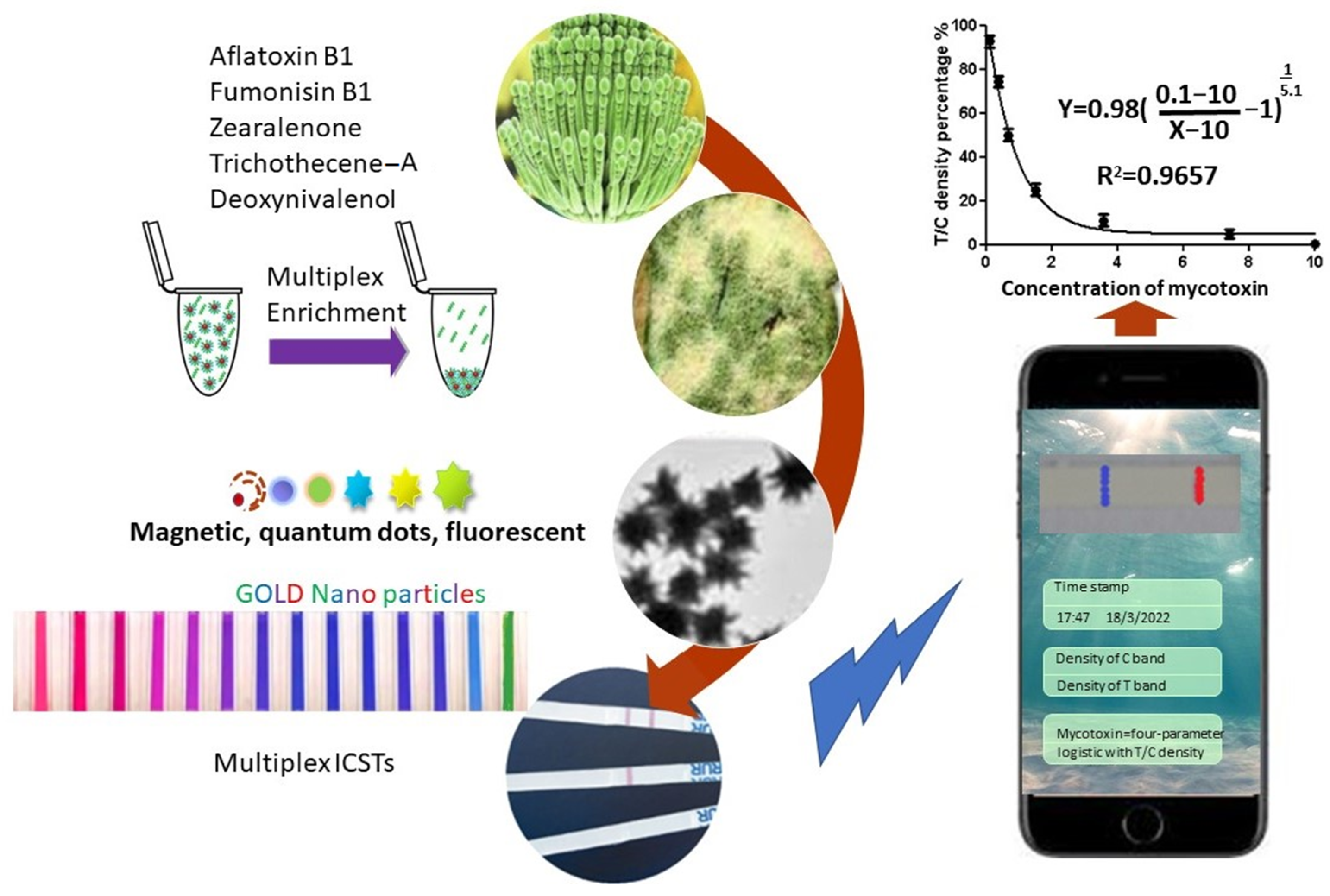
:1. Introduction
2. Research Methods
3. Results
4. Discussion
5. Conclusions
Author Contributions
Funding
Institutional Review Board Statement
Informed Consent Statement
Data Availability Statement
Conflicts of Interest
References
- Kumar, P.; Mahato, D.K.; Kamle, M.; Mohanta, T.K.; Kang, S.G. Aflatoxins: A Global Concern for Food Safety, Human Health and Their Management. Front. Microbiol. 2017, 7, 2170. [Google Scholar] [CrossRef] [PubMed] [Green Version]
- Borzekowski, A.; Anggriawan, R.; Auliyati, M.; Kunte, H.-J.; Koch, M.; Rohn, S.; Karlovsky, P.; Maul, R. Formation of Zearalenone Metabolites in Tempeh Fermentation. Molecules 2019, 24, 2697. [Google Scholar] [CrossRef] [PubMed] [Green Version]
- Rogowska, A.; Pomastowski, P.; Sagandykova, G.; Buszewski, B. Zearalenone, and its metabolites: Effect on human health, metabolism and neutralisation methods. Toxicon 2019, 162, 46–56. [Google Scholar] [CrossRef] [PubMed]
- Perincherry, L.; Lalak-Kańczugowska, J.; Stępień, L. Fusarium-Produced Mycotoxins in Plant-Pathogen Interactions. Toxins 2019, 11, 664. [Google Scholar] [CrossRef] [Green Version]
- Yu, S.; Jia, B.; Liu, N.; Yu, D.; Wu, A. Evaluation of the Individual and Combined Toxicity of Fumonisin Mycotoxins in H uman Gastric Epithelial Cells. Int. J. Mol. Sci. 2020, 21, 5917. [Google Scholar] [CrossRef]
- Adhikari, M.; Negi, B.; Kaushik, N.; Adhikari, A.; Al-Khedhairy, A.A.; Kaushik, N.K.; Choi, E.H. T–2 mycotoxin: Toxicological effects and decontamination strategies. Oncotarget 2017, 8, 33933–33952. [Google Scholar] [CrossRef] [Green Version]
- Delmulle, B.S.; De Saeger, S.M.; Sibanda, L.; Barna-Vetro, I.; Van Peteghem, C.H. Development of an immunoassay-based lateral flow dipstick for the rapid detection of aflatoxin B1 in pig feed. J. Agric. Food Chem. 2005, 53, 3364–3368. [Google Scholar] [CrossRef]
- Sun, K.; Jian, F.; Jayas, D.S.; White, N.D.G. Quality changes in high and low oil content canola during storage: Part I–Safe storage time under constant temperatures. J. Stored Prod. Res. 2014, 59, 320–327. [Google Scholar] [CrossRef]
- Navarro, J.L.; Biglione, C.; Paesani, C.; Moiraghi, M.; León, A.E.; Steffolani, M.E. Effect of wheat pearling process on composition and nutritional profile of flour and its bread-making performance. Int. J. Food Sci. 2022, 57, 249–257. [Google Scholar] [CrossRef]
- Anfossi, L.; Baggiani, C. Lateral Flow Immunoassays for Aflatoxins B and G and for Aflatoxin M1. In Aflatoxins-Recent Advances and Future Prospects; Razzaghi-Abyaneh, M., Ed.; InTech: Rijeka, Croatia, 2013; Volume 15, pp. 315–340. [Google Scholar]
- European Commission. European Commission Regulation No 1881/2006 setting maximum levels for certain contaminants in foodstuffs. Off. J. Eur. Union 2006, 364, 5–24. [Google Scholar]
- European Commission. European Commission Recommendation 2006/576/EC on the presence of deoxynivalenol, zearalenone, ochratoxin A, T–2 and HT–2 and fumonisins in products intended for animal feeding. Off. J. Eur. Union 2006, 229, 7–9. [Google Scholar]
- Liu, J.; Applegate, T. Zearalenone (ZEN) in Livestock and Poultry: Dose, Toxicokinetics, Toxicity and Estrogenicity. Toxins 2020, 12, 377. [Google Scholar] [CrossRef]
- Bertuzzi, T.; Camardo Leggieri, M.; Battilani, P.; Pietri, A. Co-occurrence of type A and B trichothecenes and zearalenone in wheat grown in northern Italy over the years 2009–2011. Food Addit. Contam. Part B 2014, 7, 273–281. [Google Scholar] [CrossRef] [PubMed]
- Tan, D.C.; Flematti, G.R.; Ghisalberti, E.L.; Sivasithamparam, K.; Chakraborty, S.; Obanor, F.; Barbetti, M.J. Mycotoxins produced by Fusarium species associated with annual legume pastures and ‘sheep feed refusal disorders’ in Western Australia. Mycotoxin Res. 2011, 27, 123–135. [Google Scholar] [CrossRef]
- Radoi, A.; Targa, M.; Prieto-Simon, B.; Marty, J.L. Enzyme-Linked Immunosorbent Assay (ELISA) based on superparamagnetic nanoparticles for aflatoxin M1 detection. Talanta 2008, 77, 138–143. [Google Scholar] [CrossRef]
- Sajid, M.; Kawde, A.-N.; Daud, M. Designs, formats and applications of lateral flow assay: A literature review. J. Saudi Chem. Soc. 2015, 19, 689–705. [Google Scholar] [CrossRef] [Green Version]
- Zhang, J.; Ramu, V.; Zhou, X.-Q.; Frias, C.; Ruiz-Molina, D.; Bonnet, S.; Roscini, C.; Novio, F. Photoactivable Ruthenium-Based Coordination Polymer Nanoparticles for Light-Induced Chemotherapy. Nanomaterials 2021, 11, 3089. [Google Scholar] [CrossRef] [PubMed]
- Huang, Y.; Du, Z.; Zheng, T.; Jing, W.; Liu, H.; Liu, X.; Mao, J.; Zhang, X.; Cai, Q.; Chen, D.; et al. Antibacterial, conductive, and osteocompatible polyorganophosphazene microscaffolds for the repair of infectious calvarial defect. J. Biomed. Mater. Res. A. 2021, 109, 2580–2596. [Google Scholar] [CrossRef]
- Khaled, M.Q.; Thalij, K.M. Effect of Aflatoxin B1 Contaminated Corn and Their Products on Some Physiology Parameters in Laboratory Rats. In IOP Conference Series: Earth and Environmental Science, Proceedings of the Fourth International Conference for Agricultural and Sustainability Sciences, Babil, Iraq, 4–5 October 2021; IOP Publishing: Bristol, UK, 2021; Volume 910. [Google Scholar]
- Mayo, M.L.; Eberly, J.O.; Crocker, F.H.; Indest, K.J. Modeling a synthetic aptamer-based riboswitch biosensor sensitive to low hexahydro-1,3,5-trinitro-1,3,5-triazine (RDX) concentrations. PLoS ONE 2020, 15, e0241664. [Google Scholar] [CrossRef]
- Cordeiro, M.F.; Nunes, T.R.S.; Bezerra, F.G.; Damasco, P.K.M.; Silva, W.A.V.; Ferreira, M.R.A.; Magalhães, O.M.C.; Soares, L.A.L.; Cavalcanti, I.M.F.; Pitta, M.G.R.; et al. Phytochemical characterization and biological activities of Plectranthus barbatus Andrews. Braz. J. Biol. 2021, 82, e236297. [Google Scholar] [CrossRef]
- Heuer, C.; Bahnemann, J.; Scheper, T.; Segal, E. Paving the Way to Overcome Antifungal Drug Resistance: Current Practices and Novel Developments for Rapid and Reliable Antifungal Susceptibility Testing. Small Methods 2021, 5, 2100713. [Google Scholar] [CrossRef]
- Knoppers, T.; Beauchamp, E.; Dewar, K.; Kimmins, S.; Bourque, G.; Joly, Y.; Dupras, C. The omics of our lives: Practices and policies of direct-to-consumer epigenetic and microbiomic testing companies. New Genet. Soc. 2021, 40, 541–569. [Google Scholar] [CrossRef]
- Cheraghi-Shahi, S.; Dadmehr, M.; Korouzhdehi, B.; Tavassoli, A. A novel colorimetric biosensor for sensitive detection of aflatoxin mediated by bacterial enzymatic reaction in saffron samples. Nanotechnology 2021, 32, 505503. [Google Scholar] [CrossRef]
- Nesakumar, N.; Lakshmanakumar, M.; Srinivasan, S.; Jayalatha, J.B.B.; Balaguru-Rayappan, J.B.A. Principles and Recent Advances in Biosensors for Pathogens Detection. ChemistrySelect 2021, 6, 10063–10091. [Google Scholar] [CrossRef]
- Hao, Z.; Chen, H.; Shi, X.; Tan, W.; Zhu, G. Fabrication for paper-based microfluidic analytical devices and saliva analysis application. Microfluid. Nanofluid. 2021, 25, 80. [Google Scholar] [CrossRef]
- Chang, C.-C.; Chen, C.-P.; Wu, T.-H.; Yang, C.-H.; Lin, C.-W.; Chen, C.-Y. Gold Nanoparticle-Based Colorimetric Strategies for Chemical and Biological Sensing Applications. Nanomaterials 2019, 9, 861. [Google Scholar] [CrossRef] [Green Version]
- Cardoso, S.; Leitao, D.C.; Dias, T.M.; Valadeiro, J.; Silva, M.D.; Chicharo, A.; Silverio, V.; Gaspar, J.; Freitas, P.P. Challenges and trends in magnetic sensor integration with microfluidics for biomedical applications. J. Phys. D Appl. Phys. 2017, 50, 213001. [Google Scholar] [CrossRef]
- Sarver, R.W.; Almy, D.J.; Bergeron, E.R.; Strong, B.F.; Steiner, B.A.; Donofrio, R.; Lupo, A.J.; Gray, R.L.; Sperry, A.K. Overview of Portable Assays for the Detection of Mycotoxins, Allergens, and Sanitation Monitoring. J. AOAC Int. 2021, 104, 39–48. [Google Scholar] [CrossRef]
- Mak, A.C.; Osterfeld, S.J.; Yu, H.; Wang, S.X.; Davis, R.W.; Jejelowo, O.A.; Pourmand, N. Sensitive giant magnetoresistive-based immunoassay for multiplex mycotoxin detection. Biosens. Bioelectron. 2010, 25, 1635–1639. [Google Scholar] [CrossRef] [Green Version]
- Liao, J.; Li, H. Lateral flow immunodipstick for visual detection of aflatoxin B1 in food using immuno-nanoparticles composed of a silver core and a gold shell. Microchim. Acta 2010, 171, 289–295. [Google Scholar] [CrossRef]
- Tittlemier, S.A.; Cramer, B.; Dall’Asta, C.; DeRosa, M.C.; Lattanzio, V.M.T.; Malone, R.; Maragos, C.; Stranska, M.; Sumarah, M.W. Developments in mycotoxin analysis: An update for 2020–2021. World Mycotoxin J. 2022, 15, 3–25. [Google Scholar] [CrossRef]
- Lu, W.; Wang, K.; Xiao, K.; Qin, W.; Hou, Y.; Xu, H.; Yan, X.; Chen, Y.; Cui, D.; He, J. Dual Immunomagnetic Nanobeads-Based Lateral Flow Test Strip for Simultaneous Quantitative Detection of Carcinoembryonic Antigen and Neuron Specific Enolase. Sci. Rep. 2017, 7, 42414. [Google Scholar] [CrossRef]
- Ngom, B.; Guo, Y.; Wang, X.; Bi, D. Development and application of lateral flow test strip technology for detection of infectious agents and chemical contaminants: A review. Anal. Bioanal. Chem. 2010, 397, 1113–1135. [Google Scholar] [CrossRef]
- Sojinrin, T.; Liu, K.; Wang, K.; Cui, D.; Bryne, H.J.; Curtin, J.F.; Tian, F. Developing Gold Nanoparticles-Conjugated Aflatoxin B1 Antifungal Strips. Int. J. Mol. Sci. 2019, 20, 6260. [Google Scholar] [CrossRef] [Green Version]
- Ji, Y.; Ren, M.; Li, Y.; Huang, Z.; Shu, M.; Yang, H.; Xiong, Y.; Xu, Y. Detection of aflatoxin B1 with immunochromatographic test strips: Enhanced signal sensitivity using gold nanoflowers. Talanta 2015, 142, 206–212. [Google Scholar] [CrossRef]
- Urusov, A.; Zherdev, A.V.; Dzantiev, B.B. Use of gold nanoparticle-labeled secondary antibodies to improve the sensitivity of an immunochromatographic assay for aflatoxin B1. Microchim. Acta 2014, 181, 1939–1946. [Google Scholar] [CrossRef]
- Di-Nardo, F.; Alladio, E.; Baggiani, C.; Cavalera, S.; Giovannoli, C.; Spano, G.; Anfossi, L. Colour-encoded lateral flow immunoassay for the simultaneous detection of aflatoxin B1 and type-B fumonisins in a single Test line. Talanta 2019, 192, 288–294. [Google Scholar] [CrossRef]
- Xiulan, S.; Xiaolian, Z.; Jian, T.; Xiaohong, G.; Jun, Z.; Chu, F.S. Development of an immunochromatographic assay for detection of aflatoxin B1 in foods. Food Control 2006, 17, 256–262. [Google Scholar] [CrossRef]
- Liu, B.-H.; Hsu, Y.-T.; Lu, C.-C.; Yu, F.-Y. Detecting aflatoxin B1 in foods and feeds by using sensitive rapid enzyme-linked immunosorbent assay and gold nanoparticle immunochromatographic strip. Food Control 2013, 30, 184–189. [Google Scholar] [CrossRef]
- Zheng, Q.; Wu, H.; Jiang, H.; Yang, J.; Gao, Y. Development of a Smartphone-Based Fluorescent Immunochromatographic Assay Strip Reader. Sensors 2020, 20, 4521. [Google Scholar] [CrossRef]
- Li, X.; Yang, F.; Wong, J.X.H.; Yu, H.Z. Integrated Smartphone-App-Chip System for On-Site Parts-Per-Billion-Level Colorimetric Quantitation of Aflatoxins. Anal. Chem. 2017, 89, 8908–8916. [Google Scholar] [CrossRef] [PubMed] [Green Version]
- Lee, S.; Kim, G.; Moon, J. Performance improvement of the one-dot lateral flow immunoassay for aflatoxin B1 by using a smartphone-based reading system. Sensors 2013, 13, 5109–5116. [Google Scholar] [CrossRef] [PubMed] [Green Version]
- Kawashima, L.M.; Vieira, A.P.; Soares, L.M.V. Fumonisin B1 and ochratoxin A in beers made in Brazil. Food Sci. Technol. 2007, 27, 317–323. [Google Scholar] [CrossRef] [Green Version]
- Wang, Z.; Li, H.; Li, C.; Yu, Q.; Shen, J.; De-Saeger, S. Development and Application of a Quantitative Fluorescence-Based Immunochromatographic Assay for Fumonisin B1 in Maize. J. Agric. Food Chem. 2014, 62, 6294–6298. [Google Scholar] [CrossRef]
- Ren, W.; Xu, Y.; Huang, Z.; Li, Y.; Tu, Z.; Zou, L.; He, Q.; Fu, J.; Liu, S.; Hammock, B.D. Single-chain variable fragment antibody-based immunochromatographic strip for rapid detection of fumonisin B1 in maize samples. Food Chem. 2020, 319, 126546. [Google Scholar] [CrossRef]
- Venkataramana, M.; Navya, K.; Chandranayaka, S.; Priyanka, S.R.; Murali, H.S.; Batra, H.V. Development and validation of an immunochromatographic assay for rapid detection of fumonisin B1 from cereal samples. J. Food Sci. Technol. 2014, 51, 1920–1928. [Google Scholar] [CrossRef] [Green Version]
- Tang, X.; Li, P.; Zhang, Q.; Zhang, Z.; Zhang, W.; Jiang, J. Time-Resolved Fluorescence Immunochromatographic Assay Developed Using Two Idiotypic Nanobodies for Rapid, Quantitative, and Simultaneous Detection of Aflatoxin and Zearalenone in Maize and Its Products. Anal. Chem. 2017, 89, 11520–11528. [Google Scholar] [CrossRef]
- Hong, X.; Mao, Y.; Yang, C.; Liu, Z.; Li, M.; Du, D. Contamination of Zearalenone from China in 2019 by a Visual and Digitized Immunochromatographic Assay. Toxins 2020, 12, 521. [Google Scholar] [CrossRef]
- Wang, D.; Zhang, Z.; Zhang, Q.; Wang, Z.; Zhang, W.; Yu, L.; Li, H.; Jiang, J.; Li, P. Rapid and sensitive double-label based immunochromatographic assay for zearalenone detection in cereals. Electrophoresis 2018, 39, 2125–2130. [Google Scholar] [CrossRef]
- Zhang, X.; He, K.; Fang, Y.; Cao, T.; Paudyal, N.; Zhang, X.F.; Song, H.H.; Li, X.L.; Fang, W.H. Dual flow immunochromatographic assay for rapid and simultaneous quantitative detection of ochratoxin A and zearalenone in corn, wheat, and feed samples. J. Zhejiang Univ. Sci. B 2018, 19, 871–883. [Google Scholar] [CrossRef]
- Sun, Y.; Xing, G.; Yang, J.; Wang, F.; Deng, R.; Zhang, G.; Hu, X.; Zhang, Y. Development of an immunochromatographic test strip for simultaneous qualitative and quantitative detection of ochratoxin A and zearalenone in cereal. J. Sci. Food Agric. 2016, 96, 3673–3678. [Google Scholar] [CrossRef]
- Qie, Z.; Shi, J.; Yan, W.; Gao, Z.; Meng, W.; Xiao, R.; Wang, S. Immunochromatographic assay for T–2 toxin based on luminescent quantum dot beads. RSC Adv. 2019, 9, 38697–38702. [Google Scholar] [CrossRef] [Green Version]
- Urusov, A.E.; Petrakova, A.V.; Bartosh, A.V.; Gubaydullina, M.K.; Zherdev, A.V.; Dzantiev, B.B. Immunochromatographic assay of T–2 toxin using labeled anti-species antibodies. Appl. Biochem. 2017, 53, 594–599. [Google Scholar] [CrossRef]
- Petrakova, A.V.; Urusov, A.E.; Voznyak, M.V.; Zherdev, A.V.; Dzantiev, B.B. Immunochromatographic test system for the detection of T–2 toxin. Appl. Biochem. 2015, 51, 688–694. [Google Scholar] [CrossRef]
- Petrakova, A.V.; Urusov, A.E.; Gubaydullina, M.K.; Bartosh, A.V.; Zherdev, A.V.; Dzantiev, B.B. “External” antibodies as the simplest tool for sensitive immunochromatographic tests. Talanta 2017, 175, 77–81. [Google Scholar] [CrossRef]
- Zhang, X.; Yu, X.; Wen, K.; Li, C.; Mujtaba-Mari, G.; Jiang, H.; Shi, W.; Shen, J.; Wang, Z. Multiplex Lateral Flow Immunoassays Based on Amorphous Carbon Nanoparticles for Detecting Three Fusarium Mycotoxins in Maize. J. Agric. Food Chem. 2017, 65, 8063–8071. [Google Scholar] [CrossRef]
- Wang, Z.; Duan, N.; Hun, X.; Wu, S. Electrochemiluminescent aptamer biosensor for the determination of ochratoxin A at a gold-nanoparticles-modified gold electrode using N-(aminobutyl)-N-ethylisoluminol as a luminescent label. Anal. Bioanal. Chem. 2010, 398, 2125–2132. [Google Scholar] [CrossRef]
- Nile, S.H.; Baskar, V.; Selvaraj, D.; Nile, A.; Xiao, J.; Kai, G. Nanotechnologies in Food Science: Applications, Recent Trends, and Future Perspectives. Nano-Micro Lett. 2020, 12, 45. [Google Scholar] [CrossRef] [Green Version]
- Liu, K.; He, Z.; Byrne, H.J.; Curtin, J.F.; Tian, F. Investigating the Role of Gold Nanoparticle Shape and Size in Their Toxicities to Fungi. Int. J. Environ. Res. Public Health 2018, 15, 998. [Google Scholar] [CrossRef] [Green Version]
- Tian, F.; Clift, M.J.; Casey, A.; Del-Pino, P.; Pelaz, B.; Conde, J.; Byrne, H.J.; Rothen-Rutishauser, B.; Estrada, G.; Fuente, J.M.; et al. Investigating the role of shape on the biological impact of gold nanoparticles in vitro. Nanomedicine 2015, 10, 2643–2657. [Google Scholar] [CrossRef] [Green Version]
- Szulc, J.; Kołodziej, A.; Ruman, T. Silver-109/Silver/Gold Nanoparticle-Enhanced Target Surface-Assisted Laser Desorption/Ionisation Mass Spectrometry-The New Methods for an Assessment of Mycotoxin Concentration on Building Materials. Toxins 2021, 13, 45. [Google Scholar] [CrossRef]
- Anfossi, L.; Di-Nardo, F.; Cavalera, S.; Giovannoli, C.; Spano, G.; Speranskaya, E.S.; Goryacheva, I.Y.; Baggiani, C. A lateral flow immunoassay for straightforward determination of fumonisin mycotoxins based on the quenching of the fluorescence of CdSe/ZnS quantum dots by gold and silver nanoparticles. Microchim. Acta 2018, 185, 94. [Google Scholar] [CrossRef]
- Asharani, P.V.; Lianwu, Y.; Gong, Z.; Valiyaveettil, S. Comparison of the toxicity of silver, gold and platinum nanoparticles in developing zebrafish embryos. Nanotoxicology 2011, 5, 43–54. [Google Scholar] [CrossRef]
- Siddiqi, K.S.; Husen, A.; Rao, R.A.K. A review on biosynthesis of silver nanoparticles and their biocidal properties. J. Nanobiotechnol. 2018, 16, 14. [Google Scholar] [CrossRef]
- Ahamed, M.; Karns, M.; Goodson, M.; Rowe, J.; Hussain, S.M.; Schlager, J.J.; Hong, Y. DNA damage response to different surface chemistry of silver nanoparticles in mammalian cells. Toxicol. Appl. Pharmacol. 2008, 233, 404–410. [Google Scholar] [CrossRef]
- Armbruster, D.A.; Pry, T. Limit of blank, limit of detection and limit of quantitation. Clin. Biochem. Rev. 2008, 29, S49–S52. [Google Scholar]
- Liu, Y.; Zhan, L.; Qin, Z.; Sackrison, J.; Bischof, J.C. Ultrasensitive and Highly Specific Lateral Flow Assays for Point-of-Care Diagnosis. ACS Nano 2021, 15, 3593–3611. [Google Scholar] [CrossRef]
- Soh, J.H.; Chan, H.-M.; Ying, J.Y. Strategies for developing sensitive and specific nanoparticle-based lateral flow assays as point-of-care diagnostic device. Nano Today 2020, 30, 100831. [Google Scholar] [CrossRef]
- Nguyen, V.-T.; Song, S.; Park, S.; Joo, C. Recent advances in high-sensitivity detection methods for paper-based lateral-flow assay. Biosens. Bioelectron. 2020, 152, 112015. [Google Scholar] [CrossRef]
- Bishop, J.D.; Hsieh, H.V.; Gasperino, D.J.; Weigl, B.H. Sensitivity enhancement in lateral flow assays: A systems perspective. Lab Chip 2019, 19, 2486–2499. [Google Scholar] [CrossRef] [Green Version]
- Nardo, F.; Chiarello, M.; Cavalera, S.; Baggiani, C.; Anfossi, L. Ten Years of Lateral Flow Immunoassay Technique Applications: Trends, Challenges and Future Perspectives. Sensors 2021, 21, 5185. [Google Scholar] [CrossRef] [PubMed]


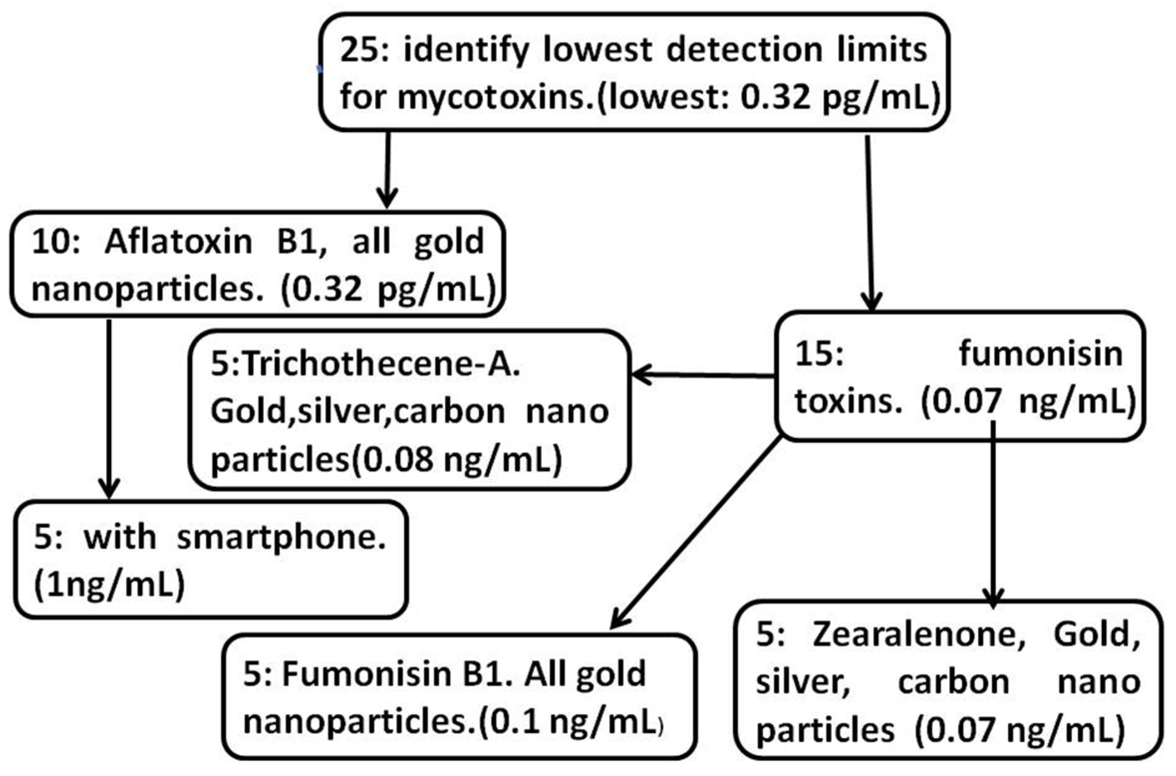
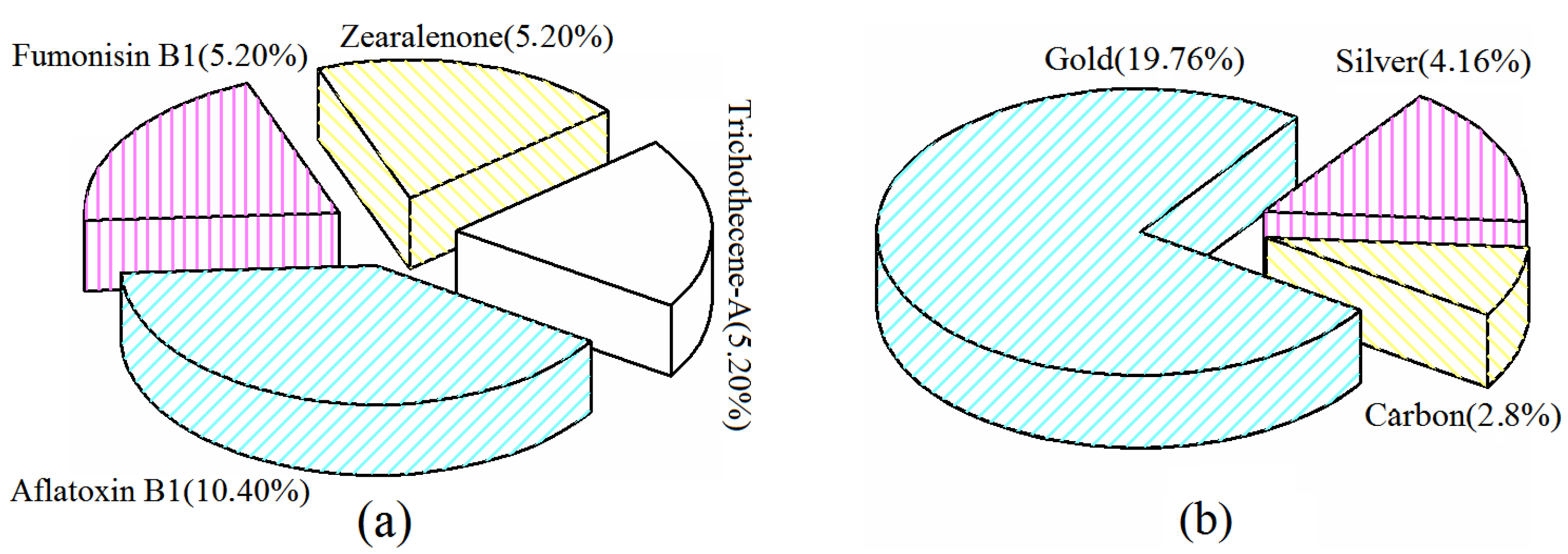
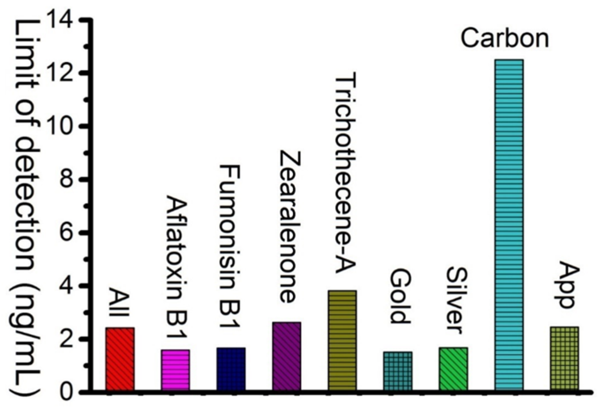
| Type | Particle | Size (nm) | Year | Replicates | Standard Deviations | Coefficient of Variation (%) | LOD (ng/mL) | Reference |
|---|---|---|---|---|---|---|---|---|
| AFB1 | Gold | na | 2013 | na | na | na | 0.1 | [10] |
| Gold + phone | 14 | 2019 | 6 | 0.01 | 2.7 | 0.3 | [36] | |
| Gold | 45 | 2015 | 6 | 0.09 | 9.47 | 0.00032 | [37] | |
| Gold | na | 2014 | na | na | na | 0.03 | [38] | |
| Gold + phone | 30 | 2019 | 6 | na | 1.5 | 2 | [39] | |
| Gold | 25 | 2006 | 11 | 0.44 | 10.4 | 2.5 | [40] | |
| Gold | na | 2013 | 3 | na | na | 2 | [41] | |
| Gold + phone | na | 2019 | 9 | na | na | 2 | [42] | |
| Gold + phone | 1.4 | 2017 | 5 | na | na | 3 | [43] | |
| Gold + phone | 40 | 2013 | 5 | na | na | 5 | [44] | |
| FB1 | Gold | na | 2007 | 12 | 0.3 | 6.67 | 0.1 | [45] |
| Gold | na | 2014 | 5 | 0.18 | 6.01 | 0.12 | [46] | |
| Gold | 30 | 2017 | 3 | na | na | 0.6 | [47] | |
| Gold | 20 | 2020 | na | na | na | 2.5 | [48] | |
| Gold | 40 | 2014 | 3 | 0.9 | 10.6 | 5 | [49] | |
| ZEN | Gold | 25 | 2017 | 3 | 0.16 | 6.2 | 0.07 | [50] |
| Gold | 17 | 2020 | 5 | 0.09 | 4.9 | 0.25 | [51] | |
| Silver | na | 2018 | 3 | na | na | 0.25 | [52] | |
| Silver | 15 | 2018 | 10 | 0.22 | 4.85 | 0.58 | [53] | |
| Carbon | 190 | 2016 | 3 | 0.05 | 3.79 | 12 | [54] | |
| T–2 | Gold | na | 2019 | 2 | na | na | 0.08 | [55] |
| Gold | 30 | 2017 | 3 | na | na | 0.1 | [56] | |
| Silver | na | 2015 | na | na | na | 0.9 | [57] | |
| Silver | 45 | 2017 | na | na | na | 5 | [58] | |
| Carbon | 120 | 2017 | na | na | na | 13 | [59] |
Publisher’s Note: MDPI stays neutral with regard to jurisdictional claims in published maps and institutional affiliations. |
© 2022 by the authors. Licensee MDPI, Basel, Switzerland. This article is an open access article distributed under the terms and conditions of the Creative Commons Attribution (CC BY) license (https://creativecommons.org/licenses/by/4.0/).
Share and Cite
Zhao, X.; Byrne, H.J.; O’Connor, C.M.; Curtin, J.; Tian, F. Limits of Detection of Mycotoxins by Laminar Flow Strips: A Review. Appl. Nano 2022, 3, 91-101. https://doi.org/10.3390/applnano3020006
Zhao X, Byrne HJ, O’Connor CM, Curtin J, Tian F. Limits of Detection of Mycotoxins by Laminar Flow Strips: A Review. Applied Nano. 2022; 3(2):91-101. https://doi.org/10.3390/applnano3020006
Chicago/Turabian StyleZhao, Xinyi, Hugh J. Byrne, Christine M. O’Connor, James Curtin, and Furong Tian. 2022. "Limits of Detection of Mycotoxins by Laminar Flow Strips: A Review" Applied Nano 3, no. 2: 91-101. https://doi.org/10.3390/applnano3020006