Technical Note

Emergence Profile Creation with CAD Technology on Vertical Edgeless Preparation (VEP)

1
Department of Medicine and Surgery, Dental School, University of Parma, Via Gramsci 14, 43126 Parma, Italy
2
Private Practice, Piazza di Porta Mascarella 7, 40126 Bologna, Italy
3
Private Practice, Via Vittorio Veneto 99, 19121 La Spezia, Italy
*
Author to whom correspondence should be addressed.
Prosthesis 2023, 5(4), 1369-1381; https://doi.org/10.3390/prosthesis5040094
Submission received: 7 November 2023 / Revised: 11 December 2023 / Accepted: 14 December 2023 / Published: 18 December 2023
(This article belongs to the Special Issue Digital Technologies, Materials and Telemedicine in Dentistry)

Abstract

:
This article proposes a digital procedure to create the emergence profile of prosthetic crowns for teeth prepared with the Vertical Edgeless Preparation (VEP) technique. This technique extends the tooth preparation below the gingival margin on the root surface of teeth with reduced periodontal attachment. The tooth prepared according to this technique does not present a defined prosthetic margin or the relative finishing line. Still, it shows an edgeless axial wall that emerges from the gingival sulcus. The digital procedure for creating a prosthetic emergence profile, although representing a novelty in absolute terms, is based on traditional concepts, as they have been known and described in the literature for a long time. This article will explain and review the various clinical and laboratory steps necessary to produce, through an entirely digital procedure, a fixed partial denture composed of three zirconia elements, which are going to be subsequently veneered in a traditional way on the vestibular side to improve the esthetic appearance.

1. Introduction

Digital technology is a consolidated reality within dental laboratories, where scanners and CAD/CAM methods have revolutionized design and production processes, combining traditional and innovative techniques and materials. It should be emphasized that the results obtained with these current digital procedures cannot disregard the knowledge of the rules and principles of traditional dentistry. As a matter of fact, in prosthodontic processes, the spread of digital technologies revealed new treatment possibilities but also proved to have some operative limitations [1,2,3]. Simplifying procedures granted by digital technologies can indeed lead to thinking that some steps can be skipped. A clear example of this includes realizing a complete digital workflow without using physical models and then adjusting the margin thickness after milling [2]. Ditching procedures, checking the margin, and other precautions are still fundamental steps in producing a precise and durable dental crown.
This work aims to show how to transfer and adapt traditional procedures and concepts onto a more modern and innovative methodology relying on digital technologies, in order to create the emergence profile of a crown prepared according to the Vertical Edgeless Preparation (VEP) technique [4,5].
This article also illustrates the complete digital workflow of a prosthetic project, from the impressions with an intraoral scanner (IoS) to the creation of a three-element fixed partial prosthesis for teeth prepared according to the VEP technique.
Different techniques for preparing complete dental crowns exist in prosthodontics, each with its characteristics, indications, advantages, and disadvantages. Complete crown preparations have been classified into vertical and horizontal preparations [6]. However, the authors have also subdivided preparation techniques into two groups: the first one includes preparations that present, at their apical part, a defined margin [7]; the second consists of the Vertical Edgeless Preparation (VEP) [4,5] and the intraoperatory prosthetic preparation, which both lack a defined margin [8].
Vertical Edgeless Preparation is the technique to which the feather edge preparation technique of the Porta Mascarella School in Bologna has been renamed. It consists of a prosthetic preparation for complete crowns, recommended for dental elements with a loss of periodontal support, and it is performed by extending the preparation under the gingival margin, in order to let the bur condition every part of the root surface which lost periodontal attachment. This preparation technique does not generate a finishing line but ends in a “finishing area” where the prosthetic margin can be positioned at different levels, ensuring an excellent marginal closure. The absence of steps, edges, or undercuts is the reason for the name “edgeless”. Clearly, the presence of a transition angle between the prepared part of the root and the apical portion is unavoidable. Still, this angle will be entirely covered by the regrowth of the soft tissues that were involuntarily but inevitably injured during the preparation process. Therefore, from a clinical perspective, its presence is irrelevant: this defines the “periodontal dominance” of the technique. The position of the final prosthetic margin can then be decided considering the thickness of the tissues, the materials used, and the esthetic needs of the area and the patient.
This not-bur-shaped technique finds its main indication in teeth with a medium/thick periodontal phenotype and a loss of periodontal attachment, which, as is well known, is not a synonym of active periodontitis. Preparing the exposed part of the dental root removes the section of dental tissue potentially contaminated by toxins and bacteria and protects it from the oral environment. Provisional crowns on VEP-prepared teeth need to be left short on the margin to let the soft tissues heal and mature without obstacles, which could lead to tissue inflammation [4,5].

2. Technique

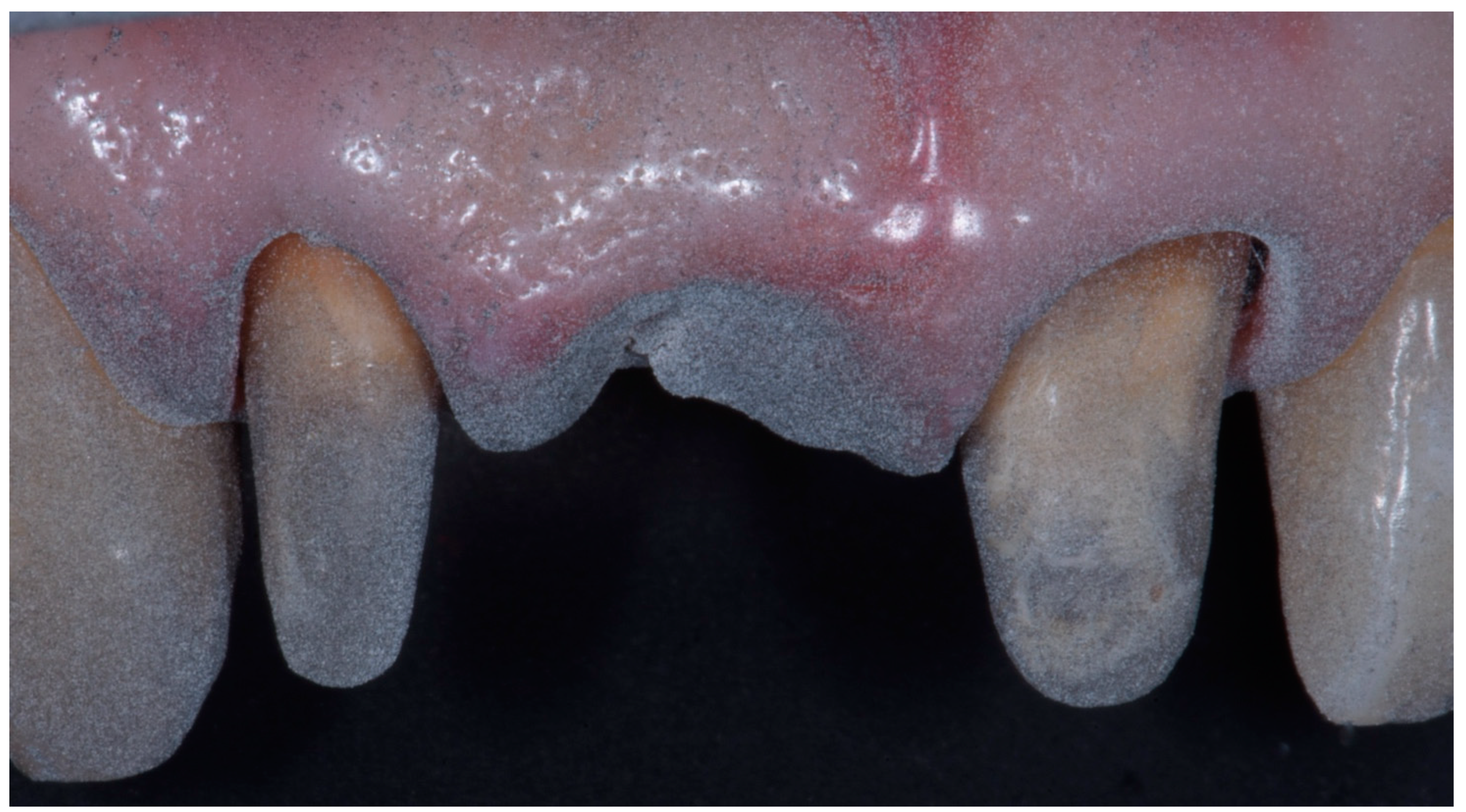
  • Prepare the abutment according to the VEP technique.
  • Reline, finish, polish, and deliver the provisional crown, following the VEP technique principles and the traditional prosthodontics concepts. The accuracy of occlusal contacts, lateral and anterior guidance, and interproximal contact points is checked.
  • After the complete healing of soft tissues, impressions are ready to be taken. Firstly, take the digital impression of the arch with the provisional in place.
  • Then, take a definitive digital impression. The double retraction cord technique is recommended to obtain a complete and clean impression of the gingival sulcus. Mattifying powder can also be used to help avoid light reflections from the teeth surfaces.
  • Take the impression of the opposite arch and the vestibular impression in maximum intercuspation position.
  • Send the obtained files to the dental lab.
  • The dental technician digitally draws the margin of the crown in the most apical visible part of the sulcus, creates the internal spacing, and uses the provisional crown shape as a starting point for the digital wax up.
  • The technician performs a digital cut back, leaving 1 mm of space on the surfaces needing layering but keeping the intrasulcular part intact.
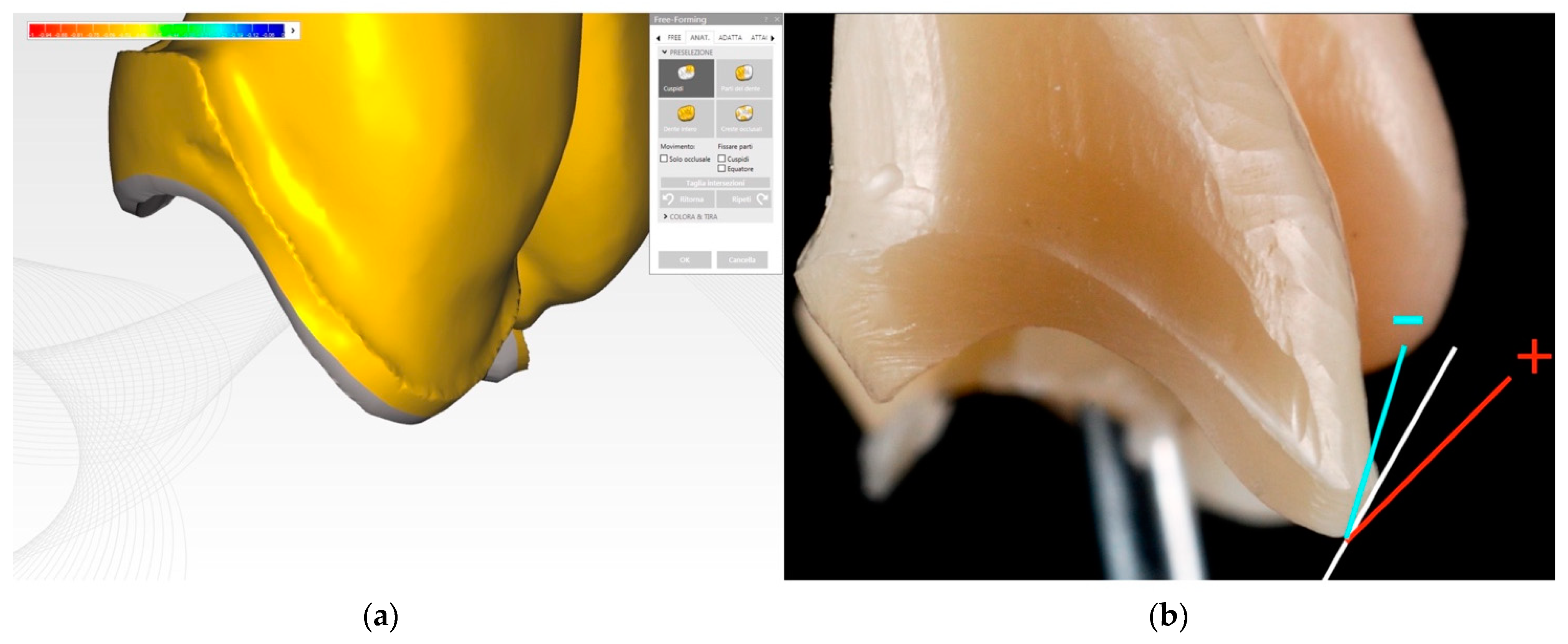
  • The resulting frame is tested on the abutment to check the accuracy of the seating and the position of the prosthetic margin, which can be shortened and arranged less apically in the sulcus. The emergence profile and its angle can be modified depending on the relationship between the abutment surface and the gingival margin, in case the gingival zenith needs to be altered or the soft tissues need more support from the prosthetic crown.
  • A 3D abutment model is printed with high precision, and the ditching procedure is performed. This allows the dental technician to check the emergence profile after designing it digitally.
  • The ceramic layering phase is performed on the 3D-printed full-mouth model obtained from the STL files.
  • The crown margin thickness must be reduced to the minimum during the finishing procedure and then polished with diamond rubbers. Intraoral checks for shape, occlusal, and interproximal contact points are performed. The congruity of the color is confirmed. The emergence profile and the depth of the crown margin within the sulcus are also verified and potentially modified.
  • The prosthetic crown is ready to be delivered following the cementation protocol of the chosen material.

2.1. Clinical Case

2.1.1. Preparation and Impressions

A fixed partial prosthesis (FPP) is taken as an example of this technique.
A 51-year-old man came to the clinic with an old metal–ceramic FPP in the esthetic area, whose margins were visible and whose esthetic appearance was insufficient. The old FPP from 1.2 to 2.1 was removed to be replaced with a new FPP made of zirconia and ceramic (Figure 1). Once the old FPP was removed, the underlying abutments were prepared according to the VEP technique (Figure 2, Figure 3 and Figure 4) and a metal–resin provisional FPP was adapted, relined, finished, and luted (Figure 5).
The provisional FPP was designed to avoid altering the patient’s maximum intercuspation position. Subsequently, the accuracy of the occlusal contacts was checked, and the protrusive and lateral guidances were functionalized.
The margins of the provisional FPP were placed at an iuxta-gingival level so that they would not interfere with the healing process of soft tissues, which were slightly but necessarily injured during the subgingival preparation procedure.
Precise impressions represent the way clinicians communicate the information of the clinical case to the dental laboratory and through which the prosthetic work is realized. Numerous preliminary conditions need to be satisfied before taking a correct impression of intraoral structures. An adequate prosthetic treatment requires the correct sequence of operative procedures and their clean execution. The accuracy in the abutment preparation, a good adaptation of the provisional restoration, and the proper management of soft tissues are crucial points in order to precisely register the information.
Digital impressions for the definitive FPP were taken four weeks after preparing, waiting for the soft tissues to heal and reach complete stability [9]. As for analogic impressions, given the type of preparation, the double cord technique was used in this case. Specifically, a silk cord 2-0 and a second silk cord 0 (Ultrapack, Ultradent Products, Inc., South Jordan, UT, USA), soaked in a 25% aluminum chloride (Racestyptine, Septodont, Saint-Maur-des-Fossés, France) solution, were used. Both cords were inserted without trauma to guarantee the displacement of the soft tissues both horizontally and vertically, allowing a better scan of the prepared dental tissues placed under the gingival margin. The digital impression was taken using the intraoral scanner Omnicam (Dentsply Sirona, Charlotte, NC, USA), and before starting with the scan, the second cord (the outermost of the two) was gently removed. A thin layer of mattifying powder (Apollo DI Speedspray, Dentsply Sirona, Charlotte, NC, USA) was applied to the teeth and the soft tissues. According to the authors’ experience, powder guarantees a more straightforward and smoother procedure and a better intrasulcular impression (Figure 6). The STL files obtained from the impression were finally sent to the dental laboratory.
The dental laboratory receives the STL files of the upper arch with the prepared abutments, the lower arch, their relationship in maximum intercuspation, and the upper arch with the provisionals in place. Then, the files are imported into the CAD software (Exocad 3.1, Exocad GmbH, Darmstadt, Germany), and the margin of the prosthetic crown, below the gingival margin, is determined. Generally, this point matches the most apical visible part of the prepared surface (Figure 7). The prosthetic axis is now defined, checking the absence of undercuts and disparallelisms between the abutments (Figure 8).
Subsequently, the software creates the internal spacing of the crown according to the length and width of the abutment and the material chosen for the prosthetic restoration. Virtual models are now articulated to check the accuracy of maximum intercuspation (Figure 9). At the same time, upper and lower models are obtained with 3D printing to be used only as control models. The impression of the prepared abutments and of the provisionals are superimposed to verify the correspondence of articulation relationships (Figure 10).
This procedure, which uses the information obtained from the provisionals, allows the technician to improve and speed up the final wax-up design (Figure 11a). Once the project is finished, the virtual cut back is performed (Figure 11b). As for analogic techniques, this allows the generation of a frame whose morphologic features guarantee an adequate resistance and an optimal and homogeneous thickness for the layering ceramic. The cut-back reduction consists of 1 mm on all the surfaces on which ceramic layering is necessary.

2.1.2. Digital Creation of the Emergence Profile

The intrasulcular part of the crown is left intact (Figure 12a). This allows for the maintenance of a margin whose properties are effective in enduring the working process and, subsequently, the load of masticatory function [6]. The prosthetic margin has a triangular form, and its dimension is determined by the area between the surface of the abutment and the gingival sulcus, which plays a crucial role in defining the anatomy of the emergency profile [10]. It is possible to modify the prosthetic margin if there is a need for corrections. It is also possible to increase or decrease the angle of the emergency profile if the harmonization of the gingival zenith is desired or more support of soft tissues is needed (Figure 12b).
During the phase of ceramic layering, the angle of the emergency profile helps to maintain the whole crown profile as defined by the digital design. The project of the zirconia frame is now ready and can be milled. The multilayered zirconia (in this case, Katana UTML, Kuraray, Chiyoda, Japan) frame is milled from a disk and then sintered in a furnace (in this case, Multimat 2sinter, Dentsply Sirona, Charlotte, NC, USA) at 1500 °C, according to the indications given by the producer (Figure 13). After sinterization, a first check is performed using the model obtained through 3D printing. The model accuracy, however, is not so high, and it is necessary to check the frame directly in the patient’s mouth. The model is used mainly as a support in the subsequent layering phases. The frame is then checked directly on the abutments, and a series of verification procedures are performed. First of all, the insertion axis is checked to verify that there are no undercuts on the prepared abutment along the insertion axis. Then, when dealing with a partial fixed prosthesis, as in the case used as an example in this article, it is necessary to check that the intermediate element guarantees the correct compression on the soft tissue and that there is no tilting.
The verification of the correct seating and the frame precision is performed through the same procedure used in the analogic workflow. A silicon paste (Fit Checker Advanced Blue, GC Corporation, Tokyo, Japan) is used to detect and eventually correct major defects (Figure 14a). Subsequently, in order to perform a more accurate check, a paste with zinc oxide and eugenol (Impression Paste Kit SS White, SS White Dental Group, Lakewood, NJ, USA) is used (Figure 14b). In this phase, the retention of the frame on the abutment is also evaluated.
During the seating verification procedures for the frame, it is possible to check the prosthetic margin in its extension, thickness, and axis. Any overextension that may interfere with the periodontal tissues can be modified and corrected. Moreover, it is possible to reduce the intrasulcular extension during the subsequent laboratory procedures if the clinician considers it necessary.
The margin thickness, measuring 0.15–0.2 mm, needs to be reduced to the minimum during the finishing procedures and then polished with diamond rubbers (Figure 15a). The ceramic layer is then placed so that it follows the emergence profile smoothly (Figure 15b).
In the case shown as an example, given the high esthetic value of the work, the ceramization of the zirconia frame is performed directly in the dental office while the patient is present (Figure 16). Before finishing this phase, the prosthesis undergoes another series of checks. First, the insertion axis is verified again, together with the suitability of the interproximal contact point. The evaluation of the emergence profile and the accessibility of the interproximal zone for hygienic maneuvers is performed.
Clearly, now that the frame is layered with ceramic, the occlusal contact points are verified first with a thin articulation paper and then with a Shimstock foil (Dental Medical, Maribor, Slovenia), which, thanks to its 8 µm thickness, allows the clinician to reach great precision. Considering that the example shown in this article involves an anterior FPP, the assessment of the incisor guidance is performed. Finally, the esthetic and phonetic analysis is performed.

3. Discussion

Vertical Edgeless Preparation (VEP) does not present a finishing line but ends in a prosthetically usable area whose extension may vary according to the depth of the periodontal sulcus. For this reason, the position of the prosthetic margin is not conditioned by finishing lines, steps, transition angles, and relative undercuts. Its position can instead be chosen on the basis of different factors, the first of which is the position of the gingival margin on the abutment [4,5].
Using intraoral scanners to make impressions of abutments prepared with the VEP technique allows for easy detection of zones with defects (preparation/scanning errors); duplication of the provisional emergence profile if necessary; and copying the form, the depth, and the width of the gingival sulcus, which is a fundamental step in the creation of the emergence profile. Moreover, all the elements obtained with a digital impression are measurable, quantifiable, and replicable. However, learning and applying the traditional and fundamental periodontal and prosthetic concepts are crucial to a successful rehabilitation. The absence of overcontour, the design of a correct emergence profile, and the right location and strength of the occlusal contact point are crucial aspects to be considered in modern prosthodontics, since materials may have changed over the years, but biology and biomechanics have not. Using an entire digital procedure may often lead to skipping essential steps in finalizing a dental crown. The fact that no physical model is produced makes it challenging to perform the verification procedure, especially when the tooth has been prepared with a vertical preparation technique [2]. Indeed, the impossibility of fulfilling the ditching procedure and the lack of control over the emergence profile can lead to huge imprecisions. Instead, with this technique, the control of the emergence profile is performed during the CAD procedure, on the 3D printed abutment model, and on the prepared abutment. Thanks to these steps, a perfect check of the emergence profile and a very thin thickness of the crown margin can be obtained. Moreover, this technique freely allows for the determination of how deep the crown margin goes in the gingival sulcus and to modify it accordingly to the patient’s esthetic needs and the gingival profile.
The precision of intraoral scanners has made significant progress in recent years, and the literature on the topic is not unanimous in defining a degree of precision compared to the traditional impression [11]. However, in vitro studies have shown that marginal and internal fit of crowns and FDP obtained from digital impressions are more precise than the ones obtained from traditional impression [12,13].
Choosing the correct material to use also requires a careful evaluation based on the literature and the clinician’s expertise [14]. Understanding and applying traditional techniques and procedures is fundamental to designing full crowns with digital tools. As a matter of fact, these digitally manufactured crowns must fulfill precision, function, and esthetic needs exactly as analogically manufactured ones. It is essential to underline that the correct use of digital procedures requires specific skills and training both for the clinician and the dental technician.

Author Contributions

Conceptualization, G.N., E.F. and R.B.; methodology, G.N., A.T. and E.M.; validation, G.M.M. and G.N.; formal analysis, A.T. and E.B.; resources, G.N. and E.M.; data curation, E.F. and R.B.; writing—original draft preparation, A.T. and E.B.; writing—review and editing, A.T., E.M. and G.M.M.; visualization, A.T.; supervision, E.M. and G.M.M.; project administration, G.M.M. All authors have read and agreed to the published version of the manuscript.

Funding

This research received no external funding.

Conflicts of Interest

The authors declare no conflict of interest.

References

  1. Richert, R.; Goujat, A.; Venet, L.; Viguie, G.; Viennot, S.; Robinson, P.; Farges, J.C.; Fages, M.; Ducret, M. Intraoral Scanner technologies: A review to make a successful impression. J. Healthc. Eng. 2017, 2017, 8427595. [Google Scholar] [CrossRef] [PubMed]
  2. Joda, T.; Zarone, F.; Ferrari, M. The complete digital workflow in fixed prosthodontics: A systematic review. BMC Oral Health 2017, 17, 124. [Google Scholar] [CrossRef]
  3. Siqueira, R.; Galli, M.; Chen, Z.; Mendonça, G.; Meirelles, L.; Wang, H.-L.; Chan, H.-L. Intraoral scanning reduces procedure time and improves patient comfort in fixed prosthodontics and implant dentistry: A systematic review. Clin. Oral Investig. 2021, 25, 6517–6531. [Google Scholar] [CrossRef] [PubMed]
  4. Noè, G.; Toffoli, A.; Foce, E.; Di Febo, G.; Carnevale, G.; Bonfiglioli, R.; Macaluso, G.M.; Manfredi, E. Vertical Edgeless Preparation: Periodontal Dominance in Prosthetic Crown Preparation. Prosthesis 2023, 5, 358–367. [Google Scholar] [CrossRef]
  5. Foce, E.; Noè, G.; Di Febo, G.; Bonfiglioli, R.; Carnevale, G. Vertical Edgeless Preparation (VEP): La Dominanza Parodontale nella Preparazione Protesica; Quintessence Publishing: Berlin, Germany, 2021; 80p. [Google Scholar]
  6. Goodacre, C.J.; Campagni, W.V.; Aquilino, S.A. Tooth preparation for complete crown: An art form based on scientific principles. J. Prosthet. Dent. 2001, 85, 363–376. [Google Scholar] [CrossRef] [PubMed]
  7. Gavelis, J.R.; Morency, J.D.; Riley, E.D.; Sozio, R.B. The effect of various finishing lines on the marginal seal and occlusal seat of full crown preparation. J. Prosthet. Dent. 1981, 45, 138–145. [Google Scholar] [CrossRef] [PubMed]
  8. Carnevale, G.; Di Febo, G.; Fuzzi, M. A retrospective analysis of the perio-prosthetic aspects of teeth re-prepared during periodontal surgery. J. Clin. Periodontol. 1990, 17, 313–316. [Google Scholar] [CrossRef] [PubMed]
  9. Sculean, A.; Gruber, R.; Bosshardt, D.D. Soft tissue wound healing around teeth and dental implants. J. Clin. Periodontol. 2014, 41 (Suppl. S15), S6–S22. [Google Scholar] [CrossRef] [PubMed]
  10. Kay, H.B. Criteria for restorative contours in the altered periodontal environment. Int. J. Periodontics Restor. Dent. 1985, 5, 42–63. [Google Scholar]
  11. Ahlholm, P.; Sipilä, K.; Vallittu, P.; Jakonen, M.; Kotiranta, U. Digital Versus Conventional Impressions in Fixed Prosthodontics: A Review. J. Prosthodont. 2018, 27, 35–41. [Google Scholar] [CrossRef]
  12. Su, T.S.; Sun, J. Comparison of marginal and internal fit of 3-unit ceramic fixed dental prostheses made with either a conventional or digital impression. J. Prosthet. Dent. 2016, 116, 362–367. [Google Scholar] [CrossRef] [PubMed]
  13. Chochlidakis, K.M.; Papaspyridakos, P.; Geminiani, A.; Chen, C.J.; Feng, I.J.; Ercoli, C. Digital versus conventional impressions for fixed prosthodontics: A systematic review and meta-analysis. J. Prosthet. Dent. 2016, 116, 184–190. [Google Scholar] [CrossRef] [PubMed]
  14. Fuzzi, M.; Tricarico, M.G.; Ferrari Cagidiaco, E.; Bonadeo, G.; Sorrentino, R.; Ferrari, M. Nanoleakage and internal adaptation of zirconia and lithium disilicate single crowns with feather edge preparation. J. Osseointegr. 2017, 9, 250–262. [Google Scholar]
Figure 1. The abutments as they were found under the old FPP.
Prosthesis 05 00094 g001
Figure 2. The periodontal mapping is performed according to the VEP protocols.
Prosthesis 05 00094 g002
Figure 3. The bur is used according to the periodontal mapping.
Prosthesis 05 00094 g003
Figure 4. The abutments prepared according to the VEP technique.
Prosthesis 05 00094 g004
Figure 5. Provisional FPP in place.
Prosthesis 05 00094 g005
Figure 6. The powder is used to take IoS impressions to obtain a more predictable result.
Prosthesis 05 00094 g006
Figure 7. The margin of the prosthetic crown is digitally placed at the most apical visible position.
Prosthesis 05 00094 g007
Figure 8. Definition of the insertion axis: the absence of disparallelism and undercuts are checked.
Prosthesis 05 00094 g008
Figure 9. Internal spacing is defined.
Prosthesis 05 00094 g009
Figure 10. Impression with the provisional FPP.
Prosthesis 05 00094 g010
Figure 11. (a) Digital wax-up and (b) digital cut-back.
Prosthesis 05 00094 g011
Figure 12. Intrasulcular part of the crown. (a) Digital and (b) milled.
Prosthesis 05 00094 g012
Figure 13. Zirconia frame.
Prosthesis 05 00094 g013
Figure 14. Correct seating and frame precision are checked with (a) silicon paste and (b) paste with zinc oxide and eugenol.
Prosthesis 05 00094 g014
Figure 15. Intrasulcular component of the frame without (a) and with (b) the ceramic layer.
Prosthesis 05 00094 g015
Figure 16. Final result.
Prosthesis 05 00094 g016
Disclaimer/Publisher’s Note: The statements, opinions and data contained in all publications are solely those of the individual author(s) and contributor(s) and not of MDPI and/or the editor(s). MDPI and/or the editor(s) disclaim responsibility for any injury to people or property resulting from any ideas, methods, instructions or products referred to in the content.

