
Digital Analysis of a Novel Impression Method Named the Biological-Oriented Digital Impression Technique: A Clinical Audit

 ,
,  and
and 
Abstract
:1. Introduction
2. Case Presentation
3. Discussion
Limitations
4. Conclusions
Author Contributions
Funding
Institutional Review Board Statement
Informed Consent Statement
Data Availability Statement
Conflicts of Interest
References
- Cötert, H.S.; Dündar, M.; Oztürk, B. The effect of various preparation designs on the survival of porcelain laminate veneers. J. Adhes. Dent. 2009, 11, 405–411. [Google Scholar]
- Sirous, S.; Navadeh, A.; Ebrahimgol, S.; Atri, F. Effect of preparation design on marginal adaptation and fracture strength of ceramic occlusal veneers: A systematic review. Clin. Exp. Dent. Res. 2022, 8, 1391–1403. [Google Scholar] [CrossRef]
- Gracis, S.; Thompson, V.P.; Ferencz, J.L.; Silva, N.R.; Bonfante, E.A. A new classification system for all-ceramic and ceramic-like restorative materials. Int. J. Prosthodont. 2015, 28, 227–235. [Google Scholar] [CrossRef]
- Malterud, M.I. Minimally invasive restorative dentistry: A biomimetic approach. Pract. Proced. Aesthet. Dent. 2006, 18, 409–414. [Google Scholar]
- Agarwal, S.; Maiti, S.; Ashok, V. Correlation of soft tissue biotype with pink aesthetic score in single full veneer crown. Bioinformation 2020, 16, 1139–1144. [Google Scholar]
- Podhorsky, A.; Rehmann, P.; Wöstmann, B. Tooth preparation for full-coverage restorations-a literature review. Clin. Oral Investig. 2015, 19, 959–968. [Google Scholar] [CrossRef]
- Łabno, P.; Drobnik, K. Comparison of horizontal and vertical methods of tooth preparation for a prosthetic crown. J. Pre Clin. Clin. Res. 2020, 14, 25–28. [Google Scholar] [CrossRef]
- Shillingburg, H.T.; Hobo, S.; Fisher, D.W. Preparation design and margin distortion in porcelain-fused-to-metal restorations. J. Prosthet. Dent. 2003, 29, 276–284. [Google Scholar] [CrossRef]
- Paniz, G.; Nart, J.; Gobbato, L.; Mazzocco, F.; Stellini, E.; De Simone, G.; Bressan, E. Clinical Periodontal Response to Anterior All-Ceramic Crowns with Chamfer or Feather-edge Subgingival Tooth Preparations: Six-Month Results and Patient Perception. Int. J. Periodontics Restor. Dent. 2017, 37, 61–68. [Google Scholar] [CrossRef]
- Giudice, A.L.; Quinzi, V.; Ronsivalle, V.; Farronato, M.; Nicotra, C.; Indelicato, F.; Isola, G. Evaluation of imaging software accuracy for 3-dimensional analysis of the mandibular condyle. A comparative study using a surface-to-surface matching technique. Int. J. Environ. Res. Public Health 2020, 17, 4789. [Google Scholar] [CrossRef]
- Loi, I.; Di Felice, A. Biologically oriented preparation technique (BOPT): A new approach for prosthetic restoration of periodontically healthy teeth. Eur. J. Esthet. Dent. 2013, 8, 10–23. [Google Scholar]
- Canullo, L.; Tallarico, M.; Pradies, G.; Marinotti, F.; Loi, I.; Cocchetto, R. Soft and hard tissue response to an implant with a convergent collar in the esthetic area: Preliminary report at 18 months. Int. J. Esthet. Dent. 2017, 12, 306–323. [Google Scholar]
- Abad-Coronel, C.; Villacís Manosalvas, J.; Palacio Sarmiento, C.; Esquivel, J.; Loi, I.; Pradíes, G. Clinical outcomes of the biologically oriented preparation technique (BOPT) in fixed dental prostheses: A systematic review. J. Prosthet. Dent. 2022. online ahead of print. [Google Scholar]
- Serra-Pastor, B.; Loi, I.; Fons-Font, A.; Solá-Ruíz, M.F.; Agustín-Panadero, R. Periodontal and prosthetic outcomes on teeth prepared with biologically oriented preparation technique: A 4-year follow-up prospective clinical study. J. Prosthodont. Res. 2019, 63, 415–420. [Google Scholar] [CrossRef] [PubMed]
- The Glossary of Prosthodontic Terms: Ninth Edition. J. Prosthet. Dent. 2017, 117, e1–e105. [CrossRef]
- Ferrari, M.; Cagidiaco, M.C.; Ercoli, C. Tissue management with a new gingival retraction material: A preliminary clinical report. J. Prosthet. Dent. 1996, 75, 242–247. [Google Scholar] [CrossRef]
- Thimmappa, M.; Bhatia, M.; Somani, P.; Kumar, D.R.V. Comparative evaluation of three noninvasive gingival displacement systems: An in vivo study. J. Indian Prosthodont. Soc. 2018, 18, 122–130. [Google Scholar] [CrossRef]
- Einarsdottir, E.R.; Lang, N.P.; Aspelund, T.; Pjetursson, B.E. A multicenter randomized, controlled clinical trial compares displacement cords, an aluminum chloride paste, and a combination of paste and cords for tissue displacement. J. Prosthet. Dent. 2018, 119, 82–88. [Google Scholar] [CrossRef] [PubMed]
- Cicciù, M.; Fiorillo, L.; D’Amico, C.; Gambino, D.; Amantia, E.M.; Laino, L.; Crimi, S.; Campagna, P.; Bianchi, A.; Herford, A.S.; et al. 3D Digital Impression Systems Compared with Traditional Techniques in Dentistry: A Recent Data Systematic Review. Materials 2020, 13, 1982. [Google Scholar] [CrossRef] [PubMed]
- Papaspyridakos, P.; Vazouras, K.; Chen, Y.W.; Kotina, E.; Natto, Z.; Kang, K.; Chochlidakis, K. Digital vs Conventional Implant Impressions: A Systematic Review and Meta-Analysis. J. Prosthodont. 2020, 29, 660–678. [Google Scholar] [CrossRef]
- Tallarico, M. Computerization and Digital Workflow in Medicine: Focus on Digital Dentistry. Materials 2020, 13, 2172. [Google Scholar] [CrossRef]
- Bambini, F.; Orilisi, G.; Quaranta, A.; Memè, L. Biological Oriented Immediate Loading: A New Mathematical Implant Vertical Insertion Protocol, Five-Year Follow-Up Study. Materials 2021, 14, 387. [Google Scholar] [CrossRef] [PubMed]
- Arora, H.; Ivanovski, S. Ten Year Clinical and Aesthetic Outcomes of an Immediately Placed and Restored Implant in the Anterior Maxilla: A Case Report. Prosthesis 2021, 3, 129–136. [Google Scholar] [CrossRef]
- Uccioli, U.; Fonzar, A.; Lanzuolo, S.; Meloni, S.M.; Lumbau, A.I.; Cicciù, M.; Tallarico, M. Tissue Recession around a Dental Implant in Anterior Maxilla: How to Manage Soft Tissue When Things Go Wrong? Prosthesis 2021, 3, 209–220. [Google Scholar] [CrossRef]
- Noè, G.; Toffoli, A.; Bonfiglioli, R.; Macaluso, G.M.; Manfredi, E. Full-Arch, Implant-Fixed Complete Dentures in Monolithic Zirconia and Titanium: A Digital Workflow to Maximize Cost Effectiveness. Prosthesis 2022, 4, 73–79. [Google Scholar] [CrossRef]


| Impression | Strategy | Scan stage (Medit Link, Medit Corp.) |
|---|---|---|
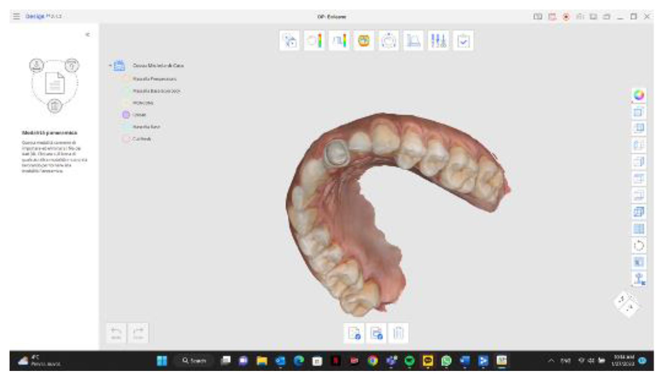
| Digital impression of the prepared dental abutment, without cords. | Taken immediately after temporary restoration removal and abutment cleaning, to transfer the information of the prepared dental abutment to the dental lab. | Pre-operative maxilla scan, using the high-resolution scan mode for scan areas that required finer information. |
| Digital impression with the interim restoration in place. | Taken after the previous impression, to transfer the macroscopic information of the interim restoration to the dental lab (size and shape). | Maxilla scan. |
| Digital impression of the interim restoration outside the patient month. | Taken after the previous impression, to transfer the customized finish line and emergence profile of the adapted interim restoration. | Scanbody scan, with high-resolution scan mode. |
| Digital impression of the antagonist arch | Taken after the previous impression with the mandible in maximal intercuspation. | Mandible scan. |
| Analog impression of the prepared dental abutment, with cords. | Taken according to the double-cord technique, after the second cord removal, and used as control. | One-step analog impression with medium and light material. |
| Digital impression of the prepared dental abutment, with cords. | Taken according to the double-cord technique, after the second cord removal, and used to evaluate the amount of soft tissue displacement. | Additional data. |
| Digital bite registration | To obtain the occlusal alignment of the arches in maximal intercuspation. | Occlusion scan. |
Disclaimer/Publisher’s Note: The statements, opinions and data contained in all publications are solely those of the individual author(s) and contributor(s) and not of MDPI and/or the editor(s). MDPI and/or the editor(s) disclaim responsibility for any injury to people or property resulting from any ideas, methods, instructions or products referred to in the content. |
© 2023 by the authors. Licensee MDPI, Basel, Switzerland. This article is an open access article distributed under the terms and conditions of the Creative Commons Attribution (CC BY) license (https://creativecommons.org/licenses/by/4.0/).
Share and Cite
Tallarico, M.; Cuccu, M.; Meloni, S.M.; Lumbau, A.I.; Baldoni, E.; Pisano, M.; Fiorillo, L.; Cervino, G. Digital Analysis of a Novel Impression Method Named the Biological-Oriented Digital Impression Technique: A Clinical Audit. Prosthesis 2023, 5, 992-1001. https://doi.org/10.3390/prosthesis5040068
Tallarico M, Cuccu M, Meloni SM, Lumbau AI, Baldoni E, Pisano M, Fiorillo L, Cervino G. Digital Analysis of a Novel Impression Method Named the Biological-Oriented Digital Impression Technique: A Clinical Audit. Prosthesis. 2023; 5(4):992-1001. https://doi.org/10.3390/prosthesis5040068
Chicago/Turabian StyleTallarico, Marco, Manuel Cuccu, Silvio Mario Meloni, Aurea Immacolata Lumbau, Edoardo Baldoni, Milena Pisano, Luca Fiorillo, and Gabriele Cervino. 2023. "Digital Analysis of a Novel Impression Method Named the Biological-Oriented Digital Impression Technique: A Clinical Audit" Prosthesis 5, no. 4: 992-1001. https://doi.org/10.3390/prosthesis5040068