Article

Comparison of the Airway Anatomy between Infants and Three Pediatric Simulators: A Radiological Study on Premature Anne, Infant AM Trainer and Simbaby Manikins

1
Department of Anesthesia and Intensive Care, “Policlinico-San Marco” University Hospital, 95123 Catania, Italy
2
Radiology Unit 1, University Hospital Policlinico “G. Rodolico-San Marco”, 95123 Catania, Italy
3
School of Anesthesia and Intensive Care, University of Catania, 95100 Catania, Italy
4
School of Anesthesia and Intensive Care, University “Magna Graecia”, 88100 Catanzaro, Italy
5
Department of Medical Surgical Sciences and Advanced Technologies “GF Ingrassia”, University of Catania, 95123 Catania, Italy
6
Department of Surgery and Medical-Surgical Specialties, University of Catania, 95123 Catania, Italy
*
Author to whom correspondence should be addressed.
These authors contributed equally to this work.
Prosthesis 2023, 5(3), 602-609; https://doi.org/10.3390/prosthesis5030042
Submission received: 10 May 2023 / Revised: 14 June 2023 / Accepted: 5 July 2023 / Published: 6 July 2023
(This article belongs to the Special Issue Innovative Prosthetic Devices Applied to the Human Body)

Abstract

:
Background: Training is required to achieve proficiency in airway management. Simulators are of utmost importance not only for the purpose of training novices, but also for evaluating newer airway devices and techniques. Growing evidence supports inadequate anatomic airway reproduction in adult and pediatric manikins. Methods: We conducted an observational study comparing 17 radiological anatomic airway measurements obtained via the computed tomography of three commercially available manikins with the same measurements obtained from a population of newborns/infant (range: 0–3 months) undergoing magnetic resonance imaging for diagnostic purposes. According to the reference (mean and standard deviation (SD) of the pediatric population), each manikin measurement was defined as adequate, partially adequate or inadequate (difference between means: ≤±1, 1.0–1.96 or >1.96 SD, respectively). The primary outcome was the number of measurements with an adequate reproduction of airways. Results: We included 27 pediatric patients (21 ± 19 days, 48% males, 46.6 ± 3.5 cm, 2.7 ± 0.5 Kg and 12.6 ± 2.9 kg/m2). All manikins had n = 11/17 measurements with inadequate airway anatomic reproduction. The three measurements with more adequate reproduction were the height of the soft palate, retropalatal airspace volume and tongue volume (adequate in two manikins, and partially adequate in the remaining one). Conclusions: In three manikins commonly used for training in pediatric airways, static dimensions do not seem anatomically correct in relation to those of pediatric patients. Such inaccuracies may introduce biases in airway device development as well as in training.

1. Introduction

Endotracheal intubation (ETI) is performed for a variety of reasons, spanning from life-saving indications to elective endotracheal tube (ETT) placement to allow elective surgical interventions in the operating room. Around 25 million ETI procedures are performed in the USA per year. Training is needed in order to reach proficiency, with some authors suggesting that up to 75 procedures are required [1], whilst ETI failure is associated with potentially life-threatening complications. Of note, ETI performed in neonates and children is even more complex and more procedures may be required to achieve expertise.
The success of ETI is also influenced not only by the number of procedures but also by the professional background of the operator with higher failure rates in emergency medical technicians and paramedics (up to 50% or 20%, respectively) compared to physicians [2,3,4,5,6]. Considering the importance of providing training to different professionals, the value of simulators (manikins) cannot be overemphasized in consideration of the ethical limitations of training on patients. Manikins have been in use since the 1960s when Laerdal® developed the first airway training manikin for mouth-to-mouth breathing during resuscitation [7]. Since then, the use of simulators has expanded allowing the reproduction of high-risk situations, which is similar in concept to training airplane pilots.
Therefore, precise reproduction of the airway anatomy is of utmost importance since inaccuracies may lead not only to negative effects on training but also to the inaccurate development of airway devices. Three studies have examined the anatomical correctness of the airways in simulators of adult humans [8,9,10]. These studies investigated both high-fidelity patient simulators as well as airway trainers, and all of them concluded that manikins do not reproduce correct anatomical dimensions in relation to humans.
To our knowledge, only one study approached the issue of correctness of manikins reproducing pediatric airway anatomy [11]. In this study, computed tomography (CT) measures of the upper airway anatomy of two SimBaby simulators were compared to images obtained from pediatric patients undergoing magnetic resonance imaging (MRI) under sedation. The study found that SimBaby anatomic features do not adequately reproduce pediatric upper airway anatomy concluding on the inadequate realism of this simulator for airway training. However, the study enrolled 20 pediatric patients aged on average 7 months, but with a wide age range (from 1 to 11 months). We performed a prospective study comparing the results of CT imaging of three commercially available manikins with the ones from MRI obtained from a larger population of newborns/infants with a smaller age range, in order to achieve more homogeneous results.

2. Materials and Methods

We conducted an observational study with the prospective design and acquisition of images from two different populations: (1) a historical cohort of newborns/infants (0–3 months) that underwent MRI for diagnostic purposes, and (2) three commercially available manikins from Laerdal®, from which CT images were obtained:
  • Premature Anne (female baby born at 25 weeks—low fidelity);
  • Infant Airway Management Trainer (three-month-old infant—low fidelity);
  • SimBaby (9-month-old pediatric patient—high fidelity).
Consecutive MRI images from newborns/infants were acquired from scans performed between January 2018 and January 2022. Patients with craniofacial malformations, an endotracheal tube in place, or with suboptimal imaging acquisitions were excluded from the study. The CT imaging of the simulators was performed in September 2022. Consent was waived by the Ethical Committee.

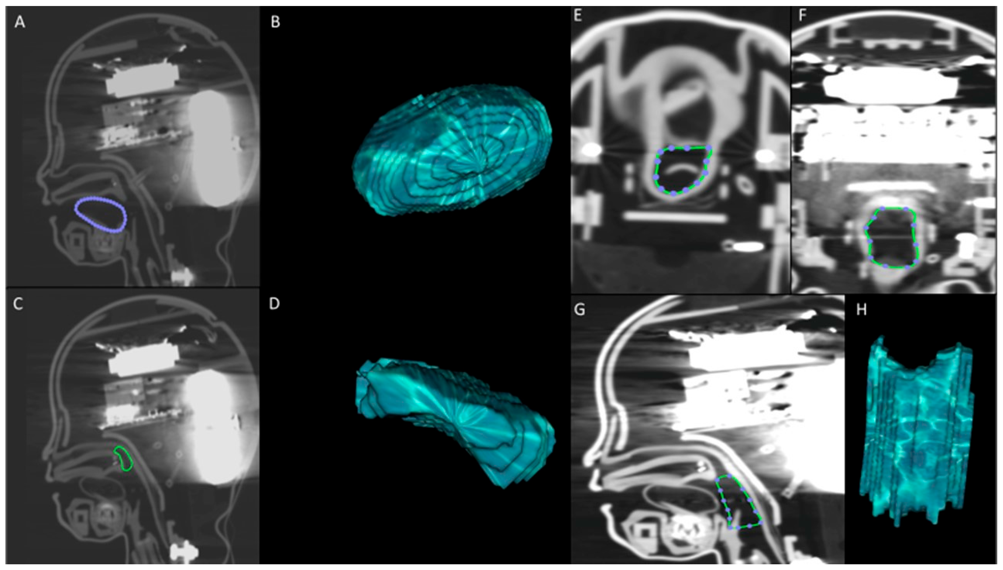
2.1. Radiological Measurements

Each MRI was acquired with the same 1.5 T scanner (Signa HD × T; GE Healthcare, Milwaukee, WI, USA) using a protocol containing at least the following features:
  • Sagittal T2 weighted TSE sequence (turbo spin echo; slice thickness: 4 mm, interspace: 1 mm, matrix: 512 × 512).
  • Sagittal T1 weighted SPGR sequence (spoiled gradient recalled echo; slice thickness: 3.5, interspace: 1 mm, matrix: 512 × 512).
  • 3D FIESTA sequence (fast imaging employing steady-state acquisition; slice thickness: 1 mm, interspace: 0, matrix: 512 × 512)
Measurements were performed on the midsagittal plane using the sequence with the best resolution and least motion artifacts. The anatomical landmarks for the measurements of distances (n = 14) and volumes (n = 3) were defined according to the scheme used by Schebesta et al. [11] (Table 1).
The image sets were evaluated by one radiologist with 6 years of experience in head and neck imaging. Calculations of distances were made in the medio-sagittal plane. The manikin airways were evaluated via CT using a standard airway acquisition protocol (120 kV and 250 mAs; slice thickness: 1 mm; pitch: 1). Volume segmentation was performed using a dedicated software (OsiriX DICOM Viewer, OxiriX Foundation®, Geneva, Switzerland); each structure was manually independently segmented by the radiologist, and the volumes were subsequently extrapolated from the segmentation. Volume segmentation on MRI was performed on 3D FIESTA sequences. Examples of the measurements performed are shown in Figure 1 (manikin) and Figure 2 (patient).
In order to increase accuracy, the radiologist took each measurement three times and on different days for each newborn/infant MRI scan as well as for the CT scans of the manikins. The three measurements were averaged and the mean value was taken as a definitive result.

2.2. Primary Outcome and Statistical Analysis

Radiological measurements are reported as mean and standard deviation (SD) for both newborns/infants and manikins. For each of the radiological measures obtained from the three simulators, we scored the adequateness of the manikin anatomy referring the mean value and SD obtained from the newborn/infant population. Precisely, for each simulator/variable we defined:
  • Adequate reproduction of airway, if the mean value was included between ±1 SD of the mean of the population;
  • Partially adequate reproduction of airway, if the mean value of the simulator was over ±1 SD but comprised ± 1.96 SD of the mean of the population;
  • Inadequate reproduction of airway, if the mean value of the simulator was <−1.96 or >1.96 SD from the mean of the population.
Our primary outcome was the number of simulator’s measurements with adequate reproduction of the human airway. Considering a total of 17 measurements, we preventively agreed that overall reproduction of airway would have been declared as satisfactory (n ≥ 9 adequate measurements), partially satisfactory (n ≤ 8 adequate measurements) or unsatisfactory (n ≤ 4 adequate measurements). Each partially adequate measurement was counted as n = 0.5. As secondary outcome we statistically compared with Student’s t-test the means of the values obtained in the pediatric population with the ones from the manikins. We considered significant a p value below 0.05.

3. Results

A total of 48 MRI scans of newborns/infants were retrieved and 21 discarded as per exclusion criteria. In the remaining 27 MRI scan included in our study, the population had a mean age of 21 ± 19 days, and 13/27 (48%) were males. Eight patients were born prematurely (30%). From anthropometric perspectives, the population mean height, weight and body mass index were 46.6 ± 3.5 cm, 2.7 ± 0.5 kg and 12.6 ± 2.9 kg/m2, respectively.
Results of each of the 17 measurements in the population and in the manikins are shown in Table 2.
Results of manikins are reported both singularly (mean of three measurements) and as an average of all three pooled together (mean and standard deviation, SD). Statistical comparison is made between the values obtained from the pediatric population and the pooled values from manikins. Comparison between values obtained from the pediatric population and the single manikin are made according to preestablished methods where for each variable we defined adequacy of reproduction of airway according to the delta between means. In particular, reproduction was adequate (green color, delta < ±1 SD of the mean of the population), partially adequate (yellow color, delta comprised between ±1 and ±1.96 SD of the mean of the population) or inadequate (red color, delta exceeded −1.96 or +1.96 SD from the mean of the population). When reproduction of airway resulted not entirely appropriate, each arrow indicates if the derangement of the manikin is in excess or in deficiency as compared to the pediatric population.
We used a color code to identify anatomical measures, in particular red, yellow and green for inadequate, partially adequate and adequate reproduction of airway, respectively. Moreover, we also inserted arrows to indicate if the value of the manikin was greater or smaller than reference values gathered from the patient’s population.
Interestingly, all the manikins had 11 out of 17 measurements classified as inadequate, but the inadequate measures were not always the same. Seven measurements were classified as inadequate in all the three manikins and none was green in all simulators. The two measurements that seemed closer to the infant population were the height of the soft palate and the tongue Volume, as these were classified as adequate in two manikins and partially adequate in the remaining one.

4. Discussion

Our study confirms that the airway anatomy of three pediatric simulators does not accurately reflect the airway of a population ranging from newborns up to 3 months. Of note, over two-thirds (n = 11) of the measurements were judged of inappropriate realism as they were outside the predefined cut-off of the mean ± 1.96 SD, so that we judged unsatisfactory the overall anatomic reproduction. Considering the age of the recruited population (mean age 21 days, but including almost one third of prematurely born), we thought that the Infant AM Trainer® or the Premature Anne® would have produced closer anatomical results than the one found in the Simbaby®, but in practice the number of inadequate measurements was similar across the manikins studied. Furthermore, when we compared measurements obtained in the pediatric population with the mean and SD pooled from the data of the three manikins, we found statistically significant differences in 12 out of 17 variables, supporting again an inadequate reproduction of airways.
In most cases, these measurements were much bigger in the simulators compared to those in the pediatric population. Among others, the retroglossal airspace was very different and our findings are similar to those of the original study conducted by Schebesta et al. [11] that used the retroglossal airspace volume as the primary outcome of their interest. Their choice was based on the special importance that this region has for fitting supraglottic devices, being therefore crucial in their design and development by the industry. They found significant differences in this space with simulators having much larger volumes (almost three times) than those of the pediatric patients. In our study, we found even bigger differences, and this could be due to either the younger age of our population (a mean age of around 3 weeks in our study, compared to that of almost 7 months in the study by Schebesta et al. [11]), or eventually to a deeper sedation level which may have produced a greater posterior displacement of the tongue. It should be noted that simulators are likely stiffer than patients and that the anterior displacement of the tongue with a direct laryngoscope may not be entirely reproduced; hence, even if the baseline retroglossal airspace volume is much bigger than that in patients, it is possible that this difference will be smaller after direct laryngoscopy.
In the study by Schebesta et al. [11], whilst the authors found the SimBaby simulator to have inadequate realism in relation to the upper airway, they also conducted a subgroup analysis according to the age of the patients. In this analysis, they showed that the age group of 5–8 months was the one with more appropriate measurements compared to the younger (1–4 months) or older (9–11 months) group. Indirectly, this finding supports our results of very poor concordance between simulators and measures of infants ranging from 0 to 3 months. Additionally, the overall airway length (distance from the outermost part of the lower lip to the pharynx) was much bigger in simulators than in patients.
Notably, the same group of authors [10] studied also the airway anatomy of adult manikins and showed that four high-fidelity patient simulators and two airway trainers did not match the upper airway anatomy of 20 adult trauma patients, suggesting for the first time a significant impact on device performances. These findings were subsequently replicated in other studies. In a large study on 13 manikins, Schalk et al. [9] found that none of the 13 included manikins for airway management matched the human anatomy in the six measurements taken by the authors. The manikin that replicated best the human airway anatomy was Laerdal Airway Management Trainer®. Another study was conducted by Blackburn et al. [8] on three manikins (SynDaver®, Laerdal® and AirSim®) and over one-third of measurements was outside two SDs from the mean of the patients. In particular, the space between the epiglottis and posterior pharyngeal wall was too big in all manikins. Furthermore, the group of Schebesta [11] conducted another original randomized crossover study where anesthesia residents performed intubation, laryngeal mask airway insertion and mask ventilation in 80 patients undergoing elective surgery and in two human manikins (HAL® and SimMan®). The authors found that manikins have adequate validity for intubation, but their fidelity and validity are much lower for laryngeal mask insertion or when mask ventilation is performed, suggesting that the results of simulation-based studies evaluating airway devices must be interpreted cautiously.
We think that it is a duty of the companies producing simulators to consider the overall findings of this other mentioned studies in order to improve the design of simulators. Further, biomedical engineers should consider the differences between human anatomy and the limitations of reproduction in manikins when developing and designing airway devices. Then, it will be the duties of anesthesiologists to test if newer manikins and/or device have improved realism and performances.
Our study has strengths and limitations. Among the strengths, we were able to recruit a larger sample size compared to that of the other study on a pediatric simulator [12]. Moreover, we recruited a more homogeneous population with patients, all of them being <3 months of age. Although the presence of sedation (for diagnostic procedures as well as for MRI scans) may be seen as a limitation because it inevitably produces changes in the anatomic conformation of the upper airway, such modifications may be very valuable for the purpose of training in airway management. Indeed, a high-fidelity simulator should reproduce the anatomy of an anaesthetized patient rather than the conformation of the airways of a non-anesthetized one. Therefore, the inclusion of sedated pediatric patients could be seen as a strength of the study.
We can observe two main limitations in our study. The first issue is the comparison of measurements performed with different radiological techniques, since MRI scans were used for patients whilst CT imaging was the only option for the simulators (metal components preclude their introduction in the MRI suite). However, Schebesta et al. already tested the accuracy of CT and MRI scanning a 9 × 9 × 9 cube, showing differences below 0.01 cm [12]. Second, we included a homogeneous population of 0–3-month-old babies but the simulators had variable targets, ranging from a 25-week-old premature baby to a 9-month-old healthy infant.
In addition to the above-described limitations, our results have also the intrinsic limitations related to the simulation environment. Whilst the role of simulation in airway training is well-demonstrated and supported [13], simulation scenarios are not able to fully reproduce several challenges experienced in clinical practice such as the occurrence of secretions and bleeding, as well as human factors (stress and/or anxiety due to clinical deterioration with desaturation and/or bradycardia) [14]. Further, only in vivo studies produce definitive answers to clinical needs and allow the proper investigation of technical aspects and human factors.

5. Conclusions

In summary, in manikins for pediatric airway management static dimensions do not seem anatomically correct in relation to the measurements obtained in pediatric patients; such inaccuracies are likely to introduce biases in airway device development as well as in training. It is also possible that these inaccuracies may lead to over-confidence in novices. Although the radiological evaluation of the anatomic realism of the simulators does not directly translate into failure regarding the “skill-enhancing” performances of simulators, our findings confirm the need for cautiousness when using simulators as a gold standard for patients to develop airway devices or technical concepts.

Author Contributions

Conceptualization, L.L.V. and D.F.; methodology, F.S.; software, F.S.; validation, A.B., B.L. and S.R.B.; formal analysis, L.L.V.; investigation, S.M. and F.M.; resources, A.B.; data curation, B.L.; writing—original draft preparation, L.L.V.; writing—review and editing, D.F.; visualization, F.M.; supervision, F.S. All authors have read and agreed to the published version of the manuscript.

Funding

This research received no external funding.

Institutional Review Board Statement

Ethical review and approval were waived for this study.

Informed Consent Statement

Not applicable.

Data Availability Statement

Data are available from corresponding author at reasonable request.

Conflicts of Interest

The authors declare no conflict of interest.

References

  1. Je, S.; Cho, Y.; Choi, H.J.; Kang, B.; Lim, T.; Kang, H. An application of the learning curve-cumulative summation test to evaluate training for endotracheal intubation in emergency medicine. Emerg. Med. J. 2015, 32, 291–294. [Google Scholar] [CrossRef] [PubMed]
  2. Garza, A.G.; Gratton, M.C.; Coontz, D.; Noble, E.; Ma, O.J. Effect of paramedic experience on orotracheal intubation success rates. J. Emerg. Med. 2003, 25, 251–256. [Google Scholar] [CrossRef] [PubMed]
  3. Peters, J.; van Wageningen, B.; Hendriks, I.; Eijk, R.; Edwards, M.; Hoogerwerf, N.; Biert, J. First-pass intubation success rate during rapid sequence induction of prehospital anaesthesia by physicians versus paramedics. Eur. J. Emerg. Med. 2015, 22, 391–394. [Google Scholar] [CrossRef] [PubMed]
  4. Sagarin, M.J.; Barton, E.D.; Chng, Y.M.; Walls, R.M.; National Emergency Airway Registry Investigators. Airway management by US and Canadian emergency medicine residents: A multicenter analysis of more than 6000 endotracheal intubation attempts. Ann. Emerg. Med. 2005, 46, 328–336. [Google Scholar] [CrossRef] [PubMed]
  5. Sayre, M.R.; Sakles, J.C.; Mistler, A.F.; Evans, J.L.; Kramer, A.T.; Pancioli, A.M. Field trial of endotracheal intubation by basic EMTs. Ann. Emerg. Med. 1998, 31, 228–233. [Google Scholar] [CrossRef] [PubMed]
  6. Stewart, R.D.; Paris, P.M.; Pelton, G.H.; Garretson, D. Effect of varied training techniques on field endotracheal intubation success rates. Ann. Emerg. Med. 1984, 13, 1032–1036. [Google Scholar] [CrossRef] [PubMed]
  7. Cooper, J.B.; Taqueti, V.R. A brief history of the development of mannequin simulators for clinical education and training. Postgrad. Med. J. 2008, 84, 563–570. [Google Scholar] [CrossRef] [PubMed]
  8. Blackburn, M.B.; Wang, S.C.; Ross, B.E.; Holcombe, S.A.; Kempski, K.M.; Blackburn, A.N.; DeLorenzo, R.A.; Ryan, K.L. Anatomic accuracy of airway training manikins compared with humans. Anaesthesia 2021, 76, 366–372. [Google Scholar] [CrossRef] [PubMed]
  9. Schalk, R.; Eichler, K.; Bergold, M.N.; Weber, C.F.; Zacharowski, K.; Meininger, D.; Byhahn, C.; Mutlak, H. A radiographic comparison of human airway anatomy and airway manikins--Implications for manikin-based testing of artificial airways. Resuscitation 2015, 92, 129–136. [Google Scholar] [CrossRef] [PubMed]
  10. Schebesta, K.; Hupfl, M.; Rossler, B.; Ringl, H.; Muller, M.P.; Kimberger, O. Degrees of reality: Airway anatomy of high-fidelity human patient simulators and airway trainers. Anesthesiology 2012, 116, 1204–1209. [Google Scholar] [CrossRef] [PubMed] [Green Version]
  11. Schebesta, K.; Hupfl, M.; Ringl, H.; Machata, A.M.; Chiari, A.; Kimberger, O. A comparison of paediatric airway anatomy with the SimBaby high-fidelity patient simulator. Resuscitation 2011, 82, 468–472. [Google Scholar] [CrossRef] [PubMed]
  12. Schebesta, K.; Spreitzgrabner, G.; Hörner, E.; Hüpfl, M.; Kimberger, O.; Rössler, B. Validity and fidelity of the upper airway in two high-fidelity patient simulators. Minerva Anestesiol. 2015, 81, 12–18. [Google Scholar] [PubMed]
  13. Kennedy, C.C.; Cannon, E.K.; Warner, D.O.; Cook, D.A. Advanced airway management simulation training in medical education: A systematic review and meta-analysis. BMC Anesthesiol. 2014, 42, 169–178. [Google Scholar] [CrossRef] [PubMed] [Green Version]
  14. Doleman, B.; Blackwell, J.; Karangizi, A.; Butt, W.; Bhalla, A.; Lund, J.N.; Williams, J.P. Anaesthetists stress is induced by patient ASA grade and may impair non-technical skills during intubation. Acta Anaesthesiol. Scand. 2016, 60, 910–916. [Google Scholar] [CrossRef] [Green Version]
Figure 1. Examples of segmentation and volume extrapolation. The volumes of interest were manually segmented via computed tomography (A,C) and then extrapolated using OsiriX DICOM Viewer (B,D). (A,B) Tongue volume. (C,D) Retropalatal airspace volume. On the right side, an example of segmentation and volume extrapolation of the retroglossal airspace is shown. (E) Axial plane. (F) Coronal plane. (G) Sagittal plane. (H) Volume extrapolated using OsiriX DICOM Viewer.
Prosthesis 05 00042 g001
Figure 2. Schematic drawing of the measurements (distances) analyzed via magnetic resonance imaging (midsagittal T2 in turbo spin echo sequence). Measurements of distances are lettered as in Table 1. (A) Horizontal distance from the outermost portion of the lower lip to the posterior pharyngeal wall (red, a); horizontal distance from the lower alveolar process to the posterior pharyngeal wall (yellow, b); oblique diameter of the tongue (blue, c). (B) Horizontal distance from the center of the tongue to the posterior pharyngeal wall (red, d); horizontal distance from the edge of the tongue to the posterior pharyngeal wall (blue, e); horizontal distance from the center of the soft palate to the posterior pharyngeal wall (yellow, f). (C) Horizontal distance from the vallecula to the posterior pharyngeal wall (red, g); horizontal distance from the tip of the epiglottis to the posterior pharyngeal wall (yellow, h); curved length of the soft palate (blue, i); distance from the vallecula to the tip of the epiglottis (orange, j). (D) Distance from the posterior base of the epiglottis to the tip of the epiglottis (red, k); height of the soft palate (orange, l); vertical distance from the base of the hard palate to the vallecula (blue, m); vertical distance base from the hard palate to the tip of the epiglottis (yellow, n).
Prosthesis 05 00042 g002
Table 1. Distances and volumes calculated for both manikins (via computed tomography) and patients (via magnetic resonance imaging).
DISTANCES
A. Horizontal distance from the outermost portion of lower lip to the posterior pharyngeal wall
B. Horizontal distance from the lower alveolar process to the posterior pharyngeal wall
C. Oblique diameter of the tongue
D. Horizontal distance from the center of the tongue to posterior pharyngeal wall
E. Horizontal distance from the edge of the tongue to posterior pharyngeal wall
F. Horizontal distance from the canter of the soft palate to posterior pharyngeal wall
G. Horizontal distance from the vallecula to posterior pharyngeal wall
H. Horizontal distance from the tip of the epiglottis to the posterior pharyngeal wall
I. Curved length of the soft palate
J. Distance from the vallecula to the tip of the epiglottis
K. Distance from the posterior base of the epiglottis to the tip of the epiglottis
L. Height of the soft palate
M. Vertical distance from the base of the hard palate to the tip of the epiglottis
N. Vertical distance from the base of the hard palate to the vallecula
VOLUMES
O. Tongue
P. Retropalatal airspace
Q. Retroglossal airspace
Table 2. Values of the distances and volumes measured in 27 pediatric patients below 3 months of age and values obtained from measurements made in three manikins (SimBaby®, Premature Anne®, Infant AM Trainer®).
Variable (Distances or Volumes)Infants
Mean ± SD
Manikins
Mean ± SD
p ValueSimBabyPremature AnneInfant AM Trainer
A. Outermost portion of lower lip to posterior pharyngeal wall (cm)4.54 ± 0.316.44 ± 0.36<0.0016.40 ↑6.86 ↑6.05 ↑
B. Lower alveolar process to posterior pharyngeal wall (cm) 3.46 ± 0.264.96 ± 0.35<0.0015.15 ↑5.23 ↑4.50 ↑
C. Oblique diameter of the tongue (cm) 2.12 ± 0.282.46 ± 0.700.192.92 ↑1.53 ↓2.92 ↑
D. Centre of the tongue to posterior pharyngeal wall (cm) 2.07 ± 0.212.83 ± 0.30<0.0012.85↑3.16 ↑2.48 ↑
E. Edge of the tongue to posterior pharyngeal wall (cm) 1.36 ± 0.261.60 ± 0.360.081.66 ↑1.97 ↑1.16
F. Canter of the soft palate to posterior pharyngeal wall (cm) 0.91 ± 0.131.26 ± 0.390.030.801.69 ↑1.29 ↑
G. Vallecula to posterior pharyngeal wall (cm) 0.74 ± 0.181.48 ± 0.27<0.0011.34 ↑1.84 ↑1.27 ↑
H. Tip of the epiglottis to posterior pharyngeal wall (cm) 0.39 ± 0.101.11 ± 0.450.0010.64 ↑1.66 ↑1.01 ↑
I. Curved length of the soft palate (cm) 2.04 ± 0.281.26 ± 0.32<0.0011.43 ↓0.84 ↓1.52 ↓
J. Vallecula to tip of the epiglottis (cm) 0.70 ± 0.151.03 ± 0.370.031.41 ↑0.571.12 ↑
K. Posterior base of the epiglottis to tip of the epiglottis (cm) 0.68 ± 0.131.13 ± 0.380.0071.58 ↑1.09 ↑0.71
L. Height of the soft palate (cm) 1.23 ± 0.321.44 ± 0.310.091.481.081.78
M. Base of the soft palate to tip of the epiglottis (cm) 1.78 ± 0.462.55 ± 0.480.0012.45 ↑2.063.13 ↑
N. Base of the soft palate to vallecula (cm) 2.26 ± 0.513.35 ± 0.760.0023.41 ↑2.474.18 ↑
O. Tongue Volume (cm3) 5.79 ± 1.216.08 ± 0.940.467.10 ↑4.946.22
P. Retropalatal airspace Volume (cm3) 1.10 ± 0.301.14 ± 0.320.741.380.72 ↓1.33
Q. Retroglossal airspace Volume (cm3) 0.97 ± 0.265.79 ± 1.59<0.0016.52 ↑7.15 ↑3.69 ↑
Disclaimer/Publisher’s Note: The statements, opinions and data contained in all publications are solely those of the individual author(s) and contributor(s) and not of MDPI and/or the editor(s). MDPI and/or the editor(s) disclaim responsibility for any injury to people or property resulting from any ideas, methods, instructions or products referred to in the content.

