Systematic Review

Computer-Guided Surgery for Dental Implant Placement: A Systematic Review

by
Elena Araujo-Corchado
and
Beatriz Pardal-Peláez
*
Department of Surgery, Faculty of Medicine and Dental Clinic, University of Salamanca, 37007 Salamanca, Spain
*
Author to whom correspondence should be addressed.
Prosthesis 2022, 4(4), 540-553; https://doi.org/10.3390/prosthesis4040044
Submission received: 10 August 2022 / Revised: 13 September 2022 / Accepted: 19 September 2022 / Published: 26 September 2022
(This article belongs to the Collection Oral Implantology: Current Aspects and Future Perspectives)

Abstract

:
Implant therapy is currently the treatment of choice for the replacement of missing teeth. Correct implant positioning is of vital importance. To this end, radiographic techniques providing 3D information and guided surgery techniques, both static and dynamic, have been developed. The primary objective of this work is to study whether placing dental implants in partially edentulous patients with guided surgery techniques results in less, equal or greater precision than placing them freehand. The secondary objectives are to gain an understanding of the advantages and disadvantages, indications, limitations, and complications of this type of surgery. A literature search was performed in Pubmed and BVS, and six randomized clinical trials meeting the marked inclusion criteria were included. The different guided surgery techniques were compared with each other and with the traditional method. Freehand surgery was found to be the least accurate, as the implants placed with this technique showed the largest deviations between the planned position and the final position, both when calculating the global (3D) deviation and when measuring the deviation in each of the axes (vertical, mesio-distal, bucco-lingual and lateral), and the angular deviation and voxel overlap. In contrast, the most accurate surgeries were fully guided and half-guided, with the smallest deviations between the planned and final implant position. With any guided surgery technique, more precise implant positioning is achieved than with freehand placement. Advantages include reduced trauma and surgery time; disadvantages include reduced primary implant stability and higher cost. This type of surgery is more indicated in cases of critical anatomy, but may encounter limitations in terms of cost, degree of buccal opening, visibility and adjustment of the guides and the need for prior familiarization with the procedure. Nevertheless, this surgical technique reduces the complication rate.

1. Introduction

Implant therapy is the treatment of choice for the replacement of missing teeth [1], as long-term follow-up of patients rehabilitated with implants has shown satisfactory results in a large number of patients [2].
The placement of implants should be as precise as possible, as they are located very close to vital structures (vessels and nerves) [3], so pre-treatment planning is essential [4]. Nowadays, implantology uses techniques that provide three-dimensional information for the optimal placement of implants taking into account prosthetic parameters. These techniques are computed tomography (CT) and cone beam computed tomography (CBCT) as well as 3D implant planning software and CAD/CAM [2,5,6,7,8].
All these advances have made the development of navigation surgery or computer-guided surgery possible [7,8,9,10]. This can be classified into static and dynamic surgery [10,11,12]. Dynamic guided surgery does not use templates [10,11,13], but is a system that, by means of video technology, allows the surgery to be performed by visualizing it live [14]. Static guided surgery uses static surgical templates to guide the procedure [10,15] which can be supported by mucosa, bone, crowns or teeth [7,11,13].
Depending on how the bone bed preparation and implant placement is performed, static guided surgery can be fully guided (FG), half-guided (HG) and freehand (FH) [11,12,13].
In fully guided surgery (FG), the template guides the entire procedure [11,13,15]; therefore, accuracy is one of its main advantages [11]. However, the guide can impede the passage of irrigation fluid and lead to tissue heating, bone resorption and impairment of the osseointegration of the implant [13].
Another modality is half-guided (HG) or partially guided surgery, which includes three variants: drill-guided surgery, which uses the guide during the entire drilling sequence but the implant is placed without a guide; pilot drill-guided (PD) surgery, where only the initial drill is passed with the guide and the rest of the drilling and implant placement is conducted freehand; and non-guided surgery, where the template is made on the basis of a wax-up or a previous prosthesis of the patient [11,13]. Finally, freehand (FH) surgery is surgery in which no surgical templates are used [11,12,13].

Objectives

The main objective of this study was to determine the degree of precision obtained when placing titanium dental implants in partially edentulous patients using computer-guided surgery (fully guided, half-guided, pilot drill-guided and dynamic guided) compared to freehand surgery (conventional).
The secondary objectives were to evaluate the advantages, disadvantages, indications, limitations and complications of computer-guided surgery compared to conventional surgery.

2. Materials and Methods

This systematic review has been prepared according to the PRISMA guidelines [16].
The following PICO (Population, Intervention, Comparison, Outcome) question was posed: “What is the degree of accuracy (Outcome) obtained when placing dental implants (Intervention) in partially edentulous patients (Population) using guided surgery compared to freehand surgery (Comparison)?”.

2.1. Eligibility Criteria

We included articles on randomized and controlled clinical trials (RCT/CCT), conducted on living humans and/or human cadavers, in partially edentulous patients, in which titanium implants were placed with guided surgery and in which accuracy was measured by comparing cone beam computed tomography (CBCT) scans. There was no time restriction and articles in both English and Spanish were selected.
On the other hand, clinical cases, case series, narrative reviews, prospective and retrospective studies were excluded, as were papers including fully edentulous patients, full denture implants placed using a mucosa- and/or bone-supported guide wire and all articles in languages other than English and Spanish.

2.2. Sources of Information and Search

A bibliographic search was carried out in PubMed and BVS (Biblioteca Virtual de Salud—Virtual Health Library), using the following combination of keywords: “dental implants AND guided surgery AND accuracy”. Details of the search are shown in Figure 1.

Study Selection and Data Collection

All articles were independently reviewed. To be included in the analysis, they had to meet all the marked inclusion criteria. Data were extracted independently and only those related to the topic and objective of the present work were considered. The following data were collected from each selected article: author, type of study, year of publication, techniques used for implant placement, measurements obtained, number of teeth rehabilitated per patient, type of edentulism, number of patients and number of implants placed in total, and deviations between the planned implant position and the final position in the different types of surgery.

2.3. Risk of Bias Assessment

The risk of bias assessment was performed with the RevMan 5 software (Review Manager (RevMan) (Computer program). Version 5.3. Copenhagen: The Nordic Cochrane Centre, The Cochrane Collaboration, 2014). Seven criteria were assessed: random sequence generation, allocation concealment, blinding of participants and staff, blinding of outcome assessment, incomplete outcome data, selective reporting and other biases, based on the Cochrane risk of bias tool for randomized clinical trials [17]. Depending on the data obtained for each criterion, they were rated as low (green +) or high (red -) risk of bias, and in those cases where the information was not sufficient to fall into the above two, they were rated as unclear (yellow ?) risk.

2.4. Quality of Evidence

GRADEpro 3.2 [18] software was used and the GRADE classification was applied to assess the quality of evidence.

2.5. Planned Methods of Analysis and Additional Analysis

Initially, a systematic review and meta-analysis (qualitative and quantitative analysis) was planned. The meta-analysis would be performed with Revman 5.3 (Review Manager (RevMan) (Computer program). Version 5.3. Copenhagen: The Nordic Cochrane Centre, The Cochrane Collaboration, 2014).

3. Results

3.1. Study Selection

Six clinical trials that met the inclusion criteria were selected. All included trials were conducted on living humans, with the exception of one [19], which was conducted on human cadaver heads. All subjects were partially edentulous and candidates for fixed implant-supported prostheses. In addition, CBCT was used in all cases to measure accuracy.

3.2. Quality of Evidence

When comparing guided surgery with freehand surgery, two different comparisons were established, separating static guided surgery from dynamic guided surgery. According to the evaluation of the six selected papers [19,20,21,22,23,24], the quality of evidence was low for both comparisons.

3.3. Risk of within-Study Bias

The risk of bias within studies is shown in Figure 2 and Figure 3, carried out with RevMan 5 (Review Manager (RevMan) (Computer program). Version 5.3. Copenhagen: The Nordic Cochrane Centre, The Cochrane Collaboration, 2014) and based on the Cochrane risk of bias tool [17]. Accordingly, it can be said that four studies [19,20,21,22] had an unclear risk of bias, as one or more parameters were listed as unclear risk. The remaining two studies [23,24] were considered at high risk of bias, as they had at least one parameter with a high risk of bias.

3.4. Risk of Bias among Studies

The bias among studies in this review was classified as unclear, as most of the information comes from studies with unclear risk of bias.

3.5. Results of Individual Studies

In total, from the 6 trials included in this review [19,20,21,22,23,24], 519 implants were placed in 247 living humans and 8 human cadaver heads (in the upper jaw). Of these, 192 were placed freehand (180 in living humans and 12 in human cadaver heads) and 327 in a guided manner (315 in living humans and 12 in human cadaver heads).
Within the guided surgery, 43 implants were placed by dynamic surgery and the other 284 by static surgery (124 fully guided, 63 partially guided and 97 by pilot-drill surgery).
Regarding the techniques used to place the dental implants, in the six included studies [19,20,21,22,23,24], implants were placed by freehand surgery in the control group. This freehand surgery technique was compared in all studies with implant placement by guided surgery (test group).
Table 1 lists the different techniques performed in each study for the placement of dental implants.

3.6. Summary of Results

Table 2 shows the data collected from the selected studies.
Table 3 and Table 4 show the values of the deviations found between the planned and final position of the implant in the different types of surgery in the six trials included [19,20,21,22,23,24]. All measurements were performed after matching the virtual planning data with the postoperative data.
In total, 12 measurements were performed by superimposing CBCTs. They are shown in Figure 4 and Figure 5.

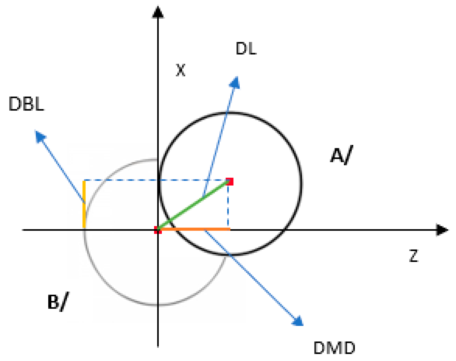
3.6.1. Global Deviation

The global deviation was obtained by measuring the 3D distance (in millimeters) from the center of the crown [19,21,22,23] or from the center of the apexes [19,20,21,22,23] of the planned and placed implants. These two measurements were named in the present work as coronal global deviation (CGD) and apical global deviation (AGD), respectively:
  • Coronal global deviation (CGD)
    When measuring the overall deviation, i.e., in 3D, between the crown of planned and placed implants in 4 studies [19,21,22,23], it was observed that the least accurate surgery was freehand surgery (1.82 mm) [23] and the most accurate was fully guided surgery (0.73 mm) [21], showing statistically significant differences.
  • Apical global deviation (AGD)
    At the apical level the global deviation, i.e., in 3D, between the apex of the planned and placed implants was also measured in 5 studies [19,20,21,22,23], and it was found that the least accurate surgery was freehand surgery (2.43 mm) [23] and the most accurate was half-guided surgery (0.93 mm) [19], with statistically significant differences.

3.6.2. Vertical Plane

The distances between the crowns of the planned and placed implants were measured [21,22], as well as the distances between the apexes of the planned and placed implants [19,21,22]. These are listed in Table 4 under the names of coronal vertical deviation (CVD) and apical vertical deviation (AVD), respectively.
In this plane, the depth of the implant was measured, using the coronal and/or apical part of the implant as reference points:
  • Coronal vertical deviation (CVD)
    When measuring the vertical distance between the crown of planned and placed implants in 2 studies [21,22], it was observed that the least accurate surgery was freehand surgery (1 mm) [22] and the most accurate was fully guided surgery (0.43 mm) [21], showing statistically significant differences.
  • Apical vertical deviation (AVD)
    When calculating the vertical distance between the apex of planned and placed implants in 3 trials [19,21,22], the same values were obtained as for coronal vertical deviation, with freehand surgery being the least accurate (1 mm) [22] and fully guided surgery the most accurate (0.43 mm) [21], but the differences were not statistically significant in this case.

3.6.3. Mesio-Distal Axis

The distance between the center of the crowns of the planned and placed implants was measured [19,22]. At the apical level, the same was conducted in these two studies [19,22], but by taking the center of the apex of both implants as a reference. In this review, these data were collected under the names of coronal mesio-distal deviation (CMDD) and apical mesio-distal deviation (MDDA), respectively:
  • Coronal mesio-distal deviation (CMDD)
    Measuring the distance between the crowns of planned and placed implants in the mesio-distal direction in 2 studies [19,22], implants placed through freehand surgery (0.6 mm) showed the lowest accuracy, [22] and the highest accuracy was observed for implants placed via both fully guided surgery [22] and half-guided surgery [19] (0.3 mm in both cases), the differences being statistically significant.
  • Apical Mesio-Distal Deviation (AMDD)
    When measuring the distance between planned and mesio-distally placed implant apexes in 2 trials [19,22], it was found that the least accurate surgery was freehand surgery (1.2 mm) [22] and the most accurate was half-guided surgery (0.43 mm) [19], the differences also being statistically significant.

3.6.4. Bucco-Lingual Axis

Two measurements were made in the two studies mentioned above [19,22]. The distance between the center of the crown of planned and placed implants was called coronal bucco-lingual deviation (CBLD) in the present review, and the distance between the center of the apexes of planned and placed implants was called apical bucco-lingual deviation (ABLD).
  • Coronal bucco-lingual deviation (CBLD)
    The bucco-lingual distance between the crown of planned and placed implants was measured in 2 studies [19,22] and the maximum deviation was found in freehand surgery (0.8 mm) [19] and the minimum in fully guided surgery (0.4 mm) [22]. Therefore, freehand surgery proved to be the least accurate and fully guided surgery the most accurate, but it should be noted that although differences were observed, they were not statistically significant in this case.
  • Apical bucco-lingual deviation (ABLD)
    When measuring the bucco-lingual distance between planned and placed implant apexes in 2 studies [19,22], the lowest accuracy was observed in freehand surgery (1.38 mm) [19] and the highest in half-guided surgery (0.53 mm) [19], but the differences were not statistically significant, as in the previous case.

3.6.5. Angular Deviation

Angular deviation (AD) (in degrees) was measured in four studies [19,22,23,24], defined as the angle formed between the center lines, i.e., the axes of the planned and placed implants.
The angle formed between the axes of planned and placed implants was measured in four studies [19,22,23,24] and the lowest accuracy was found in implants placed freehand (10.04°) [24] and the highest accuracy in implants placed by fully guided surgery (3.04°) [23], with statistically significant differences found.

3.6.6. Lateral Direction

Two measurements were made. One was calculated by drawing a line from the center of the crown of one implant to the center of the crown of the other, perpendicular to the implant axis [19,21,24]. These values were recorded in Table 3 and Table 4 under the name of coronal lateral deviation (CLD). In the three previous trials [19,21,24], the same was carried out at the apical level; a line was drawn from the center of the apex of one implant to the center of the apex of the other, also perpendicular to the axes of the two implants, and named apical lateral deviation (ALD).
  • Coronal lateral deviation (CLD)
    When measuring the distance in lateral direction between the crown of planned and placed implants in three studies [19,21,24], it was observed that the least accurate surgery was freehand surgery (1.7 mm) [24] and the most accurate was fully guided surgery (0.55 mm) [21], with statistically significant differences.
  • Apical lateral deviation (ALD)
    The distance in lateral direction between the apexes of planned and placed implants was measured in three studies [19,21,24] and the highest value was observed when performing freehand surgery (2.51 mm) [24], while the lowest value was found in implants placed with half-guided surgery (0.73 mm) [19]. Therefore, when measuring this parameter, freehand surgery was the least accurate and half-guided surgery the most accurate, with the results showing statistically significant differences.

3.6.7. Voxel Overlap

In one of the included studies [23], the overlap of voxels (VO) (in percentages) composing the image of the planned implants with the voxels resulting in the image of the placed implants according to the opacity values of each implant was calculated. This deviation was not measured in any of the other five studies included in this review [19,20,21,22,24].
This measurement was only calculated in one study, carried out by Varga et al., [23], and showed its worst result in freehand surgery (40.57%) and its best result in half-guided surgery (60.37%); therefore, freehand surgery showed the least accurate and half-guided surgery to be the most accurate. In this case, the differences were also significant.

4. Discussion

4.1. Summary of Evidence

4.1.1. Accuracy of Guided Surgery vs. Freehand Surgery

In the present systematic review, the main parameter evaluated was the degree of accuracy obtained when placing dental implants by guided surgery compared to freehand surgery. Twelve measurements were analyzed for this purpose as shown in the results section.
After analyzing all measurements, it can be said that, in general terms, the results were worse with freehand surgery than with guided surgery; significant differences were found in all the measurements of all the included studies [19,20,21,22,23,24], except in three (apical vertical deviation and coronal and apical bucco-lingual deviation), in the studies by Smitkarn et al. and Chen et al. [19,22]. However, although the differences were not significant, the results of these three measurements were also worse in freehand surgery than in guided surgery.

4.1.2. Accuracy of Different Guided Surgery Techniques

After analyzing the above 12 measurements, it was concluded that the most accurate surgeries were fully guided (FG) and half-guided (HG), as the implants placed with these two surgical techniques deviated the least from the planned position, in raw values [19,21,22,23].
Pilot drill-guided surgery (PD) performed worse than the other two static guided surgery techniques (fully guided and half-guided) [20,21,23], as the implants placed in this way deviated the most from the planned position, in raw values. It should be noted that only the global (3D), vertical and lateral deviation between crowns and apexes of the planned and placed implants, as well as the deviation of the implant axes (angular deviation) and voxel overlap were measured in this case.
Dynamic guided surgery (DG) was only performed in one study, that of Aydemir and Arisan [24], showing better results than freehand surgery when the deviations between implant axes and the distance between crowns and apexes of planned and laterally placed implants were measured. Furthermore, it should be noted that, in this study, both the implants placed with dynamic surgery and those placed freehand deviated more, in raw values, from the planned position in the lateral direction at the apical level than at the coronal level (ALD = 1.83 mm and CLD = 1.01 mm in dynamic surgery; ADL = 2.51 mm and CLD = 1.7 mm in freehand surgery).

4.1.3. Advantages and Disadvantages

The advantages of guided surgery were discussed in two studies [20,24], both of which cited reduced trauma and duration of surgery, as well as greater precision than the aforementioned freehand surgery. Disadvantages were addressed in two other studies [21,22], but these addressed different issues. While one addressed primary stability [22], the other addressed costs [21]. Regarding primary stability, Smitkarn et al. [22] observed that it was lower in implants placed using fully guided surgery than those using freehand surgery, and this was found to be due to the fact that in guided surgery, the implant was inserted completely through the surgical guide, which allowed control of vertical depth only indirectly through the guide markings, and the surgeon had very little tactile perception of implant stability other than the torque value. In contrast, in freehand surgery, the surgeon had a direct view of the bone level and vertical position of the implant, and could determine the depth in relation to the tactile perception of the primary stability. In relation to costs, Younes et al. [20] found that performing guided surgery increased the cost of treatment due to the need for a surgical guide, specific instruments and tools, as well as specific planning software.

4.1.4. Indications for Guided Surgery

Computer-guided surgery may be more indicated in cases where the implant is planned to be placed in the vicinity of a critical anatomy (proximity of the inferior dental nerve, maxillary sinus, etc.) [24]. Within guided surgery, the most suitable technique in these cases is fully guided surgery, due to its great precision. However, in cases where fully guided surgery is not possible, the pilot drill-guided surgery technique could be indicated, as it would provide better results than freehand surgery [21].

4.1.5. Limitations of Guided Surgery

This issue was addressed in one study [22], and it was concluded that, although the accuracy obtained with guided surgery is higher than with freehand surgery, it has limitations, including higher costs, the need for favorable anatomical conditions in terms of buccal opening, and adequate adjustment of the surgical guides. Two other trials [21,24] also addressed this last limitation, and found that the stability of the guide position during implant placement is important, as inadequate guide position can affect the correct positioning of the implant. On the other hand, with regard to dynamic surgery, only addressed in one study [24], it was found that surgeon familiarization with the procedure was necessary, as the entry point is visualized on the monitor instead of directly observing the patient’s jaw. As the surgeon in this study was unfamiliar with the procedure, there were frequent starts and stops during the surgery. Furthermore, in this same study [24], comparing what was said in a systematic review about static and dynamic guided surgery, it was concluded that in static guided surgery there are limitations in detecting deviations in the implant position. This is due to the closed and restrictive structure of the static guides, which does not occur in dynamic surgery because it allows complete visibility of the surgical area and the final position of the implant.

4.1.6. Complications

Freehand surgery may involve more complications than guided surgery [21]. A reflection of this is that, in this study, all the implants placed (both with guided surgery and freehand) were intended to be restored with a screw-retained restoration; however, 19.2% (5/26) of the implants that were placed freehand had to be restored with a cemented restoration, because deviations were observed and the screw holes were placed towards the buccal plane and the aesthetics were compromised. In relation to the guided surgery techniques, in this same study [21], only 4.2% (1/24) of the implants placed with pilot drill had to receive a screw-retained restoration; this was because deviations occurred when inserting the drills subsequent to the pilot drill. None of the implants placed with fully guided surgery had to be screw-retained, as they could all be restored with a cemented restoration. Another study [19] also showed a greater buccal deviation in implants placed freehand, and therefore also stated that complications were lower in guided surgery than in freehand surgery. It is important to note that, apart from complications with regard to the type of restoration, no biological complications of invasion of neighboring structures such as perforation of the floor of the maxillary sinus, the inferior dental nerve or the mental nerve were described for any of the techniques in any of the included studies [19,20,21,22,23,24].

4.2. Limitations of This Review

Among the main limitations of this work are the small number of articles included for the synthesis of the results and the absence of complete data with standard deviation values [19,20,21,22,23,24]. This prevented a weighting of the data for a quantitative analysis (meta-analysis).

5. Conclusions

Regarding the evaluation of the degree of precision obtained with guided surgery versus freehand surgery, in general terms, the degree of precision obtained when placing dental implants in partially edentulous patients with guided surgery techniques is greater than with freehand surgery, with the results of the individual studies showing statistically significant differences. Regarding static guided surgery techniques (fully guided, half-guided and pilot drill-guided), fully guided and half-guided surgery are the most accurate, while pilot drill-guided surgery is the least accurate. With regard to dynamic guided surgery, it could not be concluded whether it offers better results than static guided surgery techniques, as only one of the included studies in this review dealt with it and compared it only with freehand surgery. It would be desirable for future studies on the subject to provide unified and homogeneous data in terms of the measurements evaluated in order to be able to analyze the data more precisely.
The advantages of guided surgery include greater precision, reduced trauma to the patient and reduced duration of the surgical procedure; the disadvantages include lower primary stability of the implants and higher cost. Guided surgery is indicated in situations of critical anatomy, such as proximity to the inferior dental nerve or maxillary sinus, as it offers greater precision and a lower probability of complications. The main limitations of this type of surgery are the higher cost compared to freehand surgery, the need for a good mouth opening, the visibility and adjustment of the surgical guides and the need for prior familiarization with the procedure.

Author Contributions

Conceptualization, B.P.-P.; methodology, B.P.-P.; validation, B.P.-P.; formal analysis, E.A.-C.; investigation, E.A.-C.; resources, E.A.-C.; data curation, E.A.-C. and B.P.-P.; writing—original draft preparation, E.A.-C.; writing—review and editing, B.P.-P.; visualization, B.P.-P.; supervision, B.P.-P.; project administration, B.P.-P. All authors have read and agreed to the published version of the manuscript.

Funding

This research received no external funding.

Institutional Review Board Statement

Not applicable.

Informed Consent Statement

Not applicable.

Data Availability Statement

Not applicable.

Conflicts of Interest

The authors declare no conflict of interest.

References

  1. Tattan, M.; Chambrone, L.; González-Martín, O.; Avila-Ortiz, G. Static computer-aided, partially guided, and free-handed implant placement: A systematic review and meta-analysis of randomized controlled trials. Clin. Oral Implant. Res. 2020, 31, 889–916. [Google Scholar] [CrossRef] [PubMed]
  2. Tallarico, M.; Meloni, S. Retrospective Analysis on Survival Rate, Template-Related Complications, and Prevalence of Peri-implantitis of 694 Anodized Implants Placed Using Computer-Guided Surgery: Results between 1 and 10 Years of Follow-up. Int. J. Oral Maxillofac. Implant. 2017, 32, 1162–1171. [Google Scholar] [CrossRef] [PubMed]
  3. Bover-Ramos, F.; Viña-Almunia, J.; Cervera-Ballester, J.; Peñarrocha-Diago, M.; García-Mira, B. Accuracy of Implant Placement with Computer-Guided Surgery: A Systematic Review and Meta-Analysis Comparing Cadaver, Clinical, and In Vitro Studies. Int. J. Oral Maxillofac. Implant. 2018, 33, 101–115. [Google Scholar] [CrossRef]
  4. Schneider, D.; Marquardt, P.; Zwahlen, M.; Jung, R.E. A systematic review on the accuracy and the clinical outcome of computer-guided template-based implant dentistry. Clin. Oral Implant. Res. 2009, 20, 73–86. [Google Scholar] [CrossRef]
  5. Jung, R.E.; Schneider, D.; Ganeles, J.; Wismeijer, D.; Zwahlen, M.; Hämmerle, C.H.F.; Tahmaseb, A. Computer Technology Applications in Surgical Implant Dentistry: A Systematic Review. Int. J. Oral Maxillofac. Implant. 2009, 24, 92–109. [Google Scholar]
  6. Zhou, W.; Liu, Z.; Song, L.; Kuo, C.-L.; Shafer, D.M. Clinical Factors Affecting the Accuracy of Guided Implant Surgery—A Systematic Review and Meta-analysis. J. Evid.-Based Dent. Pract. 2018, 18, 28–40. [Google Scholar] [CrossRef] [PubMed]
  7. Raico Gallardo, Y.N.; da Silva-Olivio, I.R.T.; Mukai, E.; Morimoto, S.; Sesma, N.; Cordaro, L. Accuracy comparison of guided surgery for dental implants according to the tissue of support: A systematic review and meta-analysis. Clin. Oral Implant. Res. 2017, 28, 602–612. [Google Scholar] [CrossRef]
  8. Tahmaseb, A.; Wu, V.; Wismeijer, D.; Coucke, W.; Evans, C. The accuracy of static computer-aided implant surgery: A systematic review and meta-analysis. Clin. Oral Implant. Res. 2018, 29, 416–435. [Google Scholar] [CrossRef]
  9. Laleman, I.; Bernard, L.; Vercruyssen, M.; Jacobs, R.; Bornstein, M.M.; Quirynen, M. Guided Implant Surgery in the Edentulous Maxilla: A Systematic Review. Int. J. Oral Maxillofac. Implant. 2016, 31, s103–s117. [Google Scholar] [CrossRef]
  10. Putra, R.H.; Yoda, N.; Astuti, E.R.; Sasaki, K. The accuracy of implant placement with computer-guided surgery in partially edentulous patients and possible influencing factors: A systematic review and meta-analysis. J. Prosthodont. Res. 2022, 66, 29–39. [Google Scholar] [CrossRef]
  11. Gargallo-Albiol, J.; Barootchi, S.; Salomó-Coll, O.; Wang, H.-L. Advantages and disadvantages of implant navigation surgery. A systematic review. Ann. Anat. 2019, 225, 1–10. [Google Scholar] [CrossRef] [PubMed]
  12. Gargallo-Albiol, J.; Barootchi, S.; Marqués-Guasch, J.; Wang, H.-L. Fully Guided Versus Half-Guided and Freehand Implant Placement: Systematic Review and Meta-analysis. Int. J. Oral Maxillofac. Implant. 2020, 35, 1159–1169. [Google Scholar] [CrossRef] [PubMed]
  13. Yogui, F.C.; Verri, F.R.; de Luna Gomes, J.M.; Lemos, C.A.A.; Cruz, R.S.; Pellizzer, E.P. Comparison between computer-guided and freehand dental implant placement surgery: A systematic review and meta-analysis. Int. J. Oral Maxillofac. Surg. 2021, 50, 242–250. [Google Scholar] [CrossRef] [PubMed]
  14. Somogyi-Ganss, E.; Holmes, H.I.; Jokstad, A. Accuracy of a novel prototype dynamic computer-assisted surgery system. Clin. Oral Implant. Res. 2015, 26, 882–890. [Google Scholar] [CrossRef] [PubMed]
  15. Tallarico, M.; Martinolli, M.; Kim, Y.-J.; Cocchi, F.; Meloni, S.M.; Alushi, A.; Xhanari, E. Accuracy of computer-assisted template-based implant placement using two different surgical templates designed with or without metallic sleeves: A randomized controlled trial. Dent. J. 2019, 7, 41. [Google Scholar] [CrossRef] [PubMed]
  16. Moher, D.; Liberati, A.; Tetzlaff, J.; Altman, D.G.; Altman, D.; Antes, G.; Atkins, D.; Barbour, V.; Barrowman, N.; Berlin, J.A.; et al. Preferred reporting items for systematic reviews and meta-analyses: The PRISMA statement (Chinese edition). J. Chin. Integr. Med. 2009, 7, 889–896. [Google Scholar] [CrossRef]
  17. Higgins, J.; Green, S. (Eds.) Cochrane Handbook for Systematic Reviews of Interventions Version 5.1.0 [Updated March 2011]. In The Cochrane Collaboration; 2011; Available online: Handbook.cochrane.org (accessed on 12 September 2022).
  18. McMaster University. GRADEpro GDT: GRADEpro Guideline Development Tool [Software]. Available online: https://www.gradepro.org/ (accessed on 12 September 2022).
  19. Chen, Z.; Li, J.; Sinjab, K.; Mendonca, G.; Yu, H.; Wang, H.-L. Accuracy of flapless immediate implant placement in anterior maxilla using computer-assisted versus freehand surgery: A cadaver study. Clin. Oral Implant. Res. 2018, 29, 1186–1194. [Google Scholar] [CrossRef] [PubMed]
  20. Younes, F.; Eghbali, A.; De Bruyckere, T.; Cleymaet, R.; Cosyn, J. A randomized controlled trial on the efficiency of free-handed, pilot-drill guided and fully guided implant surgery in partially edentulous patients. Clin. Oral Implant. Res. 2019, 30, 131–138. [Google Scholar] [CrossRef]
  21. Younes, F.; Cosyn, J.; De Bruyckere, T.; Cleymaet, R.; Bouckaert, E.; Eghbali, A. A randomized controlled study on the accuracy of free-handed, pilot-drill guided and fully guided implant surgery in partially edentulous patients. J. Clin. Periodontol. 2018, 45, 721–732. [Google Scholar] [CrossRef]
  22. Smitkarn, P.; Subbalekha, K.; Mattheos, N.; Pimkhaokham, A. The accuracy of single-tooth implants placed using fully digital-guided surgery and freehand implant surgery. J. Clin. Periodontol. 2019, 46, 949–957. [Google Scholar] [CrossRef]
  23. Varga, E.; Antal, M.; Major, L.; Kiscsatári, R.; Braunitzer, G.; Piffkó, J. Guidance means accuracy: A randomized clinical trial on freehand versus guided dental implantation. Clin. Oral Implant. Res. 2020, 31, 417–430. [Google Scholar] [CrossRef] [PubMed] [Green Version]
  24. Aydemir, C.A.; Arısan, V. Accuracy of dental implant placement via dynamic navigation or the freehand method: A split-mouth randomized controlled clinical trial. Clin. Oral Implant. Res. 2020, 31, 255–263. [Google Scholar] [CrossRef] [PubMed]
Figure 1. Flow chart of included studies according to PRISMA guidelines [16].
Prosthesis 04 00044 g001
Figure 2. Risk of bias graph.
Prosthesis 04 00044 g002
Figure 3. Risk of bias summary [19,20,21,22,23,24].
Prosthesis 04 00044 g003
Figure 4. Deviations between planned and final implant position. A/ = actual implant position; B/ = planned implant position; AD = angular deviation; AGD = apical global deviation; CGD = coronal global deviation; ALD = apical lateral deviation; CLD = coronal lateral deviation; AVD = apical vertical deviation; CVD = coronal vertical deviation.
Prosthesis 04 00044 g004
Figure 5. Axis deviations from an occlusal view between the planned and final implant position. A/ = actual implant position; B/ = planned implant position; BLD = bucco-lingual deviation; DL = lateral deviation; MDD = mesio-distal deviation.
Prosthesis 04 00044 g005
Table 1. Surgical techniques compared by study.
Author/YearComparisons Made
Younes et al., 2019 [20]Freehand vs. pilot-guided and fully guided.
Younes et al., 2018 [21]Freehand vs. pilot-guided and fully guided.
Smitkarn et al., 2019 [22]Freehand vs. fully guided.
Varga et al., 2020 [23]Freehand vs. pilot-guided, half-guided and fully guided.
Aydemir and Arisan, 2020 [24]Freehand vs. dynamic guided.
Chen et al., 2018 [19]Freehand vs. half-guided.
Table 2. Data collected from the studies.
Author/YearType of StudyCompared TechniquesMeasurements (mm, Degrees and Percentage)N Teeth Rehabilitated/PatientEdentulism
Younes et al., 2019 [20]RCTFH vs. PD vs. FGGlobal deviation (AGD)N = ≥2Class III
Younes et al., 2018 [21]RCTFH vs. PD vs. FGGlobal deviation (CGD and AGD)
Vertical plane (CVD and AVD)
Lateral direction (CLD and ALD)
N = ≥2Class III
Smitkarn et al., 2019 [22]RCTFG vs. FHGlobal deviation (CGD and AGD)
Vertical plane (CVD and AVD)
Mesio-distal axis (CMDD and AMDD)
Bucco-lingual axis (CBLD and ABLD)
Deviation of the axes (DA) (°)
N = 44 patients 1 implant
N = 8 patients 2 implants
Class III
Varga et al., 2020 [23]RCTFH vs. PD vs. HG vs. FGGlobal deviation (CGD and AGD)
Deviation of the axes (AD) (°)
VO (%)
--
Aydemir and Arisan., 2020 [24]RCTDG vs. FHLateral direction (CLD and ALD)
Deviation of the axes (AD) (°)
N = ≥2Class I
Chen et al., 2018 [19]CCTHG vs. FHGlobal deviation (CGD and AGD)
Vertical plane (AVD)
Mesio-distal axis (CMDD and AMDD)
Bucco-lingual axis (CBLD and ABLD)
Deviation of the axes (AD) (°)
Lateral direction (CLD and ALD)
N = ≥2Class III
AD = angular deviation; ABLD = apical bucco-lingual deviation; CBLD = coronal bucco-lingual deviation; AGD = apical global deviation; CGD = coronal global deviation; ALD = apical lateral deviation; CLD = coronal lateral deviation; AMDD = apical mesio-distal deviation; CMDD = coronal mesio-distal deviation; AVD = apical vertical deviation; CVD = coronal vertical deviation; PD = pilot drill; DG = dynamic guided; FH = freehand; HG = half-guided; FG = fully guided; VO = voxel overlap.
Table 3. Measurements in freehand surgery VS. dynamic guided surgery.
Types of SurgeryMeasurements
(mm and Degrees)
Author/Year
Aydemir et al., 2020 [24]
Freehand Surgery (FH) (N = 192)AD (°)10.04
CLD1.7
ALD2.51
Dynamic Guided Surgery (DG) (N = 43)AD (°)5.59
CLD1.01
ALD1.83
AD = angular deviation; ALD = apical lateral deviation; CLD = coronal lateral deviation.
Table 4. Measurements in freehand surgery VS. static guided surgery.
Types of SurgeryMeasurements in Millimeters (mm), Degrees (°) and Percentages (%)Author/Year
Younes et al., 2019 [20]Younes et al., 2018 [21]Smitkarn et al., 2019 [22]Varga et al., 2020 [23]Chen et al., 2018 [19]
Freehand Surgery (FH)
(N = 192)
CGD 1.451.51.821.43
AGD2.112.112.12.432.2
CVD 0.531
AVD 0.51 0.6
CMDD 0.6 0.4
AMDD 1.2 1.12
CBLD 0.5 0.8
ABLD 1 1.38
AD (°) 6.97.036.78
CLD 1.27 1.09
ALD 1.97 2.04
VO (%) 40.57
Static Guided Surgery (SG)
(N = 284)
Fully Guided Surgery (FG)
(N = 124)
CGD 0.7311.4
AGD0.970.971.31.59
CVD 0.430.7
AVD 0.430.7
CMDD 0.3
AMDD 0.6
CBLD 0.4
ABLD 0.7
AD (°) 3.13.04
CLD 0.55
ALD 0.81
VO (%) 58.82
Half-Guided Surgery (HG)
(N = 63)
CGD 1.370.85
AGD 1.590.93
AVD 0.5
CMDD 0.3
AMDD 0.43
CBLD 0.42
ABLD 0.53
AD (°) 4.33.11
CLD 0.62
ALD 0.73
VO (%) 60.37
Pilot Drill-Guided Surgery (PD)
(N = 97)
CGD 1.12 1.57
AGD1.431.43 1.86
CVD 0.68
AVD 0.68
AD (°) 5.71
CLD 0.79
ALD 1.14
VO (%) 55.8
AD = angular deviation; ABLD = apical bucco-lingual deviation; CBLD = coronal bucco-lingual deviation; AGD = apical global deviation; CGD = coronal global deviation; ALD = apical lateral deviation; CLD = coronal lateral deviation; AMDD = apical mesio-distal deviation; CMDD = coronal mesio-distal deviation; AVD = apical vertical deviation; CVD = coronal vertical deviation; VO = voxel overlap.
Publisher’s Note: MDPI stays neutral with regard to jurisdictional claims in published maps and institutional affiliations.

