Article

Particles in Raw Sheep Milk Can Modulate the Inflammatory Response in THP-1, a Human Monocyte Cell Line, In Vitro

1
Comparative Biomedical Sciences, Royal Veterinary College, London NW1 0TU, UK
2
Department of Biological and Medical Sciences, Oxford Brookes University, Oxford OX3 0BP, UK
3
Pathobiology and Population Sciences, Royal Veterinary College, Hertfordshire AL9 7TA, UK
*
Author to whom correspondence should be addressed.
Dairy 2024, 5(1), 161-172; https://doi.org/10.3390/dairy5010013
Submission received: 30 December 2023 / Revised: 27 January 2024 / Accepted: 5 February 2024 / Published: 8 February 2024
(This article belongs to the Topic Advances in Animal-Derived Non-Cow Milk and Milk Products)

Abstract

:
Background: The UK dairy sheep industry is relatively small but growing, particularly for cheese and yogurt products. Anecdotally, sheep milk (SM) may be better tolerated by humans than cows’ milk and could have environmental as well as health benefits. All milk contains sub-micron particles called extracellular vesicles (EVs) which are mainly derived from the mammary epithelium. Physiologically, milk-derived EVs are thought to aid in the development of infant immunity and the microbiome, but may also have health benefits to adult humans. The purpose of this study was to determine whether EVs could be isolated from raw sheep milk and whether they have any effect on inflammatory responses in THP-1, a human monocyte cell line, in vitro. Methods: Using sequential ultracentrifugation, vesicles of <1 µm (LEV) followed by <200 nm (sEVs) were isolated from six individual sheep during mid-lactation. RNA was extracted and microRNA analyzed by RTqPCR for sequences previously identified in cows’ milk. Human THP-1 monocytes were differentiated into macrophages and incubated with SM-derived LEVs and sEVs in the presence of pro-inflammatory LPS to measure the effects on the secretion of the chemokine CCL-2 or in the presence of DMNQ and fluorescent dihydrorhodamine-1,2,3 to measure reactive oxygen species. Results: LEVs induced an increase in ROS in both monocytes and macrophages, whilst sEVs decreased DMNQ-mediated ROS in macrophages but not monocytes. Interestingly, the LEVs did not induce CCL2 release; however, they increased LPS-induced CCL2 secretion in monocytes but not macrophages. miR26a, miR92a, miR125b, miR155 and miR223 were identified in both sEVs and LEVs by RT-qPCR and could be responsible for the modulation of ROS and CCL2 expression. Conclusions: These findings suggest that like cows’ milk, sheep milk contains EVs, and they can influence human monocyte/macrophage responses, and so is worthy of further investigation for its potential human- and non-human-animal health benefits.

1. Introduction

In 2022, 12.4 billion liters of cows’ milk (CM) was produced, accounting for GBP 6.6bn of UK revenue [1]. However, recent years have seen a steady decline in the demand for CM, one of the reasons cited being consumer concerns about dietary intolerances [2], as well as animal welfare and sustainability [3]. The intake of small-ruminant milk, including sheep milk (SM), is, at the same time, slowly increasing This may be because it can be better tolerated by the human gut due to differences in the whey protein composition and in the lactose content [4]. In addition, cheese and yogurt made from SM is a small but growing industry in the UK, and there are indications with CM that different forms of processing for dairy products alter the EV profile [5]. SM remains relatively under-researched, and its investigation may shed light on its beneficial properties for the manufacture of dairy products and subsequent human consumption.
Extracellular vesicles (EVs) are small (<1 µm) plasma membrane-enclosed particles released from all cells into all bodily fluids that have so far been investigated [6]. They carry cargo, which can include proteins, lipids and genetic material, particularly microRNA (miRNA), and can influence recipient cell behavior either by being taken up or through receptor–ligand interactions at the cell surface [7]. EVs have recently been classified into large (200–1000 nm; LEVs; microvesicles) and small (50–150 nm; sEVs; exosomes). Whilst LEVs are mostly released via budding from the cell surface and sEVs via an endosomal pathway, there is some overlap in the size and even the expression of characteristic surface protein markers between the two categories [8]. A third class called apoptotic bodies can be up to 3 µm and are released from apoptosing cells. A fourth class are milk fat globules (MFGs), which are low-density particles released via budding from the plasma membrane of mammary epithelial cells [9] with a third membrane visible under EM. Although the majority of their cargo is lipids, they do also contain some cytoplasmic proteins and genetic material [10].
Milk-derived EVs are most likely released from the mammary epithelium together with MFGs, and physiologically, are thought to be involved in the development of the infant immune system and the infant gut microbiome (and the development of the rumen in ruminants) [11,12]. However, there is good evidence that CM-derived EVs (CMEVs) can survive the acid environment in the human stomach and can influence human cell behavior [13,14]. Cow milk is used for infant human milk supplementation. If human infants do not have access to or cannot tolerate human breast milk or cows’ milk, the option of sheep milk could aid in supplementation and provide them with the beneficial microbial effects seen from cow and goat milk [15]. This could be a more economical option than expensive formula-based products.
Considering the potential benefits that the addition of SM could have on human health for those who are unable to digest CM, we decided to explore this area. We hypothesized that SM-derived EVs (SMEVs) could have beneficial effects, including anti-inflammatory and antioxidant effects, and may harbor potentially protective miRNA, as has been described for CMEVs previously [16].

2. Materials and Methods

2.1. Materials

All plastics and all tissue culture reagents were from Thermo Fisher Scientific (Loughborough, UK). All general laboratory chemicals were from Sigma Aldrich (Merck; Poole, UK).

2.2. Milk Collection

Milk was collected from Bevistan Dairy (Valley View Farm, Bedfordshire, UK) using a standard sample collector during the normal milking parlor routine from six individual sheep at the mid-lactation point of the cycle in July 2023. Milk was immediately frozen at –20 °C at the dairy and transported frozen to the laboratory at the Royal Veterinary College for EV isolation and further analysis.

2.3. Isolation of SMEVs
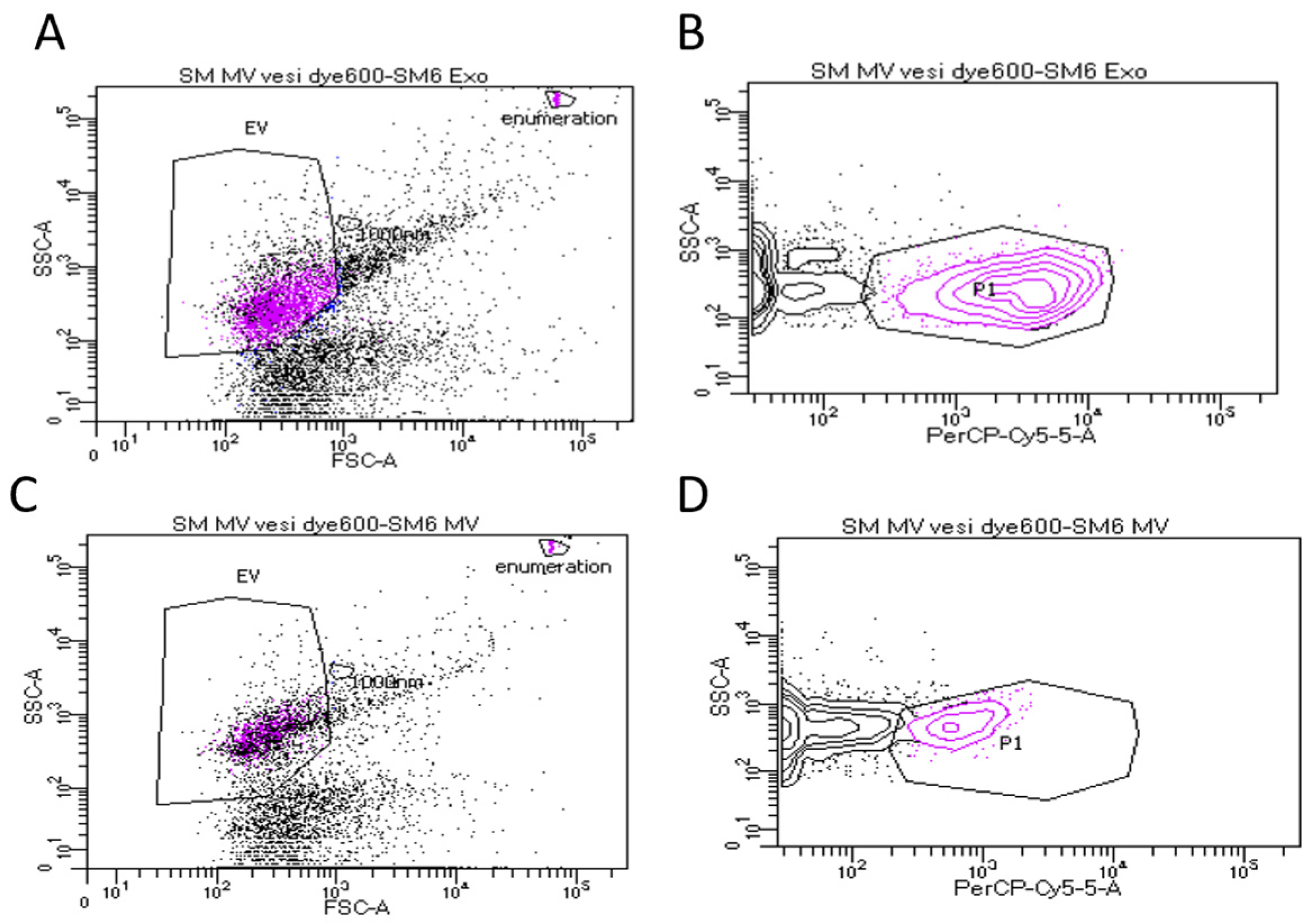
Milk was thawed quickly in a water bath at 37 °C and immediately aliquoted into two 20 mL fractions, one of which was refrozen at −80 °C for later use. The remaining 20 mL fraction was centrifuged at 3000× g for 10 min at 4 °C in a swing-out benchtop centrifuge to separate the cream and remove any cellular debris. The cream was carefully removed from the top of the samples using a spatula. The supernatant was then decanted into a clean tube, being careful not to disturb any pellets containing cellular debris. To remove casein micelles from the remaining milk fraction, glacial acetic acid was added dropwise until the pH reached 4.6 and visible precipitation was seen [17]. The precipitate was then pelleted at 3000× g for 10 min at 4 °C in a swing-out benchtop centrifuge. Using a modified method from Thery et al. [18], supernatants from the previous step were carefully decanted into clean Beckman 14 mL polyallomer ultracentrifuge tubes (Cat #331374) for further extraction. They were centrifuged in a Beckman Coulter Optima L-80-XP ultracentrifuge using a SW40Ti rotor at 17,000× g for 30 min, 4 °C, without braking, to pellet the large EV population (LEVs; microvesicles). SM-LEV pellets were resuspended in 1 mL 1 × BS and aliquoted before storage at −80 °C for further analysis. The supernatants from the previous step were decanted into clean ultracentrifuge tubes and centrifuged at 100,000× g for 90 min at 4 °C in a SW40Ti rotor, without braking, to pellet the small EV population (sEVs; exosomes). The supernatants from this centrifugation step were carefully discarded and pellets resuspended in 8 mL 1 × PBS. This was then carefully layered on top of a 30% sucrose solution and centrifuged at 100,000× g for 60 min using the SW40Ti rotor, without braking, to further purify the sEVs. The cloudy layer at the interface of the PBS and sucrose phase containing sEVs was removed using a sterile Pastette and placed in a clean ultracentrifuge tube, diluted with 1 × PBS, and centrifuged using the SW40Ti rotor, without braking, for 60 min at 100,000× g and 4 °C. The top 1 mL was collected as a negative control, and then, the remainder of the supernatant was carefully removed and discarded. The remaining pellet was resuspended in 1 mL 1 × PBS and immediately frozen in aliquots at −80 °C for further analysis.

2.4. Flow Cytometry

Five microliters of final preparation of SM-LEVs or SM-sEVs from each milk sample was diluted with 45 µL 1 × PBS. A total of 1 µL VesiDye-600 (Vesiculab, Nottingham UK; reconstituted according to the manufacturer’s instructions) was added to each and incubated at 37 °C for 1 h before further dilution to 500 µL with 1 × PBS. A total of 10 µL enumeration beads (Coulter) were added to each sample before analysis using a BD FACS CANTO, calibrated using CST beads (both BD, Witney, UK). EVs were gated by size using 1 µm NIST beads (Thermo Fisher), and the population < 1 µM was then gated by VesiDye positivity (see Figure 1 for representative PercP-Cy5.5/SSC plot). The total number of EVs was calculated using the equation EV/µL = 20 × [events of interest] × [dilution factor]/[enumeration bead count], as described previously [19].

2.5. Determination of Protein Concentration of EVs

A total of 10 µL 5xRIPA buffer was added to 40 µL of the final preparation of SM-LEVs or SM-SEVs from each milk sample. After vortexing to mix, the samples were sonicated for 15 min at 30 °C and immediately analyzed using a Pierce micro-BCA assay kit [20] (Thermo Fisher) according to the manufacturer’s instructions.

2.6. RNA Isolation and miRNA Analysis

For RNA isolation from SM-SEVs, 500 µL of the final preparations from each milk sample were used to isolate total RNA using the exoEasy total RNA kit (Qiagen; Manchester, UK) according to the manufacturer’s instructions [21]. For RNA isolation from SM-LEVs, 500 µL of the final preparations were re-centrifuged at 17,000× g for 15 min at 4 °C in a benchtop microfuge. The resulting pellet was dissolved in 700 µL Qiazol (Qiagen), and the rest of the protocol for the exoEasy total RNA kit was followed according to the manufacturer’s instructions. RNA was quantified using a DeNovix DS-11 nanodrop (DeNovix, Wilmington, DE, USA). Individual miRNAs (miR10b, miR26a, miR27a, miR92a, miR125b, miR126, miR155 and miR223) were reverse-transcribed using the TaqMan® microRNA RT kit (Cat #4366596, ABI. Thermo Fisher, UK) and the associated miRNA-specific inventoried predesigned primers (TaqMan® micro-RNA assay kit, #4427975. ABI) according to the manufacturer’s instructions. Total RNA was normalized at a concentration of 2 ng/μL, and 5 μL RNA was added to the reaction mix containing 0.15 μL 100 mM dNTP, 1 μL Reverse Transcriptase enzyme (50 U/μL), 1.5 μL 10 × RT buffer, 0.19 μL RNase inhibitor (20 U/μL), 3 μL 5× RT-specific primer and 4.16 μL DEPC-treated water to obtain a final volume of 15 μL. RT reaction conditions were as follows: primer annealing for 30 min at 16 °C, extension for 30 min at 42 °C and final incubation for 5 min at 85 °C to stop the reaction. All RT-qPCRs were carried out in triplicate according to the manufacturer’s instructions, using TaqMan™ Universal Master Mix II, no UNG (Cat# 4440040), and were performed on the Bio-Rad CFX384 Real Time PCR detection cycler system with the following conditions: an initial inactivation step at 95 °C for 10 min, followed by 40 cycles consisting of denaturing at 95 °C for 10 s, annealing and elongation at 60 °C for 60 s.

2.7. Cell Culture

THP-1 human monocytes were from ATCC. They were cultured at 37 °C in RPMI, 10% FCS, with 0.05 mM β-mercaptoethanol. For differentiation, THP-1 cells were incubated with 5 ng/mL PMA for 72 h at 37 °C.

2.8. Reactive Oxygen Species (ROS) Assays

We used a modified protocol from Heinrich et al. [19] to measure ROS. Briefly, THP-1 monocytes were pelleted, washed and resuspended at 2 × 10⁶ cells per mL in RPMI without serum. They were incubated with 5 µM dihydrorhodamine 1,2,3 for 10 min in the dark, before seeding at 10,000 cells per well in 96-well plates. SMEVs were added at a final dilution of 1:100. 2,3-Dimethoxy-1,4-naphthoquinone (DMNQ; AbCam Cambridge, UK), a well-described stimulus for ROS generation in monocytes [22], was added at a final concentration of 250 µM. Cells were incubated for 24 h at 37 °C and plates were read in a fluorescent plate reader (Tecan Cyto 400, Reading, UK) at the start and end of the experiment. All treatments were added in triplicate. Regarding THP-1 macrophages, they were plated at 100,000 cells per well in full medium together with PMA at 5 ng/mL, and incubated for 72 h at 37 °C. The medium was removed together with non-adherent cells. The adherent population was washed twice with serum-free RPMI to remove bovine-derived EVs from the serum. Dihydrorhodamine 1,2,3 was added at a final concentration of 5 µM, and the remainder of the procedure was as outlined above.

2.9. Measurement of CCL2 Secretion

THP-1 monocytes were pelleted, washed and resuspended at 2 × 10⁶ cells per mL in RPMI without serum. They were seeded at 25,000 cells per well in 24-well plates, and LPS O55:B5 was added to some wells at a final concentration of 1 µg/mL. SMEVs were added at a final dilution of 1:100. Cells were incubated for 24 h before supernatants were collected, and cells were pelleted by centrifugation at 5000× g for 10 min at 4 °C. Supernatants were carefully decanted and stored at −80 °C, followed by analysis for CCL2 using eBioscience Ready-Set-Go! ELISA (Invitrogen, Thermo Fisher) according to the manufacturer’s instructions. All treatments were added in triplicate. Regarding THP-1 macrophages, they were plated at 250,000 cells per well in full medium together with 5 ng/mL PMA and incubated for 72 h at 37 °C. The medium was removed together with non-adherent cells. The adherent population was washed twice with serum-free RPMI to remove bovine-derived EVs from the serum. Cells were incubated in serum-free RPMI with LPS (1 µg/mL) and SMEVs at a final dilution of 1:100 for 24 h at 37 °C. Supernatants were harvested as above and subjected to the same ELISA assays.

2.10. Statistical Analysis

All statistical analysis was carried out using Prism 10 (GraphPad, San Diego, CA, USA). Data were tested for normality using a Shapiro–Wilk test and one-way ANOVA followed by Fishers LSD for ROS assays and ELISA. ROS assays were carried out using the six sheep EV samples in triplicate, and CCL2 ELISAs were carried out using the six sheep EV samples in duplicate. p < 0.05 was considered statistically significant (*). ** p < 0.01, *** p < 0.001.

3. Results

3.1. Characterization of Sheep Milk Derived LEVs and sEVs

As shown in Table 1, we identified small and large EVs by flow cytometry using a lipid-permeable dye, and were able to detect measureable total protein and total RNA in small and large EVs from each sample.

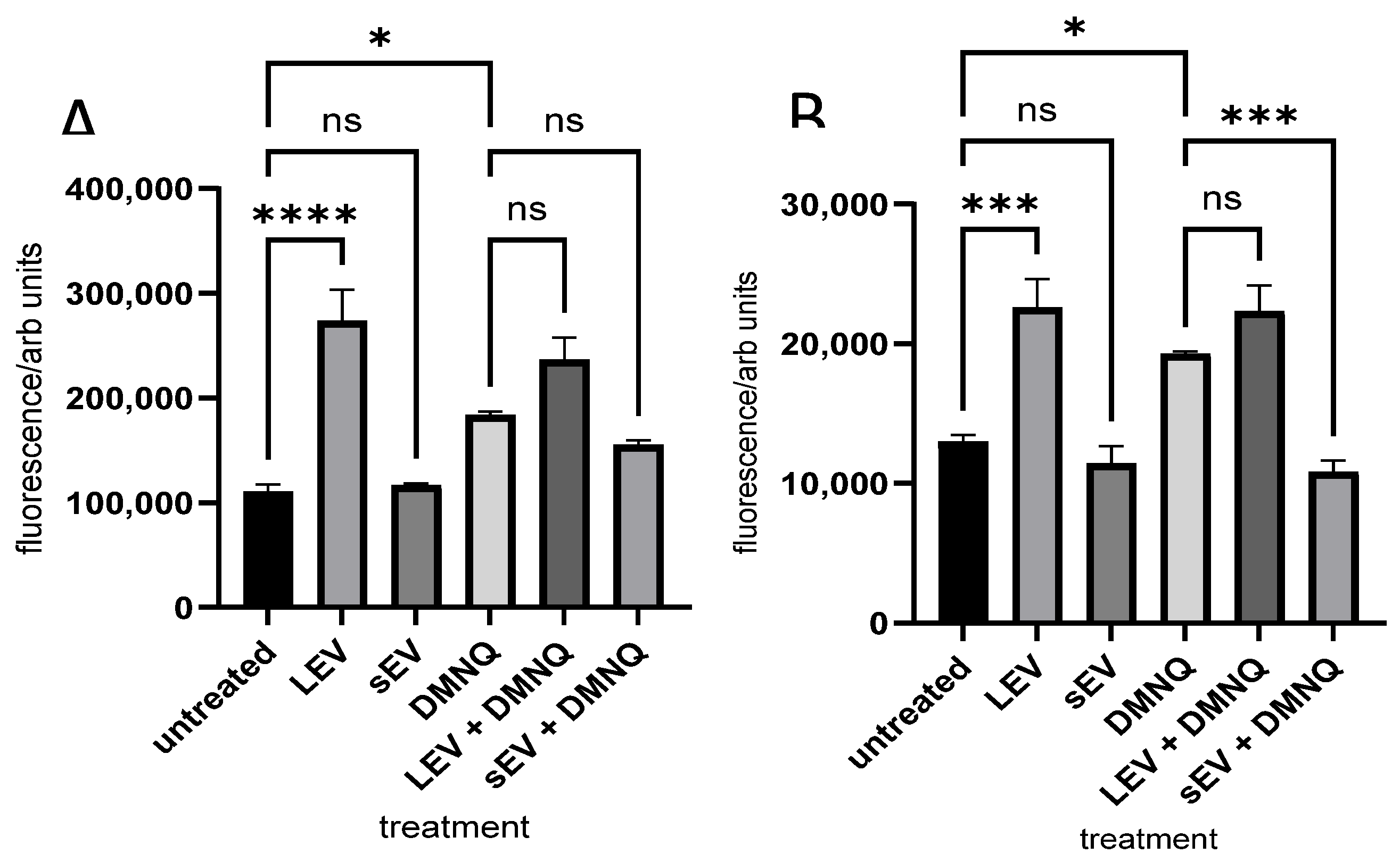
3.2. Monocyte and Macrophage ROS Production Induced by Sheep Milk-Derived LEVs and sEVs

We hypothesized that SMEVs could have an effect on leukocyte oxidant stress both in the presence and absence of other stimuli. Equal volumes of isolated LEVs or sEVs were added to THP-1 monocytes or differentiated THP-1 macrophages that had been pre-loaded with a dihydrorhodamine-1,2,3 fluorescent label and incubated for 24 h in the presence or absence of DMNQ, which is known to induce oxidative stress [22]. There was a significant increase in ROS in cells treated with LEVs, but not with sEVs, in both monocytes (Figure 2A) and macrophages (Figure 2B). Neither LEVs nor sEVs affected the DMNQ response in monocytes. sEVs significantly downmodulated the DMNQ response in macrophages.

3.3. Secretion of CCL2 Chemokine Induced by Sheep Milk-Derived LEVs and sEVs

Since SMEVs were able to modulate ROS in monocytes and macrophages and these can be an indication of wider pro-inflammatory gene expression, we next measured the secretion of the CCL2 chemokine. We chose CCL2 as it has been reported to be highly upregulated in monocytes at both the mRNA and protein level [23]. After 24 of incubation with either LEVs or sEVs (Figure 3), we found that incubation with LEVs or sEVs alone did not induce CCL2 secretion; however, in monocytes (Figure 3A), there was a significant decrease in CCL2 secretion compared to LPS alone when cells were incubated with LPS and LEVs or sEVs. This was not replicated in macrophages (Figure 3B).

3.4. miRNA Contents of Sheep Milk-Derived LEVs and sEVs

In order to determine whether there could be an influence of miRNA cargo on the increase in ROS production or modulation of CCL2 production, we measured miRNA in LEVs and sEVs from individual SM samples. We measured eight different miRNAs based on previous reports of their presence in either SM or CM and reporting in other settings of their effects on inflammatory pathways. We were able to measure expression in five of the eight miRNAs tested (miR26, 92a, 125b, 155 and 223). The expression levels of individual miRNAs was consistent between LEVs and sEVs (Figure 4). There was no detectable expression of miR10b, miR27a or miR126 in either sEVs or LEVs. There was no detectable expression of any miRNA in the water controls.

4. Discussion

As the market in the UK for sheep milk and dairy products increases, it is important to gain a greater understanding of the constitutive components that make up this complex, highly nutritious biofluid. There is an increasing awareness that milk not only contains macronutrients, which are regularly tested and reported on in commercial settings, but also includes the potential for the transfer of genetic material from sheep to humans. The secretion of extracellular vesicles by mammary epithelial cells into milk is increasingly thought to function physiologically to aid in the development of the infant immune system and gut [11], starting with intake of colostrum and continuing throughout lactation. Here, we have demonstrated that it is possible to isolate small and large EV-like particles from raw sheep milk. We have further demonstrated a functional effect on human inflammatory pathways in vitro; we believe this is the first such publication to do so. Intriguingly, we observed an increase in ROS when LEVs were applied to both THP-1 monocytes and macrophages, and this was additive when cells were treated with DMNQ, a known pro-oxidant compound. In contrast, sEVs did not induce any ROS and inhibited DMNQ-stimulated ROS, suggesting that there may be differences in the cargo, cell signaling efficiency or rates of cellular uptake for the two species of EV.
ROS production and oxidative stress are often associated directly with pro-inflammatory pathways and can lead to activation of the secretion of cytokines and chemokines, so it is important to understand whether the intake of milk-derived EVs contributes to, or can alleviate, oxidative stress, especially in human individuals with underlying chronic gastrointestinal inflammation such as ulcerative colitis and Crohn’s disease [24]. CCL2 is a key chemokine that drives the recruitment of monocytes to inflamed endothelium. It is secreted by endothelial cells, monocytes and macrophages in large amounts in several highly prevalent human chronic inflammatory conditions, including inflammatory bowel disease [25], rheumatoid arthritis [26], type 2 diabetes, insulin resistance and metabolic syndrome [27], chronic kidney disease [28], atherosclerosis [29] and endothelial dysfunction [30]. Here, we have found that though SM-LEVs increased ROS production in monocytes (Figure 2A) and macrophages (Figure 2B), they also significantly inhibited CCL2 production in monocytes (Figure 3A). SM-sEVs significantly inhibited ROS in macrophages (Figure 2B), but not monocytes (Figure 2A), though they did significantly inhibit CCL2 production in monocytes (Figure 3A). Since CCL2 has such a large impact on many different chronic inflammatory disorders, non-pharmacologic approaches to lessen its production and therefore reduce monocyte chemotaxis through simple dietary intervention could be a useful strategy. Based on these promising data, if these potential health benefits are mirrored in vivo, encouraging the consumption of sheep milk for those who are not able to tolerate cows’ milk as part of a healthy diet might meet with a positive consumer response.
We investigated the RNA cargo of the isolated SM-sEVs and LEVs and were able to identify several microRNA species that have previously been shown to be associated with chronic inflammation, either exacerbating or demonstrating the regulation of immune cell function, activation and differentiation. The most highly expressed (lowest Ct; Figure 4) in our panel was miR26a, which has previously been shown to be elevated in raw milk and to be relatively consistently expressed across lactation [31], more recently as one of the most abundant miRNAs in cows’ milk [32] and in sheep milk EVs [33]. In a separate study, miR26a has been shown to target the IFNb and IL6 genes, and was postulated to be positively related to asthma reduction in infants who drink raw milk [34].
MiR92a was the next best expressed (Figure 4). MiR92a has been identified in a bovine study of mastitis [35]. Functionally, it has been associated with STAT3, CTLA4 and TLR1 regulation, genes that have been implicated as key players in chronic inflammatory immune responses. MiR125b has also been shown to regulate STAT3 [36] and Luoreng et al. [37] found that MiR125b is a negative regulator of mastitis, with its expression significantly downregulated during the LPS stimulation of bovine mammary epithelial cells in vitro. Mastitis is a key long-standing endemic disease in sheep farming, as well as in the dairy cattle industry [38], that provides a constant source of strain on already scarce resources such as time and money [39]. Although data on the prevalence and impact of the different forms of mastitis are scarce for meat sheep in the UK, let alone dairy sheep, it is thought that the incidence of acute mastitis is 0–6.6% per flock in meat sheep [40] and that chronic mastitis can affect up to half a flock at a time [41]. Since we were able to identify expression in all six samples from healthy dairy sheep, we suggest that miR125b may hold promise as a useful biomarker of ovine mammary health and is worthy of further investigation. We detected low levels of MiR155 in both LEVs and sEVs tested, which may represent a species difference between small and large ruminants, since Izumi et al. [42] reported the presence of miR155 in bovine colostrum but not mature milk. MiR155 has been known for a long time to be a key miRNA in the regulation of both innate and adaptive immune responses [43]. For example, it has been shown to modulate the differentiation of T and B cells, as well as being a positive regulator for regulatory T cell differentiation [43]. Specifically, it has been implicated in the expression of STAT3 and CTLA4, as well as VCAM-1 and eNOS, suggesting that its presence could influence cytokine and chemokine expression in activated endothelial cells, as well as contributing to leukocyte inflammatory responses. A link has recently been established between miR155 expression and CCL2 in RAW264 murine macrophages in vitro and in a rodent model of sepsis [44]. This is intriguing and worthy of further study, given our finding that both large and small SMEVs included a cargo of miR155 but reduced, rather than exacerbated, LPS-driven CCL2 in human THP-1 cells.
In the present study, we saw the most variation in the expression of miR223 between individual sheep. MiR223 has previously been shown to be a negative regulator of neutrophil activation [43] and has recently been associated with mammary inflammation [45,46]. This could be a useful miRNA to follow up as a potential biomarker of early mastitis or other inflammatory conditions that might impact dairy sheep production or their health and welfare.
MiR10b, miR27a and miR126 have all previously been reported to be present in EVs from cows and sheep [31,33]; however, we were not able to detect these species in our samples. This may be due to differences in processing or the phase of lactation at which our samples were taken.
Sheep milk consumption is gaining popularity, and therefore, its production is becoming commercially more viable for farmers in the UK. There is continued pressure on the UK livestock industries to produce more output with less input, as well as minimizing negative impacts across the food system (all the elements, actors and activities involved in producing and consuming food), including on the environment and the welfare and health of food-producing animals and consumers, all while ensuring prices and accessibility stay at an equitable level [47,48,49]. Sheep can take advantage of lower-quality land, enabling more productive land to be used for dairy or other feedstuff production; this could allow for more milk production but lessen the overall negative effects of dairy milk production.
From a One Health and One Welfare perspective (where the health and welfare of humans, non-human animals and the ecosystem are considered to be interdependent), if sheep milk contains beneficial anti-inflammatory properties, then providing lambs with ewe milk rather than powdered lamb milk replacer could give them an advantage in being able to better ward off diseases and syndromes that are of greater prevalence in farmed settings. It will also be important in the sheep meat industry to ascertain whether these benefits also impact dietary phospholipids in the meat that would then be eaten by human consumers [50]. This represents an important move to ensure that small-ruminant health and welfare is maintained as the industry grows. There are also many questions that should be addressed that relate to the sustainability of sheep dairy enterprises, including the land use for ovine vs. bovine systems, water and land contamination from ovine waste, the production of greenhouse gases (methane) and the possible increased use of medicines such as antimicrobials. Whilst these are clearly beyond the scope of this small-molecular study, there could be a benefit to following up with an interrogation of ovine excreta to determine whether similar miRNA are present, what effect they might have on the microbiome, and whether there are ovine miRNA present that can influence soil and aquatic micro-organisms and therefore influence wider environmental questions. Additionally, it would be interesting to ascertain whether the health and welfare of sheep impact these properties; for example, if sheep have higher welfare and/or health status, does this positively impact the number or cargos of the LEVs and/or sEVs in the milk? This type of One Health–One Welfare investigation could move us more towards more win–win–win actions for more sustainable farming [51].
This study represents a first step towards understanding the potential benefits of the human ingestion of sheep milk. Further work should include an examination of milk to determine whether there are different phenotypes. These could include different stages of lactation, different breeds of dairy sheep and different management systems, additionally paying attention to ewes with evidence of mastitis or other health issues, where molecular markers measurable in milk could enable early intervention. Our approach to interrogating the molecular profile of the mammary epithelial cell via analysis of secreted EVs could also be useful to understand more about the potential health and welfare of dairy sheep (and could be applied to other small-ruminant dairy species). In particular, the finding that it is possible to measure the presence of different miRNA species could allow insight into the changes that take place, either physiologically during the progression of lactation or during mastitis, and may also be of relevance for dairy cow health. There is also potential to develop a biomarker panel based on the presence of miRNAs that are up- or downregulated in mastitis or that change at different stages of lactation.

5. Conclusions

In this study, we have shown that both small and large EV particles can be obtained from raw sheep milk; these particles are able to influence human immune cell behavior in vitro, including reactive oxygen species production and the secretion of CCL2 chemokines. We have demonstrated that it is possible to isolate sufficient miRNA cargo from individual SM-derived EVs to test using qRT-PCR. Our findings broadly suggest that the human intake of sheep milk may be beneficial for individuals with chronic inflammatory conditions. However, further work is required to fully interrogate the miRNA cargo and further explore the downstream effects that these particles have on human inflammation. This work could not only impact human health and welfare, but has the potential to also enable the investigation of the benefits to livestock and environmental health and welfare, and sustainable farming.

Author Contributions

Conceptualization, C.L.; methodology, C.L., B.S. and R.C.P.; writing—original draft preparation, C.L., B.S., L.W. and R.C.P.; writing—review and editing, C.L., B.S., L.W. and R.C.P. All authors have read and agreed to the published version of the manuscript.

Funding

This research received no external funding.

Institutional Review Board Statement

This study did not require ethical approval. Milk was collected as part of routine flock management.

Informed Consent Statement

Not applicable.

Data Availability Statement

The data that support the findings of this study are available from the corresponding author upon reasonable request.

Conflicts of Interest

The authors declare no conflicts of interest.

References

  1. AHDB. Dairy Market Dashboard. Available online: https://ahdb.org.uk/dairy-market-dashboard (accessed on 29 December 2023).
  2. Costa, A.; Lopez-Villalobos, N.; Sneddon, N.W.; Shalloo, L.; Franzoi, M.; De Marchi, M.; Penasa, M. Invited review: Milk lactose-Current status and future challenges in dairy cattle. J. Dairy. Sci. 2019, 102, 5883–5898. [Google Scholar] [CrossRef]
  3. Mee, J.F.; Boyle, L.A. Assessing whether dairy cow welfare is “better” in pasture-based than in confinement-based management systems. N. Z. Vet. J. 2020, 68, 168–177. [Google Scholar] [CrossRef]
  4. Roy, D.; Ye, A.; Moughan, P.J.; Singh, H. Composition, Structure, and Digestive Dynamics of Milk From Different Species-A Review. Front. Nutr. 2020, 7, 577759. [Google Scholar] [CrossRef]
  5. Abou El Qassim, L.; Martínez, B.; Rodríguez, A.; Dávalos, A.; López de Las Hazas, M.C.; Menéndez Miranda, M.; Royo, L.J. Effects of Cow’s Milk Processing on MicroRNA Levels. Foods 2023, 12, 2950. [Google Scholar] [CrossRef]
  6. Yáñez-Mó, M.; Siljander, P.R.; Andreu, Z.; Zavec, A.B.; Borràs, F.E.; Buzas, E.I.; Buzas, K.; Casal, E.; Cappello, F.; Carvalho, J.; et al. Biological properties of extracellular vesicles and their physiological functions. J. Extracell. Vesicles 2015, 4, 27066. [Google Scholar] [CrossRef]
  7. Fabbri, M.; Paone, A.; Calore, F.; Galli, R.; Gaudio, E.; Santhanam, R.; Lovat, F.; Fadda, P.; Mao, C.; Nuovo, G.J.; et al. MicroRNAs bind to Toll-like receptors to induce prometastatic inflammatory response. Proc. Natl. Acad. Sci. USA 2012, 109, E2110–E2116. [Google Scholar] [CrossRef] [PubMed]
  8. Kalra, H.; Simpson, R.J.; Ji, H.; Aikawa, E.; Altevogt, P.; Askenase, P.; Bond, V.C.; Borràs, F.E.; Breakefield, X.; Budnik, V.; et al. Vesiclepedia: A compendium for extracellular vesicles with continuous community annotation. PLoS Biol. 2012, 10, e1001450. [Google Scholar] [CrossRef] [PubMed]
  9. Pan, J.; Chen, M.; Li, N.; Han, R.; Yang, Y.; Zheng, N.; Zhao, S.; Zhang, Y. Bioactive Functions of Lipids in the Milk Fat Globule Membrane: A Comprehensive Review. Foods 2023, 12, 3755. [Google Scholar] [CrossRef] [PubMed]
  10. Pawlowski, K.; Lago-Novais, D.; Bevilacqua, C.; Mobuchon, L.; Crapart, N.; Faulconnier, Y.; Boby, C.; Carvalho, G.; Martin, P.; Leroux, C. Different miRNA contents between mammary epithelial cells and milk fat globules: A random or a targeted process? Mol. Biol. Rep. 2020, 47, 8259–8264. [Google Scholar] [CrossRef] [PubMed]
  11. Zempleni, J.; Aguilar-Lozano, A.; Sadri, M.; Sukreet, S.; Manca, S.; Wu, D.; Zhou, F.; Mutai, E. Biological Activities of Extracellular Vesicles and Their Cargos from Bovine and Human Milk in Humans and Implications for Infants. J. Nutr. 2017, 147, 3–10. [Google Scholar] [CrossRef] [PubMed]
  12. Jiang, X.; You, L.; Zhang, Z.; Cui, X.; Zhong, H.; Sun, X.; Ji, C.; Chi, X. Biological Properties of Milk-Derived Extracellular Vesicles and Their Physiological Functions in Infant. Front. Cell Dev. Biol. 2021, 9, 693534. [Google Scholar] [CrossRef]
  13. Benmoussa, A.; Lee, C.H.; Laffont, B.; Savard, P.; Laugier, J.; Boilard, E.; Gilbert, C.; Fliss, I.; Provost, P. Commercial Dairy Cow Milk microRNAs Resist Digestion under Simulated Gastrointestinal Tract Conditions. J. Nutr. 2016, 146, 2206–2215. [Google Scholar] [CrossRef] [PubMed]
  14. Baier, S.R.; Nguyen, C.; Xie, F.; Wood, J.R.; Zempleni, J. MicroRNAs are absorbed in biologically meaningful amounts from nutritionally relevant doses of cow milk and affect gene expression in peripheral blood mononuclear cells, HEK-293 kidney cell cultures, and mouse livers. J. Nutr. 2014, 144, 1495–1500. [Google Scholar] [CrossRef] [PubMed]
  15. Gallier, S.; Van den Abbeele, P.; Prosser, C. Comparison of the Bifidogenic Effects of Goat and Cow Milk-Based Infant Formulas to Human Breast Milk in an. Front. Nutr. 2020, 7, 608495. [Google Scholar] [CrossRef] [PubMed]
  16. Arntz, O.J.; Pieters, B.C.; Oliveira, M.C.; Broeren, M.G.; Bennink, M.B.; de Vries, M.; van Lent, P.L.; Koenders, M.I.; van den Berg, W.B.; van der Kraan, P.M.; et al. Oral administration of bovine milk derived extracellular vesicles attenuates arthritis in two mouse models. Mol. Nutr. Food Res. 2015, 59, 1701–1712. [Google Scholar] [CrossRef] [PubMed]
  17. Mukhopadhya, A.; Santoro, J.; O’Driscoll, L. Extracellular vesicle separation from milk and infant milk formula using acid precipitation and ultracentrifugation. STAR Protoc. 2021, 2, 100821. [Google Scholar] [CrossRef] [PubMed]
  18. Théry, C.; Amigorena, S.; Raposo, G.; Clayton, A. Isolation and characterization of exosomes from cell culture supernatants and biological fluids. Curr. Protoc. Cell Biol. 2006, 3, 3.22.1–3.22.29. [Google Scholar] [CrossRef]
  19. Heinrich, L.F.; Andersen, D.K.; Cleasby, M.E.; Lawson, C. Long-term high fat feeding of rats results in increased numbers of circulating microvesicles with pro-inflammatory effects on endothelial cells. Br. J. Nutr. 2015, 113, 1704–1711. [Google Scholar] [CrossRef]
  20. Smith, P.K.; Krohn, R.I.; Hermanson, G.T.; Mallia, A.K.; Gartner, F.H.; Provenzano, M.D.; Fujimoto, E.K.; Goeke, N.M.; Olson, B.J.; Klenk, D.C. Measurement of protein using bicinchoninic acid. Anal. Biochem. 1985, 150, 76–85. [Google Scholar] [CrossRef]
  21. Enderle, D.; Spiel, A.; Coticchia, C.M.; Berghoff, E.; Mueller, R.; Schlumpberger, M.; Sprenger-Haussels, M.; Shaffer, J.M.; Lader, E.; Skog, J.; et al. Characterization of RNA from Exosomes and Other Extracellular Vesicles Isolated by a Novel Spin Column-Based Method. PLoS ONE 2015, 10, e0136133. [Google Scholar] [CrossRef]
  22. Namgaladze, D.; Preiss, S.; Dröse, S.; Brandt, U.; Brüne, B. Phospholipase A2-modified low density lipoprotein induces mitochondrial uncoupling and lowers reactive oxygen species in phagocytes. Atherosclerosis 2010, 208, 142–147. [Google Scholar] [CrossRef] [PubMed]
  23. Dommel, S.; Blüher, M. Does C-C Motif Chemokine Ligand 2 (CCL2) Link Obesity to a Pro-Inflammatory State? Int. J. Mol. Sci. 2021, 22, 1500. [Google Scholar] [CrossRef] [PubMed]
  24. Sahoo, D.K.; Heilmann, R.M.; Paital, B.; Patel, A.; Yadav, V.K.; Wong, D.; Jergens, A.E. Oxidative stress, hormones, and effects of natural antioxidants on intestinal inflammation in inflammatory bowel disease. Front. Endocrinol. 2023, 14, 1217165. [Google Scholar] [CrossRef] [PubMed]
  25. Singh, U.P.; Singh, N.P.; Murphy, E.A.; Price, R.L.; Fayad, R.; Nagarkatti, M.; Nagarkatti, P.S. Chemokine and cytokine levels in inflammatory bowel disease patients. Cytokine 2016, 77, 44–49. [Google Scholar] [CrossRef]
  26. Moadab, F.; Khorramdelazad, H.; Abbasifard, M. Role of CCL2/CCR2 axis in the immunopathogenesis of rheumatoid arthritis: Latest evidence and therapeutic approaches. Life Sci. 2021, 269, 119034. [Google Scholar] [CrossRef]
  27. Pan, X.; Kaminga, A.C.; Wen, S.W.; Liu, A. Chemokines in Prediabetes and Type 2 Diabetes: A Meta-Analysis. Front. Immunol. 2021, 12, 622438. [Google Scholar] [CrossRef]
  28. Puthumana, J.; Thiessen-Philbrook, H.; Xu, L.; Coca, S.G.; Garg, A.X.; Himmelfarb, J.; Bhatraju, P.K.; Ikizler, T.A.; Siew, E.D.; Ware, L.B.; et al. Biomarkers of inflammation and repair in kidney disease progression. J. Clin. Investig. 2021, 131, e139927. [Google Scholar] [CrossRef]
  29. Georgakis, M.K.; Bernhagen, J.; Heitman, L.H.; Weber, C.; Dichgans, M. Targeting the CCL2-CCR2 axis for atheroprotection. Eur. Heart J. 2022, 43, 1799–1808. [Google Scholar] [CrossRef]
  30. Martynowicz, H.; Janus, A.; Nowacki, D.; Mazur, G. The role of chemokines in hypertension. Adv. Clin. Exp. Med. 2014, 23, 319–325. [Google Scholar] [CrossRef]
  31. Chen, X.; Gao, C.; Li, H.; Huang, L.; Sun, Q.; Dong, Y.; Tian, C.; Gao, S.; Dong, H.; Guan, D.; et al. Identification and characterization of microRNAs in raw milk during different periods of lactation, commercial fluid, and powdered milk products. Cell Res. 2010, 20, 1128–1137. [Google Scholar] [CrossRef]
  32. Li, W.; Li, W.; Wang, X.; Zhang, H.; Wang, L.; Gao, T. Comparison of miRNA profiles in milk-derived extracellular vesicles and bovine mammary glands. Int. Dairy J. 2022, 134, 105444. [Google Scholar] [CrossRef]
  33. Quan, S.; Nan, X.; Wang, K.; Jiang, L.; Yao, J.; Xiong, B. Characterization of Sheep Milk Extracellular Vesicle-miRNA by Sequencing and Comparison with Cow Milk. Animals 2020, 10, 331. [Google Scholar] [CrossRef]
  34. Kirchner, B.; Pfaffl, M.W.; Dumpler, J.; von Mutius, E.; Ege, M.J. microRNA in native and processed cow’s milk and its implication for the farm milk effect on asthma. J. Allergy Clin. Immunol. 2016, 137, 1893–1895.e1813. [Google Scholar] [CrossRef]
  35. Lai, Y.C.; Fujikawa, T.; Maemura, T.; Ando, T.; Kitahara, G.; Endo, Y.; Yamato, O.; Koiwa, M.; Kubota, C.; Miura, N. Inflammation-related microRNA expression level in the bovine milk is affected by mastitis. PLoS ONE 2017, 12, e0177182. [Google Scholar] [CrossRef] [PubMed]
  36. Yu, Y.; Ou-Yang, W.X.; Zhang, H.; Jiang, T.; Tang, L.; Tan, Y.F.; Luo, H.Y.; Xiao, Z.H.; Li, S.J. MiR-125b enhances autophagic flux to improve septic cardiomyopathy via targeting STAT3/HMGB1. Exp. Cell Res. 2021, 409, 112842. [Google Scholar] [CrossRef] [PubMed]
  37. Luoreng, Z.M.; Wei, D.W.; Wang, X.P. MiR-125b regulates inflammation in bovine mammary epithelial cells by targeting the NKIRAS2 gene. Vet. Res. 2021, 52, 122. [Google Scholar] [CrossRef] [PubMed]
  38. AHDB. Available online: https://ruminanthw.org.uk/wp-content/uploads/2021/05/Cattle-and-sheep-HW-priorities-survey-May-2021-FINAL.pdf (accessed on 29 December 2023).
  39. Whatford, L.; van Winden, S.; Häsler, B. A systematic literature review on the economic impact of endemic disease in UK sheep and cattle using a One Health conceptualisation. Prev. Vet. Med. 2022, 209, 105756. [Google Scholar] [CrossRef] [PubMed]
  40. Smith, E.M.; Willis, Z.N.; Blakeley, M.; Lovatt, F.; Purdy, K.J.; Green, L.E. Bacterial species and their associations with acute and chronic mastitis in suckler ewes. J. Dairy. Sci. 2015, 98, 7025–7033. [Google Scholar] [CrossRef] [PubMed]
  41. Grant, C.; Smith, E.M.; Green, L.E. A longitudinal study of factors associated with acute and chronic mastitis and their impact on lamb growth rate in 10 suckler sheep flocks in Great Britain. Prev. Vet. Med. 2016, 127, 27–36. [Google Scholar] [CrossRef] [PubMed]
  42. Izumi, H.; Kosaka, N.; Shimizu, T.; Sekine, K.; Ochiya, T.; Takase, M. Bovine milk contains microRNA and messenger RNA that are stable under degradative conditions. J. Dairy. Sci. 2012, 95, 4831–4841. [Google Scholar] [CrossRef] [PubMed]
  43. Lindsay, M.A. microRNAs and the immune response. Trends Immunol. 2008, 29, 343–351. [Google Scholar] [CrossRef] [PubMed]
  44. Wang, C.; Zheng, Y.; Fan, Q.; Li, Z.; Qi, X.; Chen, F.; Xu, L.; Zhou, S.; Chen, X.; Li, Y.; et al. MiR-155 promotes acute organ injury in LPS-induced endotoxemic mice by enhancing CCL-2 expression in macrophages. Shock 2023. [Google Scholar] [CrossRef] [PubMed]
  45. Zhou, M.; Barkema, H.W.; Gao, J.; Yang, J.; Wang, Y.; Kastelic, J.P.; Khan, S.; Liu, G.; Han, B. MicroRNA miR-223 modulates NLRP3 and Keap1, mitigating lipopolysaccharide-induced inflammation and oxidative stress in bovine mammary epithelial cells and murine mammary glands. Vet. Res. 2023, 54, 78. [Google Scholar] [CrossRef]
  46. Li, Y.X.; Jiao, P.; Wang, X.P.; Wang, J.P.; Feng, F.; Bao, B.W.; Dong, Y.W.; Luoreng, Z.M.; Wei, D.W. RNA-seq reveals the role of miR-223 in alleviating inflammation of bovine mammary epithelial cells. Res. Vet. Sci. 2023, 159, 257–266. [Google Scholar] [CrossRef]
  47. Boulton, A.C.; Rushton, J.; Wathes, C.M. Past trends and future challenges for a sustainable UK dairy industry. R. Agric. Soc. Engl. J. 2011, 172, 1–7. [Google Scholar]
  48. Shortall, O. Cows eat grass, don’t they? Contrasting sociotechnical imaginaries of the role of grazing in the UK and Irish diary sectors. J. Rural. Stud. 2019, 72, 45–57. [Google Scholar] [CrossRef]
  49. Trewern, J.; Chenoweth, J.; Christie, I.; Keller, E.; Halevy, S. Are UK retailers well placed to deliver ‘less and better’meat and dairy to consumers? Sustain. Prod. Consum. 2021, 28, 154–163. [Google Scholar] [CrossRef]
  50. Lordan, R.; Tsoupras, A.; Zabetakis, I. Phospholipids of Animal and Marine Origin: Structure, Function, and Anti-Inflammatory Properties. Molecules 2017, 22, 1964. [Google Scholar] [CrossRef]
  51. Scherer, L.; Behrens, P.; Tukker, A. Opportunity for a dietary win-win-win in nutrition, environment, and animal welfare. One Earth 2019, 1, 349–360. [Google Scholar] [CrossRef]
Figure 1. Representative flow cytometry to determine VesiDye-positive EVs in each sample. (A) small EVs, total events; (B) small EVs VesiDye-positive (gate P1); (C) large EVs, total events; (D) VesiDye-positive (gate P1). Purple events represent gated events.
Dairy 05 00013 g001
Figure 2. Measurement of ROS in THP-1 monocytes and macrophages after incubation with SMEVs. THP-1 monocytes (A) or PMA-differentiated THP-1 macrophages (B) were pre-loaded with dihydrorhodamine -1,2,3, and then, incubated for 24 h in the presence or absence of DMNQ. All wells were analyzed in triplicate. n = 6 LEV and sEV samples from individual sheep. One-way ANOVA followed by Fishers LSD. * p < 0.05, *** p < 0.001, **** p < 0.0001.
Dairy 05 00013 g002
Figure 3. SMEVs reduce CCL2 secreted from THP-1 monocytes but not macrophages. THP-1 monocytes (A) or PMA-differentiated THP-1 macrophages (B) were incubated for 24 h in the presence or absence of LPS and SMEVs. All wells were analyzed in triplicate. n = 6 LEV and sEV samples from individual sheep. One-way ANOVA followed by uncorrected Fishers LSD. * p < 0.05, ** p < 0.01, **** p < 0.0001.
Dairy 05 00013 g003
Figure 4. Cycle threshold measurements of miRNA in SMEVs. Total RNA was isolated from LEVs (A) and sEVs (B) and converted to cDNA, and qRT-PCR was performed for miR26a, miR92a, miR125b, miR155 and miR223. Water controls were carried out in triplicate (no signal detected for any sample). All samples were analyzed in triplicate. Individual Ct n = 6 LEV and sEV samples from individual sheep.
Dairy 05 00013 g004
Table 1. Characteristics of individual sheep milk-derived EV samples.
Small EVLarge EV
Total RNA (ng/µL)Total Protein (µg/µL)VesiDye-Positive EV/µLTotal RNA (ng/µL)Total Protein (µg/µL)VesiDye-Positive EV/µL
SMEV1 22.1145.5818,411.5259.0531675.1590,541.44
SMEV252.0140.2326,384.11159.0751915.5839,303.23
SMEV323.0153.2536,564.7179.9771915.8517,200.00
SMEV4100.3127.432,495.4792.8031414.1313,4833.3
SMEV549.4197.9523,083.854.6481810.0556,615.38
SMEV639.0136.3530,837.6154.4851618.418,077.92
Disclaimer/Publisher’s Note: The statements, opinions and data contained in all publications are solely those of the individual author(s) and contributor(s) and not of MDPI and/or the editor(s). MDPI and/or the editor(s) disclaim responsibility for any injury to people or property resulting from any ideas, methods, instructions or products referred to in the content.

