
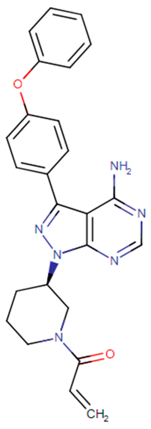
Development and Validation of a Chromatographic Method for Ibrutinib Determination in Human and Porcine Skin
 , , , and
Abstract
:1. Introduction
2. Materials and Methods
2.1. Material
2.2. Obtaining Porcine and Human Skin
2.3. Preparation of IBR Stock Solutions
2.4. Tape Stripping
2.5. Chromatographic Conditions
2.6. Validation
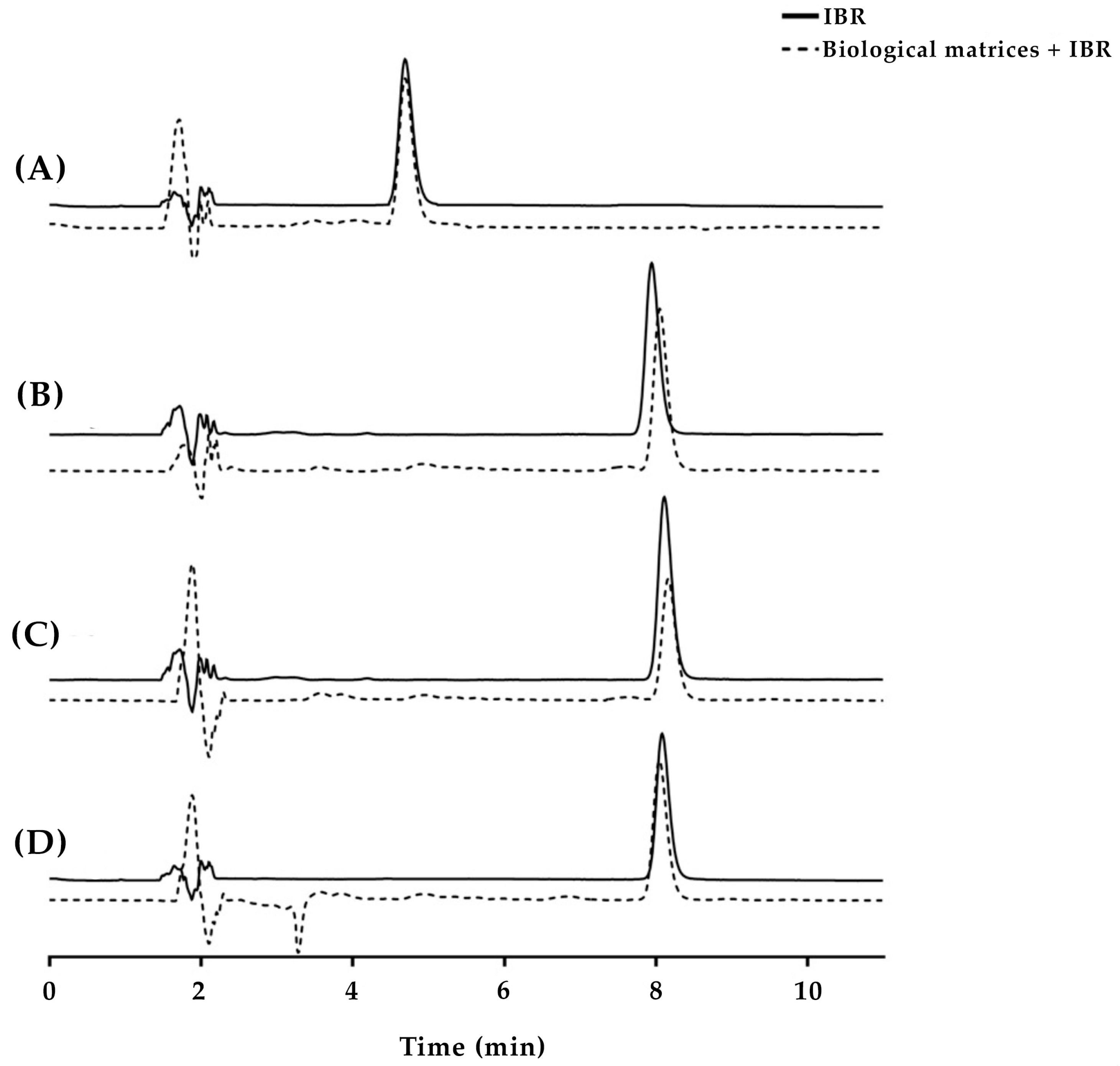
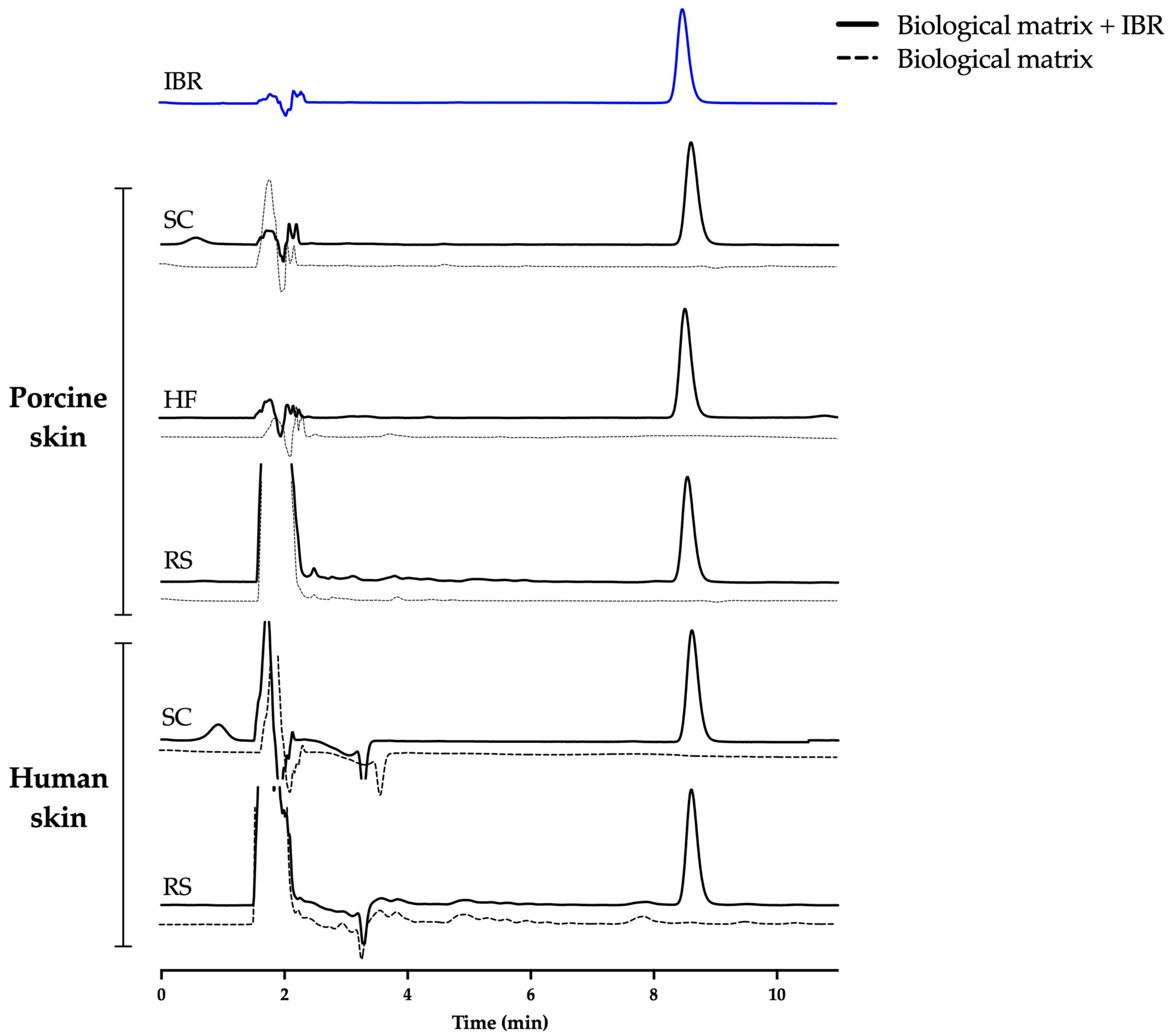
2.6.1. Selectivity
2.6.2. Linearity
2.6.3. Limit of Detection and Limit of Quantification
2.6.4. Precision
2.6.5. Accuracy
2.6.6. Robustness
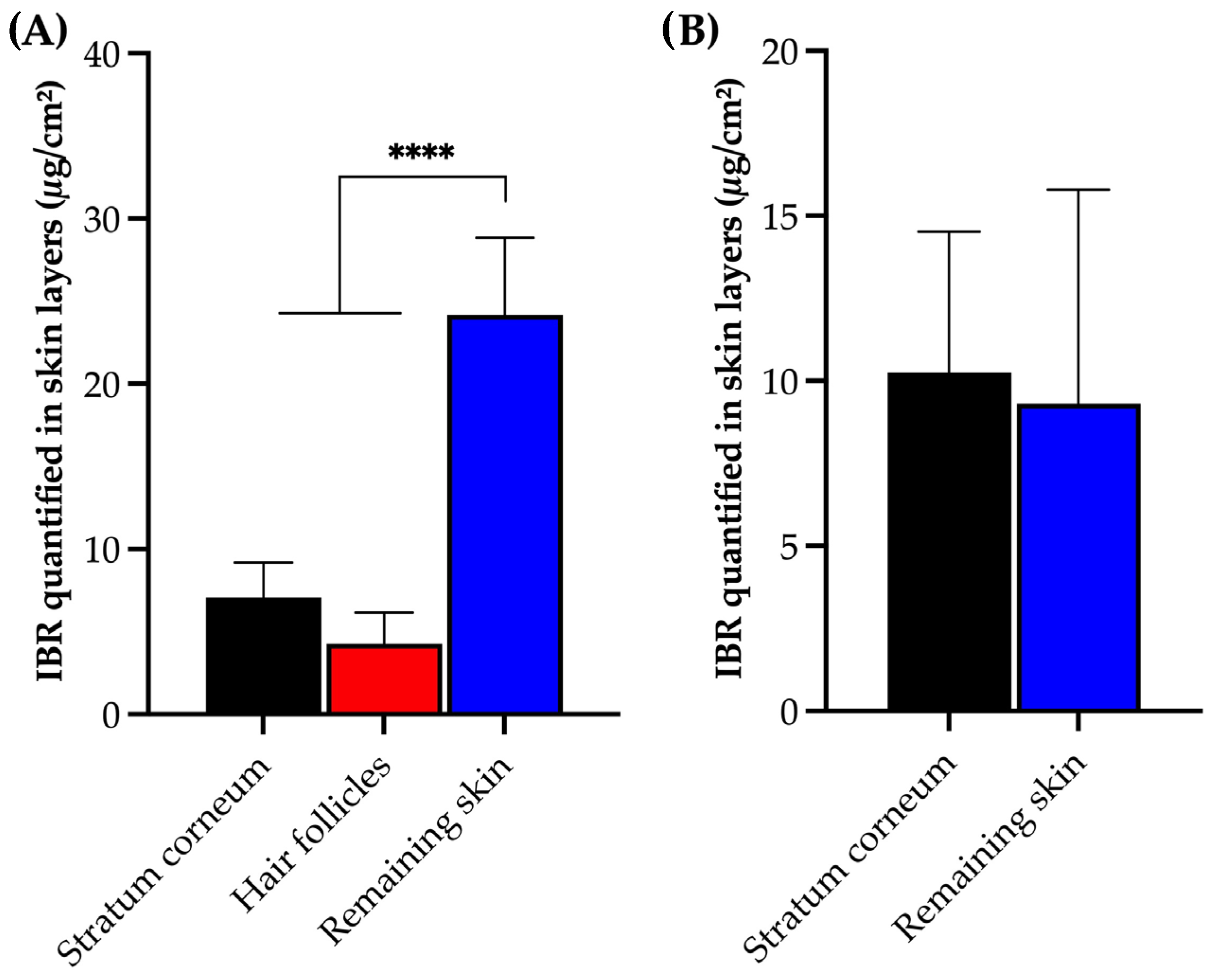
2.7. Application of the Method in Experiments of Cutaneous Permeation
3. Results and Discussion
3.1. Establishment of Chromatographic Conditions
3.2. Validation
4. Conclusions
Author Contributions
Funding
Data Availability Statement
Acknowledgments
Conflicts of Interest
References
- Molina-Cerrillo, J.; Alonso-Gordoa, T.; Gajate, P.; Grande, E. Bruton’s tyrosine kinase (BTK) as a promising target in solid tumors. Cancer Treat. Rev. 2017, 58, 41–50. [Google Scholar] [CrossRef]
- Metzler, J.M.; Burla, L.; Fink, D.; Imesch, P. Ibrutinib in gynecological malignancies and breast cancer: A systematic review. Int. J. Mol. Sci. 2020, 21, 4154. [Google Scholar] [CrossRef]
- Rangaraj, N.; Pailla, S.R.; Chowta, P.; Sampathi, S. Fabrication of ibrutinib nanosuspension by quality by design approach: Intended for enhanced oral bioavailability and diminished fast fed variability. AAPS PharmSciTech 2019, 20, 326. [Google Scholar] [CrossRef]
- Varikuti, S.; Singh, B.; Volpedo, G.; Ahirwar, D.K.; Jha, B.K.; Saljoughian, N.; Viana, A.G.; Verma, C.; Hamza, O.; Halsey, G.; et al. Ibrutinib treatment inhibits breast cancer progression and metastasis by inducing conversion of myeloid-derived suppressor cells to dendritic cells. Br. J. Cancer 2020, 122, 1005–1013. [Google Scholar] [CrossRef]
- Gomez-Rodriguez, J.; Kraus, Z.J.; Schwartzberg, P.L. Tec family kinases Itk and Rlk/Txk in T lymphocytes: Cross-regulation of cytokine production and T-cell fates: Cytokine regulation by Itk and Rlk/Txk. FEBS J. 2011, 278, 1980–1989. [Google Scholar] [CrossRef]
- Luo, Q.-Y.; Zhou, S.-N.; Pan, W.-T.; Sun, J.; Yang, L.-Q.; Zhang, L.; Qiu, M.-Z.; Yang, D.-J. A multi-kinase inhibitor APG-2449 enhances the antitumor effect of ibrutinib in esophageal squamous cell carcinoma via EGFR/FAK pathway inhibition. Biochem. Pharmacol. 2021, 183, 114318. [Google Scholar] [CrossRef] [PubMed]
- Wang, J.D.; Chen, X.Y.; Ji, K.W.; Tao, F. Targeting Btk with ibrutinib inhibit gastric carcinoma cells growth. Am. J. Transl. Res. 2016, 15, 3003–3012. [Google Scholar]
- Rangaraj, N.; Pailla, S.R.; Shah, S.; Prajapati, S.; Sampathi, S. QbD aided development of ibrutinib-loaded nanostructured lipid carriers aimed for lymphatic targeting: Evaluation using chylomicron flow blocking approach. Drug Deliv. Transl. Res. 2020, 10, 1476–1494. [Google Scholar] [CrossRef] [PubMed]
- Ashar, F.; Hani, U.; Osmani, R.A.M.; Kazim, S.M.; Selvamuthukumar, S. Preparation and optimization of ibrutinib-loaded nanoliposomes using response surface methodology. Polymers 2022, 14, 3886. [Google Scholar] [CrossRef] [PubMed]
- Yang, Z.; Du, Y.; Lei, L.; Xia, X.; Wang, X.; Tong, F.; Li, Y.; Gao, H. Co-delivery of ibrutinib and hydroxychloroquine by albumin nanoparticles for enhanced chemotherapy of glioma. Int. J. Pharm. 2023, 630, 122436. [Google Scholar] [CrossRef] [PubMed]
- Zhao, L.; Tang, B.; Tang, P.; Sun, Q.; Suo, Z.; Zhang, M.; Gan, N.; Yang, H.; Li, H. Chitosan/sulfobutylether-β-cyclodextrin nanoparticles for ibrutinib delivery: A potential nanoformulation of novel kinase inhibitor. J. Pharm. Sci. 2019, 109, 1136–1144. [Google Scholar] [CrossRef]
- Ganatra, S.; Sharma, A.; Shah, S.; Chaudhry, G.M.; Martin, D.T.; Neilan, T.G.; Mahmood, S.S.; Barac, A.; Groarke, J.D.; Hayek, S.S.; et al. Ibrutinib-Associated Atrial Fibrillation. JACC Clin. Electrophysiol. 2018, 4, 1491–1500. [Google Scholar] [CrossRef] [PubMed]
- Sibaud, V.; Beylot-Barry, M.; Protin, C.; Vigarios, E.; Recher, C.; Ysebaert, L. Dermatological Toxicities of Bruton’s Tyrosine Kinase Inhibitors. Am. J. Clin. Dermatol. 2020, 21, 799–812. [Google Scholar] [CrossRef] [PubMed]
- Csányi, E.; Bakonyi, M.; Kovács, A.; Budai-Szűcs, M.; Csóka, I.; Berkó, S. Development of topical nanocarriers for skin cancer treatment using quality by design approach. Curr. Med. Chem. 2019, 26, 6440–6458. [Google Scholar] [CrossRef] [PubMed]
- Angelo, T.; Pires, F.Q.; Gelfuso, G.M.; da Silva, J.K.; Gratieri, T.; Cunha-Filho, M.S. Development and validation of a selective HPLC-UV method for thymol deter-mination in skin permeation experiments. J. Chromatogr. B 2016, 1022, 81–86. [Google Scholar] [CrossRef] [PubMed]
- Demurtas, A.; Pescina, S.; Nicoli, S.; Santi, P.; de Araujo, D.R.; Padula, C. Validation of a HPLC-UV method for the quantification of budesonide in skin layers. J. Chromatogr. B 2020, 1164, 122512. [Google Scholar] [CrossRef] [PubMed]
- Pereira, M.N.; Matos, B.N.; Gratieri, T.; Cunha-Filho, M.; Gelfuso, G.M. Development and validation of a simple chromatographic method for simul-taneous determination of clindamycin phosphate and rifampicin in skin permeation studies. J. Pharm. Biomed. Anal. 2018, 159, 331–340. [Google Scholar] [CrossRef] [PubMed]
- Yasu, T.; Momo, K.; Yasui, H.; Kuroda, S. Simple determination of plasma ibrutinib concentration using high-performance liquid chromatography. Biomed. Chromatogr. 2019, 33, e4435. [Google Scholar] [CrossRef]
- Jain, H.; Geetanjali, D.; Dalvi, H.; Bhat, A.; Godugu, C.; Srivastava, S. Liposome mediated topical delivery of Ibrutinib and Curcumin as a synergistic approach to combat imiquimod induced psoriasis. J. Drug Deliv. Sci. Technol. 2022, 68, 103103. [Google Scholar] [CrossRef]
- Sim, Y.S.; Chong, Z.Y.; Azizi, J.; Goh, C.F. Development and validation of a gradient HPLC-UV method for mitragynine following in vitro skin permeation studies. J. Chromatogr. B 2022, 1204, 123316. [Google Scholar] [CrossRef]
- Lademann, J.; Richter, H.; Meinke, M.; Sterry, W.; Patzelt, A. Which Skin Model Is the Most Appropriate for the Investigation of Topically Applied Substances into the Hair Follicles? Skin Pharmacol. Physiol. 2010, 23, 47–52. [Google Scholar] [CrossRef] [PubMed]
- ICH. Harmonised Tripartite Guideline—Validation of Analytical Procedures Text and Methodology—Q2 (R1); ICH: Geneva, Switzerland, 2005. [Google Scholar]
- Cardoso, C.O.; Uwai, T.Y.; Gratieri, T.; Cunha-Filho, M.; Gelfuso, G.M. Chromatographic method for dacarbazine quantification in skin permeation experiments. J. Pharm. Biomed. Anal. 2023, 234, 115593. [Google Scholar] [CrossRef] [PubMed]
- Simões, M.F.; Nogueira, B.A.; Tabanez, A.M.; Fausto, R.; Pinto, R.M.A.; Simões, S. Enhanced solid-state stability of amorphous ibrutinib formulations pre-pared by hot-melt extrusion. Int. J. Pharm. 2020, 579, 119156. [Google Scholar] [CrossRef] [PubMed]
- Kazakevich, Y.V.; McNair, H.M. Low-energy interactions in high-performance liquid chromatography. J. Chromatogr. A 2000, 872, 49–59. [Google Scholar] [CrossRef] [PubMed]
- Haun, J.; Teutenberg, T.; Schmidt, T.C. Influence of temperature on peak shape and solvent compatibility: Implications for two-dimensional liquid chromatography: Liquid chromatography. J. Sep. Sci. 2012, 35, 1723–1730. [Google Scholar] [CrossRef]
- Walter, T.H.; Iraneta, P.; Capparella, M. Mechanism of retention loss when C8 and C18 HPLC columns are used with highly aqueous mobile phases. J. Chromatogr. A 2005, 1075, 177–183. [Google Scholar] [CrossRef] [PubMed]
- Mcnair, H.; Polite, L.N. 17 Troubleshooting in high performance liquid chromatography. Sep. Sci. Technol. 2007, 8, 459–477. [Google Scholar]
- ICH. Q2 (R1) Validation of Analytical Procedures: Text and Methodology; ICH: Geneva, Switzerland, 2006. [Google Scholar]
- Duarah, S.; Sharma, M.; Wen, J. Rapid and simultaneous determination of dexamethasone and dexamethasone sodium phosphate using HPLC-UV: Application in microneedle-assisted skin permeation and deposition studies. J. Chromatogr. B 2021, 1170, 122609. [Google Scholar] [CrossRef]
- Abraham, J. International Conference on Harmonisation of Technical Requirements for Registration of Pharmaceuticals for Human Use. In Handbook of Transnational Economic Governance Regimes; Tietje, C., Brouder, A., Eds.; Brill Publishers: Leiden, The Netherlands, 2010; pp. 1041–1053. [Google Scholar]
- Quintão, W.S.; Ferreira-Nunes, R.; Gratieri, T.; Cunha-Filho, M.; Gelfuso, G.M. Validation of a simple chromatographic method for naringenin quantification in skin permeation experiments. J. Chromatogr. B 2022, 1201–1202, 123291. [Google Scholar] [CrossRef]
- Tolentino, S.; Gratieri, T.; Cunha-Filho, M.; Gelfuso, G.M. Curcumin quantification in skin and mucosa: Optimization of extraction and chromatographic method validation. J. Chromatogr. B Analyt. Technol. Biomed. Life Sci. 2023, 1217, 123623. [Google Scholar] [CrossRef]


| Day | Analyst | Theoretical Concentration (μg/mL) | Measured Concentration (μg/mL) | CV 1 (%) |
|---|---|---|---|---|
| 1 | A | 1 | 1.01 ± 0.01 | 0.06 |
| 7.5 | 7.79 ± 0.17 | 2.26 | ||
| 15 | 15.47 ± 0.20 | 1.32 | ||
| B | 1 | 0.96 ± 0.03 | 1.35 | |
| 7.5 | 7.27 ± 0.17 | 1.70 | ||
| 15 | 14.86 ± 0.02 | 1.50 | ||
| 2 | A | 1 | 1.03 ± 0.01 | 2.59 |
| 7.5 | 7.90 ± 0.12 | 2.21 | ||
| 15 | 15.80 ± 0.22 | 0.12 | ||
| B | 1 | 1.00 ± 0.03 | 3.06 | |
| 7.5 | 6.94 ± 0.10 | 1.48 | ||
| 15 | 14.75 ± 0.22 | 1.53 |
| Sample | Theoretical Concentration (μg/mL) | Measured Concentration (μg/mL) | CV 1 (%) | Accuracy (%) | |
|---|---|---|---|---|---|
| Porcine skin | Stratum corneum | 1.00 | 1.03 ± 0.01 | 1.05 | 102.67 |
| 7.50 | 7.49 ± 0.15 | 2.16 | 99.91 | ||
| 15.00 | 15.34 ± 0.08 | 0.52 | 102.27 | ||
| Hair follicle | 1.00 | 1.04 ± 0.04 | 3.89 | 104.07 | |
| 7.50 | 7.88 ± 0.08 | 1.13 | 105.08 | ||
| 15.00 | 15.88 ± 0.19 | 1.26 | 105.86 | ||
| Remaining skin | 1.00 | 1.00 ± 0.03 | 2.79 | 104.32 | |
| 7.50 | 7.03 ± 0.09 | 1.34 | 93.72 | ||
| 15.00 | 13.42 ± 0.89 | 6.64 | 89.49 | ||
| Human skin | Stratum corneum | 1.00 | 1.01 ± 0.01 | 0.89 | 101.27 |
| 7.50 | 7.65 ± 0.01 | 0.10 | 102.00 | ||
| 15.00 | 14.94 ± 0.03 | 0.19 | 99.63 | ||
| Remaining skin | 1.00 | 1.00 ± 0.01 | 1.31 | 100.04 | |
| 7.50 | 6.90 ± 0.01 | 0.18 | 92.00 | ||
| 15.00 | 13.82 ± 0.01 | 0.09 | 92.12 | ||
| Parameter | Tested Conditions | IBR Retention Time (min) | Tailing Factor | Peak Resolution | Theoretical Plates |
|---|---|---|---|---|---|
| Oven temperature (°C) | 33 | 8.89 ± 0.01 | 1.18 ± 0.02 | 8.13 ± 0.2 | 8548 ± 27 |
| 35 | 8.84 ± 0.02 | 1.17 ± 0.01 | 8.23 ± 0.2 | 7888 ± 27 | |
| 37 | 8.80 ± 0.01 | 1.17 ± 0.07 | 7.87 ± 0.1 | 8702 ± 155 | |
| Acetonitrile/0.01 mol/L phosphoric acid (pH 3.5) in the mobile phase | 33:67 | 11.55 ± 0.02 | 1.13 ± 0.04 | 10.4 ± 0.4 | 9919 ± 5 |
| 35:65 | 8.84 ± 0.02 | 1.17 ± 0.01 | 8.23 ± 0.2 | 7888 ± 27 | |
| 37:63 | 7.0 ± 0.01 | 1.17 ± 0.02 | 6.0 ± 0.2 | 7983 ± 397 | |
| Flow rate (mL/min) | 0.9 | 9.78 ± 0.02 | 1.18 ± 0.02 | 9.51 ± 0.3 | 8889 ± 21 |
| 1.0 | 8.84 ± 0.02 | 1.17 ± 0.01 | 8.23 ± 0.2 | 7888 ± 27 | |
| 1.1 | 8.07 ± 0.01 | 1.17 ± 0.03 | 7.98 ±0.2 | 8364 ± 41 |
Disclaimer/Publisher’s Note: The statements, opinions and data contained in all publications are solely those of the individual author(s) and contributor(s) and not of MDPI and/or the editor(s). MDPI and/or the editor(s) disclaim responsibility for any injury to people or property resulting from any ideas, methods, instructions or products referred to in the content. |
© 2024 by the authors. Licensee MDPI, Basel, Switzerland. This article is an open access article distributed under the terms and conditions of the Creative Commons Attribution (CC BY) license (https://creativecommons.org/licenses/by/4.0/).
Share and Cite
Albuquerque, L.F.F.; Souto, M.V.; Saldanha-Araujo, F.; Carvalho, J.L.; Gratieri, T.; Cunha-Filho, M.; Gelfuso, G.M. Development and Validation of a Chromatographic Method for Ibrutinib Determination in Human and Porcine Skin. Chemistry 2024, 6, 272-282. https://doi.org/10.3390/chemistry6020014
Albuquerque LFF, Souto MV, Saldanha-Araujo F, Carvalho JL, Gratieri T, Cunha-Filho M, Gelfuso GM. Development and Validation of a Chromatographic Method for Ibrutinib Determination in Human and Porcine Skin. Chemistry. 2024; 6(2):272-282. https://doi.org/10.3390/chemistry6020014
Chicago/Turabian StyleAlbuquerque, Lucas F. F., Maria Victoria Souto, Felipe Saldanha-Araujo, Juliana Lott Carvalho, Tais Gratieri, Marcilio Cunha-Filho, and Guilherme M. Gelfuso. 2024. "Development and Validation of a Chromatographic Method for Ibrutinib Determination in Human and Porcine Skin" Chemistry 6, no. 2: 272-282. https://doi.org/10.3390/chemistry6020014