Review

Recent Advances in Influenza, HIV and SARS-CoV-2 Infection Prevention and Drug Treatment—The Need for Precision Medicine

by
Tamás Kálai
1,2,
Judit Erzsébet Pongrácz
2,3,* and
Péter Mátyus
4,*
1
Organic and Medicinal Chemistry, Faculty of Pharmacy, University of Pécs, 1 Honvéd St., H-7624 Pécs, Hungary
2
János Szentágothai Research Center, University of Pécs, 20 Ifjúság St., H-7624 Pécs, Hungary
3
Institude of Pharmaceutical Biotechnology, Faculty of Pharmacy, University of Pécs, 2 Rókus St., H-7624 Pécs, Hungary
4
E-Group ICT SOFTWARE, 11 Kacsa St., H-1027 Budapest, Hungary
*
Authors to whom correspondence should be addressed.
Chemistry 2022, 4(2), 216-258; https://doi.org/10.3390/chemistry4020019
Submission received: 22 February 2022 / Revised: 22 March 2022 / Accepted: 23 March 2022 / Published: 25 March 2022
(This article belongs to the Section Medicinal Chemistry)

Abstract

:
Viruses, and in particular, RNA viruses, dominate the WHO’s current list of ten global health threats. Of these, we review the widespread and most common HIV, influenza virus, and SARS-CoV-2 infections, as well as their possible prevention by vaccination and treatments by pharmacotherapeutic approaches. Beyond the vaccination, we discuss the virus-targeting and host-targeting drugs approved in the last five years, in the case of SARS-CoV-2 in the last one year, as well as new drug candidates and lead molecules that have been published in the same periods. We share our views on vaccination and pharmacotherapy, their mutually reinforcing strategic significance in combating pandemics, and the pros and cons of host and virus-targeted drug therapy. The COVID-19 pandemic has provided evidence of our limited armamentarium to fight emerging viral diseases. Novel broad-spectrum vaccines as well as drugs that could even be applied as prophylactic treatments or in early phases of the viremia, possibly through oral administration, are needed in all three areas. To meet these needs, the use of multi-data-based precision medicine in the practice and innovation of vaccination and drug therapy is inevitable.

Graphical Abstract

1. Introduction

Comprehensive studies confirm that the impact and burden of viral infections on health are enormous, including the loss of human lives, disease-relevant long-term health effects, and in monetary terms. In particular, the fight against RNA viruses needs to receive focused attention worldwide. There are at least two important reasons. On one hand, the well-known representatives of human RNA viruses, e.g., human influenza and human immunodeficiency viruses (IV; HIV), still pose an ongoing therapeutic challenge, and, on the other hand, the emergence of novel RNA-virus threats, such as severe acute respiratory syndrome coronavirus-2 (SARS-CoV-2), can develop especially dangerous pandemic diseases [1].
Prevention is an obvious, cost-effective way to eradicate a viral infection. This can be achieved at a population level through vaccination, which can mostly prevent the infection from occurring or can at least reduce the severity of the infection. Enormous worldwide efforts are being made today to develop effective and safe vaccinations against all three above-mentioned viruses. In particular, there is an urgent need to achieve ‘herd immunity’ against SARS-CoV-2 as the virus mutates fast and neither research nor the production of drugs can keep up with the raging pandemic. The virus infection can be treated with antiviral drugs. Although, limited prevention (e.g., in the event of a potential infection emergency) can in principle be achieved with drugs, however, such agents have been hardly available.
Thus, if prevention fails to provide effective broad-spectrum worldwide protection, despite all emerging virus mutations and variants, there is also a need for antiviral drugs. It follows from all these facts and considerations that as long as prevention fails to provide broad-spectrum worldwide protection that remains effective despite all emerging mutations and variants, there is a need for new, effective, and safe antiviral drugs that exert broad antiviral activity, and which are also suitable for bridging the period until the development of a vaccine effective against the new variant is completed.
Before describing the current state of antiviral vaccination and drug therapy, as well as the hot research areas, to provide some guidance on future directions, we must address two important aspects of the infection. First, the complex mechanism of the viral replication process and the consequences of its frequent mutations, and second, the variability of patients. Although, in the past, due to the lack of relevant data and knowledge, vaccination and drug therapy were considered as uniformly effective in all patients, we can rightly ask: is this traditional approach appropriate? Surprisingly, the ‘gut reaction’ of the scientific and medical world when facing new challenges is to aim for a single cure. The ‘one vaccine or medication fits all’ principle has been hard to eliminate both from the general public’s expectations as well as from the overall approach of the pharmaceutical and medical community. We immediately have to refer to the invalidity of the past approach to ‘uniformity’. There has been ample evidence in recent decades about how significantly, almost astonishingly, we are all unique individuals. Therefore, not just with the variability of the pathogens, but also our individual properties, and thus, the capabilities of our immune systems, as well as pharmacogenetics (cf. drug-metabolizing enzymes), in addition, drug–drug and drug–immune system interactions, influence the outcome of a serious infection and its therapy. Nowadays, disease characterization and treatment can be patient-centered according to the principles of precision medicine, utilizing state-of-the-art omics-based, and other disease- and drug-descriptor data. Although precision medicine has become an essential integral part of anticancer therapy, in the antiviral field, the practice of precision medicine is far from being exploited.
The difficulty in winning the war on viruses lies partly in a kind of ‘technicality’: viruses are not even living organisms. As viruses are unable to reproduce themselves, they need to hijack the biochemical machinery of living cells; both the human immune system, as well as the efficacy of antiviral drugs are challenged to fight the virus. The other part of the difficulty is that such fights often require the full and coordinated mobilization of our defense resources, which is not without serious health consequences.
In the present review, we focus on three RNA viruses that are among the most challenging viruses of our time: human influenza (IV) and human immunodeficiency (HIV) viruses, and severe acute respiratory syndrome coronavirus-2 (SARS-CoV-2). We briefly describe their epidemiological significance. In subsequent parts, the characteristic structural features of the three viruses, and the host immune system in terms of its response to the infection, are presented. Then, currently available, and newly developed antiviral vaccines and drugs with their mode of action are reported.
In those parts, we discuss the difficulties of seeing HIV, IV, SARS-CoV-2 infections in their high complexity of causing serious and often deadly diseases. We would also like to illustrate the race of medical and pharmaceutical research to identify ways and tools of effective prevention and treatment, causing no or minimal damage to human organs, and to possibly support the host immune system in its antiviral fight.
This content is presented in the following sections:
The toll of past and present pandemics;
RNA viruses: structures of IV, HIV, and SARS-CoV-2;
Host immune response against viruses;
Prevention of viral infection by vaccination;
Anti-influenza, anti-HIV, and anti-SARS-CoV-2 agents;
Summary and conclusion.

2. The Toll of Past and Present Pandemics

The first well-documented H1N1 influenza pandemic occurred approximately one hundred years ago, right at the end of the first world war [2]. A minimum of 15 million, and by some estimates 50 million people died of the ‘Spanish flu’, although the exact number is hard to determine [3,4]. It is also predicted that globally, nearly 500 million people were infected. The high death rate was assumed to correlate with the lack of antibiotics at the time, therefore many patients succumbed to the disease due to bacterial infections superimposed on the initial viral pathogen. The influenza virus, however, did not disappear there and then. Influenza pandemics in the 20th century claimed further lives. In 1957 (H2N2), 1.1 million, in 1968 (H3N2), 1 million, and in 2009 (H1N1), approximately 600 thousand deaths occurred [5,6]. According to the World Health Organization (WHO), an estimated 290–650 thousand people die of flu-related causes every year worldwide [7].
In the past two years, the SARS-CoV-2 pandemic has often been compared to pandemics caused by influenza viruses, but the sheer number of deaths caused by SARS-CoV-2 within 2 years of its appearance makes the SARS-CoV-2 coronavirus appear more devastating than any of the influenza strains so far. According to the WHO, from 2020 till the end of 2021, 282 million confirmed cases of SARS-CoV-2 were detected and deaths reached 5.5 million [7]. A more detailed retrospective analysis is going to provide the real answers, as the absolute numbers cannot take into consideration the drastically increasing global population and the differences in affected demographic groups in different parts of the world from, e.g., Japan to sub-Saharan Africa [8].
While both influenza and SARS-CoV-2 are airborne viruses, HIV is contracted via exchanged bodily fluids. Although to some, HIV is not anywhere near as serious of a health issue as the previously mentioned two other ribonucleic acid (RNA) viruses. According to WHO statistics, since the beginning of the HIV epidemic in the 1980s, approximately 79.3 million people have been infected with the HIV virus and 36.3 million people died. Globally, 37.7 million people were living with HIV at the end of 2020, and just in that year, 680 thousand people died of HIV-related illnesses. An estimated 0.7% of adults aged between 15–49 years are living with HIV worldwide [7]. The burden of the epidemic, however, considerably varies between countries and regions, and African regions remain most severely affected. Partly, this is due to the lack of effective prevention and treatment.
Viral infections, therefore, are a great medical challenge worldwide. Partly, as the variety of viral strains are numerous, and their ability to mutate quickly and often unpredictably, thus leading possibly to a more severe and effective infection, challenge both the human immune system and the pharmaceutical industry into a difficult and unequal duel.

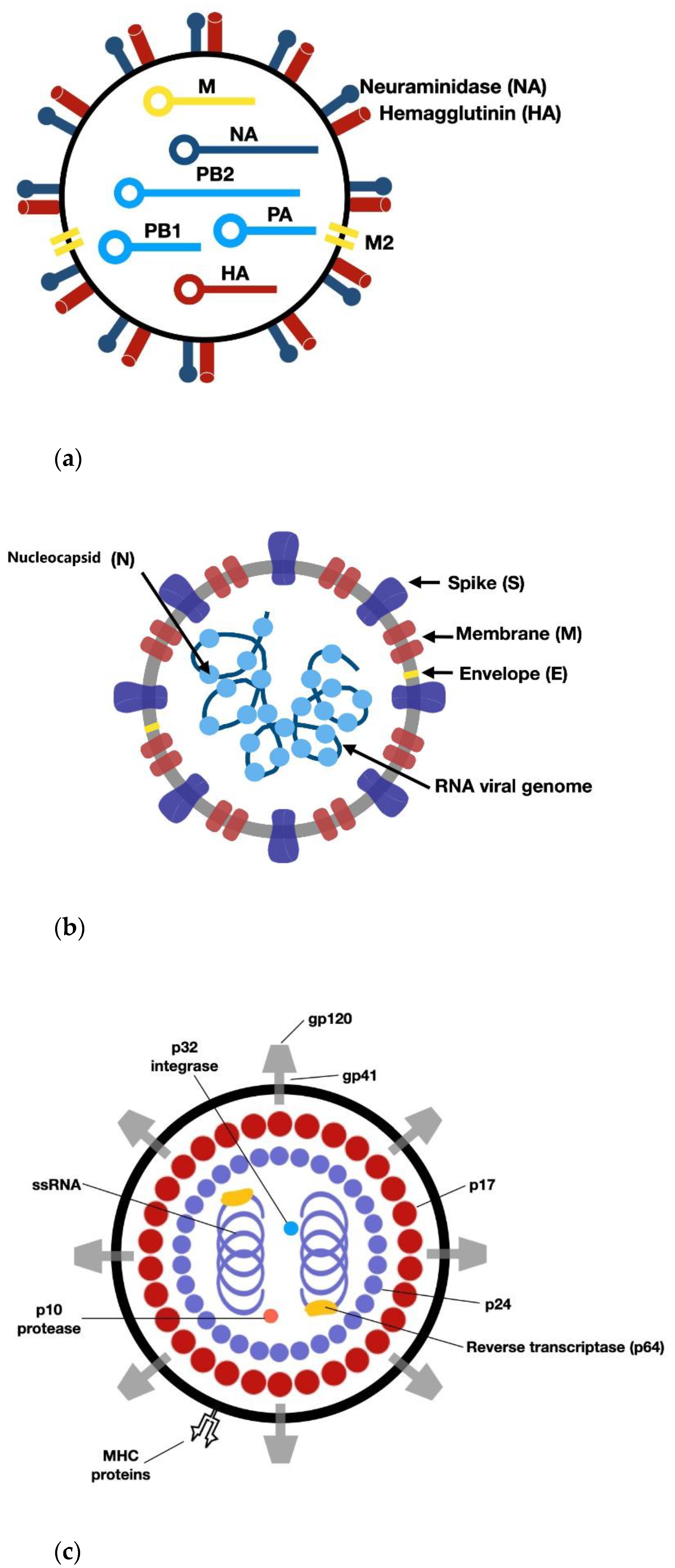
3. RNA Viruses: Structure of IV, HIV, and SARS-CoV-2

The RNA core sits within a protective protein coat called a capsid that serves for virus classification, as it may vary from one type of a virus to another (Figure 1). The capsid is made from the proteins that are encoded by viral genes within the viral genome.
Virally coded proteins will self-assemble to form a capsid once the host cell produces all the necessary components. Some viruses also have an envelope of phospholipids and proteins that surrounds the capsid and helps to protect the virus from the host’s immune system as the envelope is assembled partly from the membranes of the host’s cells. The envelope may also have protein molecules that aid in binding to host cell receptors or cell-surface proteins to facilitate the entry process into cells (Table 1).
Although virus strains differ in their sites and methods of infection, the immune response to viruses follows a series of well-defined steps (Figure 2). Viruses enter the body through cells with barrier functions, but they can also target other cell types once they have entered the organism. They bind to various cell-surface molecules that are mostly cell-surface proteins that can be ligands (e.g., IT), receptors (e.g., CCR5), enzymes (e.g., ACE2), etc. and have physiological functions in the host organism. Often, the site of viral entry is called the ‘receptor’ regardless of the actual function of the protein, as the viral infection is described from the view of the virus instead of the host. This approach can affect therapy, as individual variabilities in the host are not likely to be taken into consideration. Cells regularly internalize cell-surface proteins, regardless of their functions, therefore viruses get into the cell by binding to characteristic membrane-bound proteins of a specific cell type. Following its entry into the cell, the virus diverts the cellular machinery of nucleic acid replication and protein synthesis towards viral replication.
Although the three studied RNA viruses structurally might look very similar, there are still major differences amongst IV, SARS-CoV-2, and HIV, and the only real similarity is that all three of them carry RNA as genetic material.
In the case of IV infection, the hemagglutinin trimer binds to the sialic acid residues of the glycoproteins and glycolipids of the host cell. Following the endocytosis of the virus via a clathrin-mediated manner and facilitated by hemagglutinin, the IV can release its negative sense RNA from the endosome as rod-shaped, double-helical ribonucleoprotein complexes (RNP) to the cytosol, and they are transported into the nucleus where RNA replication takes place (Figure 2a). Once the virions are assembled, neuraminidase cleaves sialic acid residues from freshly synthesized glycoproteins of new virions and the membrane of the host cell to help the virus leave the host cell (Figure 2a) [9]. The replication cycle of SARS-CoV-2 is more complex, as binding to cell-surface proteins (ACE2 and TMPRSS2) takes place in a more elaborated manner. The S1 subunit binds to ACE2 on the surface of the host cell, then the actual viral entry into the cell depends on the activity of proteases (e.g., furin) in the host. Furin can pre-cleave the S protein between S1-S2, which can promote the activity of TMPRSS2 that cleaves the S protein between the subunits and exposes the fusion peptide in the S2 subunit. The S2 subunit facilitates the fusion of the viral membrane with the membrane of the host cell. Following fusion with the cell membrane of the host cell, the virus is endocytosed and released into the cytoplasm. The viral positive-sense RNA is released into the cytoplasm, where the RNA-dependent RNA polymerase creates the negative RNS sense sequences, replicated in the cytosol. The host’s ribosomes produce spike (s), nucleocapsid (N), membrane (M), and envelope (E) proteins, then using the endoplasmic reticulum (ER) and the endoplasmic reticulum-Golgi intermediate compartment (ERGIC) in the host cell, the N protein assembles with the newly synthesized viral RNA to create the nucleocapsid. With the viral structural proteins, new virions are built and released (Figure 2b) [10]. The viral genome is not inserted into the host genome in either case. In contrast to this mechanism, HIV contains additional enzyme proteins that allow this mechanism to take place. During infection, the viral envelope glycoprotein gp120 of HIV binds CD4 and CC-chemokine receptor 5 (CCR5) on the surface of target cells, triggering the fusion of the virus with the host cell membranes. Host genomic studies have implicated genetic variants in CCR5 (marked purple background in Figure 2c), listed as modifiers of infectivity. Reverse transcription of the single-stranded RNA genome into double-stranded DNA (dsDNA) occurs using proteins carried by the infecting virion. Viral dsDNA is trafficked to the nucleus, where it is integrated into the genome of the host cell. The transcription of viral dsDNA results in viral gene expression and genome replication. Viral mRNA is translated into polyproteins, which are cleaved by the viral protease. Functional proteins assemble with copies of the viral genome at the cell membrane and mature virions bud from the surface.
Despite the mechanism differences, the host immune system should be able to deal with each viral infection swiftly and effectively; but certain viral strains have proved to be hard to recognize and eliminate. Consequently, those infections lead to debilitating illnesses or even death. The fundamental question thus remains open: who and why is going to be more easily infected and who and how can fight off infections, while others succumb to the disease? Answering the question as accurately as possible is obviously needed to identify the risk factors and initiate a suitable therapy.
For all the viral infections discussed, the host immune system plays a prominent role.

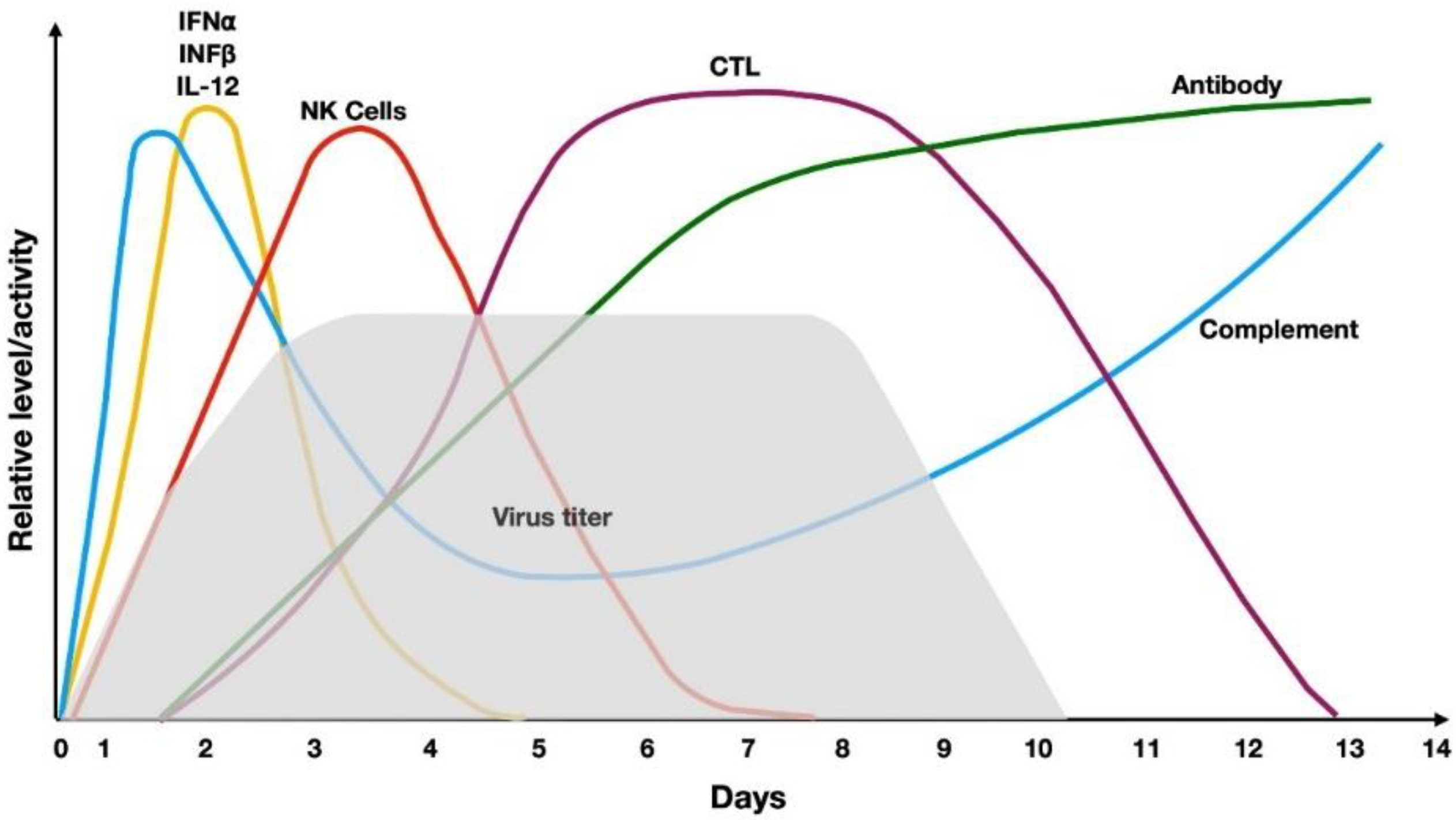
4. Host Immune Response against Viruses

4.1. The ‘One Fits All’ Immunology

Both the native (innate) and the adaptive immune systems are actively involved in the elimination of viral infections. Despite the popular belief that cytotoxic T cells play the most important role in the fight against viral infections, the elimination of viral pathogens is a complex process detailed in (Figure 3).
Fighting against a viral infection begins immediately after the virus enters the first cell. Virus-infected cells immediately produce interferon α (IFNα) and interferon β (IFNβ). These interferons inhibit the entry of the virus into neighboring cells and increase the presentation of viral proteins bound to human leukocyte antigens (HLA) class I molecules on the infected cells. Non-self-proteins presented by HLA class I molecules can trigger the activation of cytotoxic T cells. Dendritic cells secrete IL-12 which activates natural killer (NK) cells to kill the virus-infected cells. NK cells recognize those virus-infected cells which have no or little HLA class I molecules expressed on their surfaces, and the mechanism of reduced HLA expression is triggered by the virus to reduce recognition by the immune system. As NK cells are inhibited by the presence of HLA class I molecules, the lack of HLA class I molecules on the cells activates their cytotoxic function. Viruses present in the circulatory system can activate the complement system as well, both on the alternative and the lectin pathway (e.g., HIV). Activation of the complement system can result in the lysis of pathogens or the covering (opsonization) of viruses with complement fragments (mainly C3b and C4b). Opsonization of viruses facilitates ingestion by phagocytes. As viruses can be taken up by antigen-presenting cells (through recognition of opsonized viruses or phagocytosis of the infected cell), viral proteins can be presented by HLA class II molecules, which are necessary for antibody production. Antibodies that are produced against viruses have an important role, both in preventing infection and eliminating viruses. Antibodies produced against antigens in the viral envelope neutralize some of the proteins to which the virus binds, and therefore can prevent viruses from entering the cell. Opsonizing antibodies contribute to the elimination of the virus by stimulating phagocytosis. As antibodies bind to proteins of the viral envelope, the classical pathway of the complement system gets activated. The consequence of opsonization is that virus particles bind to both Fc and complement receptors (CR3, CR4), making phagocytosis and the elimination of viruses more effective. However, the phagocytosed virus is still viable, and, thanks to opsonization, it can infect more cells through Fc and complement receptors. This occasionally results in the infection of cells that lack the virus-specific receptor. Perhaps, the best known process of antiviral immunity is the function of cytotoxic T cells. Cytotoxic T cells, which are differentiated from CD8+ T cells, recognize viral peptide antigens bound to HLA class I molecules on the surface of infected cells. If the CD8+ T cell has not yet met a given virus, meaning the host has never been infected with that particular pathogen, then the CD8+ T cells need the presence of helper T cells (Th) and the cytokines (e.g., IL-2) produced by Th cells to differentiate into effector cytotoxic T cells. Cytotoxic T cells release granzymes and perforin molecules. The virus titer reaches its maximum level 3 days after the infection and starts to fall not before 7 to 8 days after infection, even in the case of full activation of the immune system. Despite all the active steps taken by our immune system, the viral titer increases continuously. While both the innate and adaptive parts of the immune system play an active role in the elimination of the pathogen, it takes about 4 days to activate the cellular (cytotoxic T cells) and the humoral immune system (antibody production), respectively. Not surprisingly, the virus can persist in the host for 8–10 days and can cause various kinds of damage in the host organisms before being eliminated. Therefore, prevention of the disease or reducing the time frame that the immune system is capable of for the elimination of the virus is highly important, especially if the virus cannot be eliminated due to inserting itself into the host genomic DNA, such as HIV. Consequently, HIV infection has long-lasting consequences. According to the presentation of symptoms, HIV infection can be divided into three main stages: (1) Immediately after infection in some individuals, influenza-like symptoms can be detected; (2) in the latent period that varies between 10 and 15 years, the virus hides in CD4+ T cells; (3) in the absence of treatment, severe symptoms appear, with full immune-suppression and its consequences (AIDS). Intensive research into the lifecycle of the virus leads to effective pharmacological intervention which can prolong the life of HIV+ patients and prevent AIDS, but the overall life expectancy is still reduced.

4.2. Individual Response to Pathogens

Although a healthy immune system in the antiviral defense makes highly similar response steps, individual differences were detected rather early on. In the 1970s, it was noted that people with different blood groups could be differentially susceptible to IV infections and similar observations were made in cases of HIV and SARS-CoV-2 (Table 2). Still, a relentless investigation has not come up with a reliable explanation as blood groups play no active part in either IV, SARS-CoV-2, or HIV infection. Indirect correlations, however, clearly exist [11] but only further genetic linkage studies can determine the exact connections.
The individual susceptibility to infection and the strength of the immune system to mount an effective immune response is strongly linked to our genetic background. One of these molecular families is the human leucocyte antigens (HLA) that present the virus-derived peptides to the effector immune cells.
The HLA-bound ‘presented’ antigen epitopes are recognized by T and B cells expressing the appropriate T and/or B cell receptors (TCR and BCR). The TCR and BCR genes and the maturation of the immune system are coded genetically as well as shaped by the modulatory effects of the environment we live in and the pathogens we are exposed to. The HLA molecules play a crucial part in the immune response, and they are coded by many individual HLA alleles on chromosome 6. The encoded HLA alleles, with more than 400 genes, are central mediators of the innate and adaptive immune responses. Within this locus, alleles at class I (HLA-A, HLA-B, HLA-C) and class II (HLA-DR, HLA-DQ, HLA-DP) are found. Studies have identified that approximately 40% of the HLA alleles are unique to single individuals, resulting in significant individual differences in immune responses to any given pathogens.
Not surprisingly, the main trends in HLA-associated virus-triggered immune responses are under intense investigation. Individual genetic variations of HLA may help to explain different immune responses to a virus across a population and identify individuals at a higher risk from specific viruses.
An IV case study [12] presented selective classical HLA class I allele and haplotype combinations that make individuals susceptible or protected against influenza A H1N1/09. A study involving broader variants of influenza viruses (H1N1, H3N2, etc.) has come to the conclusion that resistance to influenza virus infection is associated with HLA class I serologic but not genotypic homozygosity [13].
HLA susceptibility mappings were performed rather early on into the SARS-CoV-2 pandemic [14] that identified the HLA-B*46:01 allele to be associated with more severe infection, while HLA-B*15:01 was found to be associated with asymptomatic SARS-CoV-2 infection [15]. Studies are ongoing to identify further details of what part of the population would resist infection, are infected but show no clinical symptoms, or are going to become severely ill or even non-responsive to therapy.
However, the fact that HLA structures are important in response to viral infections (Figure 4) has been known for years; studies for actual individual differences have just recently provided more detailed results in the case of HIV infections [16].
Genome-wide association studies (GWAS) were used to further investigate the background of HIV resistance, although findings were largely limited to CCR5 variations [17,18]. Further genome sequencing studies of extreme exposure phenotypes [19] have shown potential associations with the immunoglobulin superfamily member CD101, important in regulatory T cell function [20], and a gene coding the ubiquitin-conjugating enzyme UBE2V1, involved in pro-inflammatory cytokine expression [21] and the HIV restriction factor TRIM5α [22]. Further analyses of GWAS showed that additional potential molecules with residual heritability have been determined, although confirmation of the results is still necessary.

5. Prevention of Viral Infections by Vaccination

While the immune system can fight off viral infections, due to individual differences, it is not always easy and not without long-lasting health consequences, even serious and sometimes irreversible damages to the host.
Prevention of viral infections, therefore, ought to be the best of the options. Ideally, not getting anywhere near the virus is the best prevention. However, overcrowded metropolitan areas, continuously increasing, and a frequently traveling world population make it almost impossible to avoid infections. The other—and theoretically outstandingly effective—prevention method is vaccination.

5.1. Essential Immunology for Vaccine Production

There are several obstacles in the vaccination process and many of them lie within the mechanism of the immune response itself:
  • A good vaccine ensures that the immune system ‘remembers’ the pathogen. The immune system, however, can only form an immune memory against proteins, therefore a viral protein has to be selected to be used as a vaccine;
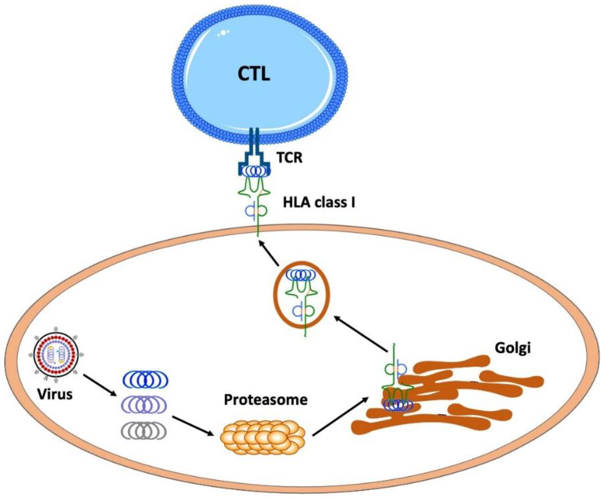
  • To trigger an immune response and consequent immune memory, the pathogenic protein has to be presented to the immune cells as short peptides with the aid of HLA class I as well as HLA class II proteins where the presented pathogenic peptides have to fit into the clefts of the presenting HLA molecules.
    The shapes of HLA clefts are therefore highly important;
    The shape depends on the structure of the HLA molecules, which is closely connected to their amino acid sequence, which is coded into our inherited genetic ‘database’ [23,24,25];
    Receptors of the immune cells (T and B cell receptors) that need to recognize the presented peptides are also shaped by both genetic background and the environment, T, and B cell development and selection, as well as exposure to pathogens [26].
Consequently, the effectiveness of an individual’s ability to present and recognize a pathogenic peptide depends on the individual’s genetic background inherited from both parents and upbringing, which determines whether we have been exposed to pathogens as exposure to pathogens determines our initial immune memories, as well as shape our T and B cell receptor repertoire. The age of the infected host is also important. It may sound irrelevant, as elderly people are adults who have been fortunate enough to successfully fight off a great variety of infectious diseases, yet they still have several disadvantages [27]. These include the reduced number of stem cells [28] that would be able to rejuvenate their pool of immune cells, as well as a reduction and modification of the microenvironments where immune cells develop, and their functional receptor pools are selected [29,30]. Finally, the presence of comorbidities also affects immune responses [31].

5.2. Challenges in Vaccine Production

  • Active vaccination (pathogen proteins to trigger an immune response)
    traditional vaccine production (attenuated and inactivated pathogens) is still amongst the actively used methods (e.g., influenza vaccines) to prevent infection or at least to reduce the seriousness of symptoms caused by infection of the wild type pathogen;
    biotechnology revolutionized vaccine production decades ago, but the revolution was only recognized when SARS-CoV-2 started its deadly spread across the world in 2019.
  • Passive vaccination (antibodies against pathogenic proteins)
    passive vaccination aims to provide anti-pathogen antibodies for immune-compromised individuals traditionally; antibodies are purified from sera of people who have recovered from the disease;
    biotechnology revolutionized antibody production, hence, the production of vaccines can use phage display to store any sequences required for the desired combination of human immunoglobulins in large volumes and great purity [32,33].
Unfortunately, viruses can change, and vaccine-induced immune memory cells may no longer recognize the virus, as the protein and consequently the peptides are not the same as they initially were. Fast spreading viruses (like IV and SARS-CoV-2) or the ones that are prone to frequent recombination (HIV) have the ability to change using two main mechanisms, namely antigen-drift and antigen-shift [34,35]:
  • Antigen-drift is the result of regularly occurring point mutations. Although antigen-drift causes relatively small-scale changes, these changes may be sufficient to ensure that the virus avoids being detected by cytotoxic memory T cells and antibodies produced by memory B cells formed during a previous infection or vaccination.
  • Antigen-shift is the result of recombination events between the RNA genomes of different types of viruses infecting the same cell at the same time. Consequently, our immune system becomes vulnerable to the new pathogen, and as the results of these random recombination events are unpredictable, they further complicate vaccine development.
Due to the high mutation rate, vaccines ought to reflect the modifications of proteins in the pathogen. That would require the ability in vaccine production to follow these modifications, but the generally used technology did not make it possible to quickly change vaccine protein sequences—not until the introduction of the mRNA-based vaccine production, which allows such fast and modified vaccine production (Table 3) [36,37].
All treatments of viral infections can be approached from two well-defined viewpoints: (i) how to aid the host’s immune system to fight off infection, and (ii) how to inhibit viral replication and consequently avoid or reduce high viral load-induced tissue damage.
(i)
Targeting the host’s immune response
Vaccination to prevent infection or to prepare the immune system to fight the disease more effectively is a well-known approach to reduce serious disease symptoms. Targeting the host’s immune response, however, has several additional possibilities if the mechanism of the viral infection is clarified. A recently published review summarizes both small molecular intervention and available monoclonal antibody therapy options in detail [38] in the case of SARS-CoV-2 infection. In general, monoclonal antibodies are used to target the hyperactive immune system at various points of entry on the host cell or characteristic proteins in the viral envelope which can block infection of the host. Furthermore, reduction in inflammatory cytokine response (e.g., IL-6) either by reducing cytokine levels (siltuximab) or blocking cytokine receptors (tocilizumab, sarilumab) on the cell’s surface can prevent further aggravation of inflammatory reactions. Unfortunately, interfering with the complement system by targeting C5a (eculizumab) [39] or neutralizing GM-CSF with gimsilumab, lenzilumab, namilumab, or otilimab risk secondary infections which are not likely to improve the therapeutic outcome. Although anti-TNF-α monoclonal antibodies (infliximab, adalimumab, and certolizumab pegol) have been shown to provide substantial benefit to patients through reductions in both localized and systemic inflammation, such treatments are not necessarily beneficial to all viral infections. Not surprisingly, the above treatments have not been approved for seriously ill patients in the ICU. In contrast, experimentally applied recombinant GM-CSF [40] in SARS-CoV-2 infected mice significantly improved survival. It has also been established that the autoantibodies against GM-CSF, which regulate the differentiation and activation of granulocytes, monocytes, macrophages, and dendritic cells [41], can seriously damage responses to infection. Further, various viral strain-induced infections need to be studied to clarify who can benefit and from what type of monoclonal or cytokine treatment.
(ii)
Pharmacotherapy of viral infection
As the effectiveness of vaccination or modulation of the host’s immune response after infection depends on various factors, antiviral drug therapy is just as essential. The main actions of antiviral drugs from the view of pathogens include (Figure 5):
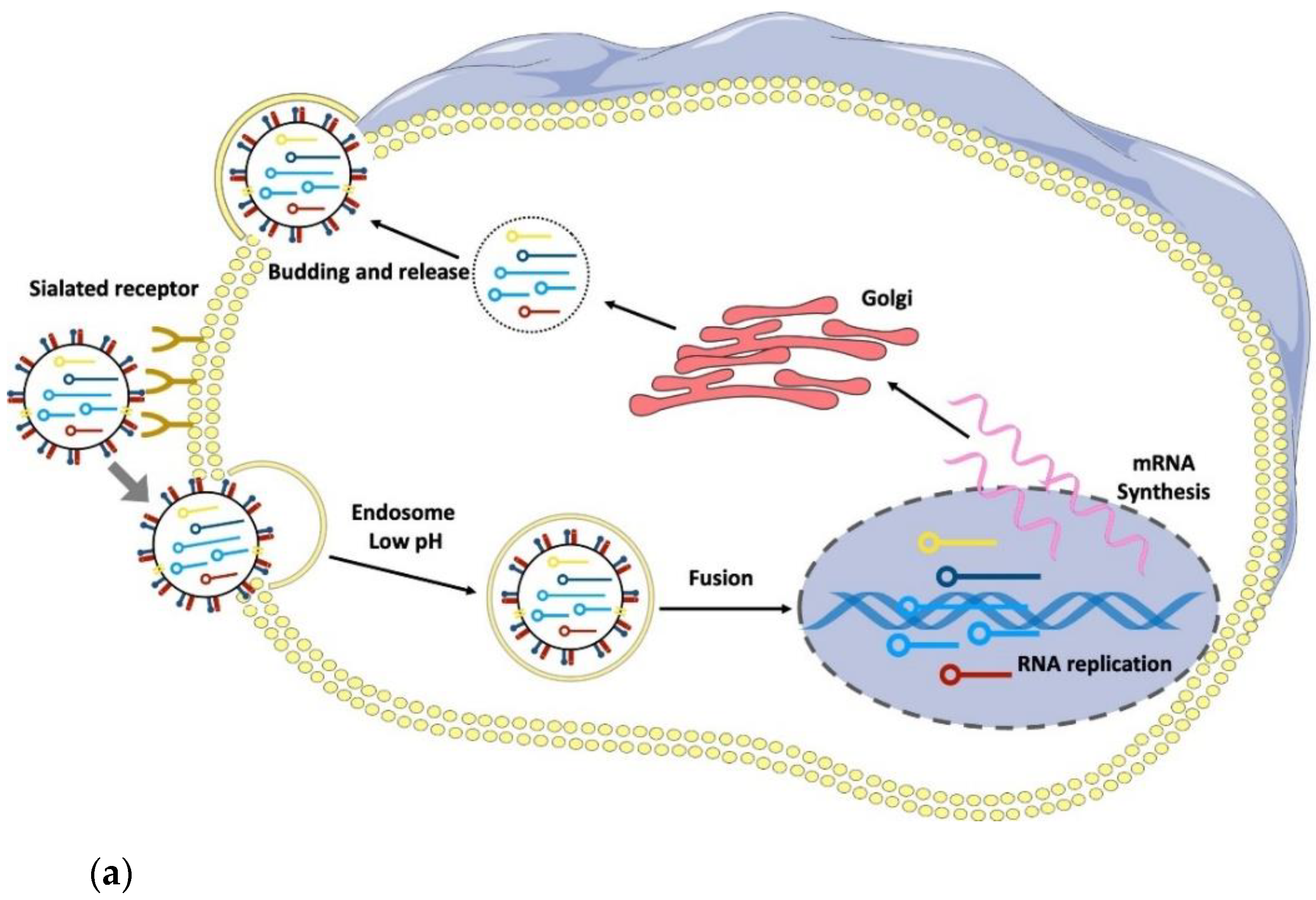
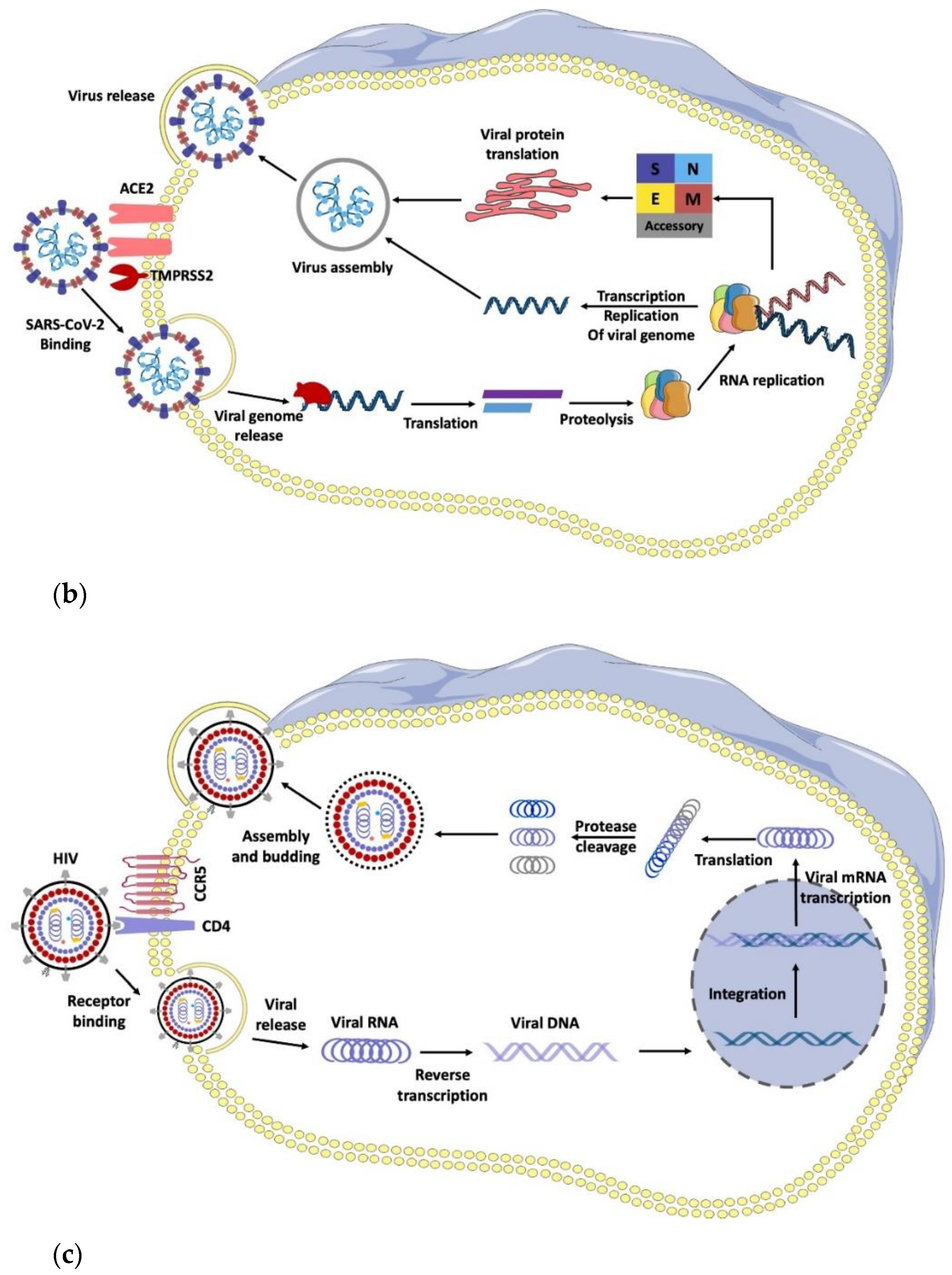
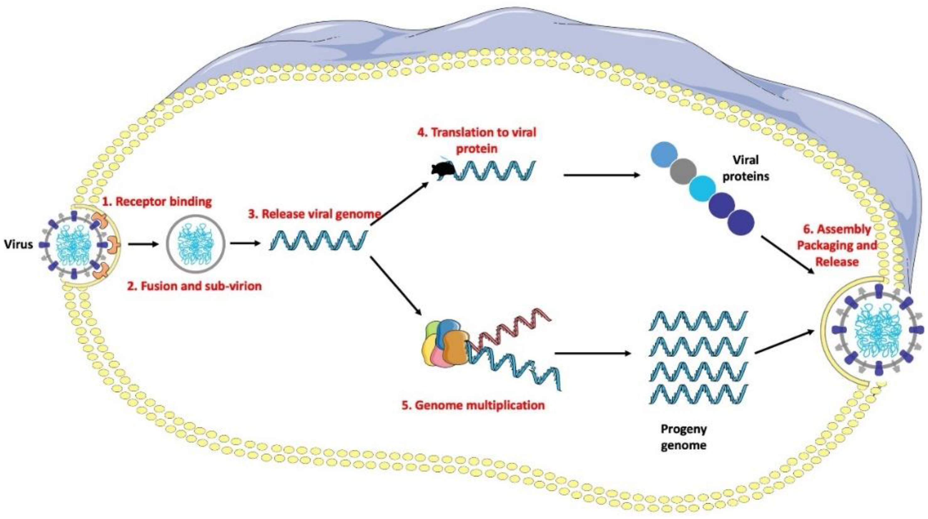
  • Binding to the host’s cellular receptors;
  • ‘Unpacking’ of the viral genome;
  • Replication of the RNA/DNA of the virus;
  • Synthesis of viral proteins;
  • Release of virions.
Figure 5. Main targets of antiviral drugs.
Chemistry 04 00019 g005
In fact, all these targets have been considered in anti-influenza, anti-HIV, and anti-SARS-CoV-2 drug therapy and research. In our discussion of the mode of action of the various therapeutic agents in the following sections, we implicitly refer to this Figure as well.

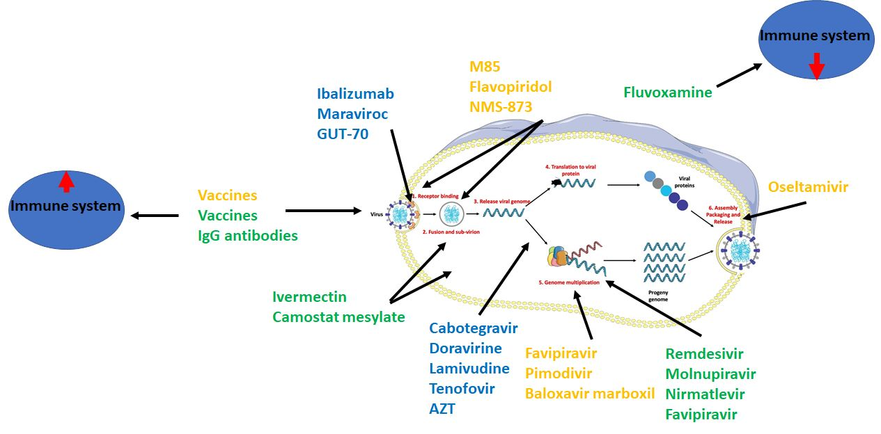
6. Anti-HIV, Anti-IV, and Anti-SARS-CoV-2 Agents

The need for vaccination is well justified in all three therapeutic areas of our subject. Vaccination and medication are, however, by no means mutually exclusive. The need for drugs to combat viral infections is also clear, as also evidenced by the current pandemic situation of COVID-19. The optimal drugs should preferably have a broad-spectrum antiviral profile, so that they are suitable for the treatment of the infection, for playing a complementary role to vaccination, and for bridging the period required to develop a new vaccine that is effective for newly emerging infective variants.
  • Virus- and host-targeting antiviral agents
In principle, antiviral drugs can target the viruses themselves or the host-dependent viral replication machinery within the host; and can also support host defense mechanisms [42]:
(i)
Direct-acting or virus-based antiviral agents that act directly on viruses to inhibit their replication.
(ii)
Host (indirect)-acting or host-based antiviral agents that act on host-based factors that are needed for viral replication.
The advantage of virus-directed agents is that their significant host toxicity is not expected in the absence of a corresponding host factor. Their therapeutic use is primarily limited by the frequent and rapid emergence of resistance to them. In particular, a significant decrease in the potency of highly selective molecules acting on a viral protein to inhibit replication is expected as a result of a viral mutation that directly modifies the drug binding site of that protein (but this may also occur as a result of a structural change distant from the binding site). Resistance may also develop with nucleotide-type virus-directed antiviral agents (through a viral proofreading process and/or mutations that may even develop into more infective variants); moreover, host effects may be unfavorable for this type, so they need to be carefully considered, mainly due to their potential host mutagenicity.
The chances of viral mutation-based resistance can be reduced with multi-targeting drug combinations or agents, which are also advantageous because they can also increase the therapeutic spectrum. This direction has proven successful primarily in the field of HIV treatment, which has brought breakthroughs in its drug therapy; this approach is receiving increasing interest in anti-influenza therapy. It is much less exploited in the anti-SARS-CoV-2 drug therapy, yet we expect the multi-targeting strategy to be considered in this area as well.
Although direct-acting antiviral drugs are the majority of antivirals in the current drug portfolios, host-based agents have recently received increasing attention. This shift in interest to host-based agents is also due to the lower risk of the development of resistance.
Host-based antiviral agents were first used in the 1990s. Interferons and some small-molecule agents have been used as immunomodulatory agents to support immune responses, regulate inflammation and cytokine storms upon virus infections and represent an important strategy to control viral infections. Maraviroc can be considered a host-based antiviral agent in the strict sense. Its introduction in 2007 to treat HIV-1 infection through the inhibition of chemokine receptor type 5 (CCR5), one of the main host chemokine receptors for viral entry into T-cells, is therefore of great importance and advance. It was hoped that resistance would not develop against host-based agents, because virus mutation to replace missing host cellular functions is less likely. However, experience has shown that resistance to certain host-based agents and therapeutic protocols may also develop, e.g., selection pressure through long-term treatment causes the virus to use an alternative host factor [43].
An important challenge for a host-based strategy is to identify the therapeutic target itself. As a target, it is advisable to select a host factor that is essential for virus replication but not mandatory for the host cell functions. However, in order to consider possible targets at all, in terms of their therapeutic suitability, molecular-level information about the host’s involvement in viral replication is also required. This can be ensured, in particular, that with the molecular biology technologies developed in the last decade and the evaluation of new data obtained on the complex pathomechanism (e.g., small interfering RNA, siRNA or clustered regularly interspaced short palindromic repeats, CRISPR, and omics, respectively), their limited availability in the past may explain the time lag of the host-based approach over direct antiviral strategy.
The main driving force and advantage of the host-based approach are that resistance hardly develops, and on the other hand, it can have the additional advantage that well-chosen host-targets can provide a broad antiviral spectrum. However, a long-term application can pose a particular challenge in terms of both resistance and safety, and therefore balancing drug safety and efficacy issues is, of course, more difficult than for direct antivirals.
In fact, all three therapeutic areas covered by our review are developing promising host-based agents, which we describe below.
Overall, we believe that both a direct and an indirect approach to control old and new emerging highly infective virus infections may be useful, especially in combination to enhance the beneficial therapeutic properties of the individual components and to minimize adverse effects.
It is also worth noting that it is important to exploit the synergies more efficiently between clinical and preclinical results and knowledge. By extending the principles and practices of precision medicine to antiviral therapy, it is possible to identify common elements of various viral replication mechanisms, as well as virus infections’ induced pathomechanisms in the host. Based on this, broad-spectrum antiviral agents effective against several virus types can be developed. From a drug-design perspective, this can be supported by designing new molecules based on the structural similarities of commonly relevant biological targets. These two pathways, therefore, need to work closely together to discover new types of antiviral agents. The initial traces of this endeavor can be seen in some of the recent examples we are also referring to, but there is room for more complete exploration.
In this report, we focus on the last 5 years for anti-influenza and anti-HIV agents and the past one year for anti-SARS-CoV-2 agents, because excellent comprehensive reviews are available for previous periods, see e.g., [42,44]. Well-established and emerging virus and host drug targets, promising drug candidates, and lead molecules, together with their molecular structures and mode of action, as well as antiviral drug discovery strategies, will be discussed.

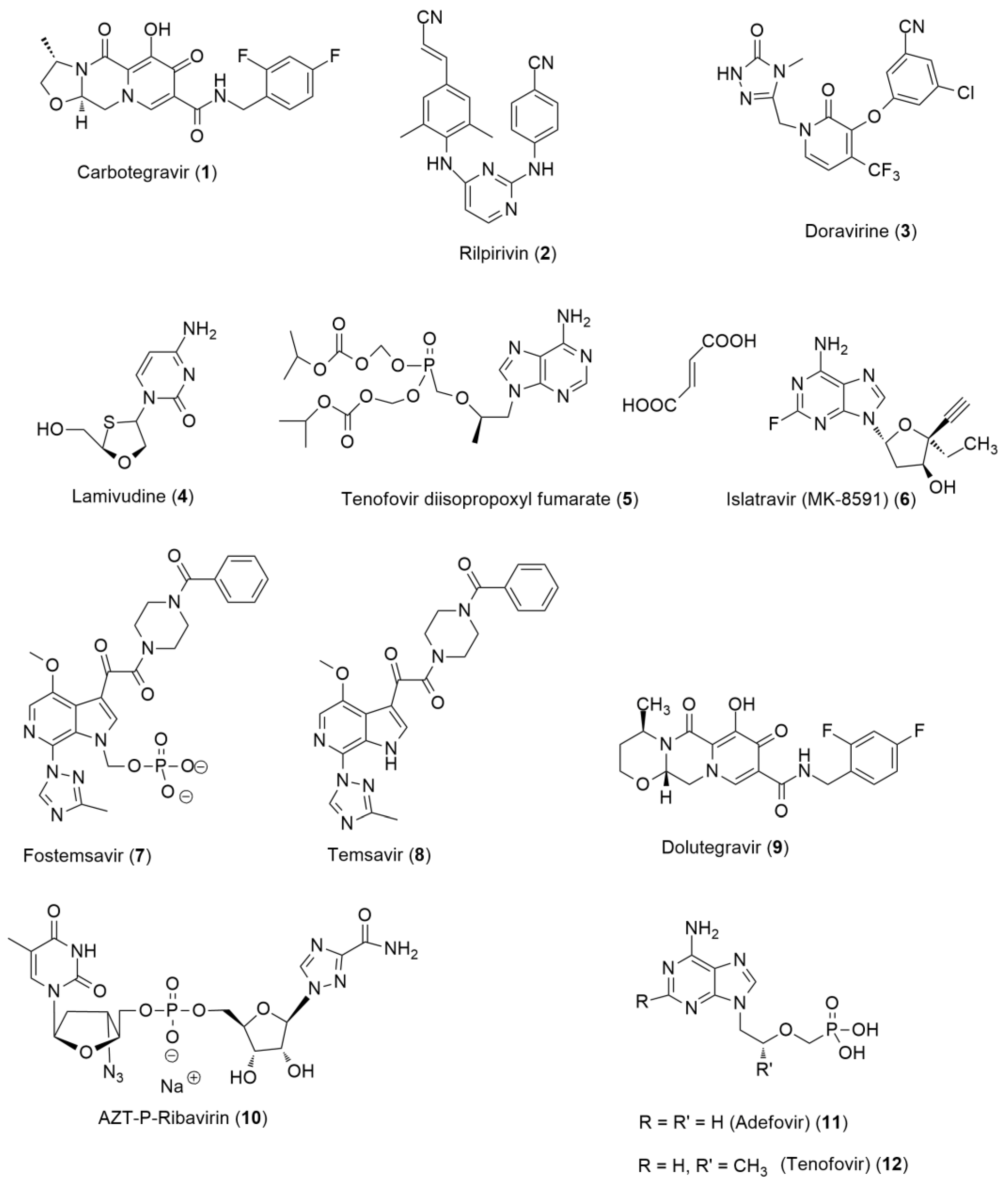
6.1. Anti-HIV Agents

The human immunodeficiency virus (HIV) belongs to the retrovirus family and is a member of the Lentiviruses. There are two main types of HIV: HIV-1 (the most common) and HIV-2 (relatively uncommon and less infectious). Human infection can result in AIDS. Another retrovirus species is the simian immunodeficiency virus (SIV) which causes persistent infections in many African non-human primates, but unlike HIV, without the development of AIDS [45]. It is believed that SIVs are the source of HIV.
HIV uses human CD4+ T cells for replication and destroys them. In humans, HIV-1 infection causes a breaking down of the immune system, leading to opportunistic infections. In addition, AIDS can also develop in the late stage of infection.
Anti-HIV drug therapy has achieved a real breakthrough by being able to treat HIV infection and prevent its progression to AIDS. Drug therapy of HIV infection started with the approval of azidothymidine (AZT) in 1987 [46], and since then, a number of highly effective antiretroviral drugs have been approved for use in monotherapy and in combination therapy as well. However, the use of monotherapy and even dual therapy often lead to the development of drug resistance and inadequate suppression of viral replication. These serious limitations directed the attention to the three-agent combination therapy. Due to its superior long-term efficacy, it has become the clinical standard of care and is now widely used for the treatment of HIV infection.
HIV drugs based on their mode of action can be grouped as follows [47,48,49]:
  • Non-nucleoside reverse transcriptase inhibitors (NNRTIs);
  • Nucleoside reverse transcriptase inhibitors (NRTIs);
  • Protease inhibitors (PIs);
  • Fusion inhibitors;
  • CCR5 antagonists;
  • Integrase strand transfer inhibitors (INSTIs);
  • Post-attachment inhibitors.
These drugs are generally used in combination in the highly active antiretroviral therapy (HAART), also called combination antiretroviral therapy (cART). This therapy is able to control the replication of HIV-1, thus, it has radically transformed the previously fatal disease into a manageable chronic disease.
Nevertheless, cART is not a curative therapy. It means that missed drug treatments may lead to the reactivation of virus reservoirs [50,51] and to the development of drug resistance. Accordingly, continuous life-long drug therapy is necessary to control the disease and achieve a functional cure. This is obviously a particular challenge for cART in terms of therapeutic efficacy, safety, and adherence.
In order to ensure a sterilizing cure, the active agent should reach all infected sites (especially in the lymph nodes and brain) in effective amounts. However, it is not easy to achieve this with currently available drugs. The relatively low solubility, bioavailability, and membrane penetration ability, and conventional drug formulations as well, often provide insufficient drug levels to achieve complete elimination of latent virus reservoirs; just as genome editing-based and monoclonal antibody-based strategies have not yet brought therapeutic breakthroughs for a sterilizing cure. Thus, although a functional cure can be achieved with the current cART approach, the sterilizing cure for HIV infection remains elusive and represents an important goal for anti-HIV drug innovation, taking the health and economic impact of HIV infection and life-long duration of treatment into consideration [52].
Currently, there are available many anti-HIV drugs in the USA and the EU as well.
Of them, four single agents and the combination products approved by the FDA in the last 5 years are below (Table 4 and Table 5, respectively).
Three of the four new drugs are small molecules and belong to the direct-acting antiviral type, whereas one new agent, it is a monoclonal antibody, ibalizumab (it is the first biologic drug introduced for HIV therapy), belongs to the indirectly-acting, host-based agents.

6.1.1. Direct-Acting Anti-HIV Agents

The chemical structures of these agents are shown in Figure 6.
Cabotegravir (1) is an integrase strand transfer inhibitor (NSTI) structurally like dolutegravir. It binds to the viral integrase active site and blocks retroviral DNA integration, which is essential for the HIV replication cycle. Injectable cabotegravir for pre-exposure prophylaxis was approved in December 2021. It is the first long-acting every two-month treatment for the prevention of HIV for individuals at risk of sexually acquired HIV-1 infection, who have a negative HIV-1 test prior to initiation [53].
It is also a breakthrough in therapy regarding drug compliance, since the combination drug, containing co-packed extended-release cabotegravir (NSTI) and extended-release rilpivirine (2) (NNRTI) in two separate injectable suspensions, is the first once-a-month preparation for HIV infection therapy (the name of the combination product is Cabenuva).
Doravirine (3) is a non-nucleoside reverse transcriptase inhibitor intended for use in combination with other anti-HIV agents. In treatment-naïve adults with HIV-1 infection, doravirine, in a combination with islatravir, exhibited a long-lasting virologic suppression without resistance, similar to the performance of doravirine’s other drug combination product (doravirine (3)/lamivudine (4)/tenofovir disoproxil fumarate (5)).
Islatravir (6) belongs to nucleoside reverse transcriptase translocation inhibitors (NRTTIs) and it is in a Phase III clinical development phase for HIV treatment. However, a clinical hold by the FDA was recently announced (December 2021), based on previous observations of decreases in total lymphocyte and CD4+ T-cell counts in some participants receiving islatravir in clinical studies.
Fostemsavir (7) is a phosphate ester prodrug of temsavir (8). Temsavir (8), the active metabolite of fostemsavir (7), is a first-in-class attachment inhibitor that binds to the viral glycoprotein gp120 (it is regarded as a conserved, resistance insensitive target), thereby inhibiting the interaction between the virus and the surface receptors on CD4 cells. Consequently, it blocks HIV entry. Importantly, no therapy-related resistance was observed [54,55,56]. Fostemsavir (7) exhibited a significant antiviral response in adults who had received multiple treatments for HIV-1 infection.
In the last few years, in addition to the introduction of the above drugs, another important therapeutic result was obtained with the integrase inhibitor dolutegravir (9), introduced nearly a decade ago; it was recently approved for the treatment of HIV-infected children [57].
The nine drug combinations approved in the last 5 years are presented in Table 5. The high number also illustrates that combination therapy has become almost predominant in current HIV therapy. It is not surprising, since the favorable multi-targeting broad-spectrum therapeutic profile due to the combination of different, often synergic mechanism components and the reduction in drug resistance potency represent important, and sometimes unique advantages over the individual components of the combination.
The simplest form of combination therapy is the co-administration of individual drugs in individual formulations (so-called cocktails). However, especially from the point of view of adherence, treatment with a fixed-dose single formulation is clearly preferred. Conventional fixed-dose combination formulations have only a limited ability to compensate for the often unfavorable physicochemical properties of each component. Advanced nanoformulations can be used to overcome this weakness, as illustrated by a series of convincing examples [58,59,60,61,62]. Increased and controlled site-specific organ penetration, and sufficient and sustained local drug concentrations can be achieved, which can also result in the eradication of virus reservoirs. Thus, enhanced and sustained drug levels in lymph nodes, HIV-host cells, and in the blood and plasma can be obtained for weeks.
However, all types of combination formulations have certain inherent disadvantages that cannot be fully compensated (chemical, pharmacokinetic and side effect incompatibilities of components of the combination). This narrows the scope and motivates the application of other multi-targeting approaches. One such strategy is based on the use of multi-targeting drugs, where a single molecule expresses multiple mechanisms of action. This approach has already been receiving much attention to developing drugs for the treatment of various complex diseases, also including, especially since the millennium, viral infections. The high therapeutic value of multi-targeting with a single agent is also demonstrated by a series of drugs, although once developed as one target-selective agent, it turned out later that their broad therapeutic profile was due to their polypharmacology. In medicinal chemistry, the incorporation of dual or multiple activities in a single molecule is called a ’designed multiple ligand’ (DML) or ’multi-target-directed ligand’ (MTDL). The chemical implementations and illustrative impressive results of this approach have been the subject of several recent comprehensive reviews, e.g., [62,63,64,65]. In short, in the case of DMLs, two or more pharmacophores responsible for the aimed pharmacological activities are combined, through ‘conjugating’ or ‘merging’ the pharmacophore moieties in various synthetic chemical ways. If available, we can select a molecule exhibiting a certain degree of the desired multi-activity as a lead compound, then, it is structurally optimized to achieve the balanced pharmacological profile. In a broader sense, the conjugation method includes the connection of two pharmacophores, which can be even drug molecules, through a direct bond or a spacer entity; well-known examples of this method can be provided by AZT-based dimers, such as AZT-P-Ribavirin (10) [66]. Another interesting dual antiviral inhibitor was developed from acyclic nucleotide phosphonate derivatives, such as adefovir (11) and tenofovir (12), for the treatment of HIV and hepatitis B virus co-infection [67]. The MTDL strategy can also be applied to combine hitherto unexplored or less explored anti-HIV activities in a single molecule by starting from a suitable lead [68]. These examples also immediately highlight one of the important advantages of DML/MTDL over combination formulations that pharmacokinetics and pharmacodynamics can be optimized, although undoubtedly, chemical lead optimization requires the synthesis of many compounds and a very thorough and detailed series of pharmacological experiments, including phenotype-based assays to evaluate the balanced profile, pharmacokinetic, and safety studies. A further disadvantage is that the development of such a multi-targeting agent, being a new molecular entity, can take a long and costly route, similar to original drug candidates. With careful consideration of the pros and cons, as well as the scope and limitations, it can be decided case-by-case whether the DML approach is a worthwhile implementation of a multi-targeting strategy.

6.1.2. Host-Based Anti-HIV Agents

Interestingly, despite the above-described possible benefits, exploration of the host-based antiviral approach lags far behind the direct-acting approach. In fact, only a few such anti-HIV drugs have been approved so far. One of the most recently approved drugs, the monoclonal antibody ibalizumab, and the discovery of some new promising small-molecule drug candidates or leads can be a sign of growing appreciation and interest in the host-based anti-HIV drug type. The structures of new host-directing small molecules are presented in Figure 7.

Dual CCR5 and CXCR4 Antagonists

To select a possible host target, the molecular mechanism, and factors of the virus replication process within the host side have to be considered. The HIV-1 entry and cycle process involves several key factors and steps. HIV-1 initiates access to the host cell by binding the viral envelope glycoprotein gp 120 to the host cell receptor cluster of differentiation 4 (CD4) glycoprotein. CD4 is highly expressed on the surface of immune cells and mediates interactions between antigen-presenting and T-cells. Further interaction with any of the two coreceptors, chemokine receptor 5 (CCR5) and CXC-chemokine receptor 4 (CXCR4), is also needed to initiate the viral cycle. Among selective and effective CCR5 antagonists, maraviroc (13) appeared as the most intriguing compound, and it is the only selective small-molecule inhibitor of CCR5 approved by the FDA so far. Its mode of action and a possible extension to a broader effect have recently also received considerable attention, in particular, to develop dual-targeting CCR5 and CXCR4 inhibitors. This approach can also be suitable for the treatment of resistant, severe dual-tropic HIV and neuro-HIV infections [69]. This line of research can be illustrated by the following interesting lead structures identified in the past few years.
The core structure of naturally occurring ingenol (14a) was modified by esterification to obtain cinnamoyl (14b), caproyl (14c), and lauryl ester (14d) derivatives. These compounds exhibited promising antiviral activity, due presumably to a dual-mode of action that is related to the protein kinase C (PKC) pathway. One of the actions of ingenols stimulates the HIV long terminal repeat (LTR), possibly via NF-κB nuclear translocation, and the other action downregulates membrane receptors CD4, CCR5, and CXCR4. Importantly, these compounds can also result in a sterilizing cure via depletion of the latent viral reservoirs, therefore, may serve, based also on their low cytotoxicity, as co-adjuvants for a ‘shock and kill’ therapy [70].
In a composite computational study, through virtual screening and a statistical approach, some polyheterocyclic derivatives were identified as active agents on both CCR5 and CXCR4. The core structures are partially saturated pyrazolopyridine (15ac) and pyrazolopyrazine (16) scaffolds [71].
Another receptor- and ligand-based computational study led also to the identification of new dual CCR5 and CXCR4 antagonist candidates [72].
An angularly fused tricyclic coumarin, GUT-70 (17), isolated from Calophyllum brasiliense, was shown to block the replication of HIV in a dose-dependent manner. It is also a dual-acting receptor blocker anti-HIV agent, but its viral entry blocking action also involves other components. It stabilizes membrane fluidity and downregulates the expression of CD4, CCR5, and CXCR4 receptors, and it also inhibits NF-kB. These interesting features make it a valuable lead compound to obtain new effective agents against HIV infection [73,74].

Purine-Receptor Antagonists

Purine receptors can be involved in the HIV-1 fusion and infection and may be used to combat the viral infection. Some compounds were identified as P2X1 receptor antagonists in an HTS campaign. Compound NF279 (18), an antagonist of this receptor, also diminished HIV-1 fusion. It was also shown that the inhibition of HIV-1 fusion by NF279 (18) did not involve CD4 or coreceptors on the cell’s surface. It seems that P2X1 is a possible target for anti-HIV therapy [75].

Post-Attachment Inhibitors

The fourth newly approved drug is ibalizumab, a humanized immunoglobulin (Ig) G4 monoclonal antibody, which is a post-attachment inhibitor. It binds CD4 extracellular domain 2 and inhibits post-attachment steps in viral entry (but not influencing normal CD4 function) [60]. Its significant adverse effects were also documented in clinical applications; immune reconstitution inflammatory syndrome was attributed eventually to the therapy, however, was only observed in one case. Ibalizumab is used as an add-on therapy for adult patients.
Leronlimab, another humanized IgG4 monoclonal antibody, is an investigational CCR5 antagonist for the treatment of HIV infection. It is currently being investigated in Phase III clinical trials for critically ill COVID patients. It is intended to be used in combination therapy [76].

New Host-Based Targets

A more recent OMICS-based analysis illustrates the great potential of data-utilization in the discovery phase. In this study, 2910 human-HIV protein–protein interactions were identified and further classified. The network-based analysis serves to determine key elements and molecular mechanisms that are involved in the HIV infection and develop new drugs to prevent, as well as to treat and cure the disease through ‘shock and kill’ and ‘block and lock’ approaches [77].

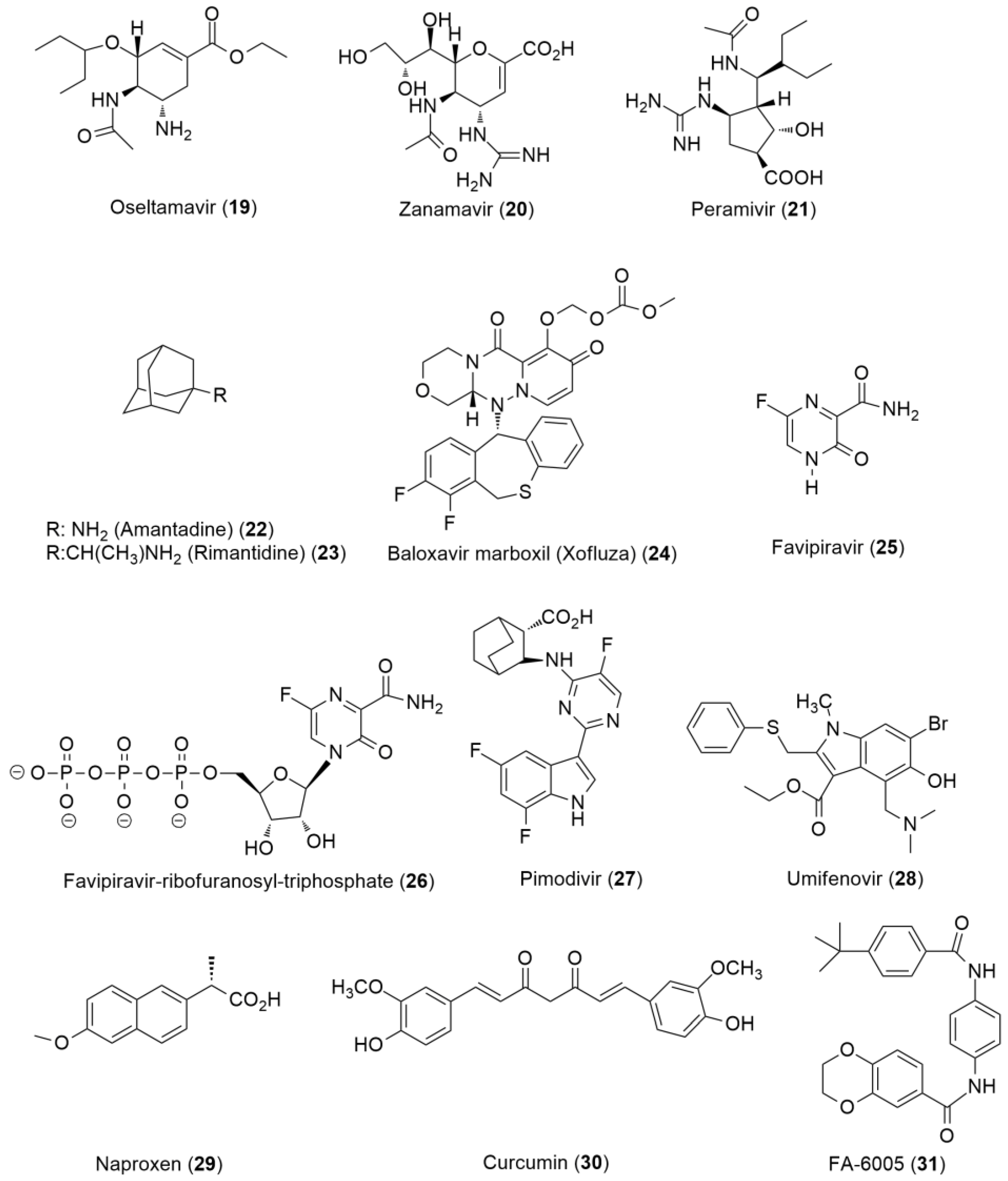
6.2. Anti-Influenza Agents

The influenza virus belongs to the Orthomyxovirus family of viruses. Influenza A, B, C, and D viruses are distinguished based on their antigenicity, of which, influenza A viruses are the most pathogenic to humans and have a wide range of hosts. Influenza B and C are weak, whereas influenza D is not pathogenic to humans (for the phylodynamics and evolutionary analyses, see [78]). The well-known manifestation of influenza illness is the seasonal flu. The most effective way to prevent seasonal flu is to get vaccinated each year. Influenza vaccination is, however, not always available and is not uniformly effective to prevent infection. Infected patients can need treatment with anti-influenza drugs approved for the treatment of acute uncomplicated influenza (and eventually, for prophylaxis).
Here, we focus on the recent achievements of antiviral drug therapy of the clinically important influenza A virus infections, especially on the host-based approaches, but referring briefly to new direct-acting agents, too (molecular structures are shown in Figure 8.
Four FDA-approved and Centers for Disease Control and Prevention (CDC) recommended anti-influenza drugs are currently used. Of them, only one drug, baloxavir marboxil (BXM) (24) has recently been approved (in 2018). Oseltamivir (19), zanamivir (20), peramivir (21) were approved before 2016. For historical reasons, the matrix 2 ion channel protein (M2) inhibitors amantadine (22) and rimantadine (23), which block viral uncoating, are also on the list, but they are presently not in use in influenza therapy, due to the resistance of many influenza viruses against these drugs.

6.2.1. Direct-Acting Anti-Influenza Agents

The approved anti-IV drugs are direct-acting, virus-based inhibitors. With the exception of baloxavir marboxil (24), they are neuraminidase inhibitors (NAIs). Hemagglutinin (HA), neuraminidase (NA), and M2 proteins are on the surface of influenza A and B viruses and play important roles in virus cell entry. The cooperative receptor-binding protein HA and the receptor-cleaving enzyme NA jointly control the multivalent interactions between the virus and the host cell, and finally the virus entry [74]. The virus-specific NA and HA also serve as antigens for antibodies. NAIs as therapeutic anti-influenza agents, which block the release of newly formed viral particles, have attracted much attention for a long time. However, therapeutic use of NAIs, due particularly to the emerging resistance of IV to the treatment, has recently been limited. Their wide use is still suggested to prevent hospitalization in pandemics when treatment has to be initiated early in the course of infection to get the most significant response [79].
It is also noteworthy that a recent thorough analysis of the therapeutic position of NA inhibitors led to a rather critical conclusion that ’in a typical influenza infection, NAIs’ efficacy is inherently not high, and even if their efficacy is improved, the effect can be negligible in practice’ [80].
The limited efficacy and risk for resistance of available agents call for new drug targets to be considered for the treatment of influenza, see for reviews [35,81,82,83,84,85,86].
Baloxavir marboxil (BXM) (24) is the first new type of drug on the anti-influenza drug list [87,88,89]. Its interesting structure contains two tricyclic ring systems and a carbonate moiety. It serves as a prodrug, from which the active form, the hydrophilic baloxavir acid (BA), is formed by hydrolysis. BA, itself does not penetrate membranes, therefore its prodrug is applied. BA strongly inhibits the cap-dependent endonuclease polymerase acidic (PA) protein of the influenza virus; thus, the virus reproduction is blocked. It is interesting that the design of BA was based on the structural similarity of IV endonuclease and HIV integrase. Therefore, an integrase inhibitor, dolutegravir (9), was taken as a model compound to design anti-IV agents; in fact, one of the tricycles of BA overlaps with the dolutegravir tricyclic system [90]. BXM (24) is now considered a valuable therapeutic agent and is used for the treatment of acute symptomatic uncomplicated influenza [90,91].
The efficacy and safety of neuraminidase inhibitors and endonuclease inhibitors for the treatment of seasonal influenza were comprehensively reviewed [92]. It was concluded that therapy with zanamivir (20) was associated with the shortest recovery time, and BXM (24) treatment was associated with a reduced rate of influenza-related complications.
There are other anti-influenza agents that have been approved or developed in certain geographic regions. Among them, favipiravir (25) is the best known.
Favipiravir (25) was approved for restricted use in Japan in 2014 for the treatment of influenza. It is a nucleoside-base analog. Favipiravir, as a prodrug, is converted in vivo into the active triphosphate nucleotide metabolite (Favipiravir-TP) (26). The active metabolite specifically inhibits the polymerase machinery of the virus. It is incorporated in the virus nucleic acid as a pseudo nucleotide, thereby inhibiting the replication. At a molecular level, it binds to the viral RNA-dependent RNA polymerase (RdRP) and induces mutagenic changes in influenza (and some other) virus RNA [93]. The mechanism of inhibition of IV by favipiravir was analyzed [35,93]. The broad antiviral spectrum of favipiravir formed the basis to also investigate its potential utility in SARS-CoV-2 infection (see in next section). Certain limitations of favipiravir’s (25) use have to also be taken into consideration. The risk of human teratogenicity and embryotoxicity is well-documented [94]. Interestingly, its antiviral mechanism is described as a potential risk factor for its therapeutic application, through its possible contribution to the emergence of novel pathogenic viral strains, being resistant to drug therapy [95].
In our opinion, the pharmacokinetics of favipiravir (25) and structurally related prodrug agents may play a significant role in the antiviral activity that is expressed by the active metabolite. The extent of metabolism can however be insufficient, due to: (i) relatively low conversion of favipiravir with the hypoxanthine-guanine phosphoribosyltransferase enzyme responsible for the formation of favipiravir-monophosphate (the first, generally rate-limiting metabolic step in the formation of favipiravir-TP (26), the active metabolite) [96]; (ii) the biochemical complexity of viral infection, especially in severe clinical conditions, can influence the rate and yield of the metabolism; (iii) individual pharmacogenetic factors may differently affect both (i) and (ii). Each of these factors can impair the effectiveness of the treatment.
The place of favipiravir in anti-influenza therapy was outlined in a recent review [35].
The viral polymerase complex can be targeted in another way. Pimodivir (27), a 3-(2-pyrimidinyl)pyrrolopyridine derivative, inhibits viral replication through binding to the PB2 subunit of viral RNA polymerase and blocking the cap-snatching activity. Pimodivir (27) was undergoing clinical development, however, the Phase III trial was halted in 2020 because of unsatisfactory efficacy results [97].
Antivirals targeting the HA protein of the influenza virus are not widely used. Umifenovir (28) belongs to this group of agents. It is currently being investigated in clinical trials in the USA (and is already marketed in China and Russia) for the treatment and prevention of influenza. Its broad-spectrum antiviral activity is based on the inhibition of virus–cell membrane fusion and membrane perturbation [83].
Another important virus-based target for anti-influenza therapy can be the viral nucleoprotein (NP) that binds to viral RNA. It is critically important for both transcription and replication processes. Interestingly, naproxen (29) [98] and curcumin (30), an anti-IV agent with multiple modes of action [99], were found to inhibit NP-RNA interactions [99,100]. Naproxen (29) also demonstrated anti-SARS-CoV-2 activity [101].
A novel NP inhibitor, FA-6005 (31), inhibited human pandemic and seasonal influenza A and B viruses in vitro. It was also active in mice against the lethal influenza A virus challenge. Its mode of action is, however, more complex. It suppresses influenza virus replication and perturbs the intracellular trafficking of viral ribonucleoproteins. Thus, it also represents a potentially valuable broad-spectrum multi-targeting anti-influenza mode of action [102].

6.2.2. Host-Based Anti-Influenza Agents

Several host factors have been identified to be involved in influenza virus replication, see, e.g., [42,103], however very few host-based anti-influenza drug candidates have entered the clinical development phase. Some interesting lead structures are also shown in Figure 9.
In a viral infection, many human kinases are hijacked by the virus to support its replication cycle. Host kinases, as targets for new anti-IV agents, have been thoroughly investigated in recent studies.
Since there are many kinase inhibitor drugs, drug repositioning and the decreasing of development time and costs is an attractive strategy to identify new antiviral and also anti-influenza drug candidates. In a high-throughput screening campaign on A549 human cells against influenza viruses, two known cyclin-dependent kinase inhibitors, flavopiridol (32) and dinaciclib (33) proved to be highly active [104].
Kinase inhibitors, as a drug repositioning source of broad-spectrum antivirals [105], and to identify new antiviral drug mechanisms, were investigated. Kinases phosphorylate are important IV proteins necessary for the replication and/or evasion/suppression of the innate immune response, and thus can be attractive targets as anti-IV agents.
Several kinases were found to be involved in the regulation of antiviral chemokines and cytokines [106].
The epidermal growth factor receptor (EGFR) and phosphoinositide 3 class II β (PIK3C2β), a newly identified factor in viral entry, are involved in the anti-influenza virus action of a triazolopyridazine derivative, M85 (34). Interestingly, its combination with oseltamivir (19) exhibited strong synergism in vitro [107].
The valosin-containing protein (VCP; p97) is also a promising target. It is a member of the AAA family of adenosine triphosphatases (AAA-ATPases) and contributes to protein homeostasis and many other important biological processes. Its inhibitors were suggested as possible therapeutic agents in cancer, in particular, multiple myeloma [108]. In addition, VCP is also involved in the replication of several viruses. Its key role in influenza virus replication was recently recognized and a small-molecule allosteric VPC inhibitor, NMS-873 (35), was shown to exhibit broad-spectrum activity against influenza viruses, including drug-resistant strains. Moreover, it inhibited influenza virus in two human lung epithelial cell lines, also confirming that VPC may be a relevant therapeutic target for anti-influenza agents [103].
In the search for new antivirals, thus anti-IV agents, data-based disease analyses to identify new targets and mechanisms, and drug repositioning to discover new clinical candidates have been increasingly applied. Another valuable result of these analyzes is that a closer and more complete description of the disease is also useful in precision therapy. In an elegant study, several new host-based anti-influenza drug candidates could be identified by utilizing transcriptomic signatures of infected patients as a signature of infection (‘clinical virogenomic profile’) and drug signatures obtained on human cells (CMAP). The subsequent detailed biological analyses led to the selection of diltiazem, a calcium channel blocker antihypertensive agent, for clinical evaluation for the treatment of influenza. A Phase II clinical study of diltiazem (36) with oseltamivir (19) in combination has recently been initiated [98,100].
Another clinical example for drug repositioning is given by nitazoxanide (37), an antiprotozoal agent that was also shown to exhibit a multi-component broad-spectrum antiviral activity. Recently, it underwent a Phase III clinical trial in non-complicated influenza patients as an anti-influenza drug candidate [109]. Surprisingly enough, a combination of nitazoxanide (37) with oseltamivir (19) was no more effective than oseltamivir alone in the treatment of hospitalized influenza patients. Nitazoxanide (37) is also being investigated as an anti-SARS-CoV-2 agent in several clinical trials.
Multi-targeting is also an attractive strategy in anti-influenza therapy. Results with drug combinations underline the importance of the careful selection of components.
Perhaps, a reduction or elimination of resistance-sensitivity can primarily be expected from the combinative treatment. Thus, a combination of oseltamivir (19) with zanamivir (20) indeed suppressed oseltamivir- and zanamivir-resistant influenza viruses, while exhibiting a comparable antiviral effect to the most effective monotherapy arm [110]. On the other hand, a triple combination of oseltamivir (19), amantadine (22), and ribavirine (38) vs. oseltamivir (19) in a Phase II trial was not convincingly superior [111].
Surprisingly, designed multi-targeting agents have been underrepresented in the anti-influenza therapeutic area. We can refer to one interesting but retrospective approach based on a thorough analysis of an anti-influenza traditional Chinese multi-component medicine. It led to the identification of several interesting multi-targeting ligands with potentially anti-influenza activities [112].
Anti-influenza strategies that are based on nanoformulations are of great interest. For anti-influenza vaccine development, some formulations have reached clinical phases. For small-molecule antivirals, most achievements have only been demonstrated in vitro. Moreso, it has been confirmed that nanoformulations can overcome unfavorable ADME-relevant physicochemical properties of the candidates. To translate it, in vivo activities and optimal target-specific delivery with acceptable safety have to be the next step. Several examples of the application of nanotechnology to anti-IV agents, demonstrating improved properties, have been reported in recent reviews [113,114].

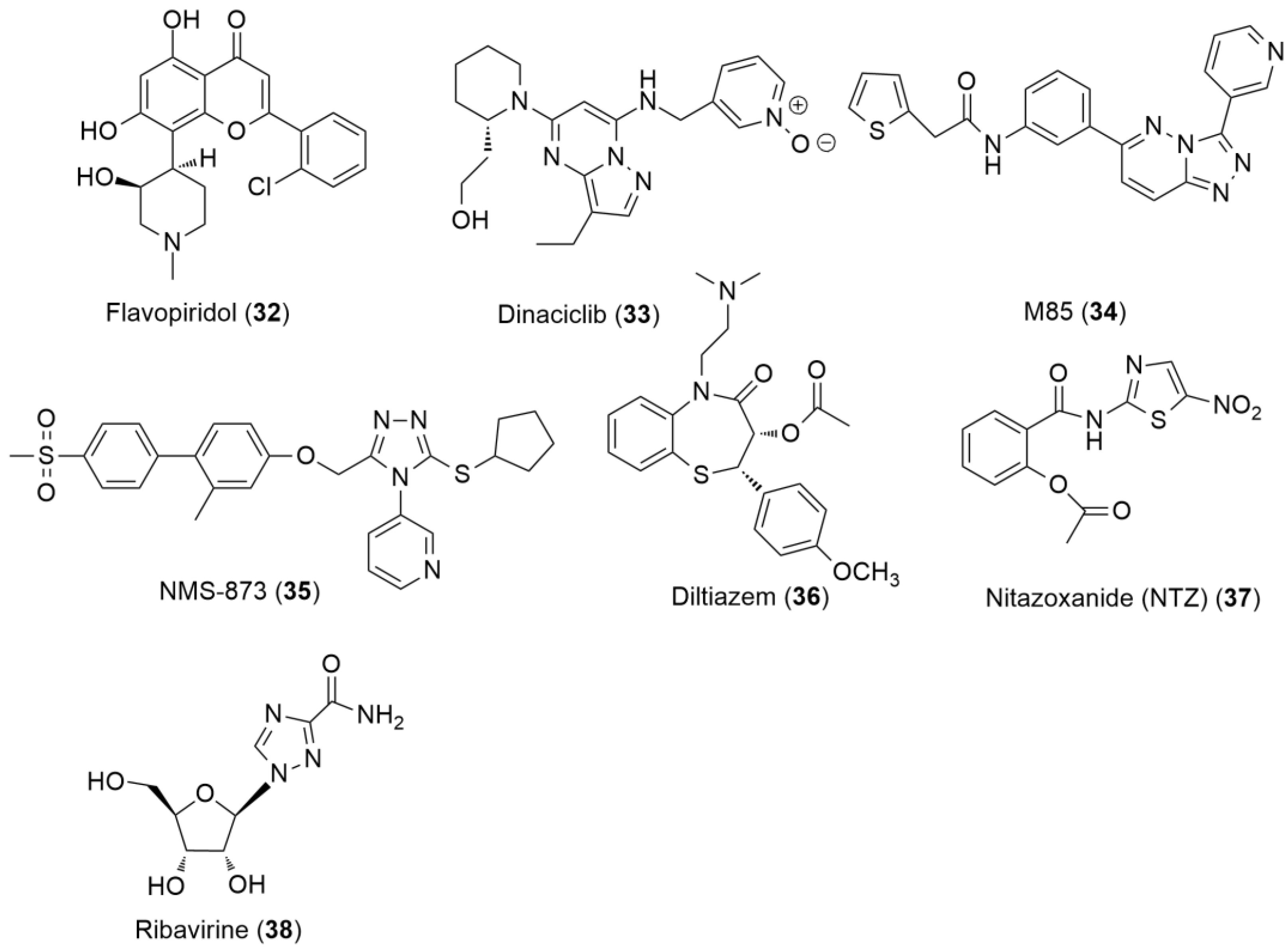
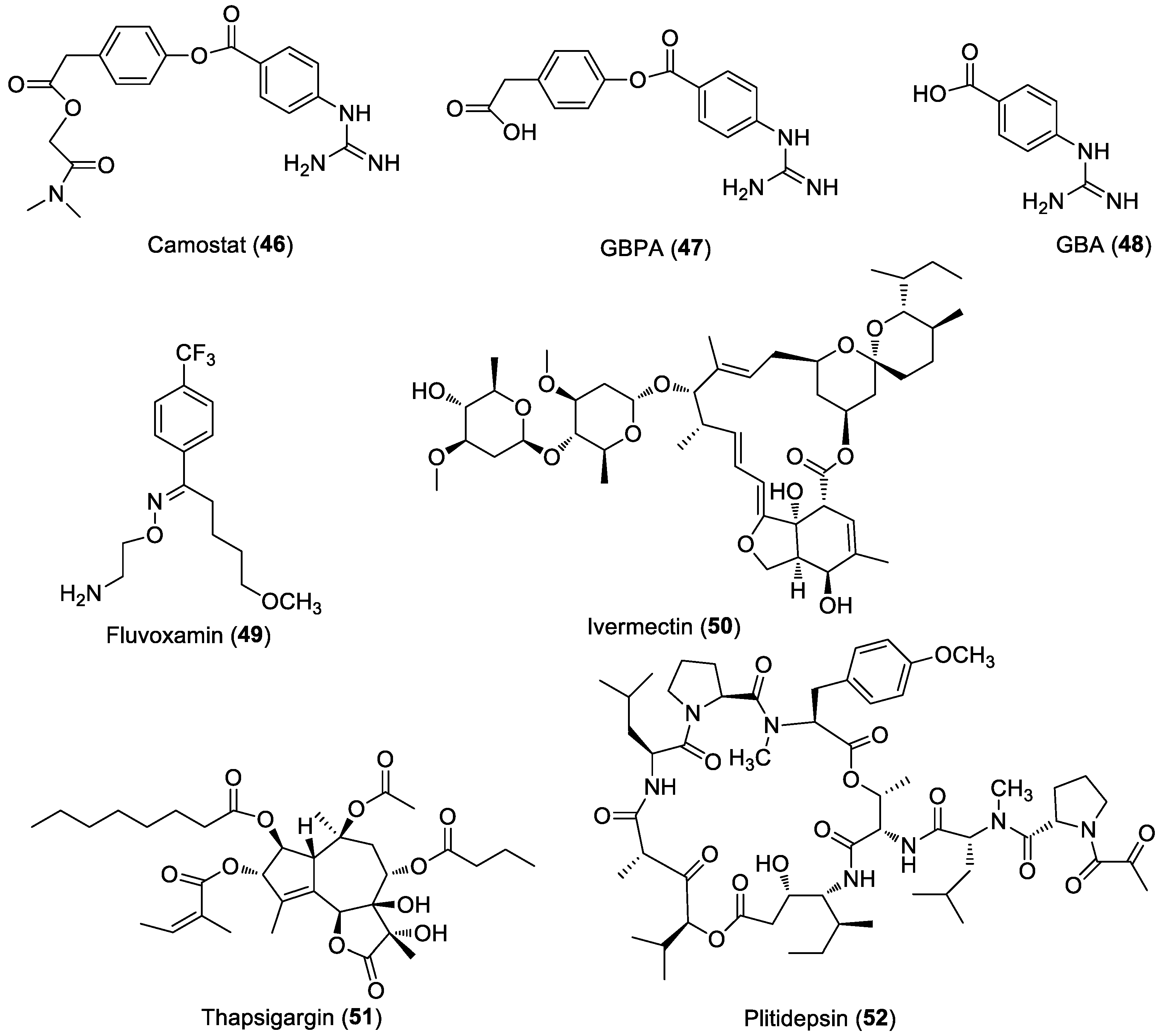
6.3. Anti-SARS-CoV-2 Agents

In this section, the goals and results of anti-SARS-CoV-2 drug research and development are presented in light of the unprecedented features and health challenges of the pandemic. Thus, lessons for the future can be outlined with greater certainty.
The many unexpected complexes and severe health burdens and challenges of the pandemic, especially in the initial phase, in 2020, demanded unprecedented performance for medicines and, of course, vaccination. Both drug innovation and vaccine development have gone through tremendous efforts. In the first phase of the pandemic, drug therapy could only rely on existing drugs. On the one hand, available agents, according to their original application, had to be properly positioned and applied to meet the typical needs of an infectious disease. On the other hand, treatment of new specific unusual symptoms of COVID-19 required new agents; this unmet and urgent need could be satisfied by a new use of existing agents, i.e., following a drug repositioning strategy. Thus, several repositioning candidates were selected based on some simple and rapid preclinical testing (it was usually an in vitro anti-SARS-CoV-2 virus assay). The scope and the limitations of these tests in their predictive competence for therapeutic suitability (defined by a balanced combination of pharmacological, pharmacokinetics, and safety factors) were often over- and underestimated, respectively. Furthermore, this approach may be criticized because it was occasionally biased and controversial in its method and content. An excellent review also addressed various aspects of the performance of in vitro assays [115]. Then, with these ‘confirmatory/promising’ preclinical results in hand, a clinical trial was initiated immediately. It would no longer seem surprising that a large number of clinical trials had hardly led to therapeutically exploitable results (cf. 4952 clinical trials, including drugs and vaccines, had been registered for COVID-19 until March 2021 [116]). It should also be added that the clinical trials themselves may be subject to strong criticism, due to modest quality in their conception, design, accomplishment, and evaluation. More so, because of this disappointing series of failures, as well as the availability of vaccination through successful development at an astonishing rate from 2021 onwards, it may have been suggested that the pandemic could be broken down and soon eradicated by vaccination only. Thus, the demand for COVID-specific drugs has declined. This was also due to the fact that, especially in the first phase of the pandemic, objective identification of the infection was often not performed in the initial stage of the disease, i.e., when an antiviral agent would have had a chance of a favorable and significant therapeutic outcome (to prevent hospitalization). Some antiviral agents have been used, especially in severe, multi-inflammatory conditions, at most as an adjunct therapy. Not at all surprising, however, is at this stage of the disease, direct-acting antiviral agents could hardly or at all be expected to halt and reverse the disease. Subsequently, many examples have confirmed this. The view that vaccination and herd immunity alone and exclusively are the solution, has become almost monopolistic. According to some experts, the vaccination campaign, which was in full swing at the beginning, seemed to be able to reach full population vaccination. It was also concluded that there was no longer a need for drugs that were ‘harmful anyway’. All of this was voiced by epidemiological ‘experts’. Widespread vaccination of the population—we ourselves also believe—is the most important means to take infections down to sporadic levels and especially to permanently prevent the pandemic. However, the growing experience gained since the introduction of vaccination makes it evident that there is also a continuous need for pharmacotherapy. We can refer to several factors in this regard, such as barriers to herd immunity, rapidly emerging new variants [117], and variant-dependent (in)efficacy of vaccines. In addition, and especially, pharmacotherapy itself has a role to play. It is necessary to provide drugs, when a vaccine is not yet available, to bridge periods between vaccine developments challenged and forced by emerging virus variants, and to treat vaccinated patients who become infected (breakthrough infection) or those who are not eligible for vaccine protection. In particular, there is a need for drugs that are effective in the early stages of the disease, capable of preventing the development of severe health conditions, and can be used orally for anti-SARS-CoV-2 therapy. All are desirable in terms of patient interest, health burden, and cost-effectiveness. Recent drug developments and results are impressive and promising in this regard; in this review, we focus on such agents. We firmly believe that such agents are of strategic importance, not only to prevent the disease from becoming serious, but also to avoid hospitalization, which is a huge burden on healthcare. We do not address drug therapy for severe conditions of the infection, which is usually aimed at reversing or treating the effects of immune dysregulation and inflammation with steroids and/or anti-cytokine drugs and/or other agents. Additionally, we also refer to the comprehensive reviews discussing therapeutic aspects of COVID-19 in detail [38,116,118].

6.3.1. Direct-Acting Antivirals for COVID-19

In Figure 5 of this review, we summarized the main steps of virus-host interactions, from entry to the release of the virion. The viral RNA translates 4 structural proteins, the spike (S), envelope (E), membrane (M), and nucleocapsid (N) proteins, and 25 non-structural (ns) and accessory proteins. The ns proteins formed from two polypeptides by the viral proteases play essential roles in replication and host interactions. Effective antiviral drugs can block viral entry and/or proliferation within the host cell. Accordingly, such targets [119], especially four non-structural proteins, including papain-like protease (PLpro), main protease (Mpro) or 3-chymotrypsin-like protease (3CLpro), helicase, and RNA-dependent RNA polymerase (RdRp) as key enzymes, have received considerable attention and are subjects of through studies to develop anti-COVID-19 agents (Mei). Remdesivir (39) and molnupiravir (40), inhibitors of replication of RNA by interaction with RdRp, and nirmatrelvir (41), an inhibitor of Mpro, are available as therapeutic agents for the treatment of COVID-19 patients in several countries. In particular, the lower susceptibility to resistance-causing viral mutations (in respect to spike-protein targeting agents) and a potentially wide spectrum of antiviral activity is of special value to these agents. Remdesivir (39) is the only FDA-approved product for the treatment of COVID-19.
The COVID-19 Treatment Guidelines Panel’s Statement from the NIH (19 January 2022) [120] on Therapies for High-Risk, Nonhospitalized Patients With Mild to Moderate COVID-19 guides clinicians on the use of oral ritonavir-boosted (42) nirmatrelvir (41) (Paxlovid) and molnupiravir (40) (both received the Emergency Use Authorization, EUA, in December 2022) and parenteral sotrovimab and remdesivir (39) medications for the treatment of nonhospitalized patients. In clinical studies in unvaccinated individuals, these agents were found and are expected to provide the greatest benefit for nonhospitalized patients who have risk factors for progression to severe COVID-19.
The drug portfolio currently available, although modest in number, is still a major step forward in the treatment of nonhospitalized patients since the outbreak and the previous year. The emergence of widely available oral formulations for patients is almost a breakthrough.
Due to space constraints, we could not undertake a comprehensive presentation of the extremely rich research and development work related to the management of COVID-19.
Here, we summarize the effects of the three direct-acting small-molecule antivirals, remdesivir (39) and the two oral formulations, the lessons learned, and experiences of their development, which are of course interesting in themselves. Together, with some promising research on new drugs (Figure 10) that will also be discussed briefly, they can provide some strategic guidance. Excellent publications are already available on the disease. For the purposes of this review, we refer in particular to the comprehensive reviews that discuss and analyze the experience to date with vaccination, drug therapy, clinical trials [116,121,122], immunotherapy guidelines [38], and potential drug targets and candidates [119,123,124,125,126,127,128,129,130,131].
Remdesivir (GS-5734) (JBC2020) (39), a cyano-substituted C-nucleoside adenosine analog, belongs to the RdRp inhibitor class of antiviral agents. RdRp is a key player in viral replication machinery. The catalytic subunit (nsp12) of RdRp is involved in RNA synthesis through the formation of phosphodiester bonds. The nsp7 and nsp8 viral proteins are also needed to process transmission replication. RdRp has a highly conserved structure in RNA viruses, but there is no homologous mammalian counterpart. All these make it an attractive virus-based drug target, although viral mutations may narrow the antiviral spectrum of drugs [132]. Remdesivir (39), a phosphoramidate prodrug, was previously shown to exhibit remarkable activities against HCV, multiple variants of Ebola, Marburg, arena, and coronaviruses, and was considered for the treatment and prevention of Ebola virus disease [133]. Remdesivir’s active triphosphate (REM-TP) metabolite, formed in several steps via a monophosphate ester, accumulates in infected cells, blocks viral replication. It exhibits a broad-spectrum antiviral effect. The mode of action of remdesivir (39) is also well understood at a molecular level. Due to the structural similarity of REM-TP to ATP, it behaves as a specific but false substrate of viral RdRp [134]. The efficient incorporation of REM-TP into viral RNA causes a delayed termination of viral RNA synthesis, which is translated into a robust in vitro anti-SARS-CoV-2 activity. Importantly, remdesivir (39) has no significant host cytotoxic effect. This unique, highly selective, and efficient viral chain-elongation terminating mechanism is apparently more favorable than that of favipiravir-ribofuranosyl-triphosphate (26), the active form of favipiravir (25), another RdRp inhibitor prodrug (see anti-influenza agents and below). We refer also to excellent in-depth comparative studies of the interaction of remdesivir (39), favipiravir (25) and molnupiravir (40) with the RdRp machinery, which highlighted differences in their mechanisms of action and also provided some hints about mechanism-based therapeutic consequences [82,135,136].
A confirmation of the inhibitory effect of remdesivir (39) against MERS, followed by a clinical study on severely ill COVID-19 patients resulting in time-shortening of hospitalization, formed the basis for EUA by the FDA, May 2021, for treatment of severely ill COVID-19 patients. Remdesivir (39) received FDA approval on 22 October 2020. It became the first approved drug for use against SARS-CoV-2 infection. Interestingly, some subsequent studies of remdesivir warned of some controversial data, as was the case in Solidarity [137]; it was critically commented in a ScienceInsider publication [138].
According to current NIH guidelines, intravenous remdesivir is also allowed for use in nonhospitalized settings, on the basis of a trial, and included 562 nonhospitalized patients. Hospitalization or death was reduced by 87% when remdesivir (39) was applied in the early phase of symptoms.
Molnupiravir (MOL, EIDD-2881; MK-4482; Lagevrio) (40) is the isopropyl ester prodrug of N4-hydroxycytidine, which inhibits the replication of human (and bat) coronaviruses, including SARS-CoV-2. MOL, due to the tautomerism of its oxime moiety, can not only mimic cytidine but also uridine. Accordingly, it can act as a false substrate and can be inserted in RdRp, replacing both natural nucleosides. Its very interesting and remarkable discovery against SARS-CoV-2 was due to G. Painter and his group at Emory University [139], who had studied and identified its anti-coronavirus effect well before the COVID-19 pandemic; they had been working on oral inhibitors of the replication of Venezuelan equine encephalitis and other infectious viruses. They designed ribonucleosides, believing in their potentially advantageous profile, thus, identifying EIDD-2881 as a promising anti-influenza and anti-MERS agent. The molecular mode of action of MOL is different from remdesivir’s mechanism of action. It is based on the deception of the RdRp to force it to make errors. More specifically, RdRp recognizes the active form, N4-hydroxycytidine, as uridine instead of cytidine, and accordingly, the polymerase incorporates adenosine instead of guanosine in the opposite position of RNA. The accumulation of high enough errors in the viral RNA leads to an error catastrophe and the virus cannot function anymore.
When COVID-19 began, Parker and his team immediately recognized the great potency of EIDD-2881 as an anti-SARS-CoV-2 agent. Following the confirmation of its promising antiviral activity in relevant preclinical assays, a collaboration with Ridgeback Biotherapeutics also started. It was extended from mid-2020, and molnupiravir (40) was developed jointly by Merck and Ridgeback Biotherapeutics.
Interestingly, an early study of MOL in 300 hospitalized patients was halted because of unsatisfactory efficacy results. In a subsequent large Phase III study on the treatment of nonhospitalized COVID-19 patients, however, convincing therapeutic benefits were seen. In 1,433 nonhospitalized adults with mild to moderate COVID-19, with at least one risk factor associated with poor outcomes, the drug reduced the hospitalization or death rate by 30% when administered within 5 days of symptom onset. This important achievement was the basis for granting EUA by the FDA.
In the case of MOL, a computational docking study suggested a low potency to the development of drug resistance. It is also noteworthy that it can also be effective in treating patients with resistance to remdesivir (39) [140].
The main concern about molnupiravir (40) therapy originates from its mutagenic potential. On the virus side, a long-term therapy could lead to the development of new, even infectious variants, whereas on the host side, it might be incorporated into human DNA, thereby becoming mutagenic.
According to NIH therapeutic guidelines, molnupiravir (40) is used for the treatment of adult patients with mild and moderate COVID-19, who are at high risk of progression to severe disease. A 5-day therapy has to be initiated in the early disease phase after a SARS-CoV-2 positive test result.
Molnupiravir’s (40) name originates from ’Mjölnir’ (Mjölnir is the hammer of the thunder god Thor in Norse mythology). Nomen est omen—let us hope it becomes a reality that molnupiravir can knock out the virus.
The antiviral agent favipiravir (25), used also for influenza therapy (Section 6.2.1), has been recommended for oral therapy of COVID-19 in several countries, such as Russia, India, Hungary, Saudi Arabia, and there are ongoing clinical trials in other countries [35]. Therapeutic experience and the results of ongoing clinical trials, as well as its efficacy against new SARS-CoV-2 virus variants, will position it in COVID-19 therapy. The interaction of favipiravir (25) with RdRp was studied by cryomicroscopy [135,136]. The molecular operating environment-based resistance scanning methodology was also used to evaluate the sensitivity of favipiravir’s (25) binding mode to mutated target structures. It was suggested that SARS-CoV-2 with only a few mutations in RdRp, could develop favipiravir (25) resistance [136]. Safety and pharmacokinetic issues of favipiravir (25) therapy were discussed in Section 6.2.1.
Nirmatlervir (PF-07321332) (41) is the result of a record-breaking innovation. It took just one year from the idea to the first clinical test [139,141,142]. The COVID-project, led by D Owen, started immediately with a focus on the main protease enzyme of SARS-CoV-2. At Pfizer, a relevant protease inhibitor drug candidate (PF-00835231) (43) was already in hands. It was originally developed for the treatment of SARS-CoV (2003). It inhibited the SARS virus’ main protease (Mpro) that, similarly to SARS-2′s protease, cleaves virus polypeptides to form viral proteins essential for replication. Importantly, binding sites of main proteases in SARS- and SARS-2 are almost overlapping in their sequences. Therefore, it could be expected that this SARS-active molecule could also inhibit the Mpro of SARS-CoV-2, and soon after, it was indeed confirmed. However, it all seemed very encouraging, but this molecule’s PK properties were not yet acceptable for good absorption and oral application. Pharmacokinetic properties had to first be improved to obtain an orally active candidate. A series of creative and structure-guided modifications, and careful consideration of chemistry (especially stereochemical stability) and synthesis (scaling-up) issues led to the selection of PF-07321332 (41). Its valuable antiviral profile was characterized by thorough in vitro and in vivo studies. Recognizing that it is extensively metabolized by CYP3A4, narrowing its oral bioavailability, another clever ‘trick’, was made to solve the problem. Namely, the co-use of a blocker agent of CYP3A4 did increase in vivo concentrations to effective therapy-dictated levels.
To make the story complete, which in this case also means a great drug candidate and a combination drug product, nirmatrelvir (41) was combined with ritonavir (42) to get Paxlovid. The co-administered ritonavir (also serving as a PK-enhancer for other protease drugs), otherwise without anti-SARS-CoV-2 activity, boosts nirmatrelvir’s (41) PK.
In the clinical study, which included 1219 patients, treatment with Paxlovid reduced hospitalization or death by 89%, when administered within 3 days of symptom onset.
Both molnupiravir (Lagevrio) (40) and Paxlovid represent important advances in outpatient therapy in nonhospitalized patients. Due to the emergence of the latest variant omicron of SARS-CoV-2, it is very important that in vitro efficacy of both agents against omicron has recently been confirmed, supporting their use also for the treatment of patients infected with this variant [143,144].
Another interesting class of inhibitors of viral main protease was developed by Kansas University researchers and collaborators [145]. This COVID-relevant project was based on two pillars. The first one was provided by a coronavirus Mpro inhibitor agent, GC 376 (44) that was highly effective for the treatment of coronavirus-induced fatal feline infectious peritonitis (FIP) in cats (it caused clinical remission); as the second pillar, a series of in vitro active peptides were available. A subsequent structure-guided (co-crystallized inhibitors with proteases) optimization led to a dipeptide derivative that was effective against multiple human coronaviruses in enzyme- and cell-based assays as well as in vivo in a mouse model of MERS-CoV. These findings may also pave the way for the development of novel SARS-CoV-2 inhibitors.
The viral RNA repair mechanism, as a part of the complex replication machinery, was also considered as a therapeutically relevant antiviral drug target [146,147]. This target was also considered for anti-SARS-CoV-2 agents. The proofreading endonuclease activity is executed for coronaviruses by the nsp14-nsp10 complex with the assistance of magnesium ions. The complex’s structure, also importantly, does not overlap with mammalian exonucleases. Screening of selected nuclease inhibitors and magnesium-chelators in a FRET assay for the ability to inhibit nsp14/10 led to the identification of some active compounds, which were also used to build a mechanistic model. On this basis, a virtual screening campaign, and in vitro and in vivo experimental studies resulted in a few candidates showing inhibitory action individually or in combination with remdesivir (39) in various anti-SARS-CoV-2 effect-relevant assays. Three hit compounds, including sofalcone (45) (Drug Bank 05197; once considered as a gastroprotective agent), could serve as leads for further optimization [147].

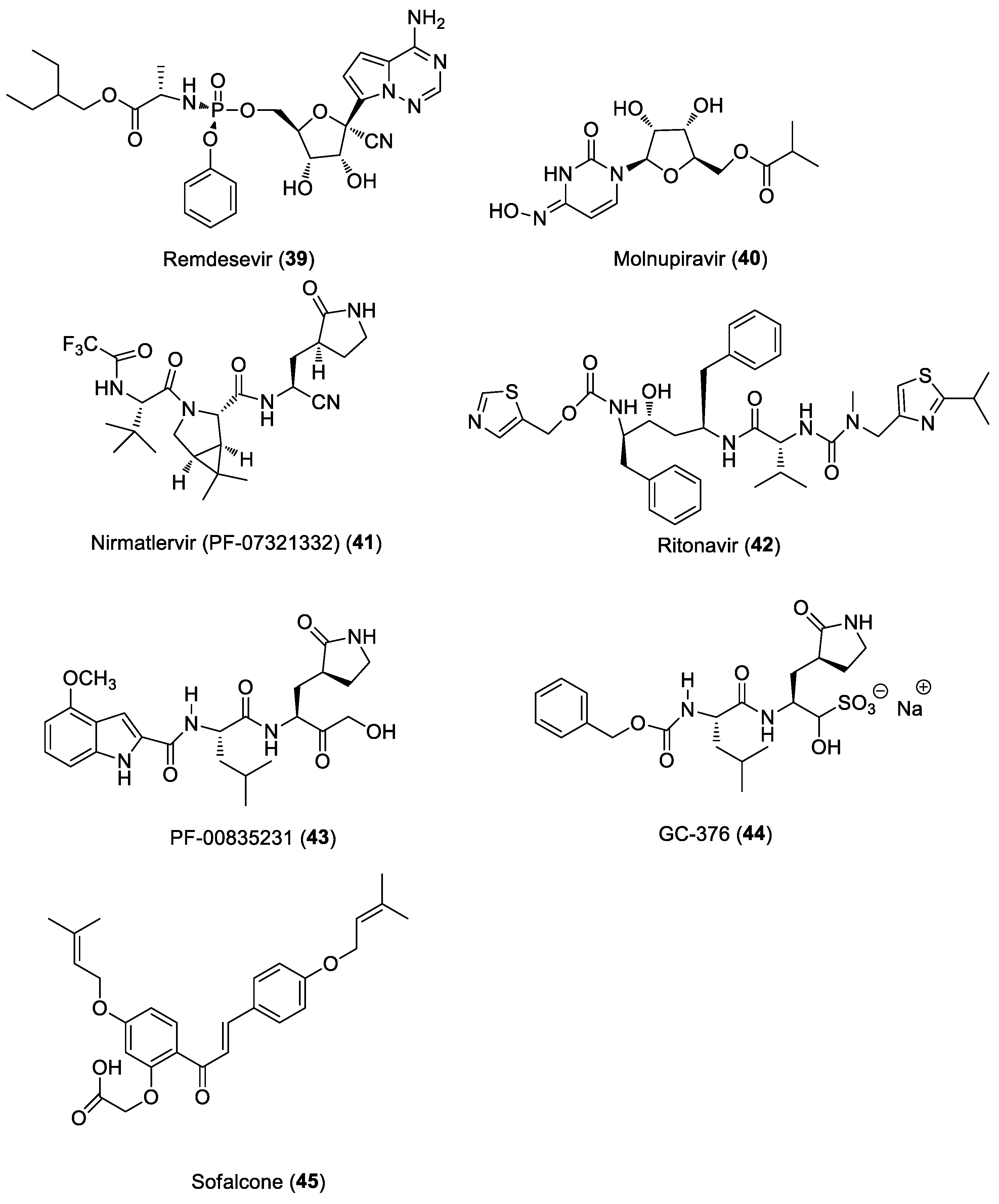
6.3.2. Host-Based Antivirals for COVID-19

The possible benefits of host-based agents over direct-acting antivirals have been suggested for RNA-virus therapies [148]. It also prompted studies with host-based antivirals agents (Figure 11) to combat COVID-19 [149,150].
One such host-based target is the transmembrane protease serine 2 (TMPRSS2). SARS-CoV-2 utilizes the TMPRSS2 enzyme for infecting the host cell. Specifically, the spike protein is cleaved by the protease enzyme to support virus entry. A well-known TMPRSS2 inhibitor, Camostat (46), approved for the treatment of reflux esophagitis and pancreatitis in Japan, was also found to inhibit the SARS-CoV-2 cell entry, especially in an assay on a human epithelial cell-line [151]. Camostat is rapidly hydrolyzed in vivo to form the also active, equally potent metabolite 4-(4-guanidinobenzoyloxy)phenylacetic acid (GBPA (47)), which is subsequently slowly converted to 4-guanidinobenzoic acid (GBA) (48). Because of its favorable antiviral activity and safety profile, camostat has also been studied in clinics for COVID-19 therapy; so far, no clearly convincing therapeutic results have been obtained [152]. There are still several ongoing trials to (re)-evaluate its efficacy on COVID-19 patients [153], which may be decisive in its role in COVID-19 therapy.
In the early phase of pandemics, to discover repurposing host-based antiviral candidates, a very elegant and comprehensive study investigated the interactions between SARS-CoV-2 and the host; through an extensive mapping method, 332 human proteins, apparently targeted by the virus, were identified. Further narrowing the list, and screening the candidates for antiviral activity, two sets of agents, inhibitors of mRNA translation, and predicted regulators of the sigma-1 and sigma-2 receptors, seemed to be especially interesting. Thus, it was suggested that some members of these classes of agents may be optimized as antiviral therapeutic agents [154].
Fluvoxamine (49), approved as an antidepressant and generally qualified as a selective serotonin reuptake inhibitor (SSRI), has also a formal connection to the above findings. In fact, fluvoxamine is an agent with a broad activity profile, many components of which are also relevant to COVID-19 therapy [155]. In particular, it also displays a high affinity to the sigma-1 receptor and has an antiviral activity; exerts immunomodulatory and anti-inflammatory effects, is related to the sigma-1 receptor and SSRI activities, and exhibits an antiplatelet action, due to the SSRI effect. This unique and interesting pharmacological profile initiated the investigation of the possible role of fluvoxamine in the treatment of COVID-19 patients [156]. In a large multicentric placebo-controlled trial (TOGETHER) for acutely symptomatic high-risk patients with COVID-19, fluvoxamine (49) (100 mg twice daily) was administered for 10 days, to evaluate its effect on preventing hospitalization. The treatment with fluvoxamine (49) reduced the need for hospitalization, defined as retention in a COVID-19 emergency setting or transfer to a tertiary hospital (absolute risk reduction of 5% and RR reduction of 32%), and importantly, without significant adverse effects.
Ivermectin (50) is probably one of the most controversial of the many candidates investigated for COVID-19 therapy, which has almost ’emotionally polarized’ both professionals and the general public. The history of its use as an antiparasitic agent [157] and its successful therapeutic application in human medicine in a huge human patient population worldwide is also well-known [158,159]. Its complex, possibly COVID-19 therapy-relevant mode of action includes anti-SARS-CoV-2 [160] and complex anti-inflammatory and immunomodulatory mechanisms with multiple components [161]. The robust, in vitro confirmed antiviral effect is due mainly to the inhibition of signal-dependent nucleocytoplasmic shuttling of the SARS–CoV nucleoplasmid protein mediated nuclear import [160]. The efficacy and safety aspects of ivermectin therapy have been the subject of many publications (for a recent review, see [159,161,162]). Clinical trials have been initiated to evaluate its therapeutic utility [153]. However, confidence in ivermectin’s (50) effectiveness in COVID-19 patients was often dictated by enthusiasm, great anticipation for success, and at most, only partially well-founded hopes instead of the results of well-designed and perfectly performed clinical trials [159,163]. In fact, despite a huge, perhaps a record-breaking number of trials, it is not yet possible to make an objective (evidence-based) assessment of its value or unsuitability in COVID-19 therapy. It is therefore advisable to wait for the results of ongoing clinical studies. Our view is also in line with the recently issued NIH guidelines on the use of ivermectin for COVID-19 (16 December 2021): ’There is insufficient evidence for the Panel to recommend either for or against the use of ivermectin for the treatment of COVID-19. Results from adequately powered, well-designed, and well-conducted clinical trials are needed to provide more specific, evidence-based guidance on the role of ivermectin (50) in the treatment of COVID-19’.
Thapsigargin (TG) (51), is another host-based antiviral agent of interest. It is a plant-derived tricyclic sesquiterpenelactone, that inhibits the sarcoplasmic or endoplasmic reticulum Ca-ATPase family of calcium pumps [164]. Due to this mechanism, more recently, inhibition of apoptosis with TG was also shown, which was interesting enough to consider TG’s structure as a lead for new anticancer agents [165]. In other studies of TG (51), the significant action against various viruses, including influenza subtypes, MERS, and the alpha-, beta- and delta-variants of SARS-CoV-2 coronaviruses was identified and carefully analyzed [166,167]. This valuable host-based broad-spectrum antiviral mechanism targeting the endoplasmic reticulum stress-unfolded protein response to inhibit the viral replication process can be concluded as to deserve further exploration, with also a focus on structural optimization to minimize TG’s host toxicity.
Plitidepsin (52) (dehydrodidemnin B), a cyclic depsipeptide originally isolated from the marine tunicate, is approved in Australia for the treatment of multiple myeloma in combination with dexamethasone. In addition, it was recently shown that it exerts a strong anti-coronavirus effect, in which the eukaryotic elongation factor 1A (eF1A) may be involved. Furthermore, eF1A is important for viral replication through its interaction with some viral proteins. A detailed study confirmed that eF1A also plays a role in the interaction with SARS-CoV-2 viral proteins. Accordingly, inhibitors of the interaction can be utilized for anti-SARS-CoV-2 action. Following this line of thinking and knowing about previous promising clinical trials of plitidepsin (52) in COVID-19 patients (PharmaMar company), it was also tested in three different (including human) cell lines for an anti-SARS-CoV-2 effect. In each case, very strong nanomolar IC90 was obtained, whereas its cell-toxic/static effect was not far comparable. The antiviral effect is mediated by eF1A, as confirmed by an impressive mechanistic study. In a mouse model, the in vivo antiviral activity was also proved. All of which together suggest that it is worthwhile to continue the clinical studies in COVID-19 patients [168].
Regarding the clinical trials of host-based agents, it is also worth making a comment. There is a particular challenge in clinical trials in non-severe patients with early-stage disease. Namely, it is difficult to evaluate these drugs clinically when the majority of untreated patients can recover. Therefore, we emphasize that in order to achieve robust results and to position these agents effectively in therapy, these studies should be performed primarily in patients with one or more risk factors, according to precision clinical considerations and well-chosen endpoints.

6.4. Multi-Targeting Approach

The multi-targeting strategic endeavor has so far been illustrated by a number of examples; these are mainly ineffective clinical trials with combinations of repurposed drugs (we refer here to an excellent review of repurposed drugs and their combinations investigated in clinical trials [116]) or have preclinical encouraging results. Of course, the significant potential of this approach may be more comprehensively assessed once the portfolio of approved anti-SARS-CoV-2 drugs is much richer.
In our view, the therapeutic value of the designed multiple ligand (DML) approach is expected to be exploited and seen in a few years, as these types of agents, representing a new chemical entity, may enter the clinical phase later.
What can we expect from the multi-targeting approach? Similar to HIV therapy, where a therapeutic breakthrough has been achieved with combination products, clinical trials may be initiated with a combination of approved anti-SARS-CoV-2 agents. The growing knowledge of the disease and the availability of mechanistically different types of agents can support this line and can be an important step forward. Thus, for example, the combination of favipiravir (25) and molnupiravir (40) showed promising results in a preclinical study [169]. The use of a combination of molnupiravir (40) and nirmatrelvir has also been suggested [143].
We would also believe that the combinations of direct agents with indirect anti-SARS-CoV-2 agents can be particularly promising, which would be worth exploring.

7. Conclusions and Future Directions

After reviewing the anti-HIV, anti-influenza, and anti-SARS-CoV-2 vaccine and drug landscape, some concluding and forward-looking comments can be made.
1. Although vaccine and drug satisfaction in these three areas are different, all experience to date confirms that vaccine and drug therapy are not mutually exclusive medical tools: their combined availability strengthens health care. This is easy to see, as discussed above, even in situations close to full population vaccination, there is a continuous need for antiviral drugs not only for the duration of vaccine development but also beyond, due to limitations of vaccination.
2. Vaccine development (and antibody drug development) is needed in all three areas. The HIV vaccine is not yet widely available. In the other fields, for influenza and COVID-19, although there are already effective vaccines—at this point too, we need to emphasize the impressive speed and effectiveness of RNA-based vaccine development for COVID-19—but all these vaccines still have limitations. The incomplete protection against infection and possible side effects; and technical limitations, such as production capacity constraints and special delivery conditions, make it difficult to quickly meet significant global demands. The frequently emerging variants are presently the biggest challenge for protection through vaccination. Thus, conceptually new broad-spectrum vaccines could be a major step forward. Such an RNA-based anti-influenza vaccine is under development; the clinical results and experiences will also be useful in other areas.
3. The low efficiency of preclinical-clinical translation is often cited as one of the major limiting factors for the productivity of drug innovation. We also drew attention to this, and criticized, among other things, preclinical methods that oversimplify and ambiguously reduce the amount of information relevant to their predictive capabilities for human use [170]. This methodological problem has occasionally occurred in the research of antiviral agents. As noted in Section 6.3, it appears that the importance and predictive values of in vitro cell-based antiviral activity are often overestimated. The drug is quickly but superficially qualified solely based on the degree of inhibition of the virus replication, while the suitability of the model is not or is hardly being considered. Namely, to conclusively evaluate such results, many model features must also be considered, thus, the cellular toxicity of the drug, the relevance of the model to the target mechanism, and the biochemical characteristics of the model (e.g., in a system expressing efflux-transporters, the antiviral efficacy of transporter substrates can be reduced). Reference is also made here to excellent publications that critically discuss such issues of in vitro antiviral assays [115,171]. Then, ’precise’ comparisons of such in vitro antiviral activities with animal/human plasma concentrations to quantitatively qualify the candidates’ in vivo antiviral abilities (and therapeutic utility) can also be misleading, as plasma concentrations do not necessarily predict target tissue or organ concentrations.
4. The role of clinical-preclinical translation, which is less frequently discussed, is emphasized by us in the context of defining drug target(s). Feedback and analysis of clinical observations and other data with data-science tools could result in an even better understanding of the molecular pathomechanism of the disease, the mode of drug action and it can lead to the identification of new targets in antiviral drug research, especially for host-based antiviral agents. By complementing these studies with phenotyping of the various stages of the disease, precision medicine principles and practices can be followed, and therapeutic treatments can be more efficiently performed. Furthermore, associative analysis of clinical diagnostic and disease-descriptive data, drug efficacy, and mechanism-related data, together with all health-relevant patient data, can be utilized for both precision medicine practice and drug innovation. We believe that it is expedient to create a common data platform to display the mutually interesting and supportive aspects of the two areas so that the data analyses for various purposes can be performed efficiently. Its implementation can result in a very huge and comprehensive ‘virus, disease, drug, treatment data platform’ supporting healthcare and innovation.
5. Precision medicine should be implemented in virus infectious therapy. Accordingly, both host and viral variabilities have to be considered in the choice of therapy. The omics-based and data association analyses, from the host- and virus-side, respectively, are thus important. As an illustration, an excellent pioneering study of severely ill COVID-19 patients is mentioned here. The involvement of genomic structural variants in the disease was investigated with a new method, optical genome mapping [172]. Such a study, together with genomic sequencing, can lead to the discovery of new associations between genomic information and disease susceptibility and pathogenesis. The early identification and characterization of viral variants require reliable, quick, and cheap, easy-to-use diagnostic tests (cf. increasing the risk of developing severe conditions without early therapy, especially in HIV and SARS-CoV-2; differential diagnosis for overlapping flu and COVID-19 symptoms).
The high importance of using precision medicine in antiviral therapy and clinical trial design is also the subject of some publications worth reading [173,174,175].
6. From the point of view of drugs and drug candidates, small molecules dominate the field of both direct-acting and host-based antiviral agents, and we believe this trend will be continued.
Interestingly, antiviral privileged scaffolds have not been recognized and reported so far. Thus, generally, lead molecule discovery is case-by-case; in certain cases, based on the structural similarity of virus protein targets of different virus types, key structural ligand elements of inhibitors identified for one target could be successfully applied to the similar target of another virus. The latter example also supports our idea regarding the importance of a comprehensive data platform. Structure-based and structure-guided molecular structure optimization is now routinely used in the development of new drug leads and candidates, see [176,177,178]. We emphasize that, in this process, the experimentally determined target-ligand structures should be preferred and used, if not exclusively, but at least in addition to the computationally obtained structures, given the not negligible limitations of traditional computational docking methods. A real new epoch-making development in this field is AlphaFold, ‘the first computational method that can predict protein structures with atomic accuracy even in cases in which no similar structure is known’ [179].
Not all antiviral agents available today have an optimal structure for ADME, especially for oral bioavailability and tissue/organ-specificity. To meet this need, the range of solutions is wide, from PK optimization of structures, using prodrugs, to innovative formulations. Furthermore, we especially consider here, as interesting alternatives, prodrugs (considering that their metabolism has to be complete and disease-independent) and nanoformulations.
However, it is important to note that precision drug therapy, besides taking drug properties into consideration, must also be relied upon for the patient’s pharmacogenetics and health condition.
In the candidate’s discovery strategy, drug repositioning also represents a great potential to offer a clinical candidate used alone or in combination with another agent; or a lead structure selected for optimization.
COVID-19 also encourages us to use all its lessons to prepare for the challenges of a possible new epidemic through broad international cooperation. One interesting and encouraging example of this is that last year, life science and venture capital firms launched projects on new broad-spectrum antiviral agents against coronaviruses and influenza viruses [180].
In the future, in the complex process of identification of new drug candidates by any strategy, the application and routine use of data-driven multi-omics- and multi-feature-based analyses will be inevitably needed. Besides single-purpose (e.g., drugs used for one single virus) studies, this approach will also allow the recognition of the close structural and functional relationships between therapeutically relevant targets of various virus types, families, and variants, as well as interoperable inhibitor structures capable of acting on them, and thus will provide valuable broad-spectrum structural starting points and drug candidates. To this end, it is worth considering that data-driven approaches supported with AI methods can be integrated into virtually all areas of antiviral drug and vaccine innovation and precision medicine to improve their effectiveness.

Author Contributions

Conceptualization, P.M., J.E.P. and T.K.; writing—original draft preparation, P.M., J.E.P. and T.K.; supervision, P.M. All authors have read and agreed to the published version of the manuscript.

Funding

This research received no external funding.

Data Availability Statement

Not applicable.

Acknowledgments

We are grateful to ElHusseiny MM Abdelwahab for creating the figures, to Mary Keen, from the University of Birmingham, Birmingham UK for language editing the manuscript, and Júlia Berki for the careful preparation and formatting of the manuscript for submission. P.M. especially thanks Elias Maccioni and Enzo Tramontano (University Cagliari) for fruitful discussions on anti-HIV drug design and for their great cooperation, and is very grateful to Anna Fadda (University Cagliari) for her valuable suggestions on drug formulation optimization and for always interesting discussions. P.M. appreciates the motivating conversations with Miklós Szócska (Semmelweis University) about the current landscape and perspectives of data-based health care and thanks Antal Kuthy (E-Group ICT Software, Hungary) for his professional advice on the application of IT methods in biomedicine.

Conflicts of Interest

The authors declare no conflict of interest.

References

  1. Woolhouse, M.E.J.; Brierley, L. Epidemiological Characteristics of Human-Infective RNA Viruses. Sci. Data 2018, 5, 180017. [Google Scholar] [CrossRef] [PubMed]
  2. Yang, W.; Petkova, E.; Shaman, J. The 1918 Influenza Pandemic in New York City: Age-Specific Timing, Mortality, and Transmission Dynamics. Influenza Other Respir. Viruses 2014, 8, 177–188. [Google Scholar] [CrossRef] [PubMed]
  3. Spreeuwenberg, P.; Kroneman, M.; Paget, J. Reassessing the Global Mortality Burden of the 1918 Influenza Pandemic. Am. J. Epidemiol. 2018, 187, 2561–2567. [Google Scholar] [CrossRef] [PubMed] [Green Version]
  4. Nickol, M.E.; Kindrachuk, J. A Year of Terror and a Century of Reflection: Perspectives on the Great Influenza Pandemic of 1918–1919. BMC Infect. Dis. 2019, 19, 117. [Google Scholar] [CrossRef] [PubMed] [Green Version]
  5. Kilbourne, E.D. Influenza Pandemics of the 20th Century. Emerg. Infect. Dis. 2006, 12, 9–14. [Google Scholar] [CrossRef] [PubMed]
  6. Dawood, F.S.; Iuliano, A.D.; Reed, C.; Meltzer, M.I.; Shay, D.K.; Cheng, P.-Y.; Bandaranayake, D.; Breiman, R.F.; Brooks, W.A.; Buchy, P.; et al. Estimated Global Mortality Associated with the First 12 Months of 2009 Pandemic Influenza A H1N1 Virus Circulation: A Modelling Study. Lancet Infect. Dis. 2012, 12, 687–695. [Google Scholar] [CrossRef] [Green Version]
  7. World Health Organization. Home Page. Available online: https://www.who.int/ (accessed on 11 December 2021).
  8. OECDiLibrary. Demography—Elderly Population. Available online: https://www.oecd-ilibrary.org/social-issues-migration-health/elderly-population/indicator/english_8d805ea1-en (accessed on 11 February 2022).
  9. Zheng, W.; Tao, Y.J. Structure and Assembly of the Influenza A Virus Ribonucleoprotein Complex. FEBS Lett. 2013, 587, 1206–1214. [Google Scholar] [CrossRef] [Green Version]
  10. V’kovski, P.; Kratzel, A.; Steiner, S.; Stalder, H.; Thiel, V. Coronavirus Biology and Replication: Implications for SARS-CoV-2. Nat. Rev. Microbiol. 2021, 19, 155–170. [Google Scholar] [CrossRef]
  11. Mackenzie, J.S.; Fimmel, P.J. The Effect of ABO Blood Groups on the Incidence of Epidemic Influenza and on the Response to Live Attenuated and Detergent Split Influenza Virus Vaccines. Epidemiol. Infect. 1978, 80, 21–30. [Google Scholar] [CrossRef] [Green Version]
  12. Falfán-Valencia, R.; Narayanankutty, A.; Reséndiz-Hernández, J.M.; Pérez-Rubio, G.; Ramírez-Venegas, A.; Nava-Quiroz, K.J.; Bautista-Félix, N.E.; Vargas-Alarcón, G.; Castillejos-López, M.D.J.; Hernández, A. An Increased Frequency in HLA Class I Alleles and Haplotypes Suggests Genetic Susceptibility to Influenza A (H1N1) 2009 Pandemic: A Case-Control Study. J. Immunol. Res. 2018, 2018, 3174868. [Google Scholar] [CrossRef]
  13. Ochoa, E.E.; Huda, R.; Scheibel, S.F.; Nichols, J.E.; Mock, D.J.; El-Daher, N.; Domurat, F.M.; Roberts, N.J. HLA-Associated Protection of Lymphocytes during Influenza Virus Infection. Virol. J. 2020, 17, 128. [Google Scholar] [CrossRef] [PubMed]
  14. Nguyen, A.; David, J.K.; Maden, S.K.; Wood, M.A.; Weeder, B.R.; Nellore, A.; Thompson, R.F. Human Leukocyte Antigen Susceptibility Map for Severe Acute Respiratory Syndrome Coronavirus 2. J. Virol. 2020, 94, e00510–e00520. [Google Scholar] [CrossRef] [PubMed] [Green Version]
  15. Augusto, D.G.; Yusufali, T.; Peyser, N.D.; Butcher, X.; Marcus, G.M.; Olgin, J.E.; Pletcher, M.J.; Maiers, M.; Hollenbach, J.A. HLA-B*15:01 Is Associated with Asymptomatic SARS-CoV-2 Infection. medRxiv 2021. preprint. [Google Scholar] [CrossRef]
  16. McLaren, P.J.; Fellay, J. HIV-1 and Human Genetic Variation. Nat. Rev. Genet. 2021, 22, 645–657. [Google Scholar] [CrossRef]
  17. McLaren, P.J.; Coulonges, C.; Ripke, S.; van den Berg, L.; Buchbinder, S.; Carrington, M.; Cossarizza, A.; Dalmau, J.; Deeks, S.G.; Delaneau, O.; et al. Association Study of Common Genetic Variants and HIV-1 Acquisition in 6300 Infected Cases and 7200 Controls. PLoS Pathog. 2013, 9, e1003515. [Google Scholar] [CrossRef] [Green Version]
  18. Lane, J.; McLaren, P.J.; Dorrell, L.; Shianna, K.V.; Stemke, A.; Pelak, K.; Moore, S.; Oldenburg, J.; Alvarez-Roman, M.T.; Angelillo-Scherrer, A.; et al. A Genome-Wide Association Study of Resistance to HIV Infection in Highly Exposed Uninfected Individuals with Hemophilia A. Hum. Mol. Genet. 2013, 22, 1903–1910. [Google Scholar] [CrossRef] [PubMed] [Green Version]
  19. Mackelprang, R.D.; Bamshad, M.J.; Chong, J.X.; Hou, X.; Buckingham, K.J.; Shively, K.; deBruyn, G.; Mugo, N.R.; Mullins, J.I.; McElrath, M.J.; et al. Whole Genome Sequencing of Extreme Phenotypes Identifies Variants in CD101 and UBE2V1 Associated with Increased Risk of Sexually Acquired HIV-1. PLoS Pathog. 2017, 13, e1006703. [Google Scholar] [CrossRef] [Green Version]
  20. Bouloc, A.; Bagot, M.; Delaire, S.; Bensussan, A.; Boumsell, L. Triggering CD101 Molecule on Human Cutaneous Dendritic Cells Inhibits T Cell Proliferation via IL-10 Production. Eur. J. Immunol. 2000, 30, 3132–3139. [Google Scholar] [CrossRef]
  21. Xia, Z.-P.; Sun, L.; Chen, X.; Pineda, G.; Jiang, X.; Adhikari, A.; Zeng, W.; Chen, Z.J. Direct Activation of Protein Kinases by Unanchored Polyubiquitin Chains. Nature 2009, 461, 114–119. [Google Scholar] [CrossRef] [PubMed] [Green Version]
  22. Pertel, T.; Hausmann, S.; Morger, D.; Züger, S.; Guerra, J.; Lascano, J.; Reinhard, C.; Santoni, F.A.; Uchil, P.D.; Chatel, L.; et al. TRIM5 Is an Innate Immune Sensor for the Retrovirus Capsid Lattice. Nature 2011, 472, 361–365. [Google Scholar] [CrossRef] [Green Version]
  23. Sturniolo, T.; Bono, E.; Ding, J.; Raddrizzani, L.; Tuereci, O.; Sahin, U.; Braxenthaler, M.; Gallazzi, F.; Protti, M.P.; Sinigaglia, F.; et al. Generation of Tissue-Specific and Promiscuous HLA Ligand Databases Using DNA Microarrays and Virtual HLA Class II Matrices. Nat. Biotechnol. 1999, 17, 555–561. [Google Scholar] [CrossRef] [PubMed]
  24. Kaur, G.; Gras, S.; Mobbs, J.I.; Vivian, J.P.; Cortes, A.; Barber, T.; Kuttikkatte, S.B.; Jensen, L.T.; Attfield, K.E.; Dendrou, C.A.; et al. Structural and Regulatory Diversity Shape HLA-C Protein Expression Levels. Nat. Commun. 2017, 8, 15924. [Google Scholar] [CrossRef] [PubMed]
  25. Reyes-Vargas, E.; Barker, A.P.; Zhou, Z.; He, X.; Jensen, P.E. HLA-DM Catalytically Enhances Peptide Dissociation by Sensing Peptide–MHC Class II Interactions throughout the Peptide-Binding Cleft. J. Biol. Chem. 2020, 295, 2959–2973. [Google Scholar] [CrossRef] [PubMed] [Green Version]
  26. Zeng, M.; Nourishirazi, E.; Guinet, E.; Nouri-Shirazi, M. The Genetic Background Influences the Cellular and Humoral Immune Responses to Vaccines. Clin. Exp. Immunol. 2016, 186, 190–204. [Google Scholar] [CrossRef] [PubMed] [Green Version]
  27. Weyand, C.M.; Goronzy, J.J. Aging of the Immune System. Mechanisms and Therapeutic Targets. Ann. ATS 2016, 13, S422–S428. [Google Scholar] [CrossRef]
  28. Warren, L.A.; Rossi, D.J. Stem Cells and Aging in the Hematopoietic System. Mech. Ageing Dev. 2009, 130, 46–53. [Google Scholar] [CrossRef]
  29. Thomas, R.; Su, D.-M. Age-Related Thymic Atrophy: Mechanisms and Outcomes. In Thymus; Rezaei, N., Ed.; IntechOpen: London, UK, 2020; ISBN 978-1-78985-133-5. [Google Scholar]
  30. Kvell, K.; Pongracz, J.E. Central Immune Senescence, Reversal Potentials. In Senescence; Nagata, T., Ed.; IntechOpen: London, UK, 2012; ISBN 978-953-51-0144-4. [Google Scholar]
  31. Farheen, S.; Agrawal, S.; Zubair, S.; Agrawal, A.; Jamal, F.; Altaf, I.; Kashif Anwar, A.; Umair, S.M.; Owais, M. Patho-Physiology of Aging and Immune-Senescence: Possible Correlates with Comorbidity and Mortality in Middle-Aged and Old COVID-19 Patients. Front. Aging 2021, 2, 748591. [Google Scholar] [CrossRef]
  32. Frenzel, A.; Schirrmann, T.; Hust, M. Phage Display-Derived Human Antibodies in Clinical Development and Therapy. mAbs 2016, 8, 1177–1194. [Google Scholar] [CrossRef] [Green Version]
  33. Alfaleh, M.A.; Alsaab, H.O.; Mahmoud, A.B.; Alkayyal, A.A.; Jones, M.L.; Mahler, S.M.; Hashem, A.M. Phage Display Derived Monoclonal Antibodies: From Bench to Bedside. Front. Immunol. 2020, 11, 1986. [Google Scholar] [CrossRef]
  34. Domingo, E.; García-Crespo, C.; Lobo-Vega, R.; Perales, C. Mutation Rates, Mutation Frequencies, and Proofreading-Repair Activities in RNA Virus Genetics. Viruses 2021, 13, 1882. [Google Scholar] [CrossRef]
  35. Johnson, A.R. In Vitro and In Vivo Assays. In Drug Discovery; Li, J.J., Corey, E.J., Eds.; John Wiley & Sons, Inc.: Hoboken, NJ, USA, 2013; pp. 67–98. ISBN 978-1-118-35448-3. [Google Scholar]
  36. Linares-Fernández, S.; Lacroix, C.; Exposito, J.-Y.; Verrier, B. Tailoring MRNA Vaccine to Balance Innate/Adaptive Immune Response. Trends Mol. Med. 2020, 26, 311–323. [Google Scholar] [CrossRef] [PubMed]
  37. Flemming, A. MRNA Vaccine Shows Promise in Autoimmunity. Nat. Rev. Immunol. 2021, 21, 72. [Google Scholar] [CrossRef] [PubMed]
  38. Van de Veerdonk, F.L.; Giamarellos-Bourboulis, E.; Pickkers, P.; Derde, L.; Leavis, H.; van Crevel, R.; Engel, J.J.; Wiersinga, W.J.; Vlaar, A.P.J.; Shankar-Hari, M.; et al. A Guide to Immunotherapy for COVID-19. Nat. Med. 2022, 28, 39–50. [Google Scholar] [CrossRef] [PubMed]
  39. Sun, S.; Zhao, G.; Liu, C.; Fan, W.; Zhou, X.; Zeng, L.; Guo, Y.; Kou, Z.; Yu, H.; Li, J.; et al. Treatment With Anti-C5a Antibody Improves the Outcome of H7N9 Virus Infection in African Green Monkeys. Clin. Infect. Dis. 2015, 60, 586–595. [Google Scholar] [CrossRef] [Green Version]
  40. Kendall, L.; Boyd, T.; Sillau, S.; Bosco-Lauth, A.; Markham, N.; Fong, D.; Clarke, P.; Tyler, K.; Potter, H. GM-CSF Promotes Immune Response and Survival in a Mouse Model of COVID-19. Res. Sq. 2022. preprint. [Google Scholar] [CrossRef]
  41. Ataya, A.; Knight, V.; Carey, B.C.; Lee, E.; Tarling, E.J.; Wang, T. The Role of GM-CSF Autoantibodies in Infection and Autoimmune Pulmonary Alveolar Proteinosis: A Concise Review. Front. Immunol. 2021, 12, 752856. [Google Scholar] [CrossRef]
  42. Li, G.; De Clercq, E. Chapter 1. Overview of Antiviral Drug Discovery and Development: Viral Versus Host Targets. In Drug Discovery; Muñoz-Fontela, C., Delgado, R., Eds.; Royal Society of Chemistry: Cambridge, UK, 2021; pp. 1–27. ISBN 978-1-78801-564-6. [Google Scholar]
  43. Kumar, N.; Sharma, S.; Kumar, R.; Tripathi, B.N.; Barua, S.; Ly, H.; Rouse, B.T. Host-Directed Antiviral Therapy. Clin. Microbiol. Rev. 2020, 33, e00168-19. [Google Scholar] [CrossRef]
  44. De Clercq, E.; Li, G. Approved Antiviral Drugs over the Past 50 Years. Clin. Microbiol. Rev. 2016, 29, 695–747. [Google Scholar] [CrossRef] [Green Version]
  45. Chahroudi, A.; Bosinger, S.E.; Vanderford, T.H.; Paiardini, M.; Silvestri, G. Natural SIV Hosts: Showing AIDS the Door. Science 2012, 335, 1188–1193. [Google Scholar] [CrossRef] [Green Version]
  46. Kolata, G. FDA Approves AZT. Science 1987, 235, 1570. [Google Scholar] [CrossRef]
  47. Meanwell, N.A.; D’Andrea, S.V.; Cianci, C.W.; Dicker, I.B.; Yeung, K.-S.; Belema, M.; Krystal, M. Antiviral Drug Discovery. In Drug Discovery; Li, J.J., Corey, E.J., Eds.; John Wiley & Sons, Inc.: Hoboken, NJ, USA, 2013; pp. 439–515. ISBN 978-1-118-35448-3. [Google Scholar]
  48. Tseng, A.; Seet, J.; Phillips, E.J. The Evolution of Three Decades of Antiretroviral Therapy: Challenges, Triumphs and the Promise of the Future: Three Decades of Antiretroviral Therapy. Br. J. Clin. Pharmacol. 2015, 79, 182–194. [Google Scholar] [CrossRef] [PubMed] [Green Version]
  49. Li, J.J. History of Drug Discovery. In Drug Discovery; Li, J.J., Corey, E.J., Eds.; John Wiley & Sons, Inc.: Hoboken, NJ, USA, 2013; pp. 1–42. ISBN 978-1-118-35448-3. [Google Scholar]
  50. Pierson, T.; McArthur, J.; Siliciano, R.F. Reservoirs for HIV-1: Mechanisms for Viral Persistence in the Presence of Antiviral Immune Responses and Antiretroviral Therapy. Annu. Rev. Immunol. 2000, 18, 665–708. [Google Scholar] [CrossRef] [PubMed]
  51. Xu, W.; Li, H.; Wang, Q.; Hua, C.; Zhang, H.; Li, W.; Jiang, S.; Lu, L. Advancements in Developing Strategies for Sterilizing and Functional HIV Cures. Biomed Res. Int. 2017, 2017, 6096134. [Google Scholar] [CrossRef] [PubMed]
  52. Oliva-Moreno, J.; Trapero-Bertran, M. Economic Impact of HIV in the Highly Active Antiretroviral Therapy Era—Reflections Looking Forward. AIDS Rev. 2019, 20, 428. [Google Scholar] [CrossRef]
  53. U.S Food & Drug Administration FDA Approves First Injectable Treatment for HIV Pre-Exposure Prevention. Available online: https://www.fda.gov/news-events/press-announcements/fda-approves-first-injectable-treatment-hiv-pre-exposure-prevention (accessed on 17 February 2022).
  54. Kozal, M.; Aberg, J.; Pialoux, G.; Cahn, P.; Thompson, M.; Molina, J.-M.; Grinsztejn, B.; Diaz, R.; Castagna, A.; Kumar, P.; et al. Fostemsavir in Adults with Multidrug-Resistant HIV-1 Infection. N. Engl. J. Med. 2020, 382, 1232–1243. [Google Scholar] [CrossRef]
  55. Lataillade, M.; Lalezari, J.P.; Kozal, M.; Aberg, J.A.; Pialoux, G.; Cahn, P.; Thompson, M.; Molina, J.-M.; Moreno, S.; Grinsztejn, B.; et al. Safety and Efficacy of the HIV-1 Attachment Inhibitor Prodrug Fostemsavir in Heavily Treatment-Experienced Individuals: Week 96 Results of the Phase 3 BRIGHTE Study. Lancet HIV 2020, 7, e740–e751. [Google Scholar] [CrossRef]
  56. Ackerman, P.; Thompson, M.; Molina, J.-M.; Aberg, J.; Cassetti, I.; Kozal, M.; Castagna, A.; Martins, M.; Ramgopal, M.; Sprinz, E.; et al. Long-Term Efficacy and Safety of Fostemsavir among Subgroups of Heavily Treatment-Experienced Adults with HIV-1. AIDS 2021, 35, 1061–1072. [Google Scholar] [CrossRef]
  57. Turkova, A.; White, E.; Mujuru, H.A.; Kekitiinwa, A.R.; Kityo, C.M.; Violari, A.; Lugemwa, A.; Cressey, T.R.; Musoke, P.; Variava, E.; et al. Dolutegravir as First- or Second-Line Treatment for HIV-1 Infection in Children. N. Engl. J. Med. 2021, 385, 2531–2543. [Google Scholar] [CrossRef]
  58. Roy, U.; Drozd, V.; Durygin, A.; Rodriguez, J.; Barber, P.; Atluri, V.; Liu, X.; Voss, T.G.; Saxena, S.; Nair, M. Characterization of Nanodiamond-Based Anti-HIV Drug Delivery to the Brain. Sci. Rep. 2018, 8, 1603. [Google Scholar] [CrossRef] [Green Version]
  59. Gao, Y.; Kraft, J.C.; Yu, D.; Ho, R.J.Y. Recent Developments of Nanotherapeutics for Targeted and Long-Acting, Combination HIV Chemotherapy. Eur. J. Pharm. Biopharm. 2019, 138, 75–91. [Google Scholar] [CrossRef]
  60. Cunha, R.F.; Simões, S.; Carvalheiro, M.; Pereira, J.M.A.; Costa, Q.; Ascenso, A. Novel Antiretroviral Therapeutic Strategies for HIV. Molecules 2021, 26, 5305. [Google Scholar] [CrossRef] [PubMed]
  61. Muheem, A.; Baboota, S.; Ali, J. An In-Depth Analysis of Novel Combinatorial Drug Therapy via Nanocarriers against HIV/AIDS Infection and Their Clinical Perspectives: A Systematic Review. Expert Opin. Drug Deliv. 2021, 18, 1025–1046. [Google Scholar] [CrossRef] [PubMed]
  62. Mátyus, P. Több Támadáspontú Gyógyszerek: Múlt, Jelen És Jövő. Orv. Hetil. 2020, 161, 523–531. [Google Scholar] [CrossRef] [PubMed]
  63. Ramsay, R.R.; Popovic-Nikolic, M.R.; Nikolic, K.; Uliassi, E.; Bolognesi, M.L. A Perspective on Multi-target Drug Discovery and Design for Complex Diseases. Clin. Transl. Med. 2018, 7, 3. [Google Scholar] [CrossRef] [Green Version]
  64. De Castro, S.; Camarasa, M.-J. Polypharmacology in HIV Inhibition: Can a Drug with Simultaneous Action against Two Relevant Targets Be an Alternative to Combination Therapy? Eur. J. Med. Chem. 2018, 150, 206–227. [Google Scholar] [CrossRef]
  65. Stelitano, G.; Sammartino, J.C.; Chiarelli, L.R. Multitargeting Compounds: A Promising Strategy to Overcome Multi-Drug Resistant Tuberculosis. Molecules 2020, 25, 1239. [Google Scholar] [CrossRef] [Green Version]
  66. Velazquez, S.; Alvarez, R.; San-Felix, A.; Jimeno, M.L.; De Clercq, E.; Balzarini, J.; Camarasa, M.J. Synthesis and Anti-HIV Activity of [AZT]-[TSAO-T] and [AZT]-[HEPT] Dimers as Potential Multifunctional Inhibitors of HIV-1 Reverse Transcriptase. J. Med. Chem. 1995, 38, 1641–1649. [Google Scholar] [CrossRef]
  67. Singal, A.K.; Anand, B.S. Management of Hepatitis C Virus Infection in HIV/HCV Co-Infected Patients: Clinical Review. World J. Gastroenterol. 2009, 15, 3713. [Google Scholar] [CrossRef]
  68. Meleddu, R.; Corona, A.; Distinto, S.; Cottiglia, F.; Deplano, S.; Sequeira, L.; Secci, D.; Onali, A.; Sanna, E.; Esposito, F.; et al. Exploring New Scaffolds for the Dual Inhibition of HIV-1 RT Polymerase and Ribonuclease Associated Functions. Molecules 2021, 26, 3821. [Google Scholar] [CrossRef]
  69. Nickoloff-Bybel, E.A.; Festa, L.; Meucci, O.; Gaskill, P.J. Co-Receptor Signaling in the Pathogenesis of NeuroHIV. Retrovirology 2021, 18, 24. [Google Scholar] [CrossRef]
  70. Abreu, C.M.; Price, S.L.; Shirk, E.N.; Cunha, R.D.; Pianowski, L.F.; Clements, J.E.; Tanuri, A.; Gama, L. Dual Role of Novel Ingenol Derivatives from Euphorbia Tirucalli in HIV Replication: Inhibition of De Novo Infection and Activation of Viral LTR. PLoS ONE 2014, 9, e97257. [Google Scholar] [CrossRef] [Green Version]
  71. Grande, F.; Occhiuzzi, M.; Rizzuti, B.; Ioele, G.; De Luca, M.; Tucci, P.; Svicher, V.; Aquaro, S.; Garofalo, A. CCR5/CXCR4 Dual Antagonism for the Improvement of HIV Infection Therapy. Molecules 2019, 24, 550. [Google Scholar] [CrossRef] [PubMed] [Green Version]
  72. Mirza, M.U.; Saadabadi, A.; Vanmeert, M.; Salo-Ahen, O.M.H.; Abdullah, I.; Claes, S.; De Jonghe, S.; Schols, D.; Ahmad, S.; Froeyen, M. Discovery of HIV Entry Inhibitors via a Hybrid CXCR4 and CCR5 Receptor Pharmacophore-based Virtual Screening Approach. Eur. J. Pharm. Sci. 2020, 155, 105537. [Google Scholar] [CrossRef] [PubMed]
  73. Marin, M.; Du, Y.; Giroud, C.; Kim, J.H.; Qui, M.; Fu, H.; Melikyan, G.B. High-Throughput HIV–Cell Fusion Assay for Discovery of Virus Entry Inhibitors. Assay Drug Dev. Technol. 2015, 13, 155–166. [Google Scholar] [CrossRef] [Green Version]
  74. Matsuda, K.; Hattori, S.; Kariya, R.; Komizu, Y.; Kudo, E.; Goto, H.; Taura, M.; Ueoka, R.; Kimura, S.; Okada, S. Inhibition of HIV-1 Entry by the Tricyclic Coumarin GUT-70 through the Modification of Membrane Fluidity. Biochem. Biophys. Res. Commun. 2015, 457, 288–294. [Google Scholar] [CrossRef]
  75. Overeem, N.J.; Vries, E.; Huskens, J. A Dynamic, Supramolecular View on the Multivalent Interaction between Influenza Virus and Host Cell. Small 2021, 17, 2007214. [Google Scholar] [CrossRef]
  76. Drugs.com. Leronlimab FDA Approval Status. Available online: https://www.drugs.com/history/leronlimab.html (accessed on 17 January 2022).
  77. Ivanov, S.; Lagunin, A.; Filimonov, D.; Tarasova, O. Network-Based Analysis of OMICs Data to Understand the HIV–Host Interaction. Front. Microbiol. 2020, 11, 1314. [Google Scholar] [CrossRef]
  78. Durães-Carvalho, R.; Salemi, M. In-Depth Phylodynamics, Evolutionary Analysis and in Silico Predictions of Universal Epitopes of Influenza A Subtypes and Influenza B Viruses. Mol. Phylogenet. Evol. 2018, 121, 174–182. [Google Scholar] [CrossRef]
  79. Hurt, A.C. Antiviral Therapy for the Next Influenza Pandemic. Trop. Med. Int. Health 2019, 4, 67. [Google Scholar] [CrossRef] [Green Version]
  80. Parra-Rojas, C.; Nguyen, V.; Hernandez-Mejia, G.; Hernandez-Vargas, E. Neuraminidase Inhibitors in Influenza Treatment and Prevention–Is It Time to Call It a Day? Viruses 2018, 10, 454. [Google Scholar] [CrossRef] [Green Version]
  81. Groaz, E.; De Clercq, E.; Herdewijn, P. Anno 2021: Which Antivirals for the Coming Decade? In Annual Reports in Medicinal Chemistry; Elsevier: Amsterdam, The Netherlands, 2021; Volume 57, pp. 49–107. ISBN 978-0-323-91511-3. [Google Scholar]
  82. Yin, H.; Jiang, N.; Shi, W.; Chi, X.; Liu, S.; Chen, J.-L.; Wang, S. Development and Effects of Influenza Antiviral Drugs. Molecules 2021, 26, 810. [Google Scholar] [CrossRef] [PubMed]
  83. Bai, Y.; Jones, J.C.; Wong, S.-S.; Zanin, M. Antivirals Targeting the Surface Glycoproteins of Influenza Virus: Mechanisms of Action and Resistance. Viruses 2021, 13, 624. [Google Scholar] [CrossRef] [PubMed]
  84. Adamson, C.S.; Chibale, K.; Goss, R.J.M.; Jaspars, M.; Newman, D.J.; Dorrington, R.A. Antiviral Drug Discovery: Preparing for the next Pandemic. Chem. Soc. Rev. 2021, 50, 3647–3655. [Google Scholar] [CrossRef] [PubMed]
  85. Lakdawala, S.S.; Brooke, C.B. What’s New with Flu? An Overview. Viruses 2019, 11, 433. [Google Scholar] [CrossRef] [PubMed] [Green Version]
  86. Kotey; Lukosaityte; Quaye; Ampofo; Awandare; Iqbal Current and Novel Approaches in Influenza Management. Vaccines 2019, 7, 53. [CrossRef] [PubMed] [Green Version]
  87. Noshi, T.; Kitano, M.; Taniguchi, K.; Yamamoto, A.; Omoto, S.; Baba, K.; Hashimoto, T.; Ishida, K.; Kushima, Y.; Hattori, K.; et al. In Vitro Characterization of Baloxavir Acid, a First-in-Class Cap-Dependent Endonuclease Inhibitor of the Influenza Virus Polymerase PA Subunit. Antivir. Res. 2018, 160, 109–117. [Google Scholar] [CrossRef]
  88. Ng, K.E. Xofluza (Baloxavir Marboxil) for the Treatment Of Acute Uncomplicated Influenza. Pharm. Ther. 2019, 44, 9–11. [Google Scholar]
  89. Hayden, F.G.; Sugaya, N.; Hirotsu, N.; Lee, N.; de Jong, M.D.; Hurt, A.C.; Ishida, T.; Sekino, H.; Yamada, K.; Portsmouth, S.; et al. Baloxavir Marboxil for Uncomplicated Influenza in Adults and Adolescents. N. Engl. J. Med. 2018, 379, 913–923. [Google Scholar] [CrossRef]
  90. Dufrasne, F. Baloxavir Marboxil: An Original New Drug against Influenza. Pharmaceuticals 2021, 15, 28. [Google Scholar] [CrossRef]
  91. Kuo, Y.-C.; Lai, C.-C.; Wang, Y.-H.; Chen, C.-H.; Wang, C.-Y. Clinical Efficacy and Safety of Baloxavir Marboxil in the Treatment of Influenza: A Systematic Review and Meta-Analysis of Randomized Controlled Trials. J. Microbiol. Immunol. Infect. 2021, 54, 865–875. [Google Scholar] [CrossRef]
  92. Liu, J.-W.; Lin, S.-H.; Wang, L.-C.; Chiu, H.-Y.; Lee, J.-A. Comparison of Antiviral Agents for Seasonal Influenza Outcomes in Healthy Adults and Children: A Systematic Review and Network Meta-Analysis. JAMA Netw. Open 2021, 4, e2119151. [Google Scholar] [CrossRef] [PubMed]
  93. Fang, Q.; Wang, D. Advanced Researches on the Inhibition of Influenza Virus by Favipiravir and Baloxavir. Biosaf. Health 2020, 2, 64–70. [Google Scholar] [CrossRef]
  94. Furuta, Y.; Komeno, T.; Nakamura, T. Favipiravir (T-705), a Broad Spectrum Inhibitor of Viral RNA Polymerase. Proc. Jpn. Acad. Ser. B Phys. Biol. Sci. 2017, 93, 449–463. [Google Scholar] [CrossRef] [Green Version]
  95. Zhirnov, O.P.; Chernyshova, A.I. Favipiravir: The Hidden Threat of Mutagenic Action. J. Microbiol. Epidemiol. Immunobiol. 2021, 98, 213–220. [Google Scholar] [CrossRef]
  96. Naesens, L.; Guddat, L.W.; Keough, D.T.; van Kuilenburg, A.B.P.; Meijer, J.; Vande Voorde, J.; Balzarini, J. Role of Human Hypoxanthine Guanine Phosphoribosyltransferase in Activation of the Antiviral Agent T-705 (Favipiravir). Mol. Pharmacol. 2013, 84, 615–629. [Google Scholar] [CrossRef] [PubMed] [Green Version]
  97. Newdrugapprovals. Tag Archives: PIMODIVIR. Available online: https://newdrugapprovals.org/tag/pimodivir/ (accessed on 11 February 2022).
  98. Pizzorno, A.; Padey, B.; Terrier, O.; Rosa-Calatrava, M. Drug Repurposing Approaches for the Treatment of Influenza Viral Infection: Reviving Old Drugs to Fight Against a Long-Lived Enemy. Front. Immunol. 2019, 10, 531. [Google Scholar] [CrossRef]
  99. Jennings, M.R.; Parks, R.J. Curcumin as an Antiviral Agent. Viruses 2020, 12, 1242. [Google Scholar] [CrossRef]
  100. Pizzorno, A.; Terrier, O.; Nicolas de Lamballerie, C.; Julien, T.; Padey, B.; Traversier, A.; Roche, M.; Hamelin, M.-E.; Rhéaume, C.; Croze, S.; et al. Repurposing of Drugs as Novel Influenza Inhibitors From Clinical Gene Expression Infection Signatures. Front. Immunol. 2019, 10, 60. [Google Scholar] [CrossRef] [Green Version]
  101. Terrier, O.; Dilly, S.; Pizzorno, A.; Chalupska, D.; Humpolickova, J.; Bouřa, E.; Berenbaum, F.; Quideau, S.; Lina, B.; Fève, B.; et al. Antiviral Properties of the NSAID Drug Naproxen Targeting the Nucleoprotein of SARS-CoV-2 Coronavirus. Molecules 2021, 26, 2593. [Google Scholar] [CrossRef]
  102. Yang, F.; Pang, B.; Lai, K.K.; Cheung, N.N.; Dai, J.; Zhang, W.; Zhang, J.; Chan, K.-H.; Chen, H.; Sze, K.-H.; et al. Discovery of a Novel Specific Inhibitor Targeting Influenza A Virus Nucleoprotein with Pleiotropic Inhibitory Effects on Various Steps of the Viral Life Cycle. J. Virol. 2021, 95, e01432-20. [Google Scholar] [CrossRef]
  103. Zhang, J.; Hu, Y.; Hau, R.; Musharrafieh, R.; Ma, C.; Zhou, X.; Chen, Y.; Wang, J. Identification of NMS-873, an Allosteric and Specific P97 Inhibitor, as a Broad Antiviral against Both Influenza A and B Viruses. Eur. J. Pharm. Sci. 2019, 133, 86–94. [Google Scholar] [CrossRef] [PubMed]
  104. Perwitasari, O.; Yan, X.; O’Donnell, J.; Johnson, S.; Tripp, R.A. Repurposing Kinase Inhibitors as Antiviral Agents to Control Influenza A Virus Replication. ASSAY Drug Dev. Technol. 2015, 13, 638–649. [Google Scholar] [CrossRef] [PubMed] [Green Version]
  105. Schor, S.; Einav, S. Repurposing of Kinase Inhibitors as Broad-Spectrum Antiviral Drugs. DNA Cell Biol. 2018, 37, 63–69. [Google Scholar] [CrossRef] [PubMed]
  106. Meineke, R.; Rimmelzwaan, G.; Elbahesh, H. Influenza Virus Infections and Cellular Kinases. Viruses 2019, 11, 171. [Google Scholar] [CrossRef] [Green Version]
  107. O’Hanlon, R.; Leyva-Grado, V.H.; Sourisseau, M.; Evans, M.J.; Shaw, M.L. An Influenza Virus Entry Inhibitor Targets Class II PI3 Kinase and Synergizes with Oseltamivir. ACS Infect. Dis. 2019, 5, 1779–1793. [Google Scholar] [CrossRef]
  108. Le Moigne, R.; Aftab, B.T.; Djakovic, S.; Dhimolea, E.; Valle, E.; Murnane, M.; King, E.M.; Soriano, F.; Menon, M.-K.; Wu, Z.Y.; et al. The P97 Inhibitor CB-5083 Is a Unique Disrupter of Protein Homeostasis in Models of Multiple Myeloma. Mol. Cancer. Ther. 2017, 16, 2375–2386. [Google Scholar] [CrossRef] [Green Version]
  109. ClinicalTrials.gov. Trial to Evaluate the Efficacy and Safety of Nitazoxanide in the Treatment of Acute Uncomplicated Influenza. Available online: https://clinicaltrials.gov/ct2/show/NCT02612922?term=NCT02612922&draw=2&rank=1 (accessed on 24 December 2021).
  110. Pires de Mello, C.P.; Drusano, G.L.; Adams, J.R.; Shudt, M.; Kulawy, R.; Brown, A.N. Oseltamivir-Zanamivir Combination Therapy Suppresses Drug-Resistant H1N1 Influenza A Viruses in the Hollow Fiber Infection Model (HFIM) System. Eur. J. Pharm. Sci. 2018, 111, 443–449. [Google Scholar] [CrossRef]
  111. Beigel, J.H.; Bao, Y.; Beeler, J.; Manosuthi, W.; Slandzicki, A.; Dar, S.M.; Panuto, J.; Beasley, R.L.; Perez-Patrigeon, S.; Suwanpimolkul, G.; et al. Oseltamivir, Amantadine, and Ribavirin Combination Antiviral Therapy versus Oseltamivir Monotherapy for the Treatment of Influenza: A Multicentre, Double-Blind, Randomised Phase 2 Trial. Lancet Infect. Dis. 2017, 17, 1255–1265. [Google Scholar] [CrossRef]
  112. Xu, L.; Jiang, W.; Jia, H.; Zheng, L.; Xing, J.; Liu, A.; Du, G. Discovery of Multitarget-Directed Ligands Against Influenza A Virus From Compound Yizhihao Through a Predictive System for Compound-Protein Interactions. Front. Cell. Infect. Microbiol. 2020, 10, 16. [Google Scholar] [CrossRef]
  113. Wieczorek, K.; Szutkowska, B.; Kierzek, E. Anti-Influenza Strategies Based on Nanoparticle Applications. Pathogens 2020, 9, 1020. [Google Scholar] [CrossRef]
  114. Chan, Y.; Ng, S.W.; Mehta, M.; Anand, K.; Kumar Singh, S.; Gupta, G.; Chellappan, D.K.; Dua, K. Advanced Drug Delivery Systems Can Assist in Managing Influenza Virus Infection: A Hypothesis. Med. Hypotheses 2020, 144, 110298. [Google Scholar] [CrossRef] [PubMed]
  115. Chen, C.Z.; Shinn, P.; Itkin, Z.; Eastman, R.T.; Bostwick, R.; Rasmussen, L.; Huang, R.; Shen, M.; Hu, X.; Wilson, K.M.; et al. Drug Repurposing Screen for Compounds Inhibiting the Cytopathic Effect of SARS-CoV-2. Front. Pharmacol. 2021, 11, 592737. [Google Scholar] [CrossRef] [PubMed]
  116. Chakraborty, C.; Sharma, A.R.; Bhattacharya, M.; Agoramoorthy, G.; Lee, S.-S. The Drug Repurposing for COVID-19 Clinical Trials Provide Very Effective Therapeutic Combinations: Lessons Learned From Major Clinical Studies. Front. Pharmacol. 2021, 12, 704205. [Google Scholar] [CrossRef] [PubMed]
  117. Callaway, E. The Coronavirus Is Mutating—Does It Matter? Nature 2020, 585, 174–177. [Google Scholar] [CrossRef] [PubMed]
  118. Cannalire, R.; Tramontano, E.; Summa, V. A Focus on Severe Acute Respiratory Syndrome (SARS) Coronavirus (SARS-CoVs) 1 and 2. In New Drug Development for Known and Emerging Viruses; Rübsamen-Schaeff, H., Buschmann, H., Eds.; Wiley: Weinheim, Germany, 2022; ISBN 978-3-527-34337-9. [Google Scholar]
  119. Jena, N.R. Drug Targets, Mechanisms of Drug Action, and Therapeutics against SARS-CoV-2. Chem. Phys. 2021, 2, 100011. [Google Scholar] [CrossRef]
  120. National Institutes of Health The COVID-19 Treatment Guidelines Panel’s Statement on Therapies for High-Risk, Nonhospitalized Patients with Mild to Moderate COVID-19. Available online: https://www.https://www.covid19treatmentguidelines.nih.gov/therapies/statement-on-therapies-for-high-risk-nonhospitalized-patients/-patients (accessed on 11 February 2022).
  121. Noor, R. A Review on the Effectivity of the Current COVID-19 Drugs and Vaccines: Are They Really Working Against the Severe Acute Respiratory Syndrome Coronavirus 2 (SARS-CoV-2) Variants? Curr. Clin. Microbiol. Rep. 2021, 8, 186–193. [Google Scholar] [CrossRef]
  122. Okoli, G.N.; Rabbani, R.; Al-Juboori, A.; Copstein, L.; Askin, N.; Abou-Setta, A.M. Antiviral Drugs for Coronavirus Disease 2019 (COVID-19): A Systematic Review with Network Meta-Analysis. Expert Rev. Anti Infect. Ther. 2022, 20, 267–278. [Google Scholar] [CrossRef]
  123. Azerang, P.; Yazdani, M.; RayatSanati, K.; Tahghighi, A. Newly Identified COVID-19 Drug Candidates Based on Computational Strategies. J. Comput. Biophys. Chem. 2022, 21, 123–137. [Google Scholar] [CrossRef]
  124. Bobrowski, T.; Chen, L.; Eastman, R.T.; Itkin, Z.; Shinn, P.; Chen, C.Z.; Guo, H.; Zheng, W.; Michael, S.; Simeonov, A.; et al. Synergistic and Antagonistic Drug Combinations against SARS-CoV-2. Mol. Ther. 2021, 29, 873–885. [Google Scholar] [CrossRef]
  125. Jeon, S.; Ko, M.; Lee, J.; Choi, I.; Byun, S.Y.; Park, S.; Shum, D.; Kim, S. Identification of Antiviral Drug Candidates against SARS-CoV-2 from FDA-Approved Drugs. Antimicrob. Agents Chemother. 2020, 64, e00819–e00820. [Google Scholar] [CrossRef]
  126. Yadav, D.K.; Singh, D.D.; Han, I.; Kumar, Y.; Choi, E.-H. Current Potential Therapeutic Approaches against SARS-CoV-2: A Review. Biomedicines 2021, 9, 1620. [Google Scholar] [CrossRef] [PubMed]
  127. Bolarin, J.A.; Oluwatoyosi, M.A.; Orege, J.I.; Ayeni, E.A.; Ibrahim, Y.A.; Adeyemi, S.B.; Tiamiyu, B.B.; Gbadegesin, L.A.; Akinyemi, T.O.; Odoh, C.K.; et al. Therapeutic Drugs for SARS-CoV-2 Treatment: Current State and Perspective. Int. Immunopharmacol. 2021, 90, 107228. [Google Scholar] [CrossRef] [PubMed]
  128. Malani, M.; Salunke, P.; Kulkarni, S.; Jain, G.K.; Sheikh, A.; Kesharwani, P.; Nirmal, J. Repurposing Pharmaceutical Excipients as an Antiviral Agent against SARS-CoV-2. J. Biomater. Sci. Polym. Ed. 2022, 33, 110–136. [Google Scholar] [CrossRef] [PubMed]
  129. Mule, S.; Singh, A.; Greish, K.; Sahebkar, A.; Kesharwani, P.; Shukla, R. Drug Repurposing Strategies and Key Challenges for COVID-19 Management. J. Drug Target. 2022, 30, 413–429. [Google Scholar] [CrossRef]
  130. Ledford, H. Hundreds of COVID Trials Could Provide a Deluge of New Drugs. Nature 2022, 603, 25–27. [Google Scholar] [CrossRef]
  131. Alencar, W.L.M.; da Silva Arouche, T.; Neto, A.F.G.; de Castro Ramalho, T.; de Carvalho Júnior, R.N.; de Jesus Chaves Neto, A.M. Interactions of Co, Cu, and Non-Metal Phthalocyanines with External Structures of SARS-CoV-2 Using Docking and Molecular Dynamics. Sci. Rep. 2022, 12, 3316. [Google Scholar] [CrossRef]
  132. Hashemian, S.M.R.; Pourhanifeh, M.H.; Hamblin, M.R.; Shahrzad, M.K.; Mirzaei, H. RdRp Inhibitors and COVID-19: Is Molnupiravir a Good Option? Biomed. Pharmacother. 2022, 146, 112517. [Google Scholar] [CrossRef]
  133. Warren, T.K.; Jordan, R.; Lo, M.K.; Ray, A.S.; Mackman, R.L.; Soloveva, V.; Siegel, D.; Perron, M.; Bannister, R.; Hui, H.C.; et al. Therapeutic Efficacy of the Small Molecule GS-5734 against Ebola Virus in Rhesus Monkeys. Nature 2016, 531, 381–385. [Google Scholar] [CrossRef]
  134. Gordon, C.J.; Tchesnokov, E.P.; Woolner, E.; Perry, J.K.; Feng, J.Y.; Porter, D.P.; Götte, M. Remdesivir Is a Direct-Acting Antiviral That Inhibits RNA-Dependent RNA Polymerase from Severe Acute Respiratory Syndrome Coronavirus 2 with High Potency. J. Biol. Chem. 2020, 295, 6785–6797. [Google Scholar] [CrossRef] [Green Version]
  135. Naydenova, K.; Muir, K.W.; Wu, L.-F.; Zhang, Z.; Coscia, F.; Peet, M.J.; Castro-Hartmann, P.; Qian, P.; Sader, K.; Dent, K.; et al. Structure of the SARS-CoV-2 RNA-Dependent RNA Polymerase in the Presence of Favipiravir-RTP. Proc. Natl. Acad. Sci. USA 2021, 118, e2021946118. [Google Scholar] [CrossRef]
  136. Padhi, A.K.; Dandapat, J.; Saudagar, P.; Uversky, V.N.; Tripathi, T. Interface-based Design of the Favipiravir-binding Site in SARS-CoV-2 RNA-dependent RNA Polymerase Reveals Mutations Conferring Resistance to Chain Termination. FEBS Lett. 2021, 595, 2366–2382. [Google Scholar] [CrossRef] [PubMed]
  137. WHO Solidarity Trial Consortium. Repurposed Antiviral Drugs for Covid-19—Interim WHO Solidarity Trial Results. N. Engl. J. Med. 2021, 384, 497–511. [Google Scholar] [CrossRef] [PubMed]
  138. Cohen, J. The ‘Very, Very Bad Look’ of Remdesivir, the First FDA-Approved COVID-19 Drug. Science 2020. [Google Scholar] [CrossRef]
  139. Cully, M. A Tale of Two Antiviral Targets—And the COVID-19 Drugs That Bind Them. Nat. Rev. Drug. Discov. 2022, 21, 3–5. [Google Scholar] [CrossRef]
  140. Pourkarim, F.; Pourtaghi-Anvarian, S.; Rezaee, H. Molnupiravir: A New Candidate for COVID-19 Treatment. Pharmacol. Res. Perspect. 2022, 10, e00909. [Google Scholar] [CrossRef]
  141. Halford, B. How Pfizer Scientists Transformed an Old Drug Lead into a COVID-19 Antiviral. Available online: https://cen.acs.org/pharmaceuticals/drug-discovery/How-Pfizer-scientists-transformed-an-old-drug-lead-into-a-COVID-19-antiviral/100/i3 (accessed on 18 February 2022).
  142. Owen, D.R.; Allerton, C.M.N.; Anderson, A.S.; Aschenbrenner, L.; Avery, M.; Berritt, S.; Boras, B.; Cardin, R.D.; Carlo, A.; Coffman, K.J.; et al. An Oral SARS-CoV-2 M pro Inhibitor Clinical Candidate for the Treatment of COVID-19. Science 2021, 374, 1586–1593. [Google Scholar] [CrossRef]
  143. Li, P.; Wang, Y.; Lavrijsen, M.; Lamers, M.M.; de Vries, A.C.; Rottier, R.J.; Bruno, M.J.; Peppelenbosch, M.P.; Haagmans, B.L.; Pan, Q. SARS-CoV-2 Omicron Variant Is Highly Sensitive to Molnupiravir, Nirmatrelvir, and the Combination. Cell Res. 2022, 32, 322–324. [Google Scholar] [CrossRef]
  144. Takashita, E.; Kinoshita, N.; Yamayoshi, S.; Sakai-Tagawa, Y.; Fujisaki, S.; Ito, M.; Iwatsuki-Horimoto, K.; Chiba, S.; Halfmann, P.; Nagai, H.; et al. Efficacy of Antibodies and Antiviral Drugs against COVID-19 Omicron Variant. N. Engl. J. Med. 2022, 386, 995–998. [Google Scholar] [CrossRef]
  145. Rathnayake, A.D.; Zheng, J.; Kim, Y.; Perera, K.D.; Mackin, S.; Meyerholz, D.K.; Kashipathy, M.M.; Battaile, K.P.; Lovell, S.; Perlman, S.; et al. 3C-like Protease Inhibitors Block Coronavirus Replication in Vitro and Improve Survival in MERS-CoV–Infected Mice. Sci. Transl. Med. 2020, 12, eabc5332. [Google Scholar] [CrossRef]
  146. Denison, M.R.; Graham, R.L.; Donaldson, E.F.; Eckerle, L.D.; Baric, R.S. Coronaviruses: An RNA Proofreading Machine Regulates Replication Fidelity and Diversity. RNA Biol. 2011, 8, 270–279. [Google Scholar] [CrossRef] [Green Version]
  147. Rona, G.; Zeke, A.; Miwatani-Minter, B.; de Vries, M.; Kaur, R.; Schinlever, A.; Garcia, S.F.; Goldberg, H.V.; Wang, H.; Hinds, T.R.; et al. The NSP14/NSP10 RNA Repair Complex as a Pan-Coronavirus Therapeutic Target. Cell. Death Differ. 2022, 29, 285–292. [Google Scholar] [CrossRef] [PubMed]
  148. Tampere, M.; Pettke, A.; Salata, C.; Wallner, O.; Koolmeister, T.; Cazares-Körner, A.; Visnes, T.; Hesselman, M.C.; Kunold, E.; Wiita, E.; et al. Novel Broad-Spectrum Antiviral Inhibitors Targeting Host Factors Essential for Replication of Pathogenic RNA Viruses. Viruses 2020, 12, 1423. [Google Scholar] [CrossRef] [PubMed]
  149. Chitalia, V.C.; Munawar, A.H. A Painful Lesson from the COVID-19 Pandemic: The Need for Broad-Spectrum, Host-Directed Antivirals. J. Transl. Med. 2020, 18, 390. [Google Scholar] [CrossRef] [PubMed]
  150. Mei, M.; Tan, X. Current Strategies of Antiviral Drug Discovery for COVID-19. Front. Mol. Biosci. 2021, 8, 671263. [Google Scholar] [CrossRef]
  151. Hoffmann, M.; Hofmann-Winkler, H.; Smith, J.C.; Krüger, N.; Arora, P.; Sørensen, L.K.; Søgaard, O.S.; Hasselstrøm, J.B.; Winkler, M.; Hempel, T.; et al. Camostat Mesylate Inhibits SARS-CoV-2 Activation by TMPRSS2-Related Proteases and Its Metabolite GBPA Exerts Antiviral Activity. EBioMedicine 2021, 65, 103255. [Google Scholar] [CrossRef]
  152. Han-soo, L. Daewoong Fails to Reach Primary Endpoint in Covid-19 Treatment Trials. Available online: http://www.koreabiomed.com/news/articleView.html?idxno=11753 (accessed on 18 February 2022).
  153. Breining, P.; Frølund, A.L.; Højen, J.F.; Gunst, J.D.; Staerke, N.B.; Saedder, E.; Cases-Thomas, M.; Little, P.; Nielsen, L.P.; Søgaard, O.S.; et al. Camostat Mesylate against SARS-CoV-2 and COVID-19—Rationale, Dosing and Safety. Basic Clin. Pharmacol. Toxicol. 2021, 128, 204–212. [Google Scholar] [CrossRef]
  154. Gordon, D.E.; Jang, G.M.; Bouhaddou, M.; Xu, J.; Obernier, K.; White, K.M.; O’Meara, M.J.; Rezelj, V.V.; Guo, J.Z.; Swaney, D.L.; et al. A SARS-CoV-2 Protein Interaction Map Reveals Targets for Drug Repurposing. Nature 2020, 583, 459–468. [Google Scholar] [CrossRef]
  155. Sukhatme, V.P.; Reiersen, A.M.; Vayttaden, S.J.; Sukhatme, V.V. Fluvoxamine: A Review of Its Mechanism of Action and Its Role in COVID-19. Front. Pharmacol. 2021, 12, 652688. [Google Scholar] [CrossRef]
  156. Hashimoto, Y.; Suzuki, T.; Hashimoto, K. Mechanisms of Action of Fluvoxamine for COVID-19: A Historical Review. Mol. Psychiatry 2022, 7, 1–10. [Google Scholar] [CrossRef]
  157. Crump, A.; Omura, S. Ivermectin, “Wonder Drug” from Japan: The Human Use Perspective. Proc. Jpn. Acad. Ser. B 2011, 87, 13–28. [Google Scholar] [CrossRef] [Green Version]
  158. Yagisawa, M.; Foster, P.J.; Hanaki, H.; Ōmura, S. Global Trends in Clinical Studies of Ivermectin in COVID-19. Jpn. J. Antibiot. 2021, 74, 44–95. [Google Scholar]
  159. Chaccour, C.; Hammann, F.; Ramón-García, S.; Rabinovich, N.R. Ivermectin and COVID-19: Keeping Rigor in Times of Urgency. Am. J. Trop. Med. 2020, 102, 1156–1157. [Google Scholar] [CrossRef] [PubMed]
  160. Caly, L.; Druce, J.D.; Catton, M.G.; Jans, D.A.; Wagstaff, K.M. The FDA-Approved Drug Ivermectin Inhibits the Replication of SARS-CoV-2 in Vitro. Antivir. Res. 2020, 178, 104787. [Google Scholar] [CrossRef] [PubMed]
  161. Zaidi, A.K.; Dehgani-Mobaraki, P. The Mechanisms of Action of Ivermectin against SARS-CoV-2—An Extensive Review. J. Antibiot. 2022, 75, 60–71. [Google Scholar] [CrossRef] [PubMed]
  162. Heidary, F.; Gharebaghi, R. Ivermectin: A Systematic Review from Antiviral Effects to COVID-19 Complementary Regimen. J. Antibiot. 2020, 73, 593–602. [Google Scholar] [CrossRef] [PubMed]
  163. Mega, E.R. Latin America’s Embrace of an Unproven COVID Treatment Is Hindering Drug Trials. Nature 2020, 586, 481–482. [Google Scholar] [CrossRef]
  164. Lytton, J.; Westlin, M.; Hanley, M.R. Thapsigargin Inhibits the Sarcoplasmic or Endoplasmic Reticulum Ca-ATPase Family of Calcium Pumps. J. Biol. Chem. 1991, 266, 17067–17071. [Google Scholar] [CrossRef]
  165. Jaskulska, A.; Janecka, A.E.; Gach-Janczak, K. Thapsigargin—From Traditional Medicine to Anticancer Drug. Int. J. Mol. Sci. 2020, 22, 4. [Google Scholar] [CrossRef]
  166. Al-Beltagi, S.; Goulding, L.V.; Chang, D.K.E.; Mellits, K.H.; Hayes, C.J.; Gershkovich, P.; Coleman, C.M.; Chang, K.-C. Emergent SARS-CoV-2 Variants: Comparative Replication Dynamics and High Sensitivity to Thapsigargin. Virulence 2021, 12, 2946–2956. [Google Scholar] [CrossRef]
  167. Shaban, M.S.; Müller, C.; Mayr-Buro, C.; Weiser, H.; Meier-Soelch, J.; Albert, B.V.; Weber, A.; Linne, U.; Hain, T.; Babayev, I.; et al. Multi-Level Inhibition of Coronavirus Replication by Chemical ER Stress. Nat. Commun. 2021, 12, 5536. [Google Scholar] [CrossRef]
  168. White, K.M.; Rosales, R.; Yildiz, S.; Kehrer, T.; Miorin, L.; Moreno, E.; Jangra, S.; Uccellini, M.B.; Rathnasinghe, R.; Coughlan, L.; et al. Plitidepsin Has Potent Preclinical Efficacy against SARS-CoV-2 by Targeting the Host Protein EEF1A. Science 2021, 371, 926–931. [Google Scholar] [CrossRef] [PubMed]
  169. Eloy, P.; Le Grand, R.; Malvy, D.; Guedj, J. Combined Treatment of Molnupiravir and Favipiravir against SARS-CoV-2 Infection: One + Zero Equals Two? EBioMedicine 2021, 74, 103663. [Google Scholar] [CrossRef] [PubMed]
  170. Chai, C.L.; Mátyus, P. One Size Does Not Fit All: Challenging Some Dogmas and Taboos in Drug Discovery. Future Med. Chem. 2016, 8, 29–38. [Google Scholar] [CrossRef] [PubMed]
  171. Yan, K.; Rawle, D.J.; Le, T.T.; Suhrbier, A. Simple Rapid in Vitro Screening Method for SARS-CoV-2 Anti-Virals That Identifies Potential Cytomorbidity-Associated False Positives. Virol. J. 2021, 18, 123. [Google Scholar] [CrossRef]
  172. Sahajpal, N.S.; Jill Lai, C.-Y.; Hastie, A.; Mondal, A.K.; Dehkordi, S.R.; van der Made, C.I.; Fedrigo, O.; Al-Ajli, F.; Jalnapurkar, S.; Byrska-Bishop, M.; et al. Optical Genome Mapping Identifies Rare Structural Variations as Predisposition Factors Associated with Severe COVID-19. iScience 2022, 25, 103760. [Google Scholar] [CrossRef] [PubMed]
  173. Liu, W.-Y.; Chien, C.-W.; Tung, T.-H. Healthcare Practice Strategies for Integrating Personalized Medicine: Management of COVID-19. World J. Clin. Cases 2021, 9, 8647–8657. [Google Scholar] [CrossRef]
  174. DeMerle, K.; Angus, D.C.; Seymour, C.W. Precision Medicine for COVID-19: Phenotype Anarchy or Promise Realized? JAMA 2021, 325, 2041. [Google Scholar] [CrossRef]
  175. Shrestha, G.S.; Paneru, H.R.; Vincent, J.-L. Precision Medicine for COVID-19: A Call for Better Clinical Trials. Crit. Care 2020, 24, 282. [Google Scholar] [CrossRef]
  176. Brogi, S.; Ramalho, T.C.; Kuca, K.; Medina-Franco, J.L.; Valko, M. Editorial: In Silico Methods for Drug Design and Discovery. Front. Chem. 2020, 8, 612. [Google Scholar] [CrossRef]
  177. Ko, Y. Computational Drug Repositioning: Current Progress and Challenges. Appl. Sci. 2020, 10, 5076. [Google Scholar] [CrossRef]
  178. D’Souza, S.; Prema, K.V.; Balaji, S. Machine Learning Models for Drug–Target Interactions: Current Knowledge and Future Directions. Drug Discov. Today 2020, 25, 748–756. [Google Scholar] [CrossRef] [PubMed]
  179. Jumper, J.; Evans, R.; Pritzel, A.; Green, T.; Figurnov, M.; Ronneberger, O.; Tunyasuvunakool, K.; Bates, R.; Žídek, A.; Potapenko, A.; et al. Highly Accurate Protein Structure Prediction with AlphaFold. Nature 2021, 596, 583–589. [Google Scholar] [CrossRef] [PubMed]
  180. Dolgin, E. The Race for Antiviral Drugs to Beat COVID—And the next Pandemic. Nature 2021, 592, 340–343. [Google Scholar] [CrossRef] [PubMed]
Figure 1. Structure of the three RNA viruses. (a) IV is an RNA virus 90–100 nm in diameter. The virions are covered with a double-layered lipid membrane derived from the host cell taken up when the virus left the cell. This envelope contains two glycoproteins: hemagglutinin and neuraminidase. The influenza virus has 3 subtypes: A, B, and C variants. The A variant can be further classified according to the combinations of different types of its hemagglutinin and neuraminidase (H1N1, H3N2, etc.) which makes a serological determination of the virus possible. (b) SARS-CoV-2 contains one single-stranded positive-sense RNA. In its membrane, the spike protein is essential for target binding; (c) HIV contains two single-stranded RNAs and two reverse transcriptase enzymes. In its membrane, there is a gp41 glycoprotein and a gp120 protein, as well as host cell-derived major histocompatibility (MHC) molecules.
Chemistry 04 00019 g001
Figure 2. Main steps of the viral life cycles in IV (a), SARS-CoV-2 (b) and HIV (c). Generally: Step 1: The virus enters the cell through binding to molecules physiologically present on the cell’s surface. Step 2: Inside the cell, the genetic material of the virus undergoes the process called ‘unpacking’ the RNA from its protective proteins. Step 3: Following unpacking of the viral genome, synthesis of viral proteins, then copying of the viral genome (RNA) take place. Step 4: Components of the virus synthesized by the host cell then organize themselves into virions and are ready to leave the cell.
Chemistry 04 00019 g002aChemistry 04 00019 g002b
Figure 3. Kinetics of innate and adaptive immune processes during viral infection.
Chemistry 04 00019 g003
Figure 4. HIV and the HLA-mediated host response. While functional proteins assemble with copies of the viral genome at the cell membrane and mature virions bud from the surface, in parallel with this process, viral proteins are digested by the host proteasome and processed through tapasin I and II (orange rectangles) into the Golgi, where the epitopes are loaded in the HLA class I molecules. The peptide-loaded HLA protein is transported to the cell’s surface and presented to cytotoxic T lymphocytes (CTLs). Variability in epitope presentation by HLA-B alleles, such as the protective allele B*57:01, and in expression levels of HLA-C and HLA-A modify the response to infection and the set-point viral load (red background). (TCR, T cell receptor).
Chemistry 04 00019 g004
Figure 6. Direct-acting anti-HIV agents.
Chemistry 04 00019 g006
Figure 7. Host-based anti-HIV agents.
Chemistry 04 00019 g007
Figure 8. Direct-acting anti-influenza agents.
Chemistry 04 00019 g008
Figure 9. Host-based anti-influenza agents.
Chemistry 04 00019 g009
Figure 10. Direct-acting anti-SARS-CoV-2 agents.
Chemistry 04 00019 g010
Figure 11. Host-based anti-SARS-CoV-2 agents.
Chemistry 04 00019 g011
Table 1. Entry points into cells by the three RNA viruses discussed in the present review.
VirusCell Surface
Entry Point
Viral GenomeRevers
Transcriptase
Primary Target Cell Type
Influenzaterminal α-sialic acidone single-stranded, negative-sense segmented RNANoEpithelial cells (primarily
bronchial)
SARS-CoV-2Angiotensin-Converting Enzyme 2 (ACE2), transmembrane
Serin protease 2 (TMPRSS2)
one single-stranded positive-sense RNANoEpithelial cell (primarily
bronchial, alveolar and intestinal)
HIVCD4, CCR5two single-stranded, non-
segmented RNA
YesT helper cell
Table 2. Susceptibility to viral infection based on blood groups.
VirusBlood Group
Susceptibility Highest to Infection
Blood Group
Susceptibility Lowest to Infection
InfluenzaA0
SARS-CoV-2A0
HIV0B
Table 3. Vaccine types.
Production Method InfluenzaSARS-CoV-2HIV
Traditional for active vaccination intact, inactivated virusYesYesYes, but any of the current vaccine trials produced no effective immune response
Biotechnology based for active vaccination1.mRNAResearch is ongoing, clinical trials recruiting soonYesResearch is ongoing, clinical trial starts recruiting
2.recombinant viral subunit protein Yes
3.recombinant adenoviral delivery vector containing viral protein sequence Yes
Traditional for passive vaccination polyclonal antibody, purified from human sera recovered from the disease Yes
Biotechnology based for passive vaccination monoclonal antibodies, effective sequences selected by phage display Yes
Table 4. FDA approved HIV medicines (2016–2021).
Drug ClassGeneric Name
(Other Names and Acronyms)
Brand NameFDA Approval Date
NNRTIdoravirinePifeltro30 August 2018
INSTIcabotegravirVocabria22 January 2021
AIfostemsavirRukobia2 July 2020
PAIibalizumab-uiykTrogarzo6 March 2018
NNRTI: non-nucleoside reverse transcriptase inhibitor; INSTI: integrase strand transfer inhibitor; AI: attachment inhibitor; PAI: post-attachment inhibitor.
Table 5. FDA approved combination HIV medicines 2016–2021. Containing two or more HIV medicines from one or more drug classes.
Generic NameBrand NameFDA Approval Date
bictegravir, emtricitabine, and tenofovir alafenamide
(bictegravir sodium/emtricitabine/tenofovir alafenamide fumarate, BIC/FTC/TAF)
Biktarvy7 February 2018
cabotegravir and rilpivirine
(CAB and RPV, CAB plus RPV, Cabenuva kit, cabotegravir extended-release injectable suspension, and rilpivirine extended-release injectable suspension)
Cabenuva22 January 2021
darunavir and cobicistat
(darunavir ethanolate/cobicistat, DRV/COBI)
Prezcobix29 January 2015
darunavir, cobicistat, emtricitabine, and tenofovir alafenamide
(darunavir ethanolate/cobicistat/emtricitabine/tenofovir AF, darunavir ethanolate/cobicistat/emtricitabine/tenofovir alafenamide, darunavir/cobicistat/emtricitabine/tenofovir AF, darunavir/cobicistat/emtricitabine/tenofovir alafenamide fumarate, DRV/COBI/FTC/TAF)
Symtuza17 July 2018
dolutegravir and lamivudine
(dolutegravir sodium/lamivudine, DTG/3TC)
Dovato8 April 2019
dolutegravir and rilpivirine
(dolutegravir sodium/rilpivirine hydrochloride, DTG/RPV)
Juluca21 November 2017
doravirine, lamivudine, and tenofovir disoproxil fumarate
(doravirine/lamivudine/TDF, doravirine/lamivudine /tenofovir DF, DOR/3TC/TDF)
Delstrigo30 August 2018
efavirenz, lamivudine, and tenofovir disoproxil fumarate
(EFV/3TC/TDF)
Symfi Lo5 February 2018
lamivudine and tenofovir disoproxil fumarate
(Temixys, 3TC/TDF)
Cimduo28 February 2018
Publisher’s Note: MDPI stays neutral with regard to jurisdictional claims in published maps and institutional affiliations.

